Dermatology · Medicine
Tinea capitis
Also known as Scalp ringworm · Kerion celsi · Favus · Black dot tinea capitis
Tinea capitis is a dermatophyte infection of the scalp hair shaft and follicle, predominantly affecting pre-pubertal children. Fellowship-level assessment demands mastery of the clinical patterns (inflammatory/non-inflammatory, black-dot, grey-patch, favus, and the boggy inflammatory kerion), the causative dermatophyte species by geography and their Wood's lamp fluorescence (Microsporum fluoresce green; Trichophyton typically does not), confirmation by microscopy/culture/trichoscopy, the absolute requirement for systemic antifungals (griseofulvin or terbinafine/itraconazole, guided by species), the often-mismanaged kerion (systemic antifungal plus consideration of adjunctive corticosteroid), and screening and treatment of asymptomatic household carriers.
On this page & tools
Your progress
Saved locally on this device.
Exam tags
Red flags
Overview
Tinea capitis is a dermatophyte infection of the scalp hair shaft and follicle, predominantly affecting pre-pubertal children (rare in adults, in whom sebum is fungistatic). Fellowship-level competence requires mastery of the clinical patterns — grey-patch, black-dot, inflammatory (kerion), and favus — the causative species by geography and their Wood's lamp behaviour, confirmation by microscopy/culture/trichoscopy, the absolute requirement for systemic antifungals, the often-mismanaged kerion (a boggy inflammatory plaque that must not be incised), and the screening and treatment of asymptomatic household carriers.[1][2][3]
Pathophysiology and microbiology
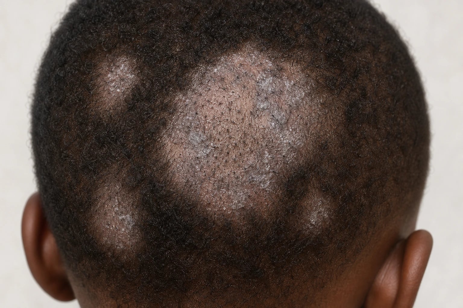
Dermatophytes invade the hair shaft (ectothrix — spores on the outside; endothrix — within the shaft) causing weakening and breakage. Causative organisms vary by geography: [1]
- Trichophyton tonsurans — the commonest cause in the Americas and UK; endothrix, non-fluorescent under Wood's lamp; produces black-dot tinea capitis and asymptomatic carriage.
- Microsporum canis — zoophilic (cats/dogs); ectothrix, bright green fluorescence; common in Europe.
- Microsporum audouinii — anthropophilic, green fluorescence; classic "grey-patch" tinea.
- Trichophyton violaceum — common in India/Asia/Africa; non-fluorescent endothrix.
- Trichophyton schoenleinii — causes favus (yellow cup-shaped crusts/scutula, permanent scarring).[2][12][14]
Transmission is by person-to-person, shared combs/hats, animals (M. canis), and fomites. Asymptomatic carriage (especially with T. tonsurans) maintains transmission within households and schools.[1]
Clinical features
Quick numbers for the examiner
CAPITIS — clinical patterns of tinea capitis
Trichoscopy hallmarks: comma hairs (slightly bent, sloping); corkscrew hairs (tightly coiled); both highly specific for tinea capitis
Up to 30% of household contacts asymptomatically carry the organism; treat with selenium sulfide shampoo
Grey-patch (Microsporum, ectothrix, green fluorescence) or black-dot (T. tonsurans, endothrix, no fluorescence)
Widespread eczematous eruption distant from infection; hypersensitivity to dermatophyte antigen; treat the infection
Boggy inflammatory mass from zoophilic species; do NOT incise; oral antibiotics and antifungals + prednisolone
Screen and treat all household members; comb/brush sharing; fomite control (bedding, hats)
Topical antifungals alone FAIL for tinea capitis; oral griseofulvin/terbinafine/itraconazole/fluconazole 6-8 weeks
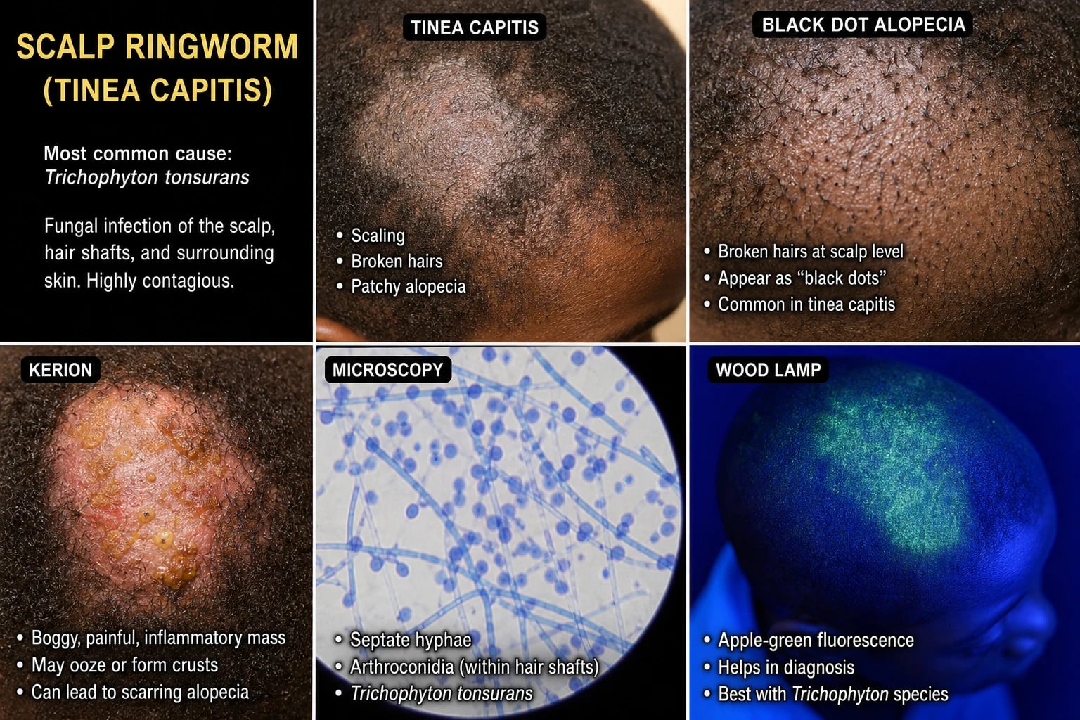
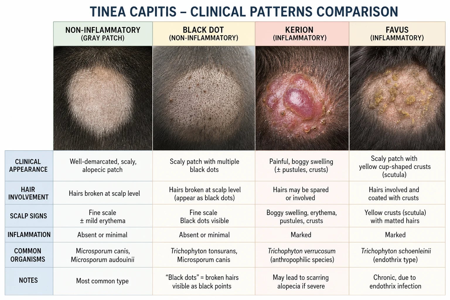
- Grey-patch (non-inflammatory) — one or more round, scaly, grey patches of partial hair loss with broken hairs; little inflammation.
- Black-dot — hairs broken at the scalp surface leave dark dots within areas of alopecia; characteristic of endothrix species (T. tonsurans, T. violaceum).[11]
- Inflammatory / kerion — a boggy, tender, purulent, elevated, inflamed mass with pustules, crusting, hair loss, and regional lymphadenopathy; a vigorous immune response to the dermatophyte (often zoophilic/inflammatory species); misdiagnosis as bacterial abscess leads to inappropriate incision; heals with possible scarring alopecia.[8]
- Favus — chronic, yellow, cup-shaped scutula around hair follicles with mousy odour, hair loss, and progressive scarring alopecia; caused by T. schoenleinii.[9]
- Occipital/cervical lymphadenopathy is a useful sign, especially with inflammatory disease.
Diagnosis

- KOH microscopy of plucked hairs / scale — ectothrix spores (outside the shaft) or endothrix spores (within).[5]
- Fungal culture of plucked hairs / brush-culture (toothbrush technique) — confirms species, guides therapy and epidemiology.
- Wood's lamp — green fluorescence with Microsporum species (not T. tonsurans, the commonest US/UK cause); useful where Microsporum predominates.
- Trichoscopy — comma hairs, corkscrew hairs, broken hairs, black dots, perifollicular scaling.[10]
Key features of tinea capitis
- Histopathology (rarely needed) — confirms in atypical or scarring cases.[13]
Treatment Specifics
[1] [1]TRICHOPHYTON — tinea capitis species and features
Endothrix; non-fluorescent; black-dot; asymptomatic carriage 30%
Peak 3-12 yr; rare in adults (sebum fungistatic)
Autoeczematisation: widespread eczematous eruption distant from infection; treat the infection
Trichoscopy hallmarks: highly specific for tinea capitis
Screen and treat all contacts; selenium sulfide shampoo 2-3x/week
Topical alone FAIL; griseofulvin 20-25 mg/kg/day or terbinafine weight-based
Don't incise kerion; treat medically; scarring alopecia permanent
After successful treatment, regrowth takes 6-12 months
Griseofulvin first-line age 2-12 yr; terbinafine from age 4
Reduces spore shedding, transmission; adjunct to oral
Endothrix; non-fluorescent; common in India, Africa
Favus (T. schoenleinii) - yellow cup-shaped scutula; permanent scarring alopecia
Differential diagnosis [1]
Alopecia areata (no scale, exclamation-mark hairs), seborrhoeic/atopic dermatitis of the scalp, psoriasis, bacterial folliculitis or abscess (kerion mimics), piedra (fungal nodules on hair shaft — black/white piedra), traction alopecia, trichotillomania, secondary syphilis ("moth-eaten"), and discoid lupus for scarring lesions. KOH and culture distinguish tinea.[3][2]
Management [1]
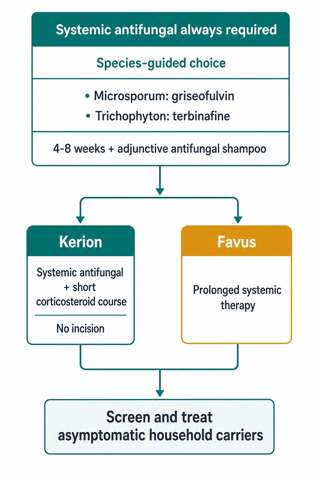
Tinea capitis always requires systemic antifungal therapy — topical agents cannot penetrate the hair shaft.[1][5][6]
- Griseofulvin — historic first-line; fungicidal for Microsporum; dosed by weight (e.g., 20-25 mg/kg/day, sometimes higher) for 6-8 weeks or longer; fat-containing food improves absorption. Meta-analyses support its efficacy, especially for Microsporum.[6][7]
- Terbinafine — first-line for Trichophyton (T. tonsurans, T. violaceum); shorter courses (2-4 weeks); granule formulation for children; less effective for Microsporum.[6][7]
- Itraconazole and fluconazole — alternatives, effective for both Microsporum and Trichophyton; pulsed dosing options.
- Adjunctive topical antifungal shampoo (ketoconazole 2%, selenium sulphide 2.5%, ciclopirox) — reduces shedding and carriage while systemic therapy takes effect.[5]
Kerion
- Systemic antifungal (griseofulvin or terbinafine, guided by species) is essential.
- Adjunctive oral corticosteroid (short tapering course) is commonly used to reduce inflammation and limit scarring, with supportive (not strong) evidence.
- Do NOT incise a kerion (does not help; worsens scarring and secondary infection).
- Treat any secondary bacterial infection.
- Reassure about gradual hair regrowth unless scarring has occurred.[8]
Public-health and carrier management
- Screen household and close contacts for tinea capitis and asymptomatic carriage (especially T. tonsurans).
- Treat carriers with antifungal shampoo (ketoconazole/selenium sulphide) ± a short course of systemic antifungal in selected cases.
- Exclude from school until treatment has started; launder combs, hats, and bedding. [1]
Kerion and id reaction: detailed management
Kerion and id reaction are the two inflammatory extremes of tinea capitis that the fellowship examiner probes to test that you understand the disease as more than a superficial infection. Both arise from a vigorous host immune response to dermatophyte antigen — not from bacterial superinfection — and both are best managed medically, not surgically. [1]
Kerion — clinical spectrum and natural history
A kerion is a boggy, tender, pustular, often crusted, inflammatory mass that develops over 1-3 weeks, most often on the vertex or occiput of a school-age child. It is typically 2-6 cm in diameter, fluctuant to palpation, studded with follicular pustules that weep serosanguinous fluid ("spongy honeycomb"), and is accompanied by regional (occipital, posterior-auricular, cervical) lymphadenopathy in most cases. Constitutional symptoms — low-grade fever, malaise, headache — occur in up to one-third of patients and often mislead clinicians towards a bacterial abscess. The lesion is sterile on routine bacterial culture in most cases; pustular fluid yields the dermatophyte on fungal culture (KOH and Sabouraud agar) but no significant bacterial growth. The histology is a dense dermal neutrophilic and granulomatous infiltrate with perifollicular abscess formation and a brisk T-cell response — essentially a delayed-type hypersensitivity reaction to fungal antigen. Misdiagnosis as bacterial abscess and incision-and-drainage remains the commonest management error and is the single most important cause of iatrogenic scarring alopecia in tinea capitis. [1]
The differential diagnosis of a boggy scalp mass in a child includes: bacterial scalp abscess/furuncle (usually solitary, with prominent tenderness and overlying erythema; bacterial culture positive), cellulitis (diffuse erythema without a discrete mass), dissecting cellulitis (rare in children; sinus tract formation), hidradenitis suppurativa (rare on scalp; axillary/groin involvement), and (very rarely) pyoderma gangrenosum or cutaneous lymphoma. KOH and fungal culture of expressed fluid or plucked hairs from the edge of the kerion confirms the diagnosis in 80-90% of cases; a negative KOH does not exclude the diagnosis and should not delay empirical antifungal therapy. [1]
Kerion — step-by-step management
Kerion management: from presentation to regrowth
Diagnose and start empirical antifungal
Add a short course of oral corticosteroid
Antibiotics only if secondary bacterial infection
Local care and analgesia
Screen and treat household contacts
Follow-up and reassure about regrowth
Id reaction (autoeczematisation)
The id reaction is a widespread, symmetrical, pruritic, eczematous (or, less commonly, papulovesicular or urticarial) eruption that develops distant from the primary dermatophyte infection, classically 1-3 weeks after the kerion or inflammatory tinea capitis has begun. It most commonly involves the trunk, limbs, and hands; the palms and soles may show dyshidrotic vesicles. The mechanism is a type III (immune-complex) hypersensitivity to fungal antigen — fungal antigen is disseminated haematogenously from the inflamed scalp and deposited in distant skin, where it triggers a localised vasculitic or spongiotic response. Critically, the id reaction is sterile — KOH and fungal culture of id lesions are negative, and the eruption does NOT contain live fungus. Misdiagnosing the id reaction as a drug eruption (because the child has just started griseofulvin) is a common pitfall; the answer is to continue the antifungal and treat the id reaction symptomatically. [1]
[1]Kerion prognosis and long-term outcomes
With timely antifungal therapy (started within 2 weeks of onset), 70-80% of kerions heal without scarring alopecia. Permanent scarring alopecia occurs in 20-30% of cases, particularly those with delayed presentation (>4 weeks), severe necrosis at presentation, prior incision, or co-existing favus (T. schoenleinii). Hair regrowth, when it occurs, is typically visible by 3 months and complete by 6-12 months. Children with permanent scarring alopecia can be referred to a paediatric dermatologist for scar camouflage, hair prosthetics, and (in selected adolescents) surgical options including scalp reduction or hair transplantation once the disease has been inactive for at least 12 months. [1]
Kerion severity and prognosis guide
Severe
Large >5 cm or multiple kerions; marked tenderness; significant systemic symptoms; delayed presentation >4 weeks. Prognosis: 50-70% heal without scarring; significant risk of permanent alopecia.
KERION — kerion management pitfalls and priorities
Id reaction is sterile and resolves with continued antifungal; stopping therapy is a common error
20-30% permanent alopecia; regrowth takes 6-12 months; honest prognosis at first visit
Kerion is sterile; incision destroys follicles and causes permanent scarring; treat medically
Reduces pain, swelling, scarring; evidence supportive but consistent across guidelines
Griseofulvin for Microsporum; terbinafine for Trichophyton; topical agents alone FAIL
30% of household contacts carry T. tonsurans; toothbrush culture and treat with shampoo
Household screening and carrier management: operational protocol
Asymptomatic carriage is the engine of transmission in tinea capitis. The clinical implication is that treating the index child without screening the household almost guarantees re-infection and ongoing school transmission. The following is an operational protocol used in many paediatric dermatology centres. [1]
Step 1 — Identify all household and close contacts of the index case: parents, siblings, grandparents, regular babysitters, and (if relevant) school playmates. Ask specifically about pets — cats and dogs for M. canis; rabbits and guinea pigs for T. mentagrophytes. [1]
Step 2 — Sample each contact's scalp using the toothbrush technique: a sterile soft toothbrush is brushed firmly across the entire scalp for 30 seconds, then the bristles are inoculated directly onto Sabouraud dextrose agar or Dermatophyte Test Medium (DTM). DTM is colour-change-based (red = dermatophyte) and allows primary-care screening without a mycology lab. Sample 1-2 cm of hair and any visible scale in addition to the brush. [1]
Step 3 — Examine for clinical disease: patchy alopecia, scaling, broken hair stubs, black dots, kerion, or occipital lymphadenopathy. Approximately 30% of T. tonsurans household contacts are culture-positive but clinically normal (carriers); 5% will develop clinical disease. [1]
Step 4 — Treat carriers with selenium sulphide 2.5% or ketoconazole 2% shampoo 2-3 times weekly for 4 weeks. The shampoo is left on for 5 minutes before rinsing. If a contact has clinical disease, treat with the same oral antifungal regimen as the index case. Repeat culture at 4 weeks; if still positive, a second 4-week shampoo course is given, and oral antifungal (single dose of itraconazole 10 mg/kg or 4 weeks of terbinafine) is considered. [1]
Step 5 — Environmental decontamination: hot wash (>60°C) of bedding, hats, pillowcases, and soft toys; soak combs and brushes in 1% hypochlorite or 70% isopropyl alcohol for 30 minutes; replace or disinfect hair accessories. Vacuum sofas and car seats. Limit sharing of combs, hats, helmets, and pillows during the treatment period. Household pets should be examined by a veterinarian; infected animals (typically cats with M. canis) should be treated concurrently. [1]
Step 6 — School and public-health action: most guidelines do NOT require school exclusion once treatment has started; the child may return after 24-48 hours of antifungal therapy. Classmates are usually NOT screened unless there is an outbreak (≥2 cases in a classroom within 4 weeks). Outbreak management involves notification to the local public health unit, classroom screening, and enhanced environmental cleaning. [1]
Household screening quick numbers for the examiner
Clinical pearl
[1]Red flags
Exam application bank (NEET-PG / INICET)
One-line answer
Tinea capitis is a dermatophyte infection of the scalp hair shaft and follicle, predominantly affecting pre-pubertal children. Fellowship-level assessment demands mastery of the clinical patterns (inflammatory/non-inflammatory, black-dot, grey-patch, favus, and the boggy inflammatory kerion), the causative dermatophyte species by geography and their Wood's lamp fluorescence (Microsporum fluoresce green; Trichophyton typically does not), confirmation by microscopy/culture/trichoscopy, the absolute requirement for systemic antifungals (griseofulvin or terbinafine/itraconazole, guided by species), the often-mismanaged kerion (systemic antifungal plus consideration of adjunctive corticosteroid), and screening and treatment of asymptomatic household carriers.
Worked stems (answer without another resource)
Stem 1 — Classic presentation. Map symptoms to mechanism; name the first investigation and first treatment step with dose/route if drug therapy is standard. [1]
Stem 2 — Unstable / complicated. List red flags that force immediate resuscitation, theatre, ICU, antidote, or reperfusion — and what you do in the first 15 minutes. [1]
Stem 3 — Atypical group. Elderly, pregnancy, child, or immunocompromised: how presentation and thresholds change. [1]
Stem 4 — Differential trap. Name the three closest mimics and one discriminator for each. [1]
Stem 5 — Disposition. Who goes home with safety-netting, who is admitted, who needs HDU/ICU/theatre, and what follow-up is mandatory. [1]
Rapid viva checklist
- Definition + classification
- Pathophysiology chain
- Bedside signs / criteria
- Score with exact components (if any)
- Emergency bundle
- Definitive therapy with doses
- Complications of disease and of treatment
- Special populations
- Guideline/trial name if classic
- Three exam traps
Coverage self-check
If you cannot answer any stem above from this page alone, re-read the matching section — the page is intended to be self-sufficient for final-prof and NEET-PG/INICET questions on Tinea capitis.
Expanded exam teaching (depth pass)
Clinical reasoning
For Tinea capitis, examiners test whether you can prioritise life threats, choose the right first test, and give specific therapy (agent, dose, route, timing). Generic phrases without numbers score poorly.
Mechanism → feature map
Build a short chain: cause → pathophysiologic intermediate → clinical feature → complication. Every major symptom in the classic vignette should sit on that chain.
Investigation strategy
- Bedside/first-line tests that change immediate management
- Confirmatory or staging tests
- What a normal result does not exclude
- When not to delay treatment for imaging (unstable patient)
Management ladder
- Resuscitation / ABC / sepsis or haemorrhage bundle as relevant
- Specific antidote / procedure / antimicrobial / reperfusion / surgery
- Supportive care and monitoring targets
- Definitive long-term therapy and secondary prevention
- Disposition and safety-net advice
Special populations
Always prepare one line each for children, pregnancy, elderly, renal/hepatic impairment, and immunocompromised patients when the topic allows.
Pitfalls that fail candidates
- Treating the number not the patient
- Missing pregnancy status when relevant
- Imaging before stabilisation
- Wrong empiric cover or wrong antidote timing
- Incomplete counselling on recurrence, adherence, or red-flag return
Tinea capitis is a dermatophyte infection of the scalp hair shaft and follicle, predominantly affecting pre-pubertal children. Fellowship-level assessment demands mastery of the clinical patterns (inflammatory/non-inflammatory, black-dot, grey-patch, favus, and the boggy inflammatory kerion), the causative dermatophyte species by geography and their Wood's lamp fluorescence (Microsporum fluoresce green; Trichophyton typically does not), confirmation by microscopy/culture/trichoscopy, the absolut [1]
[1]Asymptomatic carriers and public health
[1]Carrier and public health quick numbers
References
- [1]Gupta AK, Friedlander SF, Simkovich AJ. Tinea capitis: An update Pediatr Dermatol, 2022.PMID 35075666
- [2]Leung AKC, Hon KL, Leong KF, et al. Tinea Capitis: An Updated Review Recent Pat Inflamm Allergy Drug Discov, 2020.PMID 31906842
- [3]Gupta AK, Polla Ravi S, Wang T, et al. An update on tinea capitis in children Pediatr Dermatol, 2024.PMID 39113245
- [4]Hill RC, Gold JAW, Lipner SR. Comprehensive Review of Tinea Capitis in Adults: Epidemiology, Risk Factors, Clinical Presentations, and Management J Fungi (Basel), 2024.PMID 38786712
- [5]Caplan AS, Gold JAW, Smith DJ, et al. Diagnosis and Management of Tinea Infections Am Fam Physician, 2025.PMID 41118183
- [6]Tey HL, Tan AS, Chan YC. Meta-analysis of randomized, controlled trials comparing griseofulvin and terbinafine in the treatment of tinea capitis J Am Acad Dermatol, 2011.PMID 21334096
- [7]Fleece D, Gaughan JP, Aronoff SC. Griseofulvin versus terbinafine in the treatment of tinea capitis: a meta-analysis of randomized, clinical trials Pediatrics, 2004.PMID 15520113
- [8]Chiriac A, Diaconeasa A, Voicu C, et al. Kerion Celsi in infants and children-A narrative review 2010-2023 Mycoses, 2024.PMID 37983862
- [9]Ilkit M. Favus of the scalp: an overview and update Mycopathologia, 2010.PMID 20411336
- [10]Güleç AT. Trichoscopic Evaluation of Tinea Capitis Mycopathologia, 2023.PMID 36266555
- [11]Wang X. Black Dot Tinea Capitis N Engl J Med, 2024.PMID 39037044
- [12]Barac A, Stjepanovic M, Krajisnik S, et al. Dermatophytes: Update on Clinical Epidemiology and Treatment Mycopathologia, 2024.PMID 39567411
- [13]Kovitwanichkanont T, Chong AH. Superficial fungal infections Aust J Gen Pract, 2019.PMID 31569324
- [14]Gupta AK, Summerbell RC. Tinea capitis Med Mycol, 2000.PMID 10975696