Dermatology · Medicine
Tinea corporis
Also known as Ringworm (body) · Tinea cruris (groin) · Tinea faciei (face) · Tinea manuum (hand) · Tinea incognito · Majocchi granuloma
Tinea corporis (ringworm) is a superficial dermatophyte infection of glabrous body skin, with anatomically named variants (cruris at the groin, faciei at the face, manuum at the hand). Fellowship-level assessment demands mastery of the active scaly advancing edge with central clearing, potassium hydroxide microscopy and culture confirmation, the immunosuppression-altered forms (tinea incognito under topical steroid, Majocchi granuloma with follicular invasion), the topical and systemic antifungal ladder (allylamines, azoles, terbinafine, itraconazole), and the emerging global threat of antifungal-resistant Trichophyton indotineae.
On this page & tools
Your progress
Saved locally on this device.
Exam tags
Red flags
Overview
Tinea corporis ("ringworm") is a superficial dermatophyte infection of glabrous body skin caused by Trichophyton, Microsporum and Epidermophyton species. It is the commonest dermatophyte infection worldwide and accounts for the majority of cutaneous fungal disease seen in primary care. Anatomically named variants — tinea cruris (groin), tinea faciei (face), tinea manuum (hand), tinea barbae (beard), tinea imbricata (concentric rings seen in Polynesia and parts of South-East Asia) — share the same aetiology but have site-specific features. Fellowship-level competence requires recognition of the annular, scaly, peripherally advancing lesion with central clearing, confirmation by potassium hydroxide (KOH) microscopy and fungal culture, an understanding of the altered morphology under immunosuppression (tinea incognito under topical steroid, Majocchi granuloma with follicular invasion), the topical and systemic antifungal ladder, and the emerging global threat of antifungal-resistant Trichophyton indotineae.[1][2][4]
Tinea corporis at a glance
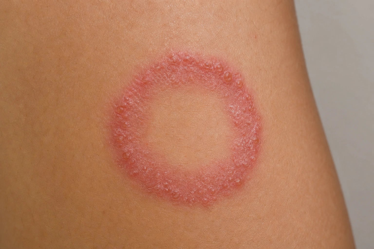
Pathophysiology and microbiology

Dermatophytes are keratinophilic fungi that digest keratin in the stratum corneum, hair and nails using secreted keratinases, elastases, lipases and other hydrolases. They cannot penetrate beyond the granular layer in immunocompetent hosts because deeper tissue is not keratinised. The principal genera affecting humans: [1]
- Trichophyton — T. rubrum (the commonest cause of chronic tinea corporis, cruris, pedis and onychomycosis globally, responsible for 60-80% of cases), T. interdigitale, T. mentagrophytes (zoophilic variant from rodents), T. tonsurans (anthropophilic, dominant cause of tinea corporis gladiatorum in North America and the leading cause of tinea capitis worldwide), and the emerging antifungal-resistant T. indotineae (formerly T. mentagrophytes ITS genotype VIII), responsible for extensive, recurrent, terbinafine-resistant epidemics in South Asia with global spread.[5][11][12]
- Microsporum — M. canis (zoophilic, from cats and dogs — commonest zoophilic cause of tinea corporis in many regions), M. audouinii (anthropophilic, historical cause of tinea capitis epidemics), M. gypseum (geophilic, soil contact), M. nanum (geophilic, swine).
- Epidermophyton floccosum — affects skin and nails, but never hair; an important cause of tinea cruris and tinea corporis.
Transmission is anthropophilic (human-to-human, often via shared towels, clothing, combs, dormitory bedding, gym mats; produces chronic, minimally inflammatory lesions), zoophilic (animal-to-human, often from kittens, puppies, rabbits, guinea pigs, cattle, horses; produces acute, intensely inflammatory, sometimes pustular lesions with marked host response) or geophilic (soil-to-human; M. gypseum from gardening). The organism proliferates in warm, moist, occluded environments, advancing centrifugally to produce the characteristic annular lesion; autoinoculation from coexisting tinea capitis, pedis or unguium maintains chronicity.[4][5][6]
The annular ring is a direct consequence of centrifugal spread: hyphae radiate outwards from the inoculation point, leaving behind a central zone where the immune response and the natural epidermal turnover (about 28 days) have already shed the infected keratin, while the active advancing edge is the zone of fresh invasion with intense inflammation, spongiosis and scale. Tinea incognito arises when topical (occasionally systemic) corticosteroid suppresses this local cell-mediated immune response: the active edge flattens, the scale disappears, the lesion becomes an ill-defined erythematous patch, often with pustules or atrophy, and the infection spreads silently, frequently to deep follicles producing Majocchi granuloma. Majocchi granuloma is a perifollicular granulomatous response to dermatophyte invasion of the hair follicle, most often triggered by topical steroid use, shaving (legs in women, beard in men), occlusion or immunosuppression; histology shows fungal hyphae within hair shafts surrounded by a mixed granulomatous infiltrate. [1]
Clinical features


High-yield features of tinea corporis
RINGWORM
Active border with fresh fungus, scale and erythema at the leading edge
Zoophilic lesions are intensely inflamed, pustular or vesicular
Treat coexisting tinea pedis/unguium to prevent relapse
If the face is involved, think tinea faciei; if groin, tinea cruris
M. canis green; T. rubrum does NOT fluoresce
Autoclave shared shoes, hats, gym gear; launder linen
SQLE F397L/L393F mutations; switch to itraconazole pulse
Follicular invasion with perifollicular papules/nodules; needs 4-6 weeks systemic therapy
- Tinea corporis — an annular (ring-shaped) erythematous plaque with a scaly, raised, vesicular or papular advancing edge and central clearing, often itchy; lesions enlarge centrifugally. May be single or multiple. The first lesion is the largest ("mother" or "herald" patch); daughter lesions appear in the surrounding skin.
- Tinea cruris (groin) — symmetrical, sharply marginated erythematous scaly plaque in the inguinal folds, extending onto the upper thighs but characteristically sparing the scrotum and penis; often coexists with tinea pedis (autoinoculation when dressing). Erythrasma (coral-red under Wood's lamp) and candida intertrigo (beefy-red, satellite pustules, involves scrotum) are the key mimics.[2]
- Tinea faciei — annular lesion on the face, often misdiagnosed as eczema, rosacea, lupus or perioral dermatitis; frequently modified by prior topical steroid use (tinea incognito).
- Tinea manuum — usually unilateral dry, scaly, fine annular lesion on one palm, often coexisting with "two feet one hand" tinea pedis/unguium; consider chronic dry palmar eczema in the differential.
- Tinea gladiatorum — typically T. tonsurans (North America) or T. interdigitale/E. floccosum (Europe), transmitted by skin-to-skin contact in wrestlers and rugby players; lesions favour the head, neck, forearms, shoulders and thighs (areas of mat contact). Exclusion from contact sport until lesions resolve with systemic therapy is mandated by NFHS, NCAA and analogous bodies.
- Tinea incognito — tinea modified by topical (or systemic) corticosteroid, losing the classical scaly edge and presenting as an atypical, often extensive, erythematous, sometimes pustular or atrophic plaque; a leading cause of "treatment-resistant eczema" and a major driver of antifungal over-use in South Asia.[7][8][9]
- Majocchi granuloma — dermatophyte follicular invasion producing follicular papules, nodules and pustules within or around a tinea lesion (often on the leg after shaving, or in immunosuppression); requires systemic antifungal because topical therapy cannot reach the follicle.[10]
- Tinea imbricata — chronic, non-resolving concentric rings caused by T. concentricum; endemic in parts of Polynesia, Melanesia, South-East Asia and Central/South America; often familial with autosomal-recessive susceptibility.
Diagnosis
- KOH microscopy of skin scrapings (from the active scaly edge) — branching septate hyphae confirm dermatophyte infection rapidly; sensitivity about 70-80%, specificity more than 90%; bedside "chewed-spaghetti" or "rail-track" appearance.[3]
- Fungal culture on Sabouraud dextrose agar with cycloheximide and chloramphenicol — confirms and identifies the species, guiding epidemiology and resistance testing; slower (2-4 weeks). Mycobiotic agar, dermatophyte test medium (DTM, red colour change) and potato dextrose agar are alternatives.
- Dermoscopy — peripheral whitish "moth-eaten" scaling with central clearing, occasional comma-shaped hairs and corkscrew vessels; useful adjunct in atypical or steroid-modified lesions.
- Wood's lamp (UV 365 nm) — green-yellow fluorescence with some Microsporum species (M. canis, M. audouinii — ectothrix infections); T. rubrum and T. indotineae do NOT fluoresce; of limited use for body tinea but very useful for tinea capitis and to distinguish from erythrasma (coral-red porphyrin fluorescence) and vitiligo (bright blue-white).[3]
- PCR / molecular methods (ITS sequencing, real-time PCR, MALDI-TOF) — increasingly available for rapid species identification and detection of T. indotineae squalene-epoxidase (SQLE) mutations conferring terbinafine resistance; turn-around 24-48 hours.[11][12]
- Skin biopsy with PAS or Grocott/GMS stain — for atypical, refractory, Majocchi or tinea incognito lesions when KOH and culture are repeatedly negative; demonstrates hyphae in stratum corneum, within hair shafts (PAS-positive) or in perifollicular granulomas.
- Baseline blood tests before systemic antifungals — LFTs (ALT, AST, ALP, bilirubin) and renal function; in patients on long courses or with hepatic risk factors also a full blood count.
Differential diagnosis
The single most useful discriminator is KOH microscopy from the active edge, supplemented by culture where needed. The major clinical mimics are summarised below.[1][6]
Tinea corporis vs nummular eczema
Both are scaly, ringed, itchy
- Raised scaly advancing edge; clear centre; KOH positive
- Often solitary or few; rapid centrifugal spread
- May have pustules at the active edge; intensely itchy
- Responds to terbinafine 1% BD 1-2 weeks
- Treat coexisting tinea pedis/unguium to prevent relapse
Tinea corporis vs erythema migrans
Both are annular, both are expanding
- Flat advancing edge; no scale; central clearing but no active scaly border
- History of tick bite, often single, more than 5 cm within days
- Flu-like prodrome, arthralgia, no itch typically
- Treat with doxycycline 100 mg BD 10-14 days (or amoxicillin)
- KOH negative; C6 ELISA positive in early disseminated disease
Tinea corporis vs annular psoriasis
Both have a sharp edge and central change
- Silvery adherent scale, not raised vesicular edge
- Extensor surfaces, scalp, nails (pitting, onycholysis)
- Negative KOH; positive family/personal history of psoriasis
- Treat with topical vitamin D analogues, steroids, systemic agents
- Often Koebner phenomenon, Auspitz sign
Tinea corporis vs granuloma annulare
Both are annular on the hand/foot
- Firm, smooth, skin-coloured to pink dermal papules in ring
- No scale, no itch, no active scaly edge
- Classic sites: dorsa of hands/feet, ankles; common in children
- Often self-limiting; topical steroid or tacrolimus for cosmesis
- KOH negative; biopsy shows necrobiotic collagen with mucin
Other important mimics include pityriasis rosea (herald patch followed by oval lesions in "Christmas-tree" distribution on the back, collarette scale), pityriasis versicolor (Malassezia, hypopigmented or hyperpigmented macules with fine scale and "spaghetti-and-meatballs" KOH, not branching septate hyphae), subacute cutaneous lupus erythematosus (photosensitive annular or polycyclic plaques, ANA/anti-Ro positive), erythema annulare centrifugum (trailing scale on the inner edge, follows infections, drugs or malignancy), secondary syphilis (palmo-plantar involvement, moth-eaten alopecia, positive RPR/VDRL), fixed drug eruption (recurs at the same site on re-exposure, well-demarcated erythema with or without bullae), and — for tinea incognito — rosacea, periorificial dermatitis, seborrhoeic dermatitis and contact dermatitis. KOH microscopy is the decisive discriminator. [1]
Management
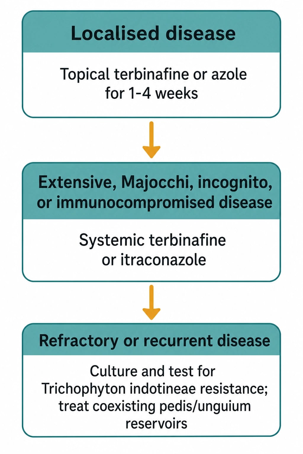
Topical antifungals (first-line for localised tinea corporis/cruris)
- Allylamines — terbinafine 1% cream applied BD for 1-2 weeks; fungicidal (superior to azoles, which are fungistatic) and the preferred topical in head-to-head trials.[4]
- Azoles — clotrimazole 1%, miconazole 2%, econazole 1%, ketoconazole 2%, sertaconazole 2%, luliconazole 1% (single-dose data), bifonazole 1%; apply BD for 2-4 weeks; useful where terbinafine is unavailable or cost-restricted.
- Other topicals — ciclopirox 0.77% (also antibacterial and anti-inflammatory), tolnaftate (less effective), selenium sulfide shampoo as adjunct.
- Apply to the lesion and 2 cm beyond, continue for 1-2 weeks after clinical resolution, and treat coexisting tinea pedis/unguium to prevent recurrence. Topical corticosteroid-antifungal combinations should be avoided — they predispose to tinea incognito and mask the disease.
Systemic antifungals (extensive, refractory, Majocchi, immunocompromise, tinea incognito)
- Terbinafine 250 mg daily for 2-4 weeks (corporis/cruris) — first-line systemic; fungicidal allylamine; inhibits squalene epoxidase causing squalene accumulation and fungal death; accumulates in keratin; monitor LFTs at baseline and at 4-6 weeks if therapy is prolonged; rare hepatotoxicity, taste/smell disturbance, severe cutaneous adverse reactions.
- Itraconazole 200 mg BD for 1 week (one pulse) OR 100-200 mg daily for 1-2 weeks; triazole that inhibits lanosterol 14α-demethylase; very broad-spectrum; pulse regimen exploits keratin affinity; check drug interactions (potent CYP3A4 inhibitor — avoid with simvastatin, ergots, colchicine, dofetilide, quinidine) and LFTs; contraindicated in heart failure.
- Fluconazole 150-300 mg weekly for 2-4 weeks — alternative; well-tolerated; useful when terbinafine and itraconazole are contraindicated.
- Griseofulvin — effective for tinea capitis but largely superseded for body tinea; needs 4-6 weeks; CYP450 inducer, teratogenic, contraindicated in pregnancy and porphyria.
- Newer agents — voriconazole and posaconazole for refractory T. indotineae in specialist hands; oteseconazole (approved for vulvovaginal candidiasis) under investigation for dermatophytosis. [1]
Antifungal pharmacology at a glance
Special considerations
- Tinea incognito — stop the corticosteroid, confirm with KOH/biopsy, and treat with systemic antifungal (often extensive and altered); warn the patient that disease may transiently worsen (unmasking) before improving.[7][9]
- Majocchi granuloma — systemic antifungal (topical cannot reach the follicle); 4-6 weeks of oral terbinafine or itraconazole; address shaving/occlusion/immunosuppression.[10]
- Antifungal-resistant T. indotineae — increasingly recognised cause of extensive, recurrent, terbinafine-refractory tinea originating in South Asia and spreading globally; culture and molecular resistance testing; switch to itraconazole pulse 200 mg BD 1 week per month for 3-4 pulses; voriconazole or posaconazole in refractory cases; consider combination topical therapy (azole plus ciclopirox plus terbinafine) and topical keratolytics (urea, salicylic acid) to enhance penetration.[11][12]
- Recurrent disease — treat coexisting tinea pedis/unguium (the reservoir), reduce occlusion and moisture, treat household/pet contacts (especially for zoophilic M. canis — vet screen the cat/dog and treat with itraconazole or terbinafine), and launder clothing/towels at temperatures above 60 °C.
- Pregnancy — topical terbinafine or azoles (clotrimazole, miconazole) are safe; avoid oral terbinafine in the first trimester; itraconazole is contraindicated.
- Paediatrics — topical therapy first-line for body tinea; oral terbinafine/itraconazole can be used for extensive or Majocchi disease at age-adjusted doses; tinea capitis always requires systemic therapy.
Anatomical variants and targeted antifungal dosing
The dermatophyte clinicopathological syndrome varies by anatomical site. Mastery of the variant list — and the site-specific drug doses and minimum treatment durations — is a high-yield fellowship topic because two-thirds of recurring "treatment failure" is simply wrong drug choice or wrong duration for that body region. [1]
- Tinea faciei — annular, often scale-poor or papular lesion on the face, frequently mimicking eczema, rosacea, seborrhoeic dermatitis, lupus or perioral dermatitis. Triggers include repeated topical corticosteroid use (so produces tinea incognito), pet contact (especially kittens — M. canis), and the same SQLE-resistant T. indotineae now endemic across the Indian subcontinent. The active edge is often subtle because facial sebum and UV exposure attenuate scale; Wood's lamp is unreliable (anthropophilic Trichophyton species do not fluoresce); biopsy with PAS is helpful when KOH repeatedly negative. Treat with topical terbinafine 1% BD for 2 weeks for solitary disease; oral terbinafine 250 mg OD for 2-4 weeks for extensive or steroid-modified disease; itraconazole pulse 200 mg BD for the first week of each month, x 3 pulses, when T. indotineae is suspected.
- Tinea cruris (jock itch) — well-demarcated erythematous, scaly plaque in the inguinal folds extending onto the upper thighs, classically sparing the scrotum and penis (a key discriminator from candida intertrigo, which involves the scrotum, and from erythrasma, which fluoresces coral-red under Wood's lamp). Commonest cause is T. rubrum, frequently coexisting with onychomycosis and tinea pedis (autoinoculation from the feet via the underwear when dressing — always examine the toes). Treat localised disease with topical terbinafine 1% or clotrimazole 1% BD for 2 weeks, extending one week beyond clinical clearance; oral terbinafine 250 mg OD for 2 weeks for extensive, recalcitrant, or follicular disease. Recurrence rate approaches 20% if the foot reservoir is not cleared.
- Tinea manuum — unilateral, dry, scaly, mildly inflammatory palmar or dorsal hand lesion, often co-existing with bilateral tinea pedis ("two feet-one hand syndrome", pathognomonic of T. rubrum). Treat aggressively with oral terbinafine 250 mg OD for 4 weeks because topical agents alone are defeated by the thick palmar stratum corneum; itraconazole 200 mg BD for the first week of each month, x 2-3 pulses, as an alternative.
- Tinea pedis (athlete's foot) — three morphological patterns: (a) interdigital — maceration, fissuring, scaling between the toes, commonly the 4th web space; (b) hyperkeratotic ("moccasin") — diffuse plantar hyperkeratosis; (c) vesiculobullous — vesicles or bullae on the instep, often with an id reaction. Treat the interdigital variant with topical terbinafine 1% BD for 1-2 weeks; the hyperkeratotic or extensive variant with oral terbinafine 250 mg OD for 2-4 weeks; the vesiculobullous variant with oral terbinafine plus a short course of oral antibiotic if secondarily infected. Always combine with foot-hygiene measures (separating the toes, antifungal powder in shoes, treating concurrent onychomycosis) to prevent recurrence.
- Tinea unguium (onychomycosis) — distal-lateral subungual hyperkeratosis and onycholysis, most often of the great toenail, caused by T. rubrum; the commonest nail dystrophy in adults but only about 50% of dystrophic nails are fungal — confirm with KOH and culture before committing to a months-long systemic course. Oral terbinafine 250 mg OD for 6 weeks (fingernails) or 12 weeks (toenails) is the most effective regimen with mycological cure rates of 70-80%; itraconazole pulse 200 mg BD for 1 week per month x 2 pulses (fingernails) or 3-4 pulses (toenails) is an effective alternative; fluconazole 150 mg once weekly for 6-9 months (fingernails) or 9-12 months (toenails) is third-line. Topical ciclopirox 8% or efinaconazole 10% nail lacquer is reserved for superficial white onychomycosis or patients who cannot tolerate systemic therapy.
- Tinea barbae — pustular, nodular, kerion-like inflammatory lesion of the beard and moustache area in adult men; almost always zoophilic (T. mentagrophytes var. mentagrophytes from cattle and dogs) and may mimic bacterial folliculitis, acne conglobata or herpetic sycosis. Treat with oral terbinafine 250 mg OD for 4 weeks (first-line) or itraconazole 200 mg BD pulse x 2-3 pulses; advise shaving to clear the infected hair reservoir, but discard the blade or razor head.
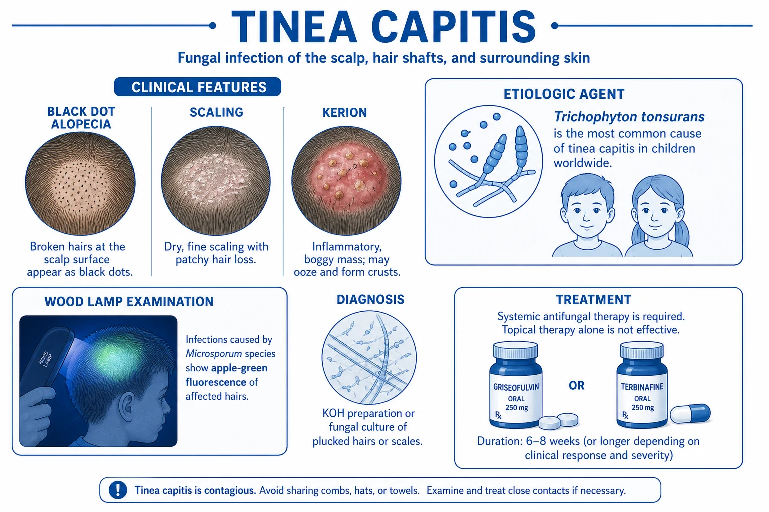
- Tinea capitis — predominantly a disease of prepubertal children; T. tonsurans dominates in North America, M. canis in Europe/Middle East, T. violaceum in Africa and South Asia. Always systemic, never topical alone, because the fungus is within the hair shaft. Griseofulvin 20-25 mg/kg per day (max 1000 mg/day) for 6-12 weeks is still the gold standard where affordable and available; oral terbinafine is now first-line in many guidelines (weight-based: 62.5 mg/day if below 20 kg, 125 mg/day if 20-40 kg, 250 mg/day if above 40 kg, for 4 weeks [T. tonsurans] to 6-8 weeks [M. canis]); alternatives include itraconazole pulse 5 mg/kg per day for 1 week per month x 2-3 pulses, or fluconazole 6 mg/kg per day for 3-6 weeks. Kerion (boggy, suppurative, painful mass with pustular discharge) also requires systemic antifungal plus a short course of oral corticosteroid (prednisolone 0.5-1 mg/kg per day for 1-2 weeks) to reduce scarring alopecia; screen and treat household contacts; comb hair with a ketoconazole 2% shampoo three times weekly.
- Tinea incognito revisited — the consequence of empirical topical steroid for an annular facial or truncal lesion that was never KOH-tested. Classical sign is loss of the raised scaly edge with persistent erythema, "eczema that just won't go away". Confirm with KOH or biopsy; stop the steroid cold; treat with oral terbinafine 250 mg OD for 4 weeks (extensive or facial disease) or itraconazole pulse 200 mg BD 1 week/month x 2-3 pulses if T. indotineae is suspected; warn the patient about unmasking.
- Trichophyton indotineae resistance (global emerging threat) — anthropophilic dermatophyte (formerly T. mentagrophytes ITS genotype VIII) originating from the Indian subcontinent and now reported across Europe, the Middle East, Australia and North America. It produces extensive, pruritic, polycyclic tinea corporis, cruris and faciei that fails standard oral terbinafine. The mechanism is point mutations in the squalene-epoxidase gene (SQLE F397L, L393F, A448T and S443P) causing structural change in the terbinafine binding pocket with up to 100-fold MIC shift. Send fungal cultures for ITS sequencing and SQLE PCR. Treat with itraconazole pulse 200 mg BD for 1 week per month x 3-4 pulses, or oral voriconazole 200 mg BD for 4-8 weeks in refractory disease, or posaconazole in pan-azole-resistant isolates. Combine with topical antifungal-corticosteroid-free regimens (sertaconazole, ciclopirox plus urea) and screen household contacts to prevent onward spread. [1]
Targeted antifungal dosing at a glance
Common anatomical tinea variants
Site dictates morphology, reservoir and first-line regimen
- Tinea incognito: steroid-modified tinea; absent scale; systemic therapy after stopping steroid
- Tinea faciei: facial, often misdiagnosed as eczema/rosacea; topical steroid worsens it
- Tinea manuum: unilateral dry palm; 'two feet-one hand' with tinea pedis is pathognomonic
- Tinea cruris: inguinal, sparing scrotum/penis; coexisting tinea pedis reservoir
- Tinea pedis: interdigital, hyperkeratotic or vesiculobullous; foot hygiene adjunct
- Tinea barbae: adult beard, zoophilic, kerion-like; oral therapy + shave reservoir
- Tinea capitis: prepubertal children; systemic always; griseofulvin or terbinafine weight-based
- Tinea unguium: oral terbinafine 6-12 wk nails; itraconazole pulse 2-4 monthly pulses
Drug resistance: T. rubrum vs T. indotineae
When terbinafine fails in tinea
- T. rubrum: SQLE wild-type; terbinafine MIC low; responds to 250 mg OD 2-4 wk
- T. indotineae: SQLE F397L or L393F mutations; terbinafine MIC 100-fold higher
- Geographic origin: T. indotineae endemic in India, Pakistan, Bangladesh, spreading globally
- Clinical clue: extensive, polycyclic, recurrent tinea failing 4 wk of standard terbinafine
- Switch: itraconazole pulse 200 mg BD 1 wk/month x 3-4 pulses; voriconazole in refractory
Anthropophilic vs zoophilic vs geophilic tinea
Reservoir dictates the clinical picture
- Anthropophilic (T. rubrum, T. interdigitale, T. tonsurans, E. floccosum): human-to-human; chronic, indolent, minimally inflamed
- Zoophilic (M. canis, T. mentagrophytes var. mentagrophytes, T. equinum, T. verrucosum): animal-to-human; acute, intensely inflamed, often pustular or kerion-like
- Geophilic (M. gypseum, M. nanum): soil contact via gardening; intermediate inflammation, often solitary lesions
- Host response inversely related to organism-host adaptation
- Pet screening and treatment essential for zoophilic disease
T. rubrum vs T. indotineae
Two anthropophilic dermatophytes — one resistant
- T. rubrum: most common dermatophyte globally; sensitive to terbinafine; classic ringworm morphology
- T. indotineae: anthropophilic dermatophyte from India/Pakistan/Bangladesh; SQLE F397L, L393F or A448T mutations conferring terbinafine resistance
- T. rubrum: respond to terbinafine 250 mg OD for 2-4 weeks (corpus) or 12 weeks (pedis/unguium)
- T. indotineae: itraconazole pulse 200 mg BD 1 week per month x 3-4 pulses; voriconazole or posaconazole in refractory cases
Prevention and household control
- Avoid sharing towels, clothing, combs, hats, gym gear and bedding; launder at temperatures above 60 °C.
- Treat household and close contacts (especially children sharing beds and wrestlers on the same mat).
- For zoophilic disease, identify and treat the animal reservoir — kittens with patchy alopecia, scaling or broken whiskers are a classic M. canis source.
- Keep skin cool and dry; cotton underwear, open footwear, weight loss in obesity, antiperspirants in hyperhidrosis-prone sites.
- Treat coexisting tinea pedis, unguium and manuum to remove the reservoir.
- Wrestlers and contact-sport athletes: skin checks before each event, exclusion until lesions are resolved, prompt systemic therapy, mat disinfection (quaternary ammonium or bleach).
- The IADVL/ICMR consensus recommends public-health awareness against the AAA (abuse, application, addiction) syndrome — the irrational over-the-counter sale of fixed-dose topical steroid + antifungal + antibacterial ("Triple Action" or "ringworm" creams) in South Asia, which is the principal driver of tinea incognito and recalcitrant dermatophytosis. [1]
Clinical pearl
[1]Bedside approach to a tinea suspect
KOHcHECK
Branching septate hyphae within 5 minutes
Find the reservoir driving recurrence
M. canis from kittens; T. indotineae from South Asia; AAAs from Indian chemists
HIV, diabetes, transplant, biologics
Screen family and pet; treat the reservoir
Clinical resolution plus 1-2 weeks of treatment beyond; mycological cure optional
SDA with cycloheximide; PCR/MALDI-TOF; SQLE mutation testing
Tinea incognito, Majocchi, secondary cellulitis, T. indotineae resistance
Red flags
Exam application bank (NEET-PG / INICET)
One-line answer
Tinea corporis (ringworm) is a superficial dermatophyte infection of glabrous body skin, with anatomically named variants (cruris at the groin, faciei at the face, manuum at the hand). Fellowship-level assessment demands mastery of the active scaly advancing edge with central clearing, potassium hydroxide microscopy and culture confirmation, the immunosuppression-altered forms (tinea incognito under topical steroid, Majocchi granuloma with follicular invasion), the topical and systemic antifungal ladder (allylamines, azoles, terbinafine, itraconazole), and the emerging global threat of antifungal-resistant Trichophyton indotineae.
Worked stems (answer without another resource)
Stem 1 — Classic presentation. Map symptoms to mechanism; name the first investigation and first treatment step with dose/route if drug therapy is standard. [1]
Stem 2 — Unstable / complicated. List red flags that force immediate resuscitation, theatre, ICU, antidote, or reperfusion — and what you do in the first 15 minutes. [1]
Stem 3 — Atypical group. Elderly, pregnancy, child, or immunocompromised: how presentation and thresholds change. [1]
Stem 4 — Differential trap. Name the three closest mimics and one discriminator for each. [1]
Stem 5 — Disposition. Who goes home with safety-netting, who is admitted, who needs HDU/ICU/theatre, and what follow-up is mandatory. [1]
Rapid viva checklist
- Definition + classification
- Pathophysiology chain
- Bedside signs / criteria
- Score with exact components (if any)
- Emergency bundle
- Definitive therapy with doses
- Complications of disease and of treatment
- Special populations
- Guideline/trial name if classic
- Three exam traps
Coverage self-check
If you cannot answer any stem above from this page alone, re-read the matching section — the page is intended to be self-sufficient for final-prof and NEET-PG/INICET questions on Tinea corporis.
[1]References
- [1]Leung AK, Lam JM, Leong KF, et al. Tinea corporis: an updated review Drugs Context, 2020.PMID 32742295
- [2]Sahoo AK, Mahajan R. Management of tinea corporis, tinea cruris, and tinea pedis: A comprehensive review Indian Dermatol Online J, 2016.PMID 27057486
- [3]Ely JW, Rosenfeld S, Seabury Stone M. Diagnosis and management of tinea infections Am Fam Physician, 2014.PMID 25403034
- [4]Barac A, Stjepanovic M, Krajisnik S, et al. Dermatophytes: Update on Clinical Epidemiology and Treatment Mycopathologia, 2024.PMID 39567411
- [5]Moskaluk AE, VandeWoude S. Current Topics in Dermatophyte Classification and Clinical Diagnosis Pathogens, 2022.PMID 36145389
- [6]Kovitwanichkanont T, Chong AH. Superficial fungal infections Aust J Gen Pract, 2019.PMID 31569324
- [7]Arenas R, Moreno-Coutiño G, Vera L, et al. Tinea incognito Clin Dermatol, 2010.PMID 20347654
- [8]Kokandi AA. Tinea Incognito Clin Cosmet Investig Dermatol, 2024.PMID 38737948
- [9]Shony S, Lobo C, Kaimal S. Tinea incognito Cleve Clin J Med, 2025.PMID 40312118
- [10]Boral H, Durdu M, Ilkit M. Majocchi's granuloma: current perspectives Infect Drug Resist, 2018.PMID 29861637
- [11]Lockhart SR, Chowdhary A, Gold JAW. The rapid emergence of antifungal-resistant human-pathogenic fungi Nat Rev Microbiol, 2023.PMID 37648790
- [12]Gupta AK, Susmita, Nguyen HC, et al. Trichophyton indotineae: Epidemiology, antifungal resistance and antifungal stewardship strategies J Eur Acad Dermatol Venereol, 2026.PMID 40613321