Dermatology · Medicine
Tinea pedis, tinea cruris and tinea unguium (onychomycosis)
Also known as Athlete's foot (tinea pedis) · Jock itch (tinea cruris) · Onychomycosis (tinea unguium) · Moccasin-type tinea pedis · Two feet, one hand syndrome
Tinea pedis (athlete's foot), tinea cruris (jock itch) and tinea unguium/onychomycosis (nail infection) are the commonest dermatophyte infections, sharing Trichophyton rubrum and the warm, occluded foot-and-shoe ecological niche. The topic covers the interdigital, moccasin, vesicobullous and acute ulcerative patterns of tinea pedis, the scrotal-sparing groin rash of tinea cruris, the four patterns of onychomycosis, the 'two feet, one hand' sign, KOH/culture/PAS diagnosis, topical allylamine and azole therapy for skin disease, systemic terbinafine and itraconazole for nails, the emerging terbinafine-resistant Trichophyton indotineae, and the public-health importance of treating the foot and nail reservoir to prevent recurrent cellulitis and groin disease.
On this page & tools
Your progress
Saved locally on this device.
Exam tags
Red flags
Overview & Definition
Dermatophytes are a group of keratinophilic fungi that colonise and invade keratinised tissue. The three most clinically important sites are the foot (tinea pedis), the groin (tinea cruris) and the nail (tinea unguium, also called onychomycosis). They are not three separate diseases so much as one ecological problem centred on the foot: the shoe is a warm, humid, occlusive incubator, the interdigital spaces stay macerated, the nail plate traps fungi, and the same organism is then transferred to the groin when the patient pulls on underclothes. This is why examination of the feet is mandatory in every case of tinea cruris, and why treatment of onychomycosis will usually fail unless the concomitant foot disease is also treated. Tinea pedis and onychomycosis are also important public-health concerns in athletes, military recruits, and institutional settings where communal showers and shared footwear drive transmission. All three conditions can usually be diagnosed and managed in primary care, but vigilance is required for the complications that arise in diabetes, immunosuppression and the emerging problem of antifungal resistance. [1][10]
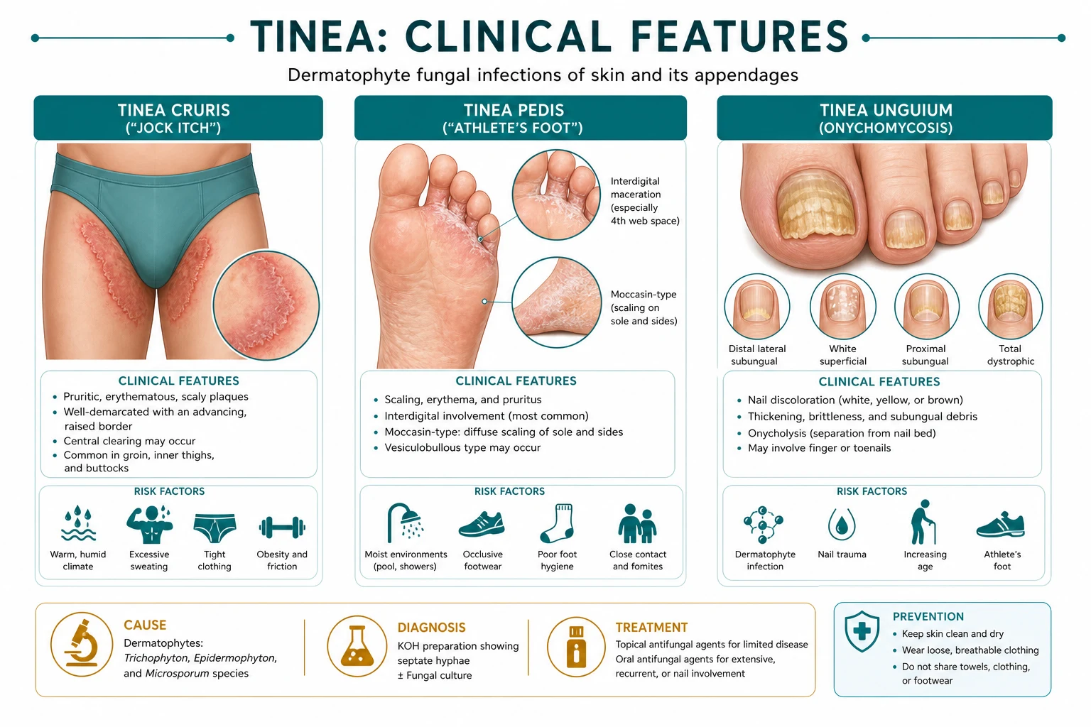
Tinea pedis (athlete's foot) is dermatophyte infection of the feet. Tinea cruris is dermatophyte infection of the groin and upper medial thighs. Tinea unguium is dermatophyte infection of the nail plate and/or nail bed, usually synonymous with onychomycosis in this context (although onychomycosis can also be caused by non-dermatophyte moulds and Candida). Because these three sites are epidemiologically, clinically and therapeutically linked, they are best understood together as the "foot-and-shoe dermatophyte syndrome."[1][2]
Classification
Tinea pedis
- Interdigital (maceration, scaling, fissuring of toe webs; commonest).
- Moccasin / chronic hyperkeratotic (diffuse silvery plantar scaling, sides of feet).
- Vesicobullous / inflammatory (pruritic vesicles and bullae, often on the arch or instep).
- Acute ulcerative (painful, macerated, malodorous erosions, usually in the lateral web spaces).
Tinea cruris
- Symmetrical, sharply marginated erythematous plaques in groin and upper inner thighs.
- Classically spares the scrotum and penis (unlike candidal intertrigo).
- Coexists with tinea pedis and onychomycosis in most cases.
Tinea unguium (onychomycosis)
- Distal-lateral subungual (DLSO) — commonest, starts under the distal or lateral nail bed.
- Superficial white (WSO) — chalky white patches on the nail plate surface.
- Proximal subungual (PSO) — rare, proximal nail fold invasion; think immunocompromise.
- Total dystrophic (TDO) — entire nail destroyed, end-stage of any pattern.
Epidemiology & Risk Factors
Tinea pedis is one of the commonest skin infections worldwide, affecting roughly 10 to 20 percent of adults at any given time and up to 70 percent of people during their lifetime. Onychomycosis is even more prevalent with advancing age, reaching over 20 percent in adults over 60 years and up to 50 percent in those over 70. Tinea cruris is far less common in isolation but coexists with tinea pedis in the majority of cases.[11][3]
Risk factors cluster around moisture, occlusion, trauma and host susceptibility. Occlusive footwear traps heat and sweat; the foot inside a shoe may reach high humidity for many hours each day. Hyperhidrosis, communal showers, swimming pools, gymnasiums, military barracks, hostels and locker rooms promote acquisition. Walking barefoot on contaminated floors is the classic route, which is why athletes, swimmers, miners, soldiers, industrial workers and schoolchildren in communal housing have high rates. Obesity increases intertriginous disease and maceration in the groin. Diabetes mellitus, peripheral vascular disease and immunosuppression increase severity, recurrence and the risk of secondary bacterial cellulitis. Family history, atopy and impaired skin-barrier function are additional host factors. Once nail disease is established, the nail plate becomes a durable reservoir that re-seeds the skin and makes cure difficult. [1][12]
Pathophysiology
The dermatophyte conidia or hyphal fragments adhere to the stratum corneum and germinate. They then secrete a family of proteolytic enzymes, including subtilisin-like proteases and metalloproteases, that degrade keratin into assimilable amino acids. The organisms grow within and between corneocytes, advancing centrifugally and producing the advancing scaly border. Inflammation is triggered by cell-wall mannans, chitin and fungal metabolites, and is partly modulated by host immunity. Acute inflammatory responses produce vesicles and bullae; chronic low-grade infection produces scaling with little erythema. The metabolic products of dermatophytes also have local irritant effects, and the superficial proteases can themselves trigger keratinocyte cytokine release, explaining the pruritus and skin-barrier disruption. In some individuals, circulating fungal antigens elicit a distant id reaction. [10]
Trichophyton rubrum is the dominant species worldwide and is responsible for most chronic tinea pedis, tinea cruris and onychomycosis. Trichophyton mentagrophytes and Trichophyton interdigitale are more commonly associated with vesicular, inflammatory, interdigital and zoonotic (animal-acquired) disease. Epidermophyton floccosum is an important cause of tinea cruris and tinea pedis, especially in shared-sport settings. Trichophyton indotineae is an emerging species of the T. mentagrophytes complex, previously known as genotype VIII, now recognised as a distinct, rapidly transmissible, antifungal-resistant pathogen that has swept across South Asia and is spreading globally.[17][16]
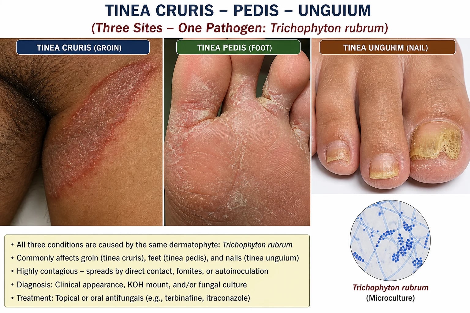
The foot-and-shoe environment creates a perfect storm: sweat raises humidity, occlusion prevents evaporation, and friction macerates the stratum corneum. The interdigital fissures that result are not only a portal for dermatophytes but also for bacterial pathogens such as Staphylococcus aureus and beta-haemolytic streptococci. This is why interdigital tinea pedis is the single most common antecedent of recurrent lower-limb cellulitis in older adults and diabetics. The nail plate, once invaded, acts as a protected fungal reservoir because topical agents and immune cells cannot penetrate well, and systemic agents must be taken for months until the nail grows out.[2][10]
Mechanism of terbinafine resistance in T. indotineae
Terbinafine inhibits squalene epoxidase (SQLE), the enzyme that converts squalene to lanosterol in the ergosterol synthesis pathway. In T. indotineae, point mutations in the SQLE gene reduce drug binding. The most commonly described mutation is Phe397Leu, with other substitutions reported. The result is high minimum inhibitory concentrations (MICs) to terbinafine and, in many isolates, reduced susceptibility to fluconazole, griseofulvin and other azoles. Itraconazole and voriconazole may retain activity, but clinical breakpoints have not been established, making antifungal susceptibility testing difficult to interpret.[17][16]
Clinical Presentation
Tinea pedis
The four recognised patterns are not mutually exclusive; patients often have overlap. [1]
Interdigital tinea pedis is the commonest form. It produces maceration, scaling, fissuring and erythema, most often in the 3rd to 4th and 4th to 5th toe webs. Itching is variable; some patients complain more of soreness from the fissures. The lateral web spaces, especially between the 4th and 5th toes, are preferentially affected because they are the deepest and most occluded. A central bacterial superinfection can produce malodour, maceration and pain, and the interdigital fissure becomes the portal through which skin flora invade the dermis. This is the pattern that most often predisposes to recurrent cellulitis. [1]
Moccasin-type tinea pedis is a chronic hyperkeratotic pattern. It presents as fine, diffuse, silvery-white scaling on the plantar surface, the heel and the sides of the feet, extending up to the malleoli in a "moccasin" distribution. The arch is often spared. It is usually bilateral, may be minimally itchy, and is frequently associated with onychomycosis. The scale is thick and recurrent, and treatment requires longer courses than interdigital disease. Patients may dismiss it as dry skin and delay presentation for years. [1][10]
Vesicobullous or inflammatory tinea pedis is an acute, intensely pruritic eruption of vesicles and bullae, usually on the instep, arch or medial foot. It can be confused with pompholyx, contact dermatitis or bullous impetigo. Ruptured vesicles predispose to secondary bacterial infection. A dermatophytid (id) reaction may appear as vesicles on the fingers, palms or elsewhere in response to fungal antigens at the primary site. The id reaction is sterile, so KOH of the distant lesions is negative; the clue is the active foot infection. [15]
Acute ulcerative tinea pedis is the most severe pattern. It presents with painful, malodorous, macerated erosions and ulcers, often in the lateral toe webs, with surrounding erythema and oedema. It is strongly associated with bacterial infection, lymphangitis and cellulitis, especially in diabetes, peripheral vascular disease and immunosuppression. It is a dermatological emergency in the compromised host. [1]
Tinea cruris
Tinea cruris is usually symmetrical, with a well-demarcated, erythematous, scaly, advancing border and central clearing. The inguinal folds and upper medial thighs are affected, and the rash may extend onto the buttocks or lower abdomen. The scrotum and penis are characteristically spared, a key distinction from candidal intertrigo, which often involves the scrotum and produces satellite pustules. Almost all patients with tinea cruris have coexisting tinea pedis or onychomycosis, because the groin is inoculated from the feet when the patient puts on underclothes. The patient may complain of itch, burning, or odour.[1][15]
Onychomycosis (tinea unguium)
Onychomycosis is usually a slow, chronic process. The nail becomes thickened, discoloured (yellow, white, brown or black), brittle and dystrophic, with subungual hyperkeratosis and onycholysis. Toenails are involved more often than fingernails because they grow more slowly, are occluded by footwear, and are more exposed to trauma and fungal contamination. Fingernail disease often occurs in the context of chronic hand wet-work, immunosuppression, or as part of the two feet-one hand syndrome. The diagnosis should be confirmed before committing to months of systemic therapy, because up to half of dystrophic nails are not fungal. The cosmetic and functional burden is significant: thickened nails can press on shoes, cause pain, and act as a source of reinfection for the skin. [2][3]
The four patterns are clinically important: [1]
- Distal-lateral subungual onychomycosis (DLSO) is the commonest. Infection starts at the distal and/or lateral hyponychium and spreads proximally under the nail bed. The nail plate thickens, turns yellow-white, and separates from the bed (onycholysis). Subungual debris accumulates.
- Superficial white onychomycosis (SWO) is caused most often by T. mentagrophytes or T. interdigitale. It appears as chalky white, sharply outlined patches on the dorsal nail plate that can be scraped off. It is more common in HIV infection and toenails, and may progress to total dystrophy.
- Proximal subungual onychomycosis (PSO) is rare. The fungus enters through the proximal nail fold, producing leukonychia that spreads distally. It is strongly associated with immunosuppression, including HIV, and should prompt HIV testing if no other cause is apparent.
- Total dystrophic onychomycosis (TDO) is the end-stage, with complete destruction and thickening of the entire nail plate and nail bed. It is usually the result of long-standing DLSO or SWO.[4][7]
The "two feet, one hand" sign
Unilateral palmar scaling and erythema (tinea manuum) accompanying bilateral tinea pedis and often onychomycosis is the classic "two feet, one hand" syndrome. The hand is infected by repeated scratching or drying of the feet with one hand. This is a high-yield clinical clue: the hand is the sentinel, but the foot is the reservoir. Treating only the hand will fail; the foot and nails must be treated simultaneously. The patient often finds the hand rash puzzling because it is unilateral; asking about foot symptoms and examining the feet and nails usually resolves the mystery. KOH of the hand and foot will confirm the diagnosis. [13][15]
Differential Diagnosis
Tinea pedis vs erythrasma
- Erythrasma is caused by Corynebacterium minutissimum and fluoresces coral-red under Wood's lamp (coproporphyrin III).
- Tinea pedis does not fluoresce; KOH shows branching septate hyphae.
Tinea pedis vs contact dermatitis / eczema
- Contact dermatitis often follows new footwear or topical products; vesicles are often more acute, and distribution matches the allergen.
- Atopic dermatitis has a history of atopy, flexural distribution, and may have lichenification; KOH is negative.
Tinea cruris vs candidal intertrigo
- Candidiasis involves the scrotum and penis and shows satellite pustules; KOH shows budding yeasts and pseudohyphae.
- Tinea cruris spares the scrotum and has a well-defined scaly border; KOH shows hyphae.
Onychomycosis vs psoriasis / trauma / melanoma
- Psoriatic nails show pitting, oil-drop changes, onycholysis with salmon patches, and usually have skin or scalp psoriasis.
- Trauma causes longitudinal ridging or onycholysis limited to the injured nail.
- Subungual melanoma presents as longitudinal pigmented streaks with Hutchinson sign (periungual spread) and must be biopsied.
Other important mimics include pitted keratolysis for moccasin tinea pedis (pitted pits on the soles, caused by Corynebacterium, Kayser-Fleischer-like odour), juvenile plantar dermatosis in children (shiny, symmetrical erythema and fissuring of the weight-bearing forefoot, KOH negative), dyshidrotic eczema for vesicular tinea pedis (tapioca-like vesicles on the sides of the fingers and feet, negative KOH), and bacterial cellulitis which can coexist with or be triggered by interdigital tinea. For nails, lichen planus, yellow nail syndrome, chronic paronychia, and squamous cell carcinoma of the nail unit are additional considerations. Psoriatic nails may show pitting, oil-drop changes, and onycholysis with salmon patches; if the skin findings are absent or the nail is single and atypical, confirm with KOH, PAS or biopsy. Pigmented or single-nail dystrophy should always be biopsied to exclude malignancy. [1][2][15]
Clinical & Bedside Assessment
The examination should be deliberate and complete. Inspect the toe webs, especially the lateral webs, for maceration, fissuring and scale. Examine the plantar surface, heels and sides of the feet for moccasin-type scaling. Look for vesicles, bullae, pustules and erosions. Check the nails for thickening, discolouration, onycholysis and subungual debris. Always examine the groin, even if the patient does not volunteer symptoms, because tinea cruris often coexists with foot disease. Palpate the foot for warmth, tenderness and lymphadenopathy, which point to secondary infection. Ask the patient to walk: a limp, guarded gait, or inability to bear weight suggests deeper soft-tissue involvement.[15]
Wood's lamp examination is useful only for specific questions. Most dermatophytes do not fluoresce. Erythrasma, however, classically shows a coral-red fluorescence in the groin or toe webs due to coproporphyrin III produced by Corynebacterium minutissimum. A negative Wood's lamp does not exclude tinea. Look for the Hutchinson sign in pigmented nail streaks: pigment extending onto the proximal or lateral nail fold strongly suggests subungual melanoma and warrants biopsy. In onychomycosis, a positive "scrape test" with a curette yielding crumbly chalky debris is a useful bedside clue, and the debris should be sent for KOH and culture.[15]
Investigations
The diagnosis of dermatophyte infection can usually be confirmed at the bedside or clinic with rapid, inexpensive tests. [1]
Potassium hydroxide (KOH) wet mount remains the cornerstone. Scrapings from the advancing edge of a skin lesion, the floor of a vesicle, or subungual debris are placed on a slide with 10 to 20 percent KOH, gently heated, and examined microscopically. Dermatophytes show branching, septate hyphae. Candida shows budding yeasts and pseudohyphae. The sensitivity of KOH for tinea pedis is reasonable, but for onychomycosis it can be as low as 50 to 70 percent because nail sampling is difficult and fungal elements may be scant. Taking samples from the most proximal diseased nail, the subungual debris, and the nail bed improves yield. [1]
Fungal culture confirms the species and allows susceptibility testing. It is slower, taking 2 to 6 weeks, but is essential when the diagnosis is uncertain, the disease is severe or refractory, or when T. indotineae is suspected. Culture is more sensitive than KOH but requires proper collection and transport. Nail samples should include nail clippings and subungual debris scraped from the most proximal affected area. Use a curette or scalpel to collect the crumbly material from beneath the nail plate; clean the nail surface first with alcohol to remove surface contaminants. For suspected T. indotineae, molecular identification by internal transcribed spacer (ITS) sequencing is required because it is morphologically indistinguishable from other T. mentagrophytes complex species. [1]
Periodic acid-Schiff (PAS) stain of nail clippings is the most sensitive single test for onychomycosis, with sensitivities generally reported above 70 to 80 percent and higher than KOH. It stains fungal cell walls magenta and is faster than culture. It is the preferred test when a quick, objective confirmation is needed before committing a patient to long systemic therapy. A negative PAS does not fully exclude disease, but it should make the clinician reconsider the diagnosis and biopsy if the presentation is atypical. [1]
Dermoscopy can help in onychomycosis (jagged proximal edge, longitudinal striae, subungual hyperkeratosis) and may help distinguish onychomycosis from other nail dystrophies. In suspected T. indotineae infection, antifungal susceptibility testing (AFST) with SQLE mutational analysis can guide therapy, but clinical breakpoints are not established, so results must be interpreted cautiously and with expert mycology support. [1]
Management — Resuscitation
Although dermatophyte infection is usually chronic and indolent, acute complications require urgent action. The resuscitation equivalent in this topic is acute ulcerative tinea pedis with secondary bacterial infection and spreading cellulitis. [1]
- Assess for systemic toxicity, fever, lymphangitic streaking, crepitus and bullae. Check vital signs and blood glucose in diabetics.
- Elevate the limb, mark the erythema margin with a pen, and obtain wound cultures if purulence is present.
- Start empiric antistaphylococcal and antistreptococcal antibiotics. For mild-moderate disease, oral flucloxacillin or cephalexin are appropriate. For penicillin allergy, clindamycin is an option. For systemic toxicity, intravenous therapy is needed.
- Treat the fungal portal simultaneously with topical or oral antifungal therapy; without this, bacterial cellulitis will recur.
- In diabetes or peripheral vascular disease, involve the diabetic foot or vascular team early; these patients can deteriorate rapidly into deep soft-tissue infection or ulceration.
- Urgent imaging (MRI or ultrasound) is indicated if deep-space infection, abscess or necrotising fasciitis is suspected. Surgical debridement may be required. [1]
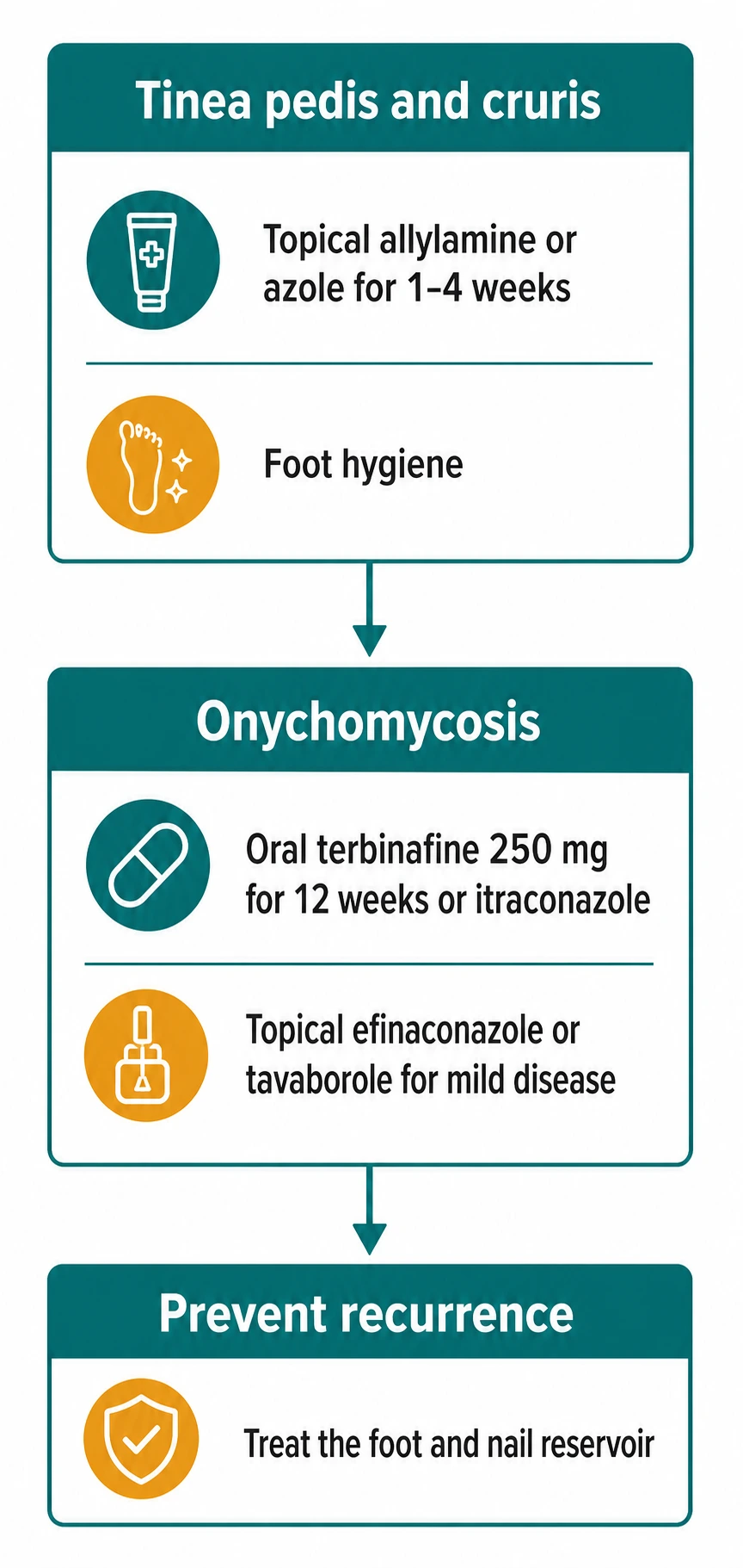
The same principle applies to recurrent lower-limb cellulitis without an obvious skin break: always look for interdigital tinea pedis and onychomycosis as the silent portal. Treating the fungal reservoir has been shown to reduce recurrent cellulitis episodes in observational studies and is a cost-effective intervention. [1]
Management — Definitive & Stepwise
Skin disease: tinea pedis and tinea cruris
For limited skin disease, topical therapy is first-line. Allylamines are preferred over azoles because they are fungicidal and have shorter courses. [1]
Topical terbinafine 1% cream or gel: apply once or twice daily for 1 week for interdigital tinea pedis and for 1 to 2 weeks for tinea cruris. Moccasin-type tinea pedis often needs 2 to 4 weeks, and some patients require longer or oral therapy because the scale is thick. The allylamine class works by inhibiting squalene epoxidase, leading to squalene accumulation and fungal cell death. [1]
- Topical naftifine, butenafine, ciclopirox olamine or azoles such as clotrimazole, ketoconazole, miconazole and econazole are effective alternatives. Azole creams are usually applied twice daily for 2 to 4 weeks. The azoles inhibit fungal CYP450-dependent lanosterol 14-alpha-demethylase, blocking ergosterol synthesis and increasing toxic membrane sterols.
- Continue treatment for at least 1 week after clinical clearance to reduce recurrence.
- Treat the foot disease when treating the groin; otherwise tinea cruris will relapse. [1]
Systemic therapy for skin disease is reserved for extensive or refractory moccasin tinea pedis, recurrent tinea cruris, or disease that fails topical therapy. Oral terbinafine 250 mg daily for 2 to 4 weeks or itraconazole 200 mg daily for 1 week per month (pulse) or continuously for 2 to 4 weeks are standard options. For T. indotineae skin disease, itraconazole 200 mg daily for 1 to 12 weeks or voriconazole may be preferred, but susceptibility should be guided by local resistance patterns and expert advice. [16][17]
Nail disease: onychomycosis
Onychomycosis almost always requires systemic therapy because topical agents cannot penetrate the nail plate adequately. Exceptions are mild, distal disease, limited number of nails, or patients who cannot take systemic therapy. [1]
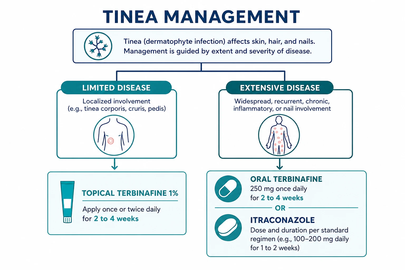
The following table summarises the drug, dose, route, duration and rationale for the common agents used in skin and nail dermatophyte infection. Each entry is a high-yield exam fact. [1]
| Agent | Route | Indication | Dose and duration | Rationale and caveats |
|---|---|---|---|---|
| Terbinafine 1% | Topical | Tinea pedis/cruris | Once or twice daily for 1–4 weeks | Fungicidal allylamine; shorter course than azoles. |
| Clotrimazole 1% | Topical | Tinea pedis/cruris | Twice daily for 2–4 weeks | Azole; widely available and inexpensive. |
| Ciclopirox 8% lacquer | Topical nail | Mild onychomycosis | Daily for up to 48 weeks | Poor nail penetration; lower cure rates. |
| Efinaconazole 10% solution | Topical nail | Mild distal onychomycosis | Once daily for 48 weeks | Better nail penetration than lacquers. |
| Tavaborole 5% solution | Topical nail | Mild distal onychomycosis | Once daily for 48 weeks | Oxaborole antifungal; modest efficacy. |
| Terbinafine 250 mg | Oral | Onychomycosis | 6 weeks fingernails, 12 weeks toenails | First-line; fungicidal; best network meta-analysis evidence. |
| Itraconazole 200 mg | Oral | Onychomycosis | Continuous 200 mg daily for 12 weeks, or pulse 200 mg BD for 1 week per month | Alternative; watch CYP3A4 and heart failure. |
| Fluconazole 150–300 mg | Oral | Onychomycosis | Once weekly for 6–12 months | Alternative; generally less effective. |
| Griseofulvin | Oral | Tinea capitis in children | 10–20 mg/kg/day for 6–8 weeks | Not for nails or T. indotineae. |
Oral terbinafine 250 mg once daily is the first-line treatment for toenail and fingernail onychomycosis caused by dermatophytes. The recommended duration is 6 weeks for fingernails and 12 weeks for toenails. It is fungicidal, well absorbed, and has the best evidence for mycological cure and cost-effectiveness in network meta-analyses. Baseline liver function tests are recommended, and therapy should be avoided in active liver disease or during pregnancy. Periodic monitoring of liver function is prudent in patients with risk factors. Terbinafine is generally well tolerated; gastrointestinal upset, rash and taste disturbance are the commonest adverse effects. Rarely, it causes hepatotoxicity, neutropenia, or severe skin reactions. [1]
Oral itraconazole is the main alternative. It can be given as continuous therapy (200 mg daily for 12 weeks for toenails) or as pulse therapy (200 mg twice daily for 1 week per month; two pulses for fingernails, three or four pulses for toenails). Itraconazole is fungistatic, requires gastric acid for absorption, has many CYP3A4-mediated drug interactions, and is contraindicated in heart failure with reduced ejection fraction. It is preferred when the causative organism is a non-dermatophyte mould such as Scopulariopsis brevicaulis or Aspergillus, or when terbinafine is contraindicated. In areas with T. indotineae, itraconazole may be more effective than terbinafine, although resistance is also emerging. [1]
Oral fluconazole (150 to 300 mg once weekly for 6 to 12 months) is an alternative but is generally less effective than terbinafine or itraconazole for dermatophyte nails. Griseofulvin was historically the first oral antifungal but is now largely reserved for tinea capitis in children because it requires very long courses and is less effective for nails and chronic foot disease. It is not effective against T. indotineae. [1]
Topical nail solutions are useful for mild disease or when systemic therapy is contraindicated. Efinaconazole 10% topical solution is applied once daily for 48 weeks. Tavaborole 5% topical solution is applied once daily for 48 weeks. Ciclopirox 8% lacquer is applied daily with periodic removal of lacquer build-up; it has lower cure rates and requires longer therapy. Amorolfine 5% lacquer is used in some countries once or twice weekly. None of these penetrate total dystrophic or matrix disease well, and their efficacy is substantially lower than oral therapy. Cost and adherence are major barriers: a patient using a topical nail solution for 48 weeks must be counselled that success is modest and the application is daily. [1]
Adjunctive measures improve outcomes: thin the thickened nail plate with urea 40% ointment under occlusion, reduce onycholytic nail, and keep the nail short. Treat concomitant tinea pedis aggressively. The foot is the source; clearing the nail without clearing the skin is likely to fail. For patients with very thick nails, podiatric debridement improves drug penetration and reduces discomfort. [2]
Hygiene, footwear and public health
Recurrence is the norm unless the reservoir is controlled. Advice must be practical and specific. Keep the feet dry: dry carefully between the toes after bathing, using a separate towel for the feet and groin. Change socks daily, preferably twice daily if feet sweat; choose cotton or moisture-wicking socks. Wear breathable footwear such as leather or mesh and rotate shoes so each pair dries completely before being worn again; consider antifungal powder or spray inside shoes. Avoid walking barefoot in communal areas; use flip-flops in gyms, pools and hostel showers. Do not share towels, nail clippers, socks or shoes. Treat household members with tinea pedis simultaneously if transmission is occurring. Wash socks, towels and bed linen at 50 degrees Celsius or hotter and heat-dry if possible. For athletes, military personnel and industrial workers, ensure boots are well ventilated and that feet are inspected regularly. For patients with recurrent disease, treating the shoes with antifungal powder or ultraviolet shoe sanitizers may help, although evidence for shoe decontamination alone is limited. [1]
At the population level, antifungal stewardship is becoming urgent. The widespread over-the-counter availability of topical corticosteroid-antifungal combinations in some countries has contributed to modified, atypical and resistant dermatophytosis. Public education, appropriate prescribing, and access to fungal culture and molecular diagnostics are essential to slow the spread of T. indotineae and other resistant species. [2][12]
Specific Subtypes & Scenarios
Interdigital tinea pedis
Treat with topical allylamine (terbinafine or naftifine) for 1 to 2 weeks. If maceration is severe, add an astringent soak (e.g., dilute aluminium acetate or potassium permanganate) to dry the web spaces, then apply antifungal cream. If there is bacterial superinfection, add oral antistaphylococcal antibiotics. In diabetics, treat early and monitor closely because the web space is a cellulitis portal.[1]
Moccasin-type tinea pedis
Topical therapy often fails unless the hyperkeratosis is addressed. Start with keratolytic agents such as urea 20 to 40% cream, salicylic acid preparations, or lactic acid, then apply topical terbinafine. If disease is extensive or unresponsive, use oral terbinafine 250 mg daily for 2 to 4 weeks. Because nails are often involved, look for and treat onychomycosis.[1][15]
Vesicobullous tinea pedis
Cool compresses, aluminium acetate soaks, and topical or oral antifungal therapy are used. Topical corticosteroids are not appropriate as monotherapy because they can mask the infection. A short course of systemic corticosteroid may be considered for severe inflammatory dermatophytid reactions, but only together with antifungal therapy. Secondary bacterial infection is common and should be treated.[15]
Acute ulcerative tinea pedis
This is the emergency pattern. Cleanse the web spaces, culture any purulence, start systemic antibiotics for suspected bacterial superinfection, and add oral or topical antifungal therapy. Elevation, offloading, and diabetic or vascular review are essential. Hospitalisation is needed if there is systemic toxicity, deep soft-tissue infection, or critical limb ischaemia.[1]
Distal-lateral subungual onychomycosis
First-line is oral terbinafine 250 mg daily for 12 weeks (toenails) or 6 weeks (fingernails). Debride the nail before or during therapy. If there is matrix involvement, total dystrophy, or failure after a full course, consider itraconazole pulse or combination topical plus systemic therapy, and rule out non-dermatophyte moulds or T. indotineae by culture and molecular testing.[7][14]
Superficial white onychomycosis
T. mentagrophytes or T. interdigitale are common causes. Scrape the white patches and send for KOH/culture. Treatment may be topical (efinaconazole, tavaborole, ciclopirox) if the disease is limited to the surface, but oral terbinafine is often needed if the disease is extensive or involves the nail matrix. In HIV-positive patients, evaluate immune status.[4]
Proximal subungual onychomycosis
PSO is rare and strongly associated with immunosuppression. It is a red flag for HIV, transplant status, or other immunodeficiency. Oral itraconazole may be preferred if the organism is not a dermatophyte, but dermatophyte PSO is treated with systemic terbinafine or itraconazole. Always investigate the underlying cause.[4]
Total dystrophic onychomycosis
TDO is end-stage disease. Complete cure is difficult and may require prolonged systemic therapy, mechanical or chemical nail avulsion, and sometimes permanent nail removal. Amorolfine or ciclopirox lacquer alone is rarely adequate. Set realistic expectations; patients should understand that even after successful therapy, the nail may take 12 to 18 months to look normal.[2][7]
Majocchi granuloma
A deep follicular dermatophyte infection, usually of the legs, caused by trauma that inoculates dermatophytes into the dermis or hair follicles. It presents as perifollicular papules, pustules, nodules or plaques. It does not respond to topical therapy alone; oral terbinafine or itraconazole for 4 to 8 weeks is required. It is more common in immunosuppressed women who shave their legs.[15]
Complications & Pitfalls
The most important complication is secondary bacterial infection. Interdigital fissures and onychomycosis act as portals for Staphylococcus aureus and beta-haemolytic streptococci, leading to cellulitis, erysipelas, lymphangitis, and, in diabetics, foot ulceration and amputation. The risk is substantially reduced by treating the foot reservoir. Recurrent cellulitis of the lower limb should always trigger examination of the feet and nails. In patients with two or more episodes of cellulitis per year, dermatologists and infectious diseases physicians increasingly recommend screening for and treating tinea pedis and onychomycosis as part of the prevention strategy. [1]
Tinea incognito is dermatophyte infection modified by topical or systemic corticosteroids. The typical well-defined scaly border is lost, the rash becomes widespread, atypical and less itchy, and diagnosis is delayed. KOH and fungal culture may be falsely negative. This is a common pitfall in misdiagnosed groin rashes and "treatment-resistant eczema," especially in regions where over-the-counter corticosteroid-antifungal combination creams are used. Management is to stop steroids and treat with systemic antifungal therapy. [15]
Dermatophytid reaction is a sterile, immunologically mediated eruption distant from the primary infection, often vesicles or papules on the hands, fingers or trunk. It resolves when the primary infection is treated. It must be distinguished from a true secondary infection or drug eruption. The id reaction is a clue that the primary dermatophyte infection is active, not a separate disease. [1]
Recurrence of onychomycosis is common, reported in 20 to 50 percent of patients after successful therapy. Factors include failure to treat the foot reservoir, reinfection from contaminated footwear or household contacts, advanced age, poor circulation, diabetes, immunosuppression, and incomplete nail penetration of drug. Setting expectations and reinforcing hygiene are essential. Some patients require maintenance therapy or periodic pulse therapy, although evidence for long-term suppressive regimens is limited. [2]
Pitfalls in immunocompromised patients include atypical presentations, deeper invasion (Majocchi granuloma), and the need for longer or higher-dose therapy. In patients with extensive, inflammatory, treatment-resistant disease, especially with recent travel to South Asia, T. indotineae must be suspected. Misdiagnosis leads to months of ineffective terbinafine, onward transmission to family members, and worsening inflammatory disease. [17][16]
Prognosis & Disposition
With appropriate therapy, the prognosis for skin disease is excellent. Interdigital tinea pedis usually clears within 1 to 2 weeks of topical therapy. Tinea cruris responds similarly. Moccasin disease may take 4 to 6 weeks and may require oral therapy. Onychomycosis is curable but slow; mycological cure rates with oral terbinafine are roughly 70 to 80 percent for fingernails and 60 to 75 percent for toenails, with complete clinical cure taking 12 to 18 months because of slow nail growth. The big toe nail grows at approximately 1 to 1.5 mm per month, while fingernails grow at 3 to 4 mm per month, which explains why toenail treatment takes longer. [1]
Recurrence is the main long-term problem. A patient with onychomycosis can be considered "cured" when the nail has grown out and repeat KOH and culture are negative. Until then, the foot must remain under preventive care. Disposition depends on severity: uncomplicated skin disease can be managed in primary care; extensive or refractory disease, total dystrophic onychomycosis, suspected T. indotineae, diabetic foot complications, or recurrent cellulitis warrant dermatology, podiatry or infectious diseases referral. In resource-limited settings, treatment may be constrained by the cost of systemic therapy and the need for monitoring; prioritise patients with complications, diabetes, immunosuppression, or recurrent cellulitis. [2]
Special Populations
Pregnancy and lactation. Topical azoles and allylamines are generally considered safe in pregnancy because systemic absorption is minimal. The amount of drug reaching the systemic circulation after topical application to limited skin areas is very low. Oral terbinafine and itraconazole are avoided in pregnancy unless absolutely necessary. Griseofulvin is contraindicated in pregnancy. During lactation, topical therapy is preferred; oral agents should be used cautiously and only if clearly indicated, with discussion of the limited data on excretion into breast milk. [15]
Children. Tinea pedis and cruris are less common in pre-pubertal children but occur, particularly in those who participate in swimming or communal sports. Tinea capitis is more common in childhood than tinea pedis. Topical therapy is preferred. Griseofulvin is the traditional oral agent for tinea capitis in children; oral terbinafine is also effective and increasingly used. For onychomycosis in children, oral terbinafine is effective but dosing should be weight-based and specialist-led. The nail grows faster in children, which may improve outcomes. [15]
Elderly. Onychomycosis is more prevalent and more difficult to treat because of slower nail growth, impaired circulation, diabetes and drug interactions. Drug-drug interactions with itraconazole are a major concern in elderly patients on polypharmacy. Terbinafine is usually preferred if there is no liver disease. Falls and infection risk should be considered. Recurrence is common, and maintenance may be unrealistic. [7]
Immunocompromised. HIV, transplant recipients, biologic therapy, long-term corticosteroids and diabetes predispose to more extensive, atypical and resistant disease. PSO and T. indotineae should be considered. Systemic therapy is often needed, and longer courses may be required. Monitoring for drug interactions and hepatotoxicity is essential. In HIV infection, superficial white onychomycosis and proximal subungual onychomycosis are red flags for advanced immunosuppression. [4][17]
Diabetes. Diabetes is the single most important risk factor for complications. Interdigital tinea pedis and onychomycosis are portals for cellulitis and limb-threatening infection. Active treatment of both skin and nail disease is recommended, with close podiatric follow-up. If oral therapy is needed, liver function should be monitored and drug interactions reviewed. The diabetic foot team should be involved if there is any ulceration, neuropathy, or peripheral vascular disease. [2]
Athletes. Tinea pedis and onychomycosis are common in athletes due to occlusive footwear, communal facilities, and repetitive trauma. Prevalence is particularly high in swimmers, runners, and team-sport players. Prevention through footwear hygiene, drying between toes, and avoiding shared towels is central to management. Treating onychomycosis improves foot comfort and reduces bacterial superinfection. In high-risk sports, prophylactic antifungal powder may be useful. [12]
Evidence, Guidelines & Regional Differences
Network meta-analyses consistently support oral terbinafine as the most effective monotherapy for dermatophyte toenail onychomycosis, with superior mycological cure rates and lower recurrence compared with itraconazole, fluconazole and griseofulvin. A 2020 network meta-analysis by Gupta and colleagues found terbinafine 250 mg daily for 12 weeks ranked highest for complete cure and mycological cure, followed by itraconazole pulse and continuous regimens. Itraconazole pulse is a reasonable alternative with better tolerability in some patients but requires careful monitoring of drug interactions and heart failure risk. Topical efinaconazole and tavaborole have lower efficacy than oral agents but offer a safer option when systemic therapy is contraindicated.[5][6][9]
Regional differences are increasingly important. In South Asia, the Middle East and increasingly in Europe and North America, T. indotineae has caused epidemics of extensive, terbinafine-resistant dermatophytosis. The first reports came from India in the mid-2010s, where the combination of high humidity, overcrowding, over-the-counter use of topical corticosteroid combinations, and inappropriate antifungal prescribing created a selective environment. In these regions, clinicians may need to start with itraconazole, use combination therapy, or seek expert mycology advice and susceptibility testing. Travel history, previous treatment failure, widespread inflammatory disease, and involvement of the genitalia or trunk are red flags. Antifungal stewardship — avoiding unnecessary topical corticosteroid combinations, limiting empiric terbinafine when resistance is suspected, treating contacts, and discouraging sharing of towels and clothing — is now a global priority.[17][16]
Guideline bodies including the Infectious Diseases Society of America (IDSA) and European dermatology groups recommend confirming onychomycosis before systemic therapy, using oral terbinafine first-line for dermatophyte disease, and considering topical agents for mild disease or poor surgical candidates. Indian and South Asian guidelines increasingly emphasise T. indotineae surveillance and alternative regimens. Cost and access vary: oral terbinafine is generally the most cost-effective systemic option, while topical nail solutions are expensive and require prolonged use. Shared decision-making should include discussion of recurrence, duration, monitoring, and cost. [7][14]
Exam Pearls
DERM
Exam application bank (NEET-PG / INICET)
One-line answer
Tinea pedis (athlete's foot), tinea cruris (jock itch) and tinea unguium/onychomycosis (nail infection) are the commonest dermatophyte infections, sharing Trichophyton rubrum and the warm, occluded foot-and-shoe ecological niche. The topic covers the interdigital, moccasin, vesicobullous and acute ulcerative patterns of tinea pedis, the scrotal-sparing groin rash of tinea cruris, the four patterns of onychomycosis, the 'two feet, one hand' sign, KOH/culture/PAS diagnosis, topical allylamine and azole therapy for skin disease, systemic terbinafine and itraconazole for nails, the emerging terbinafine-resistant Trichophyton indotineae, and the public-health importance of treating the foot and nail reservoir to prevent recurrent cellulitis and groin disease.
Worked stems (answer without another resource)
Stem 1 — Classic presentation. Map symptoms to mechanism; name the first investigation and first treatment step with dose/route if drug therapy is standard. [1]
Stem 2 — Unstable / complicated. List red flags that force immediate resuscitation, theatre, ICU, antidote, or reperfusion — and what you do in the first 15 minutes. [1]
Stem 3 — Atypical group. Elderly, pregnancy, child, or immunocompromised: how presentation and thresholds change. [1]
Stem 4 — Differential trap. Name the three closest mimics and one discriminator for each. [1]
Stem 5 — Disposition. Who goes home with safety-netting, who is admitted, who needs HDU/ICU/theatre, and what follow-up is mandatory. [1]
Rapid viva checklist
- Definition + classification
- Pathophysiology chain
- Bedside signs / criteria
- Score with exact components (if any)
- Emergency bundle
- Definitive therapy with doses
- Complications of disease and of treatment
- Special populations
- Guideline/trial name if classic
- Three exam traps
Coverage self-check
If you cannot answer any stem above from this page alone, re-read the matching section — the page is intended to be self-sufficient for final-prof and NEET-PG/INICET questions on Tinea pedis, tinea cruris and tinea unguium (onychomycosis).
[1]References
- [1]Leung AK, Barankin B, Lam JM, et al. Tinea pedis: an updated review Drugs Context, 2023.PMID 37415917
- [2]Lipner SR, Scher RK. Onychomycosis: Treatment and prevention of recurrence J Am Acad Dermatol, 2019.PMID 29959962
- [3]Gupta AK, Stec N, Summerbell RC, et al. Onychomycosis: a review J Eur Acad Dermatol Venereol, 2020.PMID 32239567
- [4]Leung AKC, Lam JM, Leong KF, et al. Onychomycosis: An Updated Review Recent Pat Inflamm Allergy Drug Discov, 2020.PMID 31738146
- [5]Gupta AK, Daigle D, Paquet M. Therapies for onychomycosis a systematic review and network meta-analysis of mycological cure J Am Podiatr Med Assoc, 2015.PMID 25032982
- [6]Gupta AK, Foley KA, Mays RR, et al. Monotherapy for toenail onychomycosis: a systematic review and network meta-analysis Br J Dermatol, 2020.PMID 31120134
- [7]Frazier WT, Santiago-Delgado ZM, Stupka KC 2nd. Onychomycosis: Rapid Evidence Review Am Fam Physician, 2021.PMID 34652111
- [8]Gupta AK, Polla Ravi S, Talukder M, et al. Effectiveness and safety of oral terbinafine for dermatophyte distal subungual onychomycosis Expert Opin Pharmacother, 2024.PMID 38221907
- [9]Yousefian F, Smythe C, Han H, et al. Treatment Options for Onychomycosis: Efficacy, Side Effects, Adherence, Financial Considerations, and Ethics J Clin Aesthet Dermatol, 2024.PMID 38495549
- [10]Leyden JL. Tinea pedis pathophysiology and treatment J Am Acad Dermatol, 1994.PMID 8077505
- [11]Havlickova B, Czaika VA, Friedrich M. Epidemiological trends in skin mycoses worldwide Mycoses, 2008.PMID 18783559
- [12]Daggett C, Brodell RT, Daniel CR, et al. Onychomycosis in Athletes Am J Clin Dermatol, 2019.PMID 31111408
- [13]Mizumoto J. Two Feet-One Hand Syndrome Cureus, 2021.PMID 35111444
- [14]Falotico JM, Lipner SR. Updated Perspectives on the Diagnosis and Management of Onychomycosis Clin Cosmet Investig Dermatol, 2022.PMID 36133401
- [15]Ely JW, Rosenfeld S, Seabury Stone M. Diagnosis and management of tinea infections Am Fam Physician, 2014.PMID 25403034
- [16]Sonego B, Corio A, Mazzoletti V, et al. Trichophyton indotineae, an Emerging Drug-Resistant Dermatophyte: A Review of the Treatment Options J Clin Med, 2024.PMID 38930086
- [17]Gupta AK, Susmita, Nguyen HC, et al. Trichophyton indotineae: Epidemiology, antifungal resistance and antifungal stewardship strategies J Eur Acad Dermatol Venereol, 2026.PMID 40613321