Dermatology · General Medicine
Tinea & Dermatophytosis (Ringworm)
Also known as Tinea · Dermatophytosis · Ringworm · Athletes foot · Tinea corporis · Tinea cruris · Tinea pedis · Tinea capitis · Tinea unguium · Tinea incognito · Kerion
Tinea (dermatophytosis, ringworm) is a superficial fungal infection of keratinised tissue by dermatophyte moulds of three genera — Trichophyton (skin, hair, nails), Microsporum (hair, skin; many fluoresce green under Wood's lamp), and Epidermophyton floccosum (skin and nails, never hair). Dermatophytes secrete keratinases that digest keratin in the stratum corneum, hair shafts, and nails; they cannot invade living tissue. The clinical lesion is the annular, erythematous, scaly plaque with a raised, advancing, scaly border (active edge) and central clearing — the ringworm pattern — and is named by site: corporis, cruris, pedis (interdigital/moccasin/vesicular), capitis (black-dot/kerion/favus), unguium (onychomycosis), manuum, faciei, barbae, and incognito (steroid-modified). The diagnosis rests on clinical morphology + KOH mount of the active border showing branching septate hyphae, supported by fungal culture on Sabouraud dextrose agar, Wood's lamp (Microsporum green hair fluorescence), dermatoscopy (comma/corkscrew hairs; Morse-code nails), and PAS/GMS stain on biopsy. Topical antifungals (clotrimazole 1% BD 4 weeks, terbinafine 1% OD one to two weeks) cure most localised skin disease; ORAL therapy is mandatory for tinea capitis, onychomycosis, extensive/recurrent/steroid-modified tinea, Majocchi granuloma, and immunosuppression. Terbinafine 250 mg OD is first-line systemic (allylamine, fungicidal, inhibits squalene epoxidase); griseofulvin remains the gold standard for Microsporum tinea capitis in children; itraconazole (pulse for nails) and fluconazole 150 mg weekly are alternatives. Kerion — boggy inflammatory tinea capitis — needs oral antifungal plus oral corticosteroid, NOT incision. Tinea incognito from misuse of fixed-dose corticosteroid-antifungal-antibacterial creams is a major Indian epidemic (AAA syndrome: Abuse, Application, Addiction), requires oral antifungal and steroid withdrawal. Recurrence is reduced by treating coexisting tinea pedis (the reservoir), washing clothing/bedding, addressing diabetes/HIV, and treating household and animal contacts.
On this page & tools
Your progress
Saved locally on this device.
Exam tags
Red flags
Overview & Definition
Tinea (dermatophytosis, ringworm) is one of the commonest skin conditions in the world — its lifetime prevalence approaches a quarter of the global population, and in tropical and subtropical regions, including India and South Asia, it has reached epidemic proportions.[10] It is caused by a specialised group of keratinophilic moulds called dermatophytes that digest keratin in the stratum corneum, hair shafts, and nails for nutrition, and is therefore confined to keratinised tissue — it does not invade living cells and does not infect mucosa (a feature that immediately separates it from candidiasis).[1]
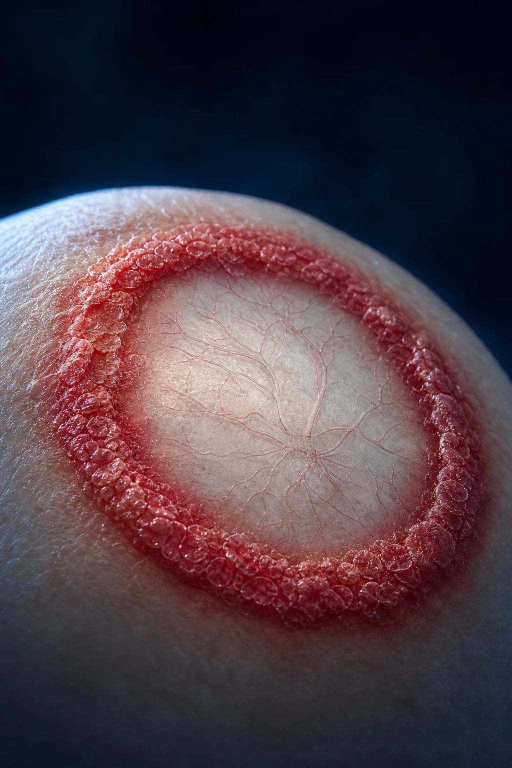
The genus dermatophyte is divided into three genera — Trichophyton, Microsporum, and Epidermophyton — distinguished by their tissue tropism and, in part, by their ecological source (anthropophilic, zoophilic, or geophilic). The clinical lesion they produce is one of the most instantly recognisable in medicine: the annular, erythematous, scaly plaque with a raised, advancing scaly border and central clearing that gives the condition its popular name, ringworm. Lesions are named by the site they occupy (tinea corporis for body, cruris for groin, pedis for feet, capitis for scalp, unguium for nails, manuum for hand, faciei for face, barbae for beard) and each site carries its own differential diagnosis, treatment duration, and prognosis.[1][2]
The two clinical skills that decide almost every tinea question — at viva, in the MCQ, and at the bedside — are confirming the diagnosis with a potassium hydroxide (KOH) mount of the active border (avoiding the catastrophic error of mistaking tinea for eczema and treating it with topical corticosteroids), and knowing when topical therapy is sufficient versus when oral antifungal therapy is mandatory. Tinea capitis and onychomycosis always require oral therapy; so do extensive, recurrent, or steroid-modified disease; so do Majocchi granuloma and immunosuppression.[2][3] Three scenarios deserve particular vigilance: the kerion (a boggy, tender, purulent inflammatory tinea capitis in a child) which is an emergency requiring oral antifungal plus oral corticosteroid — and which is never incised like an abscess; the tinea incognito that arises from misuse of fixed-dose corticosteroid-antifungal-antibacterial creams (the Indian AAA syndrome — Abuse, Application, Addiction), an epidemic that produces atypical, non-annular, infiltrative plaques that mimic eczema, psoriasis, rosacea, and lupus; and the two-feet-one-hand syndrome in which bilateral tinea pedis is the silent reservoir for a unilateral tinea manuum.[10][11][12]
Classification
Tinea is classified on two axes — the body site infected (which dictates the clinical name and the differential diagnosis) and the causative genus and ecological origin (which dictates the inflammatory response and the choice of antifungal). Both must be mastered. [1]
Classification by site
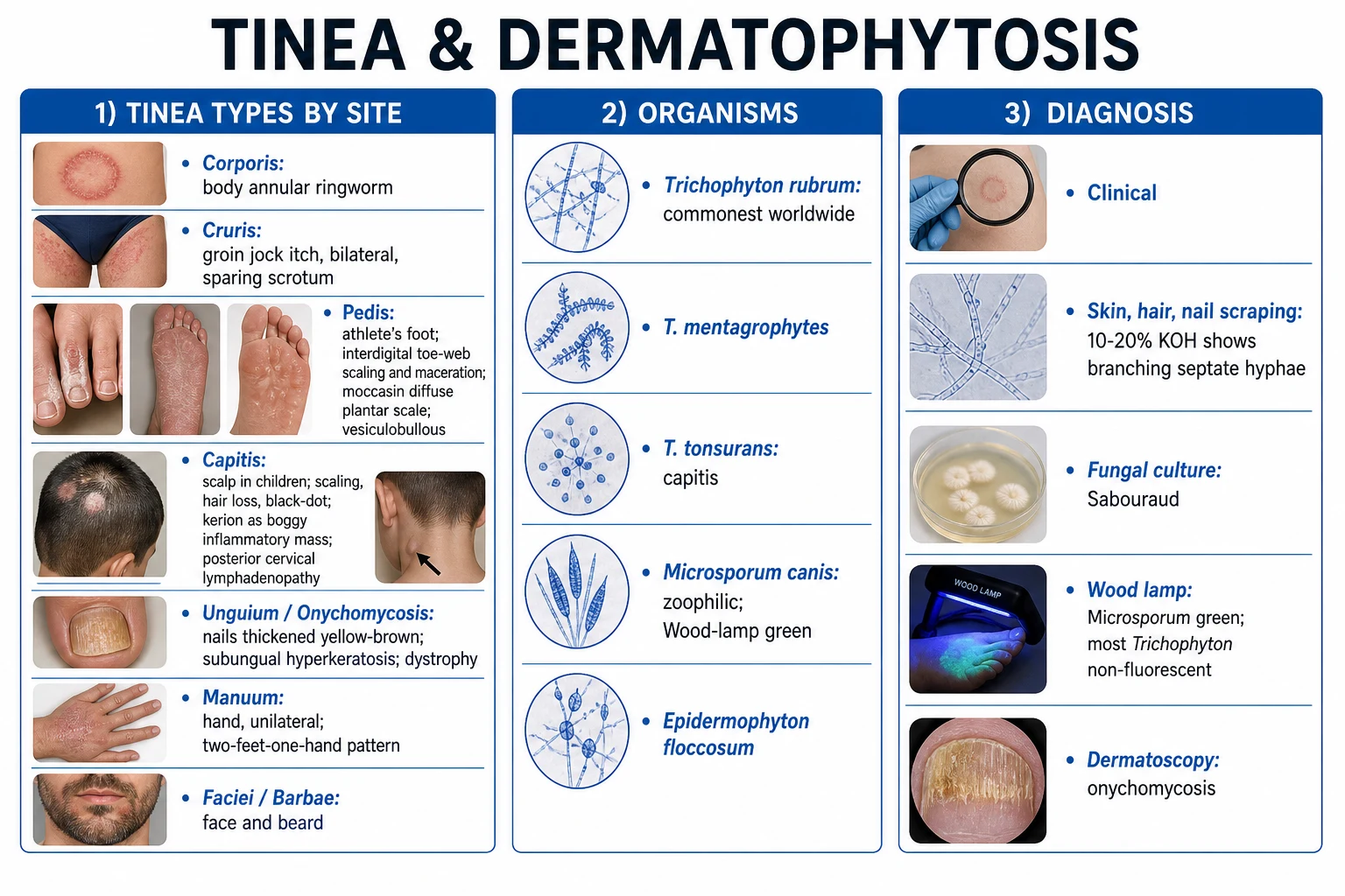
- Tinea corporis — body; the classic annular ringworm with active scaly border and central clearing.
- Tinea cruris — groin ('jock itch'); symmetric, sharply marginated, spares the scrotum.
- Tinea pedis — athlete's foot; three patterns: interdigital (toe-web scaling, maceration; commonest), moccasin (diffuse hyperkeratotic plantar scale, often T. rubrum), vesiculobullous/inflammatory (vesicles on instep, often zoophilic).
- Tinea capitis — scalp; subtypes: black-dot (endothrix Trichophyton), grey patch (ectothrix Microsporum), kerion celsi (intense boggy inflammation, usually zoophilic), favus (T. schoenleinii, scutula, permanent alopecia).
- Tinea unguium / onychomycosis — nails; patterns: distal lateral subungual (commonest), proximal subungual (think HIV), white superficial (T. mentagrophytes), endonyx, total dystrophic.
- Tinea manuum — hand; typically unilateral; classically part of two-feet-one-hand syndrome.
- Tinea faciei — face; often misdiagnosed as eczema, lupus, rosacea; KOH is mandatory.
- Tinea barbae — beard; deep inflammatory nodules from zoophilic species.
- Tinea incognito — steroid-modified tinea; atypical, non-annular, infiltrative; from misuse of topical corticosteroid combination creams. [1]
Epidemiology & Risk Factors
Tinea is among the most prevalent of all human skin conditions. Lifetime prevalence is in the range of 20 to 25 percent globally, and in tropical climates — especially India, South-East Asia, and sub-Saharan Africa — it has reached epidemic proportions in the last decade, driven by heat, humidity, dense urban living, occlusive clothing, and the catastrophic misuse of fixed-dose corticosteroid-antifungal-antibacterial creams.[10][12]
The dominant species has shifted over decades. Trichophyton rubrum is now the global workhorse, responsible for most chronic tinea pedis, onychomycosis, tinea cruris, and the two-feet-one-hand pattern; T. mentagrophytes/interdigitale is a close second and is the species driving the emerging terbinafine resistance in India (the ITS genotype VIII clone, with mutations in the squalene epoxidase gene).[12] In children, Microsporum species (M. canis from kittens and puppies, M. audouinii human-to-human) and Trichophyton tonsurans dominate tinea capitis; the species mix is regional and shifts over time.
The host and environmental risk factors cluster into environmental (heat, humidity, sweating, occlusive footwear and clothing, communal showers, swimming pools, gyms, contact sports — the term 'tinea gladiatorum' for wrestlers), host (diabetes mellitus, peripheral arterial disease, atopy, obesity, immunosuppression — HIV, transplant, chemotherapy, chronic corticosteroid), and occupational (military, athletes, miners, farmers, swimmers, animal handlers).[2][3]
Tinea capitis is predominantly a disease of prepubertal children (peak ages three to seven years) and is rare after puberty — pubertal sebum is rich in fungistatic saturated fatty acids (sapienic acid) that inhibit dermatophyte growth.[4] Animal reservoirs are the source of zoophilic species and the explanation for more inflammatory disease: Microsporum canis from kittens and puppies, Trichophyton verrucosum from cattle (kerion and inflammatory tinea barbae in farmers), T. mentagrophytes from rodents. Diabetics and immunocompromised patients are at markedly higher risk of extensive, recurrent, and atypical disease — a patient with recurrent tinea should be screened for diabetes (HbA1c) and HIV.[3]
Pathophysiology
Dermatophytes are obligately keratinophilic fungi that live in the non-living, keratinised layer of the skin (stratum corneum), the hair shaft (cortex and cuticle), and the nail plate. They cannot survive in living tissue. Their defining metabolic feature is the secretion of keratinases and other proteases (subtilisins, metalloproteases) that break keratin down into peptides and amino acids the fungus can use as nitrogen and carbon sources.[1] This single fact explains why tinea affects only skin, hair, and nails — and never mucosa, which is non-keratinised and bathed in antifungal defensins.
The three ecological classes — and why they matter clinically
Dermatophytes are classified ecologically by their natural reservoir, which predicts their inflammatory intensity and their epidemiology: [1]
- Anthropophilic (human-to-human) — e.g. Trichophyton rubrum, T. tonsurans, T. violaceum, Microsporum audouinii, Epidermophyton floccosum. These have co-evolved with humans and produce chronic, low-grade inflammation, large plaques, and high transmissibility in households and communal facilities. They are the principal drivers of recurrent tinea pedis, cruris, and onychomycosis.
- Zoophilic (animal-to-human) — e.g. Microsporum canis (dogs, cats), Trichophyton verrucosum (cattle), T. mentagrophytes (rodents), T. equinum (horses). These provoke an intense, often suppurative inflammatory response — kerion celsi, vesiculobullous tinea pedis, inflammatory tinea barbae — because the host immune system has not adapted to them. The inflammation is so intense it can self-limit the infection, but it also produces scarring alopecia in kerion.
- Geophilic (soil-dwelling) — e.g. Microsporum gypseum. Acquired from soil, rare, produces geographic, sporadic inflammatory lesions.[1][3]
Why the plaque is annular with central clearing — the immune explanation
The classic ringworm pattern is a direct readout of the host immune response. Dermatophyte antigens (mannans, proteases) trigger a T-cell mediated delayed-type hypersensitivity reaction in the skin: keratinocytes release IL-1, IL-17, IL-8, and β-defensins, recruiting neutrophils and activated T-cells that clear the fungus.[1] Clearance happens first in the centre of the plaque (where the infection is oldest and immunity is highest) and slowest at the advancing border (where hyphae are growing fastest into naive skin). The result is the annular plaque with central clearing and an active, scaly, advancing leading edge — the border is where the live hyphae are, which is why the KOH scraping is taken from the active border, not the centre.
Hair invasion patterns — endothrix vs ectothrix
When dermatophytes invade hair, the pattern of arthroconidial invasion is genus-specific and diagnostically useful: [1]
- Ectothrix — arthroconidia form a sheath around the outside of the hair shaft; hair breaks a few millimetres above the scalp, leaving a stump surrounded by a spore sheath. Classic of Microsporum species and some Trichophyton. Many ectothrix species (M. canis, M. audouinii, M. ferrugineum) fluoresce bright green under Wood's lamp because tryptophan metabolites (pteridine-like compounds) produced in the hair cortex emit at 365 nm.[4]
- Endothrix — arthroconidia fill the inside of the hair shaft; the hair breaks flush with the scalp, leaving a black dot in the follicle. Classic of Trichophyton tonsurans, T. violaceum, T. soudanense, T. schoenleinii. Endothrix species do not fluoresce under Wood's lamp.
- Favic — hyphae and air spaces within the hair shaft; T. schoenleinii; produces the yellow cup-shaped scutulum of favus.[4]
Mechanism of tinea incognito — why topical corticosteroids cause catastrophe
Topical corticosteroids cause tinea incognito through several converging mechanisms: (1) they suppress local cell-mediated immunity (T-cell activation, cytokine release, defensin production) that is the host's main defence against dermatophytes; (2) they reduce keratinocyte proliferation and impair barrier integrity, allowing deeper hyphal penetration; (3) they mask the inflammation that would otherwise prompt the patient to seek care — so the fungus proliferates unopposed for months; (4) they produce skin atrophy and telangiectasia that further breach the barrier and predispose to Majocchi granuloma (dermal invasion).[10][11][12] Systemic absorption from extensive application of potent steroids (clobetasol, betamethasone) can produce iatrogenic Cushing's syndrome and hypothalamic-pituitary-adrenal suppression — documented in Indian patients using fixed-dose combination creams on large body surface areas.[11]
Drug mechanism — the targets examiners test
| Drug class | Target | Effect | Fungicidal/fungistatic |
|---|---|---|---|
| Allylamines (terbinafine, naftifine) | Squalene epoxidase | Squalene accumulation (toxic) + ergosterol depletion | Fungicidal |
| Azoles (clotrimazole, miconazole, ketoconazole, itraconazole, fluconazole) | Lanosterol 14α-demethylase (CYP51) | Ergosterol depletion, membrane dysfunction | Fungistatic |
| Griseofulvin | Microtubules (tubulin binding) + mitotic spindle | Inhibits mitosis; also inhibits chitin synthesis | Fungistatic |
| Echinocandins (not used in tinea) | β-1,3-glucan synthase | Cell wall disruption | Fungicidal |
Terbinafine's fungicidal action via squalene accumulation is why a 1 to 2 week course of topical terbinafine equals a 4 week course of clotrimazole, and why oral terbinafine (250 mg OD) is first-line systemic therapy.[3]
Clinical Presentation
The cardinal morphology of tinea is the annular, erythematous, scaly plaque with a raised, advancing scaly border (the active edge) and central clearing. Pruritus is variable — typically mild to moderate in anthropophilic infection, severe in zoophilic. The site-specific features are examinable in detail. [1]
Tinea corporis
Annular or arcuate, sharply demarcated, erythematous, scaly plaque on the trunk, limbs, or neck. The leading edge is raised, scaly, and may have tiny vesicles or pustules; the centre clears with post-inflammatory hyperpigmentation. Lesions may be single or multiple; they coalesce into polycyclic shapes. Pruritus is mild to moderate. The "active border" is the diagnostic clue and the site of KOH sampling.[1]
Tinea cruris
Symmetric, sharply marginated, erythematous, scaly, half-moon-shaped plaques in the groin and inner upper thighs, extending onto the buttocks and lower abdomen. The scrotum is characteristically spared — a high-yield discriminator from candidal intertrigo, which involves the scrotum and produces satellite pustules.[2] Common in obese males, athletes, and in hot, humid climates; frequently coexists with tinea pedis (auto-inoculation from feet to groin when dressing).
Tinea pedis
The most common dermatophytosis worldwide. Three clinical patterns:[2]
- Interdigital — commonest; scaling, maceration, fissuring, and whitening of the toe-webs, especially the fourth interdigital space; usually T. rubrum or T. mentagrophytes. Pruritus and malodour. Complicated by secondary Gram-negative infection, cellulitis (especially in diabetics), and the dermatophytid (id) reaction.
- Moccasin (chronic hyperkeratotic) — diffuse, fine, silvery scale covering the plantar surface, sides of the foot, and heel, often unilateral; classically Trichophyton rubrum; frequently coexists with onychomycosis of the same foot. Topical therapy often fails because of poor penetration of the hyperkeratotic sole — oral therapy is frequently needed.
- Vesiculobullous (inflammatory) — vesicles, bullae, and erosions on the instep, often bilateral; usually zoophilic T. mentagrophytes; intense inflammation and pruritus; self-limiting but recurrent. [1]
Tinea capitis
Predominantly a disease of prepubertal children (peak ages three to seven years).[4] Presentation varies with the species and the hair-invasion pattern:
- Black-dot tinea capitis — endothrix Trichophyton (T. tonsurans in the Americas/UK; T. violaceum in India and the Mediterranean); hairs break flush with the scalp, leaving black dots within patches of alopecia; scaling and occipital lymphadenopathy. Wood's lamp is negative (endothrix does not fluoresce).
- Grey-patch tinea capitis — ectothrix Microsporum (M. canis, M. audouinii); hairs break a few millimetres above the scalp, leaving a grey, scaly, slightly erythematous patch with short broken hairs; Wood's lamp shows bright green fluorescence.[4]
- Kerion celsi — an intense, boggy, purulent, tender inflammatory mass on the scalp of a child, caused by a zoophilic or geophilic species (M. canis, T. verrucosum, T. mentagrophytes); posterior cervical lymphadenopathy, sinus formation, hair loss; may be misdiagnosed as a bacterial abscess — DO NOT INCISE.[4]
- Favus — chronic infection by Trichophyton schoenleinii; yellow, cup-shaped scutula around hair follicles, mousy odour, diffuse scaling, and permanent scarring alopecia. Wood's lamp shows dull blue-green hair fluorescence. Now rare but a classic exam entity.[4]
Tinea unguium / onychomycosis
A fungal infection of the nail plate, of which tinea unguium (dermatophyte, usually T. rubrum) is the commonest cause. The nail is thickened, discoloured (yellow-brown), dystrophic, with subungual hyperkeratosis and onycholysis.[7][8] Patterns:
- Distal lateral subungual — commonest; starts at the distal-lateral nail fold and spreads proximally; T. rubrum.
- Proximal subungual — starts under the proximal nail fold; a marker of HIV immunosuppression (and now also biologic therapy); T. rubrum.
- White superficial — chalky white superficial nail plate; T. mentagrophytes (and non-dermatophyte moulds).
- Endonyx — lamellar splitting within the nail plate; T. soudanense.
- Total dystrophic — the entire nail plate is thickened, dystrophic, and destroyed. [1]
Toenails are far more commonly affected than fingernails; onychomycosis of the great toenail is a frequent reservoir for recurrent tinea pedis, cruris, and corporis.[2]
Tinea manuum
Typically unilateral, dry, scaly, mildly erythematous, hyperkeratotic palm — the two-feet-one-hand syndrome when associated with bilateral tinea pedis (and often onychomycosis of both feet). Auto-inoculation from a chronic T. rubrum reservoir on the feet to the dominant hand that scratches is the mechanism.[2]
Tinea faciei
Erythematous, often annular plaque on the face; frequently misdiagnosed as eczema, subacute cutaneous lupus erythematosus, rosacea, or contact dermatitis. Photosensitivity may be present. KOH is mandatory before any topical steroid is applied to a facial rash.[3]
Tinea barbae
Deep inflammatory nodules, pustules, abscesses, and hair loss in the beard area of adult males; usually zoophilic T. verrucosum (from cattle) or T. mentagrophytes (from horses); common in farmers and animal handlers.[3]
Tinea incognito (steroid-modified tinea)
The morphology is altered beyond recognition by the topical corticosteroid. The classic annular ringworm pattern is lost: plaques are non-annular, infiltrated, erythematous, scaly, with telangiectasia and atrophy; pustules may be present (deep folliculitis, Majocchi granuloma); the active scaly border is absent; the rash extends widely and chronically.[10][12] It mimics eczema, psoriasis, rosacea, subacute cutaneous lupus erythematosus, contact dermatitis, and steroid-induced acne. The diagnosis is made by a high index of suspicion and a KOH mount — every patient with a chronic facial, groin, or body rash that is not behaving as expected should be asked about pharmacy-bought creams.[10]
Atypical presentations in the immunocompromised
In HIV, transplant recipients, and patients on chronic corticosteroids or biologics, tinea can be extensive, rapidly spreading, atypical in morphology, and may invade deeply (Majocchi granuloma, dermal and subcutaneous invasion). Proximal subungual onychomycosis is an AIDS-defining indicator. Multiple sites (capitis + corporis + unguium + manuum simultaneously) suggest immunodeficiency.[3]
Exam application bank (NEET-PG / INICET)
One-line answer
Tinea (dermatophytosis, ringworm) is a superficial fungal infection of keratinised tissue by dermatophyte moulds of three genera — Trichophyton (skin, hair, nails), Microsporum (hair, skin; many fluoresce green under Wood's lamp), and Epidermophyton floccosum (skin and nails, never hair). Dermatophytes secrete keratinases that digest keratin in the stratum corneum, hair shafts, and nails; they cannot invade living tissue. The clinical lesion is the annular, erythematous, scaly plaque with a raised, advancing, scaly border (active edge) and central clearing — the ringworm pattern — and is named by site: corporis, cruris, pedis (interdigital/moccasin/vesicular), capitis (black-dot/kerion/favus), unguium (onychomycosis), manuum, faciei, barbae, and incognito (steroid-modified). The diagnosis rests on clinical morphology + KOH mount of the active border showing branching septate hyphae, supp
Worked stems (answer without another resource)
Stem 1 — Classic presentation. Map symptoms to mechanism; name the first investigation and first treatment step with dose/route if drug therapy is standard. [1]
Stem 2 — Unstable / complicated. List red flags that force immediate resuscitation, theatre, ICU, antidote, or reperfusion — and what you do in the first 15 minutes. [1]
Stem 3 — Atypical group. Elderly, pregnancy, child, or immunocompromised: how presentation and thresholds change. [1]
Stem 4 — Differential trap. Name the three closest mimics and one discriminator for each. [1]
Stem 5 — Disposition. Who goes home with safety-netting, who is admitted, who needs HDU/ICU/theatre, and what follow-up is mandatory. [1]
Rapid viva checklist
- Definition + classification
- Pathophysiology chain
- Bedside signs / criteria
- Score with exact components (if any)
- Emergency bundle
- Definitive therapy with doses
- Complications of disease and of treatment
- Special populations
- Guideline/trial name if classic
- Three exam traps
Coverage self-check
If you cannot answer any stem above from this page alone, re-read the matching section — the page is intended to be self-sufficient for final-prof and NEET-PG/INICET questions on Tinea & Dermatophytosis (Ringworm).
Differential Diagnosis
The differential of tinea is wide because the annular scaly plaque is a final common pathway for many skin diseases. The high-yield strategy is to triage by morphology (annular, scaly, central clearing, active border), by site, and by KOH mount, then to confirm. [1]
The annular scaly plaque — the differential
RINGWORM
Site-specific differentials include: tinea capitis vs alopecia areata (smooth hairless patch with exclamation-mark hairs; no scale; KOH negative), seborrhoeic dermatitis, atopic dermatitis, and trichotillomania; onychomycosis vs psoriatic nail (pitting, oil-drop, onycholysis with skin psoriasis), lichen planus nail (brittle, dorsal pterygium), and traumatic nail dystrophy; tinea incognito vs asteatotic eczema, rosacea, SCLE, steroid-induced acne, and contact dermatitis.[3][10]
Clinical & Bedside Assessment
The bedside assessment of suspected tinea has one non-negotiable step: the KOH mount of the active scaly border. Every other step serves to confirm the morphology, identify the source and reservoirs, and screen for complications and comorbidities.[3]
Step 1 — recognise the morphology. Document whether the lesion is annular (ring-shaped) with a raised, advancing, scaly border and central clearing — the tinea signature. Note the site-specific pattern (interdigital toe-web maceration; moccasin plantar scale; boggy kerion; black-dot capitis; dystrophic nail; unilateral scaly palm). Examine the whole skin — never examine only the lesion — because tinea pedis is the silent reservoir for cruris, corporis, and manuum, and two-feet-one-hand syndrome is missed if you examine only the hand. [1]
Step 2 — perform the KOH scraping. Clean the lesion with 70% alcohol to remove surface contaminants. Using a #15 scalpel blade, scrape the active scaly border (the leading edge), collecting scale onto a glass slide. For tinea capitis, pluck affected hairs with forceps (or use a sterile toothbrush or hair-brush culture technique in children). Add 10 to 20% potassium hydroxide solution with dimethyl sulfoxide (DMSO) to speed keratin dissolution, coverslip, gentle heating (not boiling), and examine under 10x then 40x within 10 to 15 minutes.[1][3]
Step 3 — interpret the KOH mount. Branching septate hyphae are the diagnostic centrepiece of dermatophyte infection — they cross cell walls regularly, branch at acute angles, and may show arthroconidia. In tinea capitis, look for endothrix (spores inside the hair shaft) or ectothrix (spore sheath around the shaft). In onychomycosis, irregular hyphal fragments and onycholytic debris are typical. A negative KOH does not exclude tinea — repeat, or proceed to culture. [1]
Step 4 — Wood's lamp examination. A 365 nm UV light in a darkened room. Microsporum species (M. canis, M. audouinii, M. ferrugineum) — bright green hair fluorescence (ectothrix); Trichophyton schoenleinii (favus) — dull blue-green; T. tonsurans, T. rubrum, T. violaceum — negative (endothrix). Useful in children for screening scalp infection, identifying asymptomatic carriers, and choosing the site of hair sampling.[4]
Step 5 — dermatoscopy. Increasingly used. Tinea capitis: comma hairs (curved broken hair shafts) and corkscrew hairs. Onychomycosis: Morse-code sign (longitudinal white streaks with transverse bands). Tinea corporis: peripheral scaling with a leading edge. Dermoscopy helps distinguish tinea from eczema/psoriasis and select the biopsy site.[3]
Step 6 — screen for source, reservoirs, and comorbidities. Ask about animal contacts (pets with alopecia, farm animals), household contacts with similar lesions, communal facility use (gyms, pools, dormitories), cosmetic cream use (the AAA syndrome — ask specifically about pharmacy-bought 'fairness' or 'ringworm' creams), and diabetes and HIV risk. In recurrent tinea, send HbA1c and HIV serology.[3]
Investigations
| Investigation | Indication | Findings | Sensitivity/Specificity |
|---|---|---|---|
| KOH mount | First-line for any suspected tinea | Branching septate hyphae | Sensitivity 70 to 85%, specificity high |
| Fungal culture (Sabouraud dextrose agar with chloramphenicol + cycloheximide) | Recurrent/resistant disease, before long-term oral therapy, tinea capitis, suspected resistance | Colony morphology and microscopy identify genus/species in 2 to 4 weeks at 25 to 30 °C | Sensitivity high but slow (gold standard) |
| Wood's lamp (365 nm) | Tinea capitis screening; erythrasma; pityriasis versicolor | Microsporum = green hair; favus = blue-green; erythrasma = coral-red; Malassezia = pale yellow | Useful only for ectothrix species |
| Dermatoscopy | Tinea capitis, onychomycosis | Comma/corkscrew hairs; Morse-code sign | Adjunct; non-invasive |
| Skin biopsy with PAS or GMS stain | Atypical or refractory disease; suspected Majocchi granuloma | Hyphae in stratum corneum; in Majocchi, hyphae/arophoconidia within hair follicle with granulomatous dermal infiltrate | Specific; invasive |
| PCR/molecular (ITS region sequencing) | Rapid species ID; outbreak investigation; suspected resistance | Identifies genus/species in 2 to 3 days; multiplex panels available | Emerging; rapid |
The diagnostic algorithm: clinical suspicion → KOH mount of the active border (rapid, cheap, high yield). If KOH is negative but suspicion remains, repeat KOH or proceed to fungal culture. If tinea capitis is suspected, Wood's lamp first (helps choose the sampling site and screen contacts) then culture (essential to guide species-specific oral therapy, esp. Microsporum vs Trichophyton). If atypical or refractory, biopsy with PAS/GMS and consider PCR.[1][3]
Adjunct investigations in recurrent or atypical tinea: HbA1c or fasting glucose (diabetes), HIV serology, CBC, and — before long-term oral therapy — LFTs and U&E (baseline for terbinafine/itraconazole monitoring). In suspected iatrogenic Cushing's from extensive topical steroid use, a morning cortisol and short synacthen test assess HPA-axis suppression.[11]
Management — Resuscitation
Most tinea is managed in the outpatient setting. Three scenarios require urgent action: [1]
Kerion celsi — an emergency
A child with a boggy, tender, purulent scalp mass requires immediate oral antifungal therapy to prevent permanent scarring alopecia from follicular destruction. The fungus (often zoophilic) is in the hair shaft and cannot be reached by topical therapy or by surgical drainage. The bundle is:[4]
- Oral antifungal — griseofulvin (microsize 20 to 25 mg/kg/day for 6 to 8 weeks) or terbinafine (weight-based, 4 weeks) by species.
- Adjunct oral corticosteroid — prednisolone 1 to 2 mg/kg/day for 1 to 2 weeks, then taper, to dampen the destructive inflammation. The evidence is observational, but it reduces pain, speeds resolution, and probably reduces scarring.
- Analgesia — paracetamol or ibuprofen.
- Treat secondary bacterial infection — send swab; empiric cloxacillin/flucloxacillin if Staphylococcus aureus cellulitis.
- DO NOT INCISE OR DRAIN — it does not cure the fungus, causes additional scarring, delays antifungal therapy, and prolongs alopecia. [1]
Tinea incognito with iatrogenic Cushing's syndrome
Extensive misuse of potent topical corticosteroids on large body surface areas can cause systemic absorption, HPA-axis suppression, and iatrogenic Cushing's syndrome documented in Indian cohorts.[11] Management: urgent dermatology referral, oral terbinafine 250 mg OD for 4 to 6 weeks for the dermatophytosis, gradual taper of the topical steroid (sudden withdrawal risks adrenal crisis), morning cortisol and short synacthen test to assess HPA axis, and patient education to prevent re-use. The combination creams must be stopped and replaced with a plain antifungal.
Secondary bacterial infection
Tinea pedis and kerion can be complicated by impetiginisation, cellulitis, or lymphangitis (especially in diabetics). Send a swab, start empiric anti-staphylococcal antibiotic (flucloxacillin/cloxacillin 500 mg QID orally; clindamycin if penicillin-allergic), elevate the limb, mark the margin of erythema, and reassess in 24 to 48 hours. Treat the underlying tinea in parallel.[3]
Safety-net counselling before discharge: treat all coexisting sites (especially tinea pedis as reservoir); wash towels, bedding, and clothing in hot water and dry thoroughly in sunlight; do not share towels or footwear; treat household and animal contacts; check HbA1c if recurrent; avoid pharmacy-bought corticosteroid combination creams.[3]
Management — Definitive & Stepwise
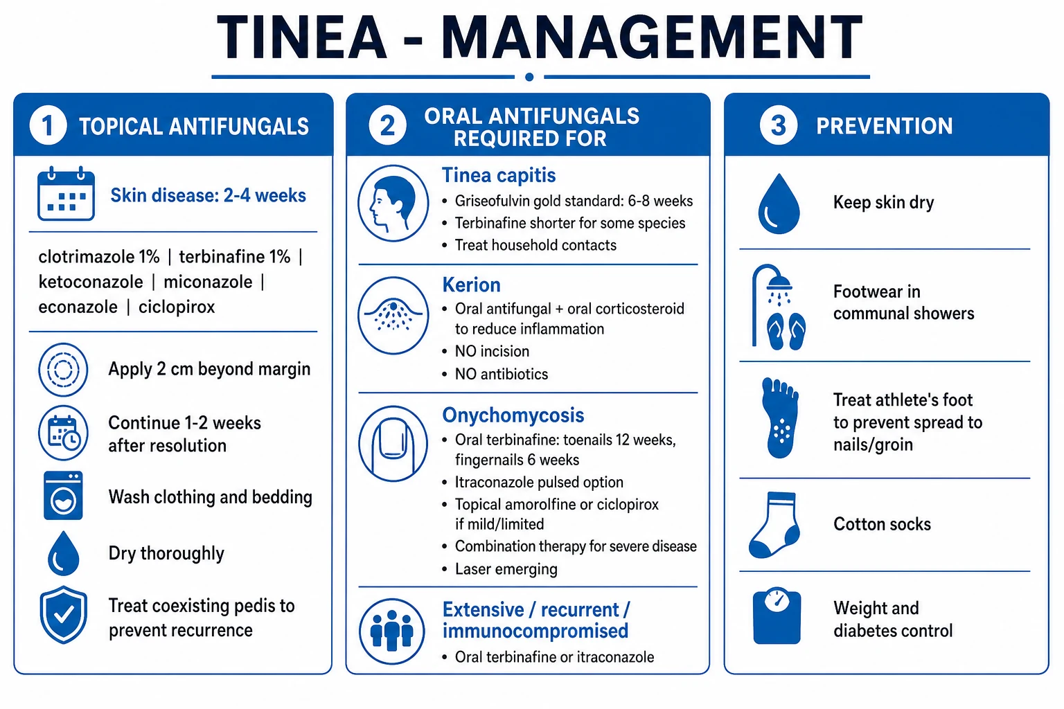
The treatment ladder depends on the site and the severity. Localised skin disease responds to topical antifungals. Tinea capitis, onychomycosis, extensive/recurrent disease, tinea incognito, Majocchi granuloma, and immunosuppression all require oral antifungal therapy.[2][3]
Topical antifungals — the workhorses for skin tinea
Application rules for topicals: apply to the lesion and 2 cm beyond the margin; apply once or twice daily as above; continue for 1 to 2 weeks after clinical resolution to prevent relapse; for tinea cruris extend to the inner thighs, gluteal fold, and lower abdomen; for tinea pedis interdigital treat the whole plantar surface and all toe-webs, not just the visible lesion.[2]
Oral antifungals — when topicals are insufficient
Indications for oral therapy (must-know): (1) tinea capitis — always; (2) onychomycosis; (3) extensive or recurrent tinea corporis/cruris/pedis; (4) tinea incognito / steroid-modified tinea; (5) Majocchi granuloma; (6) immunosuppression; (7) tinea barbae.[3]
[1]Site-specific regimens
- Tinea corporis, cruris, pedis (interdigital), faciei — localised: topical terbinafine 1% OD × 1 to 2 weeks (preferred; fungicidal) or clotrimazole 1% BD × 4 weeks. Extensive/recurrent/incognito: oral terbinafine 250 mg OD × 2 to 4 weeks.[2]
- Tinea pedis (moccasin) — topical often fails (hyperkeratotic sole blocks penetration); oral terbinafine 250 mg OD × 2 to 6 weeks plus keratolytic emollient (salicylic acid 6 to 12% or urea 20 to 40%).[2]
- Tinea capitis — always oral. Choice by species:[5][6]
- Trichophyton (T. tonsurans, T. violaceum): terbinafine weight-based OD × 4 weeks (shorter, fungicidal).
- Microsporum (M. canis, M. audouinii): griseofulvin microsize 20 to 25 mg/kg/day × 6 to 12 weeks (gold standard; terbinafine is inferior for Microsporum).[6]
- Adjunct selenium sulphide 2.5% or ketoconazole 2% shampoo twice weekly to reduce shedding and contagion.
- Treat household contacts (asymptomatic carriers) and examine and treat pets (especially for M. canis).
- Exclude from school until 1 week of treatment completed.
- Kerion celsi — see Resuscitation. Oral antifungal × 6 to 8 weeks plus oral prednisolone 1 to 2 mg/kg/day × 1 to 2 weeks; do not incise.[4]
- Onychomycosis (tinea unguium) — oral terbinafine 250 mg OD × 12 weeks (toenail) or 6 weeks (fingernail) is first-line (fungicidal; superior mycological cure). Alternative: itraconazole pulse 200 mg BD × 1 week/month × 3 to 4 pulses. Topical efinaconazole 10% or tavaborole 5% for mild, distal, single-nail disease only (cure 15 to 35%). Combine with chemical or surgical debridement of the dystrophic nail. Mycological cure 50 to 70% at 12 months; recurrence 20 to 50% (worse for toenails, diabetics, immunosuppressed).[7][8]
- Tinea incognito — STOP the corticosteroid combination cream; oral terbinafine 250 mg OD × 4 to 6 weeks; topical terbinafine as adjunct; address HPA-axis suppression if extensive.[10][11]
- Two-feet-one-hand syndrome — treat all three sites (both feet + dominant hand) with oral terbinafine 250 mg OD × 2 to 6 weeks; treat coexisting onychomycosis.[2]
- Majocchi granuloma — oral terbinafine 250 mg OD × 4 to 8 weeks; topical therapy is insufficient because the fungus has invaded the dermis.[3]
- Tinea barbae — oral terbinafine or itraconazole × 4 to 6 weeks; topical is insufficient for deep follicular involvement.
Adjunctive and preventive measures
- Wash towels, bed linen, and clothing in hot water and dry in sunlight (UV kills dermatophytes).
- Wear cotton socks and change them daily; rotate footwear; use antifungal powders (miconazole, tolnaftate) in shoes.
- Treat coexisting tinea pedis first — it is the reservoir for cruris, corporis, and manuum recurrence.
- Treat household contacts (asymptomatic capitis carriers; family members with shared tinea pedis) and pets (M. canis).
- Address obesity, sweating, occlusive clothing, diabetes, and HIV.
- Public health in India: ban/regulate fixed-dose corticosteroid-antifungal-antibacterial creams; pharmacist education; public awareness of the AAA syndrome.[11]
Escalation triggers
- Failure of topical at 4 weeks → switch topical class or escalate to oral.
- Recurrence → oral + investigate diabetes and HIV + treat reservoirs and contacts.
- Suspected resistance (especially in Indian T. mentagrophytes genotype VIII) → culture and antifungal susceptibility; switch terbinafine to itraconazole.[12]
- Kerion → oral antifungal + oral steroid; DO NOT incise.
Specific Subtypes & Scenarios
Tinea incognito and the Indian AAA syndrome deserves separate emphasis because it is now a major public health problem in India and a favourite exam topic.[10][11][12] The AAA acronym stands for Abuse, Application, and Addiction to fixed-dose corticosteroid-antifungal-antibacterial creams sold over the counter in pharmacies and cosmetic shops across India and South Asia. The corticosteroid (often clobetasol or betamethasone) suppresses local immunity, the fungus proliferates and invades deeper (Majocchi granuloma), the classic annular pattern is lost, and the patient presents with atypical, infiltrative, erythematous, scaly plaques with telangiectasia and atrophy that mimic eczema, psoriasis, rosacea, and lupus. Systemic absorption of the steroid can cause iatrogenic Cushing's syndrome and HPA-axis suppression, documented in case series from India.[11] Management: stop the combination cream, oral terbinafine 250 mg OD for 4 to 6 weeks, gradual topical steroid taper if HPA suppression is suspected, and patient education. Regional policy: IADVL and the Indian Drugs Act now restrict these combinations; pharmacists must be educated; the public must be warned.[11][12]
Tinea capitis in children is a particular scenario because it is always oral therapy. The choice between griseofulvin and terbinafine is species-guided: the Fleece 2004 meta-analysis (Pediatrics) and the Chen 2016 Cochrane review confirm that terbinafine is superior for Trichophyton (shorter course, 4 weeks) and griseofulvin is superior for Microsporum (longer course, 6 to 12 weeks).[5][6] The Bonifaz 2024 review addresses the practical problem of griseofulvin shortages in some countries — itraconazole is the substitute, with terbinafine as an alternative for Trichophyton.[13] Adjunct selenium sulphide or ketoconazole shampoo twice weekly reduces shedding and contagion; household contacts must be screened and treated (asymptomatic carriers are common in T. tonsurans); pets with alopecia should be examined by a vet (M. canis); the child is excluded from school until 1 week of treatment.[4]
Kerion celsi — see Resuscitation. Always remember: boggy purulent scalp mass in a child + cervical lymphadenopathy = kerion, NOT abscess; treat with oral antifungal + oral corticosteroid; never incise.[4]
Onychomycosis is a chronic, relapsing scenario that frustrates patients and clinicians. The Gupta 2020 network meta-analysis (BJD) confirms terbinafine 250 mg OD as having the highest mycological and complete cure, with itraconazole pulse a close alternative.[7] Topical efinaconazole and tavaborole have low cure rates (15 to 35%) and are reserved for mild, distal, single-nail disease or for patients who cannot take oral therapy.[8] Recurrence (20 to 50%) is reduced by treating coexisting tinea pedis, using topical antifungal nail lacquer as maintenance, debriding the dystrophic nail, and addressing diabetes and vascular disease. Diabetics with onychomycosis are at high risk of secondary bacterial infection, cellulitis, and diabetic foot ulcer — podiatry referral is essential.[3]
Two-feet-one-hand syndrome is a classic exam stem. The mechanism is auto-inoculation: chronic bilateral tinea pedis (often with onychomycosis of both great toenails) seeds the dominant hand that the patient uses to scratch the feet. The responsible organism is almost always Trichophyton rubrum. The treatment is oral terbinafine 250 mg OD for 2 to 6 weeks, treating all three sites (both feet + the affected hand) plus the onychomycosis.[2]
Complications & Pitfalls
Disease-related complications include secondary bacterial infection (impetiginisation, cellulitis, lymphangitis — especially in diabetics with tinea pedis, leading to diabetic foot ulcer), Majocchi granuloma (deep granulomatous folliculitis from dermal dermatophyte invasion after shaving, trauma, or topical steroid use), kerion scarring alopecia (permanent hair loss from follicular destruction if treatment is delayed), onychomycosis nail dystrophy (permanent nail deformity, pain, mobility impairment), post-inflammatory hyperpigmentation, and the dermatophytid (id) reaction.[3]
The dermatophytid (id) reaction is a symmetric, sterile, vesicular or eczematous eruption on the hands, fingers, or forearms triggered by intense inflammation at a distant primary site (typically inflammatory tinea pedis or kerion). It is a hypersensitivity reaction to circulating fungal antigens; the lesions themselves are KOH-negative. Treatment is of the primary tinea (with oral antifungal); symptomatic treatment with topical corticosteroid for the id reaction itself is acceptable. Recognising the id reaction prevents the mistake of treating the hands (which are sterile) as primary tinea.[3]
The single most dangerous pitfall is misdiagnosis of tinea as eczema and treatment with topical corticosteroids — producing tinea incognito, deeper invasion (Majocchi granuloma), eventual recalcitrance, and (in extensive use) iatrogenic Cushing's syndrome.[10][11] The corollary: every pruritic annular plaque should have a KOH mount before a topical corticosteroid is prescribed.
Misdiagnosis of kerion as a bacterial abscess is the second great pitfall. Incision and drainage does not cure the fungus (which is in the hair shaft), causes additional scarring, delays antifungal therapy, and prolongs alopecia.[4]
Drug-related adverse effects of long-term oral antifungals:
- Terbinafine: hepatotoxicity (rare but serious), ageusia (taste loss, often reversible), neutropenia, drug interactions via CYP2D6 (reduce dose of tricyclics, beta-blockers, SSRIs). Monitor LFTs at baseline and 6 weeks.
- Itraconazole: hepatotoxicity, negative inotropic effect (avoid in heart failure), hypokalaemia, major CYP3A4 drug interactions (rhabdomyolysis with statins, bleeding with warfarin, sedation with benzodiazepines, oedema with calcium channel blockers).
- Griseofulvin: headache, GI upset, hepatotoxicity, photosensitivity, lupus-like syndrome, teratogenic (avoid in pregnancy and in SLE/porphyria).
- Fluconazole: hepatotoxicity, QT prolongation, CYP2C9/2C19 interactions.[3]
Pitfalls of treatment choice: treating onychomycosis with topical alone (low cure, long duration, poor penetration); treating moccasin tinea pedis with topical alone (hyperkeratotic sole blocks penetration); not treating the tinea pedis reservoir (recurrence guaranteed); ignoring animal/pet reservoirs (re-infection with M. canis); missing emerging terbinafine resistance in Indian T. mentagrophytes genotype VIII (culture and susceptibility, switch to itraconazole).[12]
Prognosis & Disposition
Skin tinea (corporis, cruris, pedis, faciei) has an excellent prognosis with appropriate topical therapy — cure rates of 70 to 90% — but recurrence is common if reservoirs (especially tinea pedis), household contacts, and animal contacts are not addressed.[2]
Tinea capitis has a good prognosis with oral therapy, but kerion may leave scarring alopecia if treatment is delayed, and favus leaves permanent alopecia.[4]
Onychomycosis has a guarded prognosis — mycological cure 50 to 70% at 12 months, complete cure 35 to 50%, recurrence 20 to 50% (worse for toenails, diabetics, immunosuppressed). Lifelong topical nail lacquer maintenance is sometimes used.[7][8]
Tinea incognito has a guarded prognosis — chronic, recalcitrant, often requiring prolonged oral therapy; the underlying skin atrophy and telangiectasia may be permanent; HPA-axis suppression may take months to recover.[10][11]
Predictors of poor prognosis: extensive disease, immunosuppression, diabetes, toenail onychomycosis, tinea incognito, topical steroid misuse, poor adherence, untreated reservoirs (tinea pedis, onychomycosis, household/pet contacts), and emerging terbinafine resistance.[3]
Disposition: most tinea is managed in primary care; dermatology referral for recalcitrant, extensive, incognito, paediatric capitis, suspected resistance, or immunocompromised patients; urgent primary care or dermatology for kerion (start oral antifungal same day); paediatrics for childhood tinea capitis; podiatry for diabetic onychomycosis; veterinary assessment of pets for M. canis.[3]
Monitoring on oral therapy: baseline LFTs and U&E; repeat LFTs at 6 weeks for terbinafine/itraconazole; clinical review at end of therapy; re-culture at end of therapy and at 12 months for onychomycosis. Antifungals are discontinued 1 to 2 weeks after clinical resolution for skin disease and after the fixed course for onychomycosis (the dystrophic nail grows out over 12 to 18 months; clinical appearance at end of therapy is not a marker of cure).[7]
Special Populations
Children — tinea capitis: the commonest dermatophytosis of childhood. Always oral therapy; topical cannot penetrate the hair shaft. Griseofulvin microsize 20 to 25 mg/kg/day with a fatty meal for 6 to 8 weeks (Microsporum up to 12 weeks) remains the gold standard, especially for Microsporum; terbinafine weight-based OD for 4 weeks is preferred for Trichophyton and is shorter and better tolerated. Adjunct selenium sulphide or ketoconazole shampoo twice weekly. Screen and treat household contacts and pets; exclude from school until 1 week of therapy.[4][5]
Pregnancy: topical azoles and allylamines (clotrimazole 1%, terbinafine 1%) are considered safe. Avoid oral terbinafine especially in the first trimester; griseofulvin and itraconazole are teratogenic and contraindicated. For onychomycosis in pregnancy, defer oral therapy until after delivery; use topical efinaconazole or wait.[3]
Breastfeeding: topical therapy is preferred. Oral terbinafine is considered compatible with breastfeeding; avoid griseofulvin and itraconazole.[3]
Diabetes: extensive and recurrent tinea; screen HbA1c in any patient with recurrent tinea. Oral therapy earlier; aggressive treatment of tinea pedis to prevent diabetic foot ulcer and cellulitis; podiatry referral for onychomycosis.[3]
Immunocompromised (HIV, transplant, chemotherapy, chronic corticosteroid): more extensive, atypical, deeper (Majocchi granuloma, dermal/subcutaneous invasion). Oral therapy from the outset. Lower threshold for biopsy and culture. Check drug interactions with antiretrovirals (terbinafine and itraconazole interact with several PIs and NNRTIs) and calcineurin inhibitors (itraconazole markedly raises tacrolimus/ciclosporin levels). Proximal subungual onychomycosis is an AIDS-defining indicator.[3]
Elderly: onychomycosis is common; oral therapy requires LFT monitoring and drug-interaction review (polypharmacy). Falls and mobility impact (dystrophic great-toenail impedes gait). High recurrence; topical for limited disease preferred. Screen for peripheral arterial disease and immunosuppression.[3]
Obese patients: intertriginous tinea cruris and corporis, often with candidal co-infection; oral therapy for extensive disease; weight reduction; antifungal powder (miconazole) in skin folds; treat associated diabetes.[2]
Patients on warfarin or DOACs: griseofulvin reduces warfarin effect (enzyme induction); itraconazole and fluconazole markedly raise INR (CYP3A4/2C9 inhibition); terbinafine has less interaction. Choose terbinafine and monitor INR closely. For DOACs, itraconazole raises dabigatran/rivaroxaban/apixaban levels — prefer terbinafine.[3]
Evidence, Guidelines & Regional Differences
Cochrane and meta-analytic evidence dominates the tinea literature: [1]
- Cochrane review of systemic antifungal therapy for tinea capitis in children (Chen 2016) — terbinafine and griseofulvin are comparable overall, but terbinafine is superior for Trichophyton and griseofulvin is superior for Microsporum; cure rates 60 to 90% depending on species.[5]
- Fleece 2004 meta-analysis (Pediatrics) — griseofulvin vs terbinafine for tinea capitis; same species-specific conclusion.[6]
- Cochrane review of topical treatments for fungal infections of the toenails (Foley 2020) — topical efinaconazole, tavaborole, and ciclopirox have modest efficacy (cure 15 to 35%); inferior to oral therapy.[8]
- Network meta-analysis of toenail onychomycosis monotherapy (Gupta 2020, BJD) — terbinafine and itraconazole have the highest complete and mycological cure; terbinafine tends to edge out itraconazole.[7]
- Cochrane review of oral treatments for fungal infections of the skin of the foot (Bell-Syer 2012) — oral terbinafine 250 mg OD for 2 weeks is the most effective oral regimen; azoles also effective; oral therapy reserved for refractory or extensive disease.[9]
Regional guideline differences are shown in the Region block above. The pivotal regional issue is the Indian epidemic of tinea incognito driven by the AAA syndrome — addressed by IADVL consensus, the Indian Drugs Act restrictions on fixed-dose corticosteroid-antifungal-antibacterial creams, pharmacist education, and emerging antifungal stewardship programs.[10][11][12]
Emerging terbinafine resistance in Indian Trichophyton mentagrophytes/interdigitale ITS genotype VIII is a major contemporary controversy. Single-nucleotide polymorphisms in the squalene epoxidase (SQLE) gene (especially Phe397Leu) reduce terbinafine binding and confer resistance. Management: culture and antifungal susceptibility testing; switch from terbinafine to itraconazole 100 to 200 mg OD for 4 to 6 weeks. This is a key exam topic in Indian boards.[12]
Griseofulvin shortages (Bonifaz 2024, Expert Review of Anti-infective Therapy) — in countries where griseofulvin is no longer available, itraconazole (3 to 4 weeks for Trichophyton; 6 to 8 weeks for Microsporum) and terbinafine (Trichophyton only) are the substitutes, with species-guided choice critical.[13]
Controversies: the role of combination topical + oral therapy in severe disease (no clear evidence of superiority over oral alone; reserved for extensive/recalcitrant); prophylactic antifungal in recurrent onychomycosis (limited evidence; topical efinaconazole maintenance and chemical nail avulsion sometimes used); adjunct oral corticosteroid in kerion (observational evidence only; reduces pain and probably scarring but not infection duration); length of terbinafine therapy for toenail onychomycosis (12 weeks standard; 6 months with doubtful added benefit).[3][4]
Exam Pearls
[1]References
- [1]Leung AK, Lam JM, Leong KF. Tinea corporis: an updated review Drugs Context, 2020.PMID 32742295
- [2]Sahoo AK, Mahajan R. Management of tinea corporis, tinea cruris, and tinea pedis: A comprehensive review Indian Dermatol Online J, 2016.PMID 27057486
- [3]Caplan AS, Gold JAW, Smith DJ. Diagnosis and Management of Tinea Infections Am Fam Physician, 2025.PMID 41118183
- [4]Leung AKC, Hon KL, Leong KF. Tinea Capitis: An Updated Review Recent Pat Inflamm Allergy Drug Discov, 2020.PMID 31906842
- [5]Chen X, Jiang X, Yang M, et al. Systemic antifungal therapy for tinea capitis in children Cochrane Database Syst Rev, 2016.PMID 27169520
- [6]Fleece D, Gaughan JP, Aronoff SC. Griseofulvin versus terbinafine in the treatment of tinea capitis: a meta-analysis of randomized, clinical trials Pediatrics, 2004.PMID 15520113
- [7]Gupta AK, Foley KA, Mays RR, et al. Monotherapy for toenail onychomycosis: a systematic review and network meta-analysis Br J Dermatol, 2020.PMID 31120134
- [8]Foley K, Gupta AK, Versteeg S. Topical and device-based treatments for fungal infections of the toenails Cochrane Database Syst Rev, 2020.PMID 31978269
- [9]Bell-Syer SE, Khan SM, Torgerson DJ. Oral treatments for fungal infections of the skin of the foot Cochrane Database Syst Rev, 2012.PMID 23076898
- [10]Dutta B, Rasul ES, Boro B. Clinico-epidemiological study of tinea incognito with microbiological correlation Indian J Dermatol Venereol Leprol, 2017.PMID 28366916
- [11]Thakran P, Agrawal S, Singal A, Verma S, Madhu SV. Iatrogenic Cushing's Syndrome in Patients with Superficial Dermatophytosis Indian Dermatol Online J, 2021.PMID 33959519
- [12]Verma SB, Vasani R. Male genital dermatophytosis - clinical features and the effects of the misuse of topical steroids and steroid combinations - an alarming problem in India Mycoses, 2016.PMID 27028087
- [13]Bonifaz A, Lumbán-Ramírez P, García-Sotelo RS, et al. Now that griseofulvin is not available, what to do with tinea capitis treatments? Expert Rev Anti Infect Ther, 2024.PMID 39297581