Dermatology · Medicine
Transient neonatal pustular melanosis (TNPM)
Also known as Transient neonatal pustular melanosis (TNPM) · Transient pustular melanosis of the newborn · Lentigines neonatorum
Transient neonatal pustular melanosis (TNPM) is a benign, self-limiting, vesiculopustular dermatosis of the newborn that is PRESENT AT BIRTH. It predominantly affects Black infants (4 to 5 percent) and is rare in Caucasian infants (0.1 to 0.6 percent). Each lesion evolves through three morphological stages — a superficial vesiculopustule, rupture leaving a collarette of scale, and a residual hyperpigmented macule — over days to weeks. Distribution includes the trunk, proximal limbs, face, neck and crucially the PALMS AND SOLES (which distinguishes it from erythema toxicum). The baby is otherwise entirely well. Wright stain of pustule contents shows NEUTROPHILS with no organisms. No treatment is needed; the macules fade over weeks to 3 to 6 months.
On this page & tools
Your progress
Saved locally on this device.
Exam tags
Red flags
Overview & Definition
Transient neonatal pustular melanosis (TNPM), formerly known as transient pustular melanosis of the newborn and historically lumped with lentigines neonatorum, is a benign, transient, self-limiting vesiculopustular dermatosis that is present at birth. It is one of the most distinctive — and most frequently misdiagnosed — eruptions of the neonatal period because it mimics infection yet requires no treatment.[1][2][6]
[1]The condition was first clearly delineated by Ramamurthy and colleagues in 1976, who separated it from erythema toxicum neonatorum (ETN) and from infectious neonatal pustuloses on the basis of its presence at birth, palmoplantar involvement, neutrophilic (rather than eosinophilic) pustule contents, and residual hyperpigmentation.[5] The modern term reflects the three-stage evolution — the transient pustule, the collarette scale, and the melanosis (post-inflammatory hyperpigmentation) — and is the preferred name internationally.
The clinical importance of TNPM lies almost entirely in recognition: missing it leads to unnecessary septic workups, antibiotics, antivirals, and parental distress; conversely, over-calling it in a baby who actually has neonatal herpes, staphylococcal pustulosis, or congenital candidiasis may delay life-saving therapy. The diagnosis is clinical, made at the bedside on five features (timing, distribution, well baby, evolution, collarette), and confirmed, when in doubt, by a Wright or Giemsa smear showing neutrophils and no organisms.[3][7]
Classification
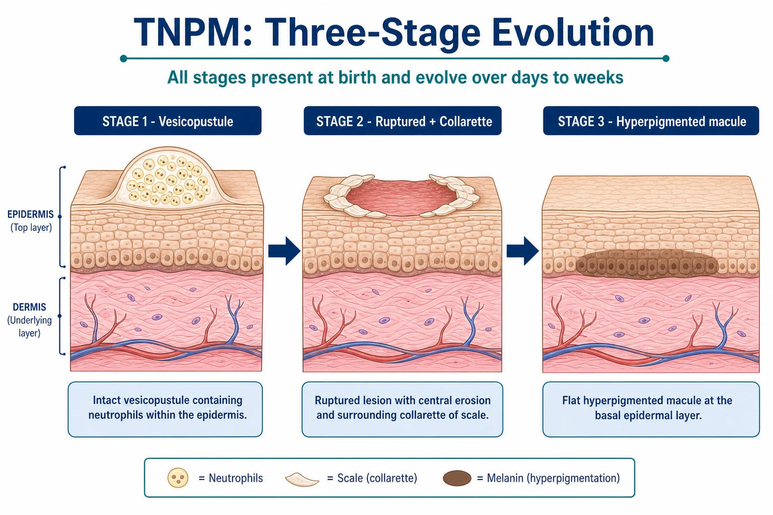
TNPM is best classified by the morphological stage of the individual lesion, because at any one moment a baby may display all three stages side by side on different parts of the body (lesions do not evolve synchronously).[3][4]
-
Stage 1 — Vesicular / vesiculopustular phase. Superficial, 2 to 5 mm, flaccid-to-tense vesicles and pustules on otherwise normal (or only faintly erythematous) skin. The pustule is non-erythematous at its base (a useful distinction from staphylococcal pustulosis and herpes, which sit on an erythematous base). The roof is fragile and easily ruptured by handling and clothing. [1]
-
Stage 2 — Collarette-of-scale phase. Rupture leaves a central superficial erosion with a characteristic fine, whitish, ring-shaped collar of scale (the collarette of scale — detached stratum corneum around the lesion). This phase may be visible from the first hours of life and lasts approximately 1 to 2 weeks. [1]
-
Stage 3 — Hyperpigmented-macular phase. The residual lesion is a brown, flat, smooth macule of post-inflammatory hyperpigmentation — more obvious and more persistent in skin of colour. These macules fade slowly over weeks to 3 to 6 months, occasionally up to a year. [1]
A useful descriptive fourth category is the "pigmented macules only at birth" presentation: when the vesiculopustules have already resolved in utero, the baby is born with widespread brown macules and no intact pustules. This is the form historically called lentigines neonatorum and can be mistaken for congenital melanocytic lesions until the characteristic collarette remnants and the fading time-course are recognised.[5]
Epidemiology & Risk Factors
The single most striking epidemiological feature of TNPM is its predilection for Black (African-descent) newborns.[1][4] Early series from the United States reported an overall incidence of approximately 0.2 to 0.5 percent of all live births but a rate of 4 to 5 percent among Black newborns — a twenty-fold enrichment.[5] TNPM is correspondingly rare in Caucasian (0.1 to 0.6 percent), Hispanic, and East-Asian infants.[2][3]
Risk factors / predispositions: [1]
- Skin of colour (the dominant risk factor). This is partly biological and partly ascertainment: post-inflammatory hyperpigmentation is far more visible in skin of colour, so the third (pigmented) phase is more easily recognised and longer-lasting.
- Term gestation (37 weeks or more). TNPM is essentially a phenomenon of mature-term newborns; it is rare in very premature infants, whose stratum corneum is too immature to support the characteristic pustule-and-collarette morphology.
- No sex predilection. Males and females are equally affected.
- No maternal, obstetric, or familial risk factor has been consistently identified. There is no link to maternal infection, prolonged rupture of membranes, chorioamnionitis, mode of delivery, or specific HLA haplotype. TNPM is not inherited and does not recur in siblings at a rate above population baseline.
- Presence at birth. Unlike ETN (which appears at 24 to 48 hours), TNPM lesions are fully formed at delivery, indicating an in utero onset. The exact gestational age at which the eruption begins is unknown. [1]
- Sub-Saharan Africa, the African diaspora in the Americas and the Caribbean, and Black British populations — TNPM is a common postnatal-ward diagnosis (4 to 5 percent of Black newborns); clinicians are familiar with the pigmented-macular phase.
- South Asia (IADVL practice) — TNPM is recognised with increasing frequency as the pigmented phase is highly visible in brown skin; misdiagnosis as neonatal acne or miliaria is common in primary care.
- Caucasian-majority populations (Northern Europe, North America) — TNPM is rare; the vesiculopustular phase is more easily missed, and the pigmented phase is subtle. Diagnostic confidence is lower, and over-investigation is more common.
Pathophysiology
TNPM is a benign, transient, idiopathic, neutrophilic pustulosis of the neonatal epidermis. It is not infectious, not autoimmune, and not allergic. The four negative investigations — sterile bacterial culture, sterile fungal culture, negative herpes PCR, and negative DIF — define the entity as much as any positive feature does.[3][7]
Subcorneal / intraepidermal neutrophilic pustule. The histological lesion is a subcorneal or superficial intraepidermal cleft filled almost exclusively with neutrophils, with a few neutrophilic fragments and occasional eosinophils. There is no acantholysis (which distinguishes it from pemphigus), no viral cytopathic effect (which distinguishes it from herpes), and no bacterial cluster (which distinguishes it from impetigo).[5][8]
The cellular contrast with erythema toxicum is the mechanistic signature of TNPM. ETN — the other great benign neonatal pustulosis — fills its pustules with eosinophils, not neutrophils, and is associated with peripheral eosinophilia, hair-follicle centring, and an absence of pigment change. TNPM, by contrast, is a sterile neutrophilic process, frequently not follicle-centred, that leaves post-inflammatory hyperpigmentation.[3][7]
Why neutrophils? The leading hypothesis is transient, dysregulated activation of the innate neonatal neutrophil as the skin transitions from the sterile amniotic environment to a colonised, dry, extra-uterine surface. Neonatal neutrophils are functionally immature (reduced chemotaxis, reduced adhesion, exaggerated oxidative bursts), and a transient, self-limiting wave of neutrophil recruitment into the upper epidermis — possibly triggered by the first contact with commensal organisms or by friction during delivery — generates the pustule. The process is self-limiting because the neutrophil activation does not propagate and there is no persistent antigen or autoantibody.[11]
Why the palms and soles? The palmoplantar epidermis has a thick stratum corneum and an abundant neutrophilic trafficking pathway (the same anatomical feature that produces palmoplantar pustulosis in adults). The mechanical stress of delivery and the unique adhesion-molecule profile of palmoplantar skin together favour neutrophil collection there. ETN, in contrast, is follicular and therefore cannot involve the follicle-free palms and soles.[7]
Why post-inflammatory hyperpigmentation? When the pustule resolves, neutrophil-derived mediators and the transient basement-membrane injury stimulate basal keratinocytes and melanocytes to increase melanogenesis and to shed melanin into the upper dermis, where it is taken up by melanophages. In skin of colour, basal melanogenesis is constitutively higher and melanin packages (melanosomes) are larger and more dispersed, so the resulting pigmented macule is more pronounced and longer-lasting. This is a post-inflammatory event, identical in mechanism to the darkening that follows any superficial skin injury in skin of colour, and explains why the macules fade only as melanophages clear the deposited pigment (a slow process taking weeks to months).[3]
Why is it sterile? The pustule represents neutrophil activation in the absence of a viable pathogen. The neutrophils are responding either to a transient mechanical/chemical stimulus (amniotic fluid, vernix, friction) or to commensal bacterial products that do not establish infection. Cultures are therefore sterile, and Gram and KOH stains reveal no organisms. [1]
Clinical Presentation
The clinical picture of TNPM is so characteristic that a confident diagnosis is usually possible from across the cot. Five features define it.[1][4]
1. Timing — present at birth. This is the single most discriminating feature. Lesions are fully developed at the moment of delivery, indicating onset in utero. ETN, by contrast, appears at 24 to 48 hours of age (and almost never in the first 24 hours). A pustular eruption noted in the delivery room is TNPM until proven otherwise (or incontinentia pigmenti, which is also present at birth but follows Blaschkoid lines). [1]
2. Distribution — widespread, including palms and soles. Lesions favour the trunk (especially the shoulders, upper back and buttocks), the proximal extremities, the chin, the forehead, the nape of the neck, and — crucially — the palms and soles. Palmoplantar involvement is the second cardinal feature: ETN spares palms and soles because it is follicular and these sites lack hair follicles. Staphylococcal pustulosis may involve palms and soles but sits on an erythematous base and yields a positive culture. [1]
3. Morphology — three stages, all of which may coexist. Individual lesions are 2 to 5 mm superficial vesicles and pustules with a fragile roof, an absence of surrounding erythema, and a tendency to rupture within hours. Rupture leaves a central superficial erosion surrounded by a fine, white, ring-shaped collarette of scale — detached stratum corneum — which is virtually pathognomonic. The third stage is a brown, smooth, flat hyperpigmented macule at the same site, more obvious in skin of colour. [1]
4. Evolution — a single lesion progresses over days to weeks. The vesiculopustular phase lasts hours to a few days; the collarette-of-scale phase approximately 1 to 2 weeks; the hyperpigmented-macular phase weeks to 3 to 6 months, occasionally up to a year. [1]
5. The baby is entirely well. This is the third cardinal feature: normal feeding, normal temperature, normal tone, normal behaviour, no irritability, no respiratory distress. Any systemic sign — fever, lethargy, poor feeding, irritability, seizures, respiratory distress — invalidates the diagnosis and mandates investigation for infection (herpes, staphylococcal, candidal, bacterial sepsis). [1]
Mucosal involvement is usually absent; the oral mucosa, conjunctiva and genital mucosa are typically spared. When present, oral lesions are rare and minor.[5]
Lesion count. The eruption may be extensive (hundreds of lesions over trunk and limbs) or localised (a handful of lesions on the chin, forehead, or nape). When only a few lesions are present and the diagnosis is in doubt, the bedside tests below clinch it. [1]
Atypical presentations. Three atypical forms deserve mention:[3][6]
- "Pigmented macules only at birth" — vesiculopustules resolved in utero; baby born with widespread brown macules with or without a residual collarette. Easily mistaken for congenital melanocytic naevi or café-au-lait macules.
- Localised TNPM — lesions confined to one region (e.g., the chin, or the forehead), which can be confused with neonatal acne or miliaria.
- Coexistence with erythema toxicum — the two conditions can co-occur, with TNPM lesions present at birth and typical ETN lesions erupting on day 2. [1]
Differential Diagnosis
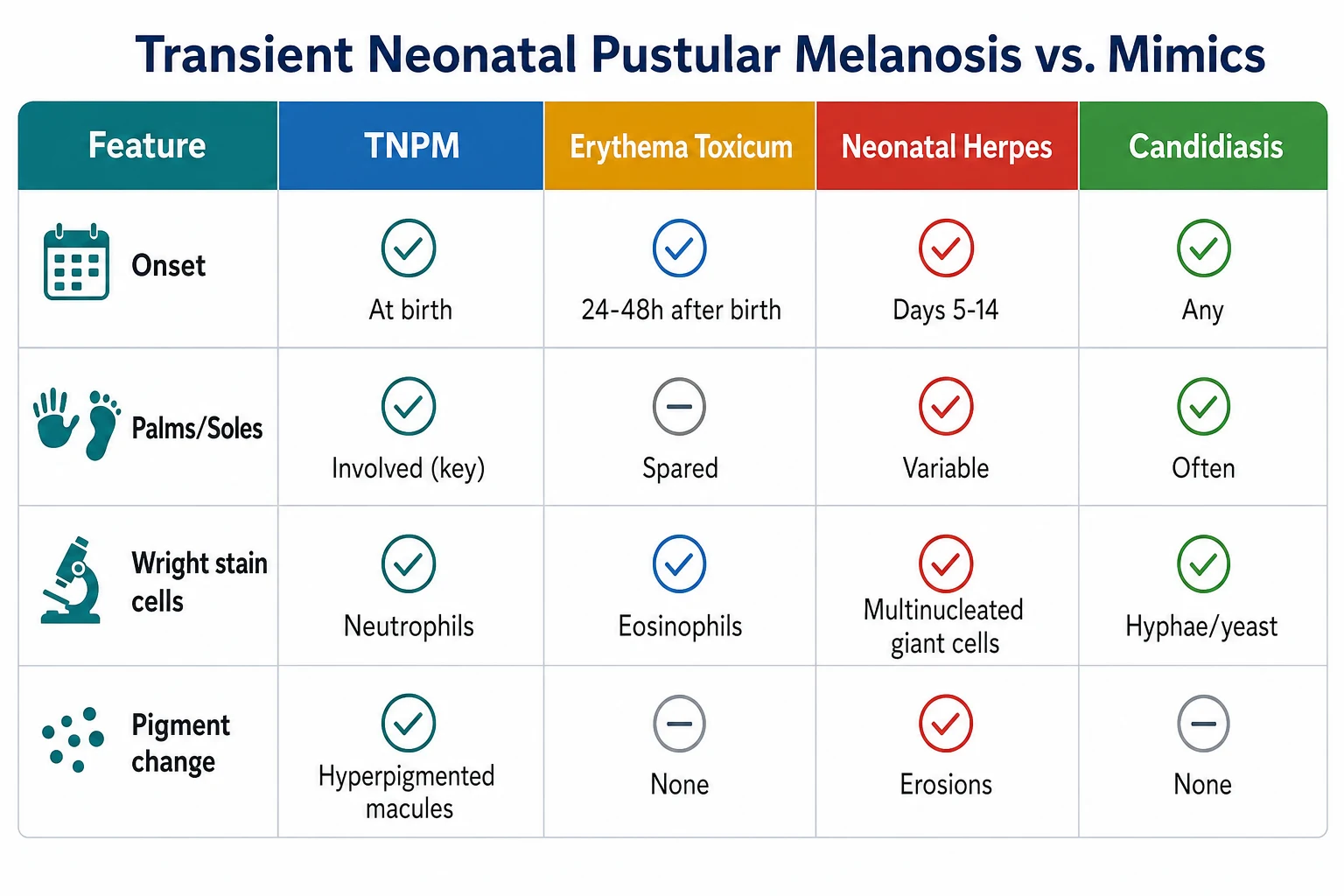
The differential of a neonatal vesiculopustular eruption is broad, but most candidates are eliminated by the four discriminators: timing, palms and soles, Wright stain cells, and pigment change.[3][7]
Three most discriminating features, by condition: [1]
- Erythema toxicum neonatorum (ETN). The closest mimic. (1) ETN appears 24 to 48 hours after birth, never at birth; (2) ETN spares the palms and soles; (3) Wright stain of an ETN pustule shows eosinophils, not neutrophils; ETN leaves no pigment change.[2][4]
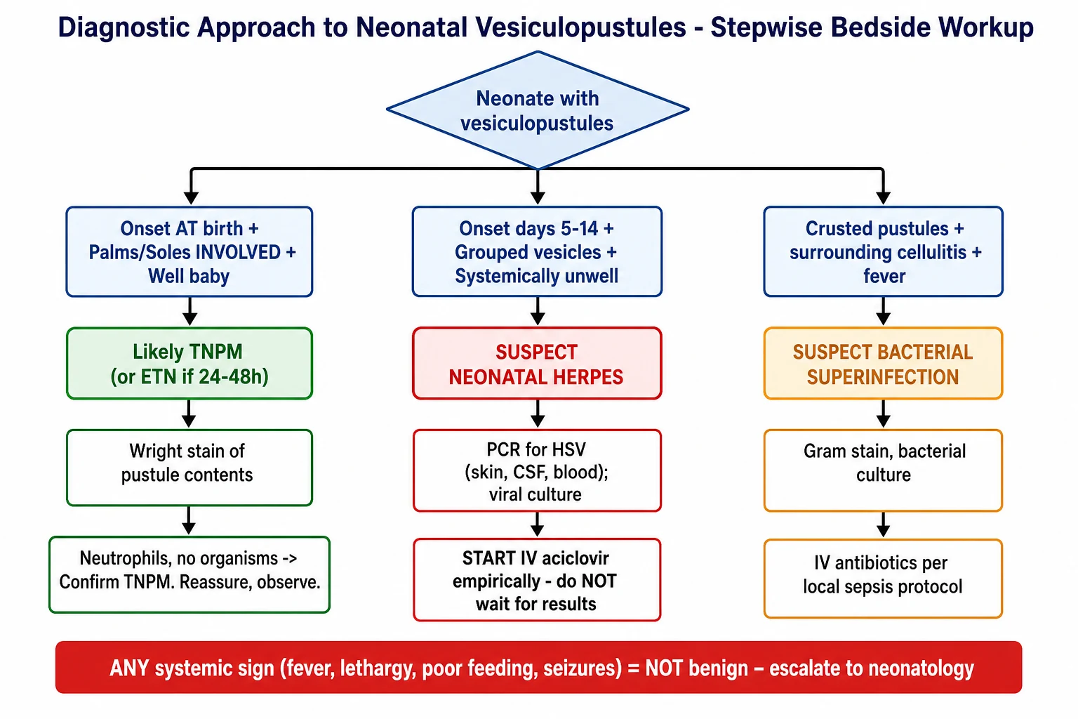
- Neonatal herpes simplex. The most dangerous mimic. (1) HSV appears days 5 to 14 after birth, almost never at birth; (2) HSV lesions are grouped vesicles on an erythematous base, often at a scalp electrode site or on the face; (3) the baby is usually systemically unwell (fever, lethargy, poor feeding, hepatitis, seizures); Tzanck shows multinucleated giant cells, HSV PCR is positive. Medical emergency — IV aciclovir.[3][6]
- Infantile acropustulosis (acropustulosis of infancy). (1) Onset at 2 to 10 months of age, never at birth; (2) lesions are intensely pruritic (TNPM is asymptomatic); (3) recurrent crops on palms and soles; responds to dapsone.[7]
- Congenital / neonatal candidiasis. (1) Often associated with prolonged rupture of membranes or maternal vaginal candidiasis; (2) diffuse erythematous papules and pustules with oral thrush (white plaques) and frequently paronychia; (3) KOH positive (budding yeast and pseudohyphae).[9]
- Staphylococcal pustulosis (impetigo neonatorum). (1) Larger, lax pustules on an erythematous base with crusting and surrounding cellulitis; (2) Gram stain shows Gram-positive cocci in clusters, culture grows Staphylococcus aureus; (3) the baby may be systemically unwell.[3]
- Incontinentia pigmenti (Stage I). (1) Linear / Blaschkoid distribution of vesicles present at birth; (2) X-linked dominant (lethal in males; almost exclusively female infants); (3) evolution through vesicular → verrucous → hyperpigmented → atrophic stages, with multisystem involvement (eye, teeth, CNS); biopsy shows eosinophilic spongiosis and apoptotic keratinocytes.[10]
- Eosinophilic pustular folliculitis of infancy. Onset in the first weeks to months; pruritic; recurrent crops on the scalp; histology shows eosinophilic folliculitis; responds to topical corticosteroids.
- Epidermolytic ichthyosis (formerly epidermolytic hyperkeratosis). Present at birth as widespread bullae (not pustules); skin becomes thickened and hyperkeratotic within months; autosomal dominant KRT1/KRT10 mutation.
Neonatal vesiculopustule — the NESTI mnemonic
Clinical & Bedside Assessment
The diagnosis of TNPM is made at the bedside, on five features:[1][6]
- Timing — lesions present at birth (not developing over the first 24 to 48 hours).
- Distribution — widespread including palms and soles.
- Systemic state — the baby is entirely well (normal feeding, temperature, tone).
- Evolution — three stages (vesicopustule → collarette scale → hyperpigmented macule), all of which may coexist.
- Collarette — a fine ring of scale around a ruptured pustule, virtually pathognomonic. [1]
Bedside tests (when diagnostic doubt): [1]
- Wright or Giemsa smear of a freshly unroofed pustule: shows neutrophils and no organisms. The single most useful confirmatory test. (ETN shows eosinophils; HSV shows multinucleated giant cells on Tzanck; candidiasis shows budding yeast and pseudohyphae on KOH.)[3]
- Gram stain of pustule contents: sterile, no bacteria. (Staphylococcal pustulosis shows Gram-positive cocci in clusters.)
- KOH preparation: negative — no budding yeast or pseudohyphae. (Candidiasis shows both.)
- Tzanck smear (if herpes is suspected): negative for multinucleated giant cells. Send HSV PCR of skin, swab, CSF and blood if any systemic concern.
Maternal and birth history (to exclude the dangerous mimics): [1]
- Maternal HSV status (history of genital herpes, recent primary infection in the third trimester — high transmission risk).
- Duration of rupture of membranes (prolonged ROM increases risk of ascending bacterial and candidal infection).
- Maternal GBS status (group B streptococcal sepsis can present with pustular lesions).
- Mode of delivery (vaginal delivery in active maternal HSV lesions increases neonatal HSV risk).
- Scalp electrode use or fetal scalp blood sampling (a portal of entry for HSV).
- Gestational age and birthweight (TNPM is a phenomenon of term infants; candidiasis can be systemic in very-low-birthweight infants). [1]
Investigations
The diagnosis of TNPM is clinical. Investigations are reserved for atypical presentations, systemically unwell babies, and lesions that fail to follow the expected time-course.[1][7]
Cytology (the highest-yield bedside test). [1]
- Wright or Giemsa stain of pustule contents: neutrophils predominate, with no organisms and no multinucleated giant cells. The cellular composition is the strongest non-invasive discriminator from ETN (eosinophils), HSV (multinucleated giant cells) and candidiasis (yeast).[3]
Microbiology. [1]
- Gram stain of pustule contents: sterile, no bacteria. Bacterial culture (aerobic and anaerobic): sterile.
- KOH preparation: no budding yeast, no pseudohyphae. Fungal culture: sterile.
- HSV PCR of skin lesion swab, conjunctival swab, oropharyngeal swab, CSF and blood: negative. (Send if the baby is unwell, if maternal HSV is positive, or if grouped vesicles are present.)
- Viral culture (vesicle fluid): negative for HSV and VZV. [1]
Histopathology (skin biopsy — rarely needed). [1]
Reserve for atypical morphology, persistent lesions, or diagnostic doubt despite bedside tests. The histological picture is:[5][8]
- An intraepidermal (subcorneal) pustule filled with neutrophils, occasional eosinophils, and neutrophilic fragments.
- No acantholysis (rules out pemphigus).
- No viral cytopathic effect (rules out herpes — no multinucleation, no ground-glass inclusions).
- No bacterial clusters (rules out impetigo).
- Hair follicles not preferentially involved (TNPM is not folliculocentric, in contrast to ETN).
- In the pigmented-macular phase: increased basal melanogenesis and melanophages in the upper dermis — the histological correlate of post-inflammatory hyperpigmentation. [1]
Direct immunofluorescence (DIF). Negative — no intercellular or basement-membrane-zone immunoglobulin or complement deposition. This excludes IgA pemphigus, linear IgA disease, and pemphigoid gestationis, which can rarely present in the neonatal period.[8]
Bloods (only if systemically unwell). [1]
- FBC with differential (eosinophilia supports ETN; leukocytosis or left shift supports bacterial infection).
- CRP and blood culture.
- Liver function tests (hepatitis in disseminated HSV).
- CSF analysis (cell count, protein, glucose, HSV PCR) — if seizures, lethargy, or any sign of encephalitis. [1]
Imaging. Not required for TNPM. Consider chest X-ray, cranial ultrasound, or MRI brain if disseminated HSV is suspected. [1]
Management — Resuscitation
TNPM does not require any acute intervention. It is benign and self-limiting.[1][6] The "resuscitation" of a baby with suspected TNPM is therefore not medical but diagnostic and parental:
- Confirm the well-baby state before discharge from the postnatal ward — normal temperature, normal feeding, normal tone, normal behaviour, no respiratory distress.
- Confirm the diagnosis clinically using the five features above; perform the Wright/Giemsa smear if any doubt.
- Counsel the parents — explain the benign, transient nature, the expected time-course of resolution (pustules in days, scale in 1 to 2 weeks, pigmented macules over weeks to 3 to 6 months), and the absence of long-term sequelae, scarring, or recurrence.
- Safety-net — explicit instructions to return immediately if the baby develops fever, poor feeding, lethargy, irritability, new grouped vesicular lesions, or any systemic sign. [1]
Escalation triggers (any one of these invalidates the diagnosis of TNPM and mandates escalation): [1]
- Any systemic sign (fever ≥ 38.0 °C rectal, hypothermia, lethargy, poor feeding, irritability, seizures, respiratory distress).
- Widespread skin erosion suggesting a more severe bullous or erosive disorder.
- Grouped vesicles on an erythematous base (suspicious for HSV) — initiate IV aciclovir empirically pending PCR.
- Surrounding cellulitis or crusting with a positive Gram stain (staphylococcal) — send culture and start appropriate antibiotic.
- Maternal active HSV lesions at delivery with neonatal lesions — treat as HSV until proven otherwise. [1]
Management — Definitive & Stepwise
The definitive management of TNPM is observation and reassurance.[1][2][9]
Step 1 — Confirm the diagnosis and reassure. The cornerstone is parental reassurance after a confident clinical diagnosis. Explain that the eruption is cosmetically prominent but medically trivial, that it will fade without treatment, and that it does not scar, recur, or portend any systemic disease. Where appropriate, show photographs of the expected evolution to set realistic expectations. [1]
Step 2 — Gentle skin care. [1]
- Gentle cleansing with water and a mild, fragrance-free baby cleanser. Avoid harsh soaps, antiseptics (chlorhexidine, hexachlorophene), and rubbing.
- Emollients (e.g., white soft paraffin / liquid paraffin mixtures, or a fragrance-free ointment emollient) for any dry or scaling skin after rupture of pustules. Emollients soothe and reduce transepidermal water loss but do not alter the natural history.
- Routine bathing — no restriction; bathing does not worsen or spread the eruption. [1]
Step 3 — Explicit non-treatments (do NOT prescribe). [1]
- NO topical corticosteroids — not indicated; risk of skin atrophy, telangiectasia, and masking of infection in a neonate.
- NO topical or systemic antibiotics — pustules are sterile; antibiotics select for resistance and disturb the developing skin microbiome.
- NO topical or systemic antifungals — KOH and culture are negative.
- NO antivirals (aciclovir) — HSV PCR is negative; reserve for proven or strongly suspected HSV.
- NO phototherapy, laser, or skin-lightening agents for the hyperpigmented macules — they fade spontaneously; intervention risks dyspigmentation and scarring. [1]
Step 4 — Treat secondary infection (if it occurs). [1]
Rarely, over-manipulation of lesions by parents or caregivers produces a secondary bacterial infection (usually Staphylococcus aureus). This presents as enlarged, crusted pustules with surrounding cellulitis. Send a swab for Gram stain and culture and treat with oral flucloxacillin (12.5 mg/kg four times daily for 5 to 7 days) or, in penicillin-allergic babies, an oral cephalosporin or macrolide per local guidance. [1]
Step 5 — Review and follow-up. [1]
- Routine neonatal check at 24 to 48 hours and at 6 to 8 weeks. No specific dermatology follow-up is required for classical TNPM.
- Refer to paediatric dermatology only if: (i) the morphology is atypical; (ii) lesions persist beyond 6 months without fading; (iii) new grouped vesicles appear; (iv) diagnostic doubt remains despite bedside tests; or (v) the baby develops any systemic sign.
- Safety-net advice: parents should return immediately with fever, poor feeding, lethargy, irritability, new grouped vesicles, or any other concern.[6]
Counselling on the hyperpigmented macules. In skin of colour, the brown macules may be striking and the source of significant parental anxiety. Reassure that they will fade over weeks to 3 to 6 months (occasionally up to a year), that they do not represent naevi or birthmarks, and that no cosmetic intervention is appropriate. [1]
Specific Subtypes & Scenarios
-
Classical TNPM in a Black term newborn. The textbook case: hundreds of vesicopustules present at birth over the trunk, proximal limbs, face and palms/soles; evolution through collarette scale to hyperpigmented macules; resolution over weeks to months; well baby throughout. [1]
-
"Pigmented macules only at birth" subtype. Vesiculopustules have already ruptured in utero; the baby is born with widespread brown macules with or without a residual collarette of scale. Easily mistaken for congenital melanocytic naevi or multiple café-au-lait macules. The diagnostic clue is the fading time-course (true naevi and CALMs persist) and the typical collarette remnants.[5]
-
Localised TNPM. A handful of lesions confined to one site — most often the chin, the forehead, the nape of the neck, or the lower back. Easily confused with neonatal acne (which appears at 2 to 4 weeks and is comedonal-pustular on the cheeks), miliaria pustulosa, or a localised contact dermatitis. The collarette of scale and the spontaneous evolution clinch the diagnosis. [1]
-
TNPM in premature infants. Rare, because the immature stratum corneum of the very premature infant does not support the characteristic pustule-and-collarette morphology. When present in a late-preterm infant, the picture is the same as in term babies. [1]
-
TNPM in skin of colour versus Caucasian skin. In skin of colour, the pigmented-macular phase is highly visible and the dominant concern of parents; counselling is essential. In Caucasian skin, the pigmented phase is subtle or absent and the vesiculopustular phase may be the only manifestation. [1]
-
Coexistence with erythema toxicum. TNPM and ETN can coexist in the same baby: TNPM lesions present at birth, ETN lesions erupting at 24 to 48 hours. The two patterns are easily separated at the bedside by their morphology and distribution. [1]
-
TNPM with superimposed secondary infection. Rare; presents as crusted, enlarging pustules with surrounding cellulitis. Treat the secondary infection as above; the underlying TNPM resolves as expected. [1]
Complications & Pitfalls
Complications of TNPM itself: [1]
- Transient post-inflammatory hyperpigmentation — the most common "complication"; actually an expected phase of the disease, fading over weeks to 3 to 6 months.
- Secondary bacterial superinfection — rare; usually from over-manipulation of lesions; presents as crusted pustules with surrounding cellulitis.
- Parental anxiety and cosmetic distress — particularly in skin of colour, where the pigmented phase is striking; an under-recognised but important morbidity.
- No long-term sequelae — no scarring, no recurrence, no association with later skin or systemic disease. [1]
Diagnostic pitfalls: [1]
The TNPM diagnostic pitfalls — the MISS mnemonic
Management pitfalls: [1]
- Unnecessary septic workup, antibiotics and isolation in a benign self-limiting condition — exposes the baby to antibiotic side effects, disturbs the developing microbiome, separates mother and baby, and consumes resources.
- Over-treatment with topical corticosteroids — risk of skin atrophy, telangiectasia, and masking of infection in a neonate.
- Misdiagnosing incontinentia pigmenti stage I as TNPM — missing the ocular (retinal vascular abnormalities), dental (peg-shaped teeth, hypodontia), and neurological (seizures, developmental delay) sequelae that define the long-term prognosis of IP.[10]
- Misdiagnosing the "pigmented macules only at birth" subtype as multiple congenital melanocytic naevi or neurofibromatosis (CALMs) — leading to unnecessary investigation and parental anxiety. The fading time-course and the residual collarette are diagnostic.
Prognosis & Disposition
Prognosis is excellent. TNPM is a benign, self-limiting condition with complete resolution and no scarring.[1][2]
Time-course of resolution: [1]
- Vesiculopustules: resolve within hours to a few days.
- Collarette of scale: resolves within 1 to 2 weeks.
- Hyperpigmented macules: fade over weeks to 3 to 6 months; occasionally up to a year, especially in skin of colour. [1]
No long-term sequelae. No scarring, no atrophy, no recurrence, no association with later skin disease (including atopic dermatitis, psoriasis, or pigmentary disorders). The baby's growth, development, immunisation schedule, feeding, and bathing routines are entirely normal. [1]
Disposition. Routine postnatal care with the neonate rooming-in with the mother; no restriction of feeding, bathing, or skin-to-skin contact. Normal immunisation schedule. No specific dermatology follow-up is required for classical TNPM. [1]
Safety-net advice for parents. Return immediately if the baby develops: [1]
- Fever (≥ 38.0 °C rectal, or hypothermia < 36.5 °C).
- Poor feeding or refusal of feeds.
- Lethargy, irritability, or reduced tone.
- New grouped vesicular lesions or rapid spread of the eruption.
- Any systemic concern. [1]
Special Populations
-
Term versus preterm neonates. TNPM is essentially a phenomenon of mature-term newborns (37 weeks or more). It is rare in very premature infants because the immature, poorly keratinised stratum corneum cannot form the characteristic subcorneal pustule and collarette. When present in a late-preterm infant, the morphology and evolution are the same. [1]
-
Skin of colour. The pigmented-macular phase is more pronounced and more persistent because basal melanogenesis is constitutively higher and melanosomes are larger and more dispersed. Counselling must explicitly address the cosmetic prominence and the expected time-course (up to 6 months, occasionally longer). [1]
-
Neonates with a positive maternal HSV history or recent maternal primary HSV. Even if the lesions look like TNPM, a low threshold for HSV PCR (skin, oropharyngeal, conjunctival swabs, CSF and blood) and empirical IV aciclovir is appropriate until PCR results are negative. Maternal primary HSV in the third trimester carries the highest neonatal transmission risk.[3]
-
Adopted, surrogate, or IVF neonates with limited maternal history. A cautious approach: a confident clinical diagnosis of TNPM can still be made at the bedside; if the maternal HSV and ROM history is unavailable, a low threshold for HSV PCR is appropriate. [1]
-
Very-low-birthweight (< 1500 g) or immunocompromised neonates. Cutaneous candidiasis in this group can be systemic and life-threatening; in any VLBW baby with pustular lesions, send fungal culture and treat empirically for disseminated candidiasis until proven otherwise — TNPM is not the diagnosis to make first in this population.[9]
-
Resource-limited settings. A confident clinical diagnosis can be made with only the Wright/Giemsa smear, Gram stain and KOH preparation as bedside tests. This avoids unnecessary hospitalisation, IV antibiotics, and separation of mother and baby in settings where resources are scarce. The single most important intervention is parental reassurance and safety-netting. [1]
Evidence, Guidelines & Regional Differences
Strength of evidence
The evidence base for TNPM is almost entirely descriptive — case series, reviews, and consensus expert opinion — rather than interventional. There are no randomised controlled trials, because TNPM is a self-limiting condition that requires no treatment and is therefore not amenable to interventional study. The largest contemporary evidence comes from the 2026 two-part JAAD review of pustular dermatoses (Hong and Feldman, Parts I and II), which consolidates TNPM within the broader framework of benign and pathological neonatal and adult pustular eruptions; the 2025 Acta Medica Portuguesa case series (Valente and colleagues), which highlights TNPM as a benign mimicker of neonatal infection; and the long-running Indian and American dermatology overviews.[6][7][8][3]
Guidelines
There is no single authoritative international guideline for TNPM. Practice is informed by: [1]
- American Academy of Dermatology (AAD) and American Academy of Pediatrics (AAP) neonatal skin-care consensus statements, which emphasise clinical diagnosis, restraint in investigation, and avoidance of unnecessary topical therapies.[2]
- British Association of Dermatologists (BAD) and UK Paediatric Dermatology consensus, which mirror the AAP approach and stress parental reassurance.
- Indian Association of Dermatologists, Venereologists and Leprologists (IADVL) task-force guidance, which emphasises the high prevalence in skin of colour and the avoidance of topical steroids and skin-lightening agents.[3]
- RANZCP / RACP neonatal guidance (Australia and New Zealand), concordant with the above.
- United States (AAP/AAD) — emphasis on minimal handling, breast-feeding, rooming-in, and avoidance of antiseptic baths (no hexachlorophene due to neurotoxicity; chlorhexidine not recommended for routine full-body neonatal bathing). TNPM is managed expectantly.
- United Kingdom (NICE/BAD) — skin-to-skin contact encouraged from birth; bathing delayed for at least the first 24 hours; water-only or mild cleanser for the first month. TNPM managed with parental reassurance.
- South Asia (IADVL) — TNPM is a common postnatal-ward diagnosis given the skin-of-colour predominance; emphasis on avoiding topical steroids, antibiotic creams, and "skin-lightening" preparations sometimes used culturally for the pigmented phase.
- Universal — across all regions, the cardinal rule is invariant: TNPM needs no treatment; investigate only the atypical or systemically unwell baby; and safety-net for HSV and sepsis.
Controversies
- Spectrum versus distinct entity. Whether TNPM and ETN represent opposite ends of a single spectrum of benign transient neonatal pustulosis (varying only in cellular infiltrate — neutrophilic versus eosinophilic — and in pigment outcome) or are truly distinct entities remains debated. Most contemporary reviews treat them as distinct on the basis of timing (at birth vs 24 to 48 h), cell type (neutrophil vs eosinophil), and pigment outcome (hyperpigmentation vs none), but the boundary is not absolute.[7]
- Mechanism of neutrophil recruitment. The leading hypothesis — transient dysregulated activation of the immature neonatal neutrophil on first contact with commensal organisms — remains unproven. The role of the developing infant cutaneous microbiome is an active area of research, but no causal organism or molecular trigger has been identified.[11]
- Investigation threshold. The threshold for sending HSV PCR and treating empirically with aciclovir varies between units. Most paediatric dermatologists favour a very low threshold for HSV PCR — the cost of missing neonatal herpes (high mortality and morbidity) vastly exceeds the cost of an empirical course of aciclovir pending PCR.[6]
Exam Pearls
TNPM — the high-yield exam checklist
Red Flags
Exam application bank (NEET-PG / INICET)
One-line answer
Transient neonatal pustular melanosis (TNPM) is a benign, self-limiting, vesiculopustular dermatosis of the newborn that is PRESENT AT BIRTH. It predominantly affects Black infants (4 to 5 percent) and is rare in Caucasian infants (0.1 to 0.6 percent). Each lesion evolves through three morphological stages — a superficial vesiculopustule, rupture leaving a collarette of scale, and a residual hyperpigmented macule — over days to weeks. Distribution includes the trunk, proximal limbs, face, neck and crucially the PALMS AND SOLES (which distinguishes it from erythema toxicum). The baby is otherwise entirely well. Wright stain of pustule contents shows NEUTROPHILS with no organisms. No treatment is needed; the macules fade over weeks to 3 to 6 months.
Worked stems (answer without another resource)
Stem 1 — Classic presentation. Map symptoms to mechanism; name the first investigation and first treatment step with dose/route if drug therapy is standard. [1]
Stem 2 — Unstable / complicated. List red flags that force immediate resuscitation, theatre, ICU, antidote, or reperfusion — and what you do in the first 15 minutes. [1]
Stem 3 — Atypical group. Elderly, pregnancy, child, or immunocompromised: how presentation and thresholds change. [1]
Stem 4 — Differential trap. Name the three closest mimics and one discriminator for each. [1]
Stem 5 — Disposition. Who goes home with safety-netting, who is admitted, who needs HDU/ICU/theatre, and what follow-up is mandatory. [1]
Rapid viva checklist
- Definition + classification
- Pathophysiology chain
- Bedside signs / criteria
- Score with exact components (if any)
- Emergency bundle
- Definitive therapy with doses
- Complications of disease and of treatment
- Special populations
- Guideline/trial name if classic
- Three exam traps
Coverage self-check
If you cannot answer any stem above from this page alone, re-read the matching section — the page is intended to be self-sufficient for final-prof and NEET-PG/INICET questions on Transient neonatal pustular melanosis (TNPM).
References
- [1]Wilson JL, Coughlin CC, et al. Neonatal Dermatology Prim Care, 2025.PMID 40835282
- [2]Chadha A, O'Connor NR, et al. Common Neonatal Rashes Pediatr Ann, 2019.PMID 30653638
- [3]Ghosh S, Batalla A, et al. Neonatal pustular dermatosis: an overview Indian J Dermatol, 2015.PMID 25814724
- [4]O'Connor NR, McLaughlin MR, Ham P, et al. Newborn skin: Part I. Common rashes Am Fam Physician, 2008.PMID 18236822
- [5]Van Praag MC, Van Rooij RW, Folkers E, et al. Diagnosis and treatment of pustular disorders in the neonate Pediatr Dermatol, 1997.PMID 9144701
- [6]Valente JP, Goncalves AC, et al. Transient Neonatal Pustular Melanosis: A Benign Mimicker of Infection Acta Med Port, 2025.PMID 40952891
- [7]Hong S, Feldman SR, et al. Pustular Dermatoses. Part II. Infectious, Genetic, and Transient Neonatal J Am Acad Dermatol, 2026.PMID 42176705
- [8]Hong S, Feldman SR, et al. Pustular Dermatoses. Part I. Inflammatory and Autoimmune J Am Acad Dermatol, 2026.PMID 42176709
- [9]Patrizi A, Raone B, Neri I, et al. Advances in pharmacotherapeutic management of common skin diseases in neonates and infants Expert Opin Pharmacother, 2017.PMID 28429969
- [10]Cetinarslan T, Sezer E, et al. Incontinentia pigmenti Stage 1 is not simply vesiculo-bullous but vesiculo-pustular Pediatr Dermatol, 2024.PMID 38284782
- [11]Schoch JJ, et al. The infantile cutaneous microbiome: A review Pediatr Dermatol, 2019.PMID 31332846