Dermatology · Medicine
Tungiasis and myiasis
Also known as Tungiasis · Myiasis · Sand flea disease · Jiggers · Botfly infestation · Cutaneous larva migrans
Tungiasis: gravid female Tunga penetrans (sand flea) burrows into the stratum corneum of the feet → a white-yellow nodule with a central black dot (the flea's posterior respiratory cone); endemic Caribbean/sub-Saharan Africa/South America; treatment is sterile needle/curette extraction en bloc plus antiseptic and tetanus prophylaxis, with oral ivermectin 200 mcg/kg for heavy infestation. Myiasis: infestation of living tissue by dipteran fly larvae — furuncular (Dermatobia hominis botfly, Central/South America, painful nodule with a breathing pore; Cordylobia anthropophaga tumbu fly, sub-Saharan Africa), wound (Lucilia/Calliphora/Cochliomyia in neglected wounds), migratory, and nasopharyngeal; treatment is occlude the pore with petrolatum/bacon to asphyxiate the larva → extract with forceps, plus debridement for wound myiasis. Cutaneous larva migrans: dog/cat hookworm larvae (Ancylostoma braziliense) → serpiginous pruritic track migrating 1 to 2 cm/day; treat with oral albendazole 400 mg for 3 to 5 days or ivermectin 200 mcg/kg single dose.
On this page & tools
Your progress
Saved locally on this device.
Exam tags
Red flags
Tungiasis, myiasis, and cutaneous larva migrans (CLM) are the three skin infestations a dermatology candidate must be able to separate at sight. They share a tropical geography and a returning-traveller presentation, but their biology — and therefore their treatment — diverges sharply. Tungiasis is a flea that burrows into the skin and must be dug out. Myiasis is a fly larva that is already in the skin and must be coaxed out. CLM is a hookworm that wanders under the skin and is killed by anthelmintics rather than extracted. Confusing the three loses marks and mistreats the patient: incising a botfly nodule releases the larva into the wound, while curetting a CLM track yields nothing.[1][4][10]
All three belong to the broader family of ectoparasitoses — organisms that live on or in the superficial skin — alongside scabies (Sarcoptes scabiei), pediculosis (lice), and tick infestation. This topic covers tungiasis and myiasis as the primary focus, with CLM as the key differential, and ends with an ectoparasite comparison table that anchors the board answer. [1]
Overview & Definitions
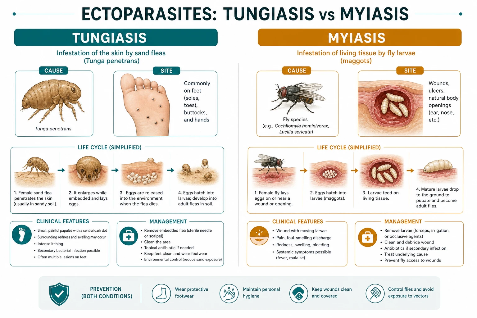
Tungiasis is an inflammatory skin disease caused by the gravid female sand flea Tunga penetrans (and rarely T. trimamillata), which burrows head-first into the stratum corneum of its host, feeds on dermal blood, swells to the size of a pea, expels eggs through a posterior opening, and then dies in situ. The flea does not leave the host during the parasitic phase; it must be physically removed. The disease is endemic across the tropics and is recognised by the World Health Organization as a neglected tropical disease in endemic countries.[1][3][10]
Myiasis (from the Greek myia, fly) is the infestation of living or dead vertebrate tissue by the larvae (maggots) of two-winged flies (order Diptera). It is classified by the anatomical site of infestation and, within the skin, by the behaviour of the larva. The clinical importance of myiasis lies in distinguishing furuncular disease (a single larva in a boil-like lesion, removable at the bedside) from wound/cavitary disease (many larvae in neglected tissue, needing debridement) and from rare but dangerous nasopharyngeal, aural, and ophthalmic forms.[4][6]
Cutaneous larva migrans (CLM), the most common tropically acquired skin disease in returning travellers, is a creeping eruption produced when animal hookworm larvae (classically Ancylostoma braziliense of dogs and cats) penetrate human skin but cannot complete their life cycle in an abnormal host, so they wander aimlessly in the epidermis until they die weeks to months later. Humans are a dead-end host; the parasite never reaches the gut, so the disease is self-limiting but intensely pruritic.[7][8]
Classification
Myiasis and CLM each have a classification the examiner tests deliberately. For myiasis, the clinically useful axis is anatomical (where the larva lives) cross-referenced with behavioural (obligate, facultative, or accidental parasite). For CLM there is one entity with a defined set of mimics. [1]
Cutaneous — furuncular
- A single larva in a boil-like nodule with a central breathing pore
- *Dermatobia hominis* (human botfly) — Central and South America; transported by a mosquito (phoresy)
- *Cordylobia anthropophaga* (tumbu fly) — sub-Saharan Africa; eggs laid on soil/soiled clothing
- Treatment: occlude the pore, then extract the intact larva with forceps
Cutaneous — wound
- Multiple larvae colonise open wounds, ulcers, gangrene, or neglected surgical sites
- *Lucilia* (greenbottle), *Calliphora* (bluebottle), *Cochliomyia hominivorax* (New World screw-worm)
- Facultative (most) or obligate (*C. hominivorax* burrows into living tissue)
- Treatment: irrigation, debridement, removal of all larvae
Cutaneous — migratory (creeping)
- Larva tunnels through the skin producing a wandering track
- *Gasterophilus* (horse botfly) or *Hypoderma* (cattle botfly/ox warble)
- Mimics CLM but the track is longer, less itchy, and the larva is a fly larva not a hookworm
- Treatment: extract the larva or ivermectin
Cavitary / systemic
- Nasopharyngeal, aural, ophthalmic, urogenital, intestinal myiasis
- Screw-worm flies and other calliphorids in neglected orifices or debilitated patients
- Often a surgical/ENT emergency with tissue destruction
- Treatment: removal under anaesthesia plus antibiotics for secondary infection
A second distinction the examiner rewards is between obligate myiasis flies (which require a host to complete the life cycle — Dermatobia hominis, Cordylobia anthropophaga, Cochliomyia hominivorax) and facultative myiasis flies (which normally lay eggs in carrion or decaying matter but opportunistically infest wounds — Lucilia, Calliphora, Sarcophaga).[4][9]
Epidemiology & Risk Factors
Tungiasis
- Endemic in the Caribbean, Central and South America, sub-Saharan Africa, and under-reporting on the Indian subcontinent and Madagascar
- Poverty-driven: barefoot children and elderly in resource-poor communities; ~23 endemic countries per WHO
- Risk factors: barefoot walking, earthen floors, cracked walls, keeping dogs and pigs indoors, dry season peaks
- Severe neglected disease; morbidity from secondary infection (lymphangitis, abscess, tetanus)
Furuncular myiasis
- *Dermatobia hominis*: neotropical — Mexico to northern Argentina; commonest travel-associated myiasis returning from Central/South America
- *Cordylobia anthropophaga*: sub-Saharan Africa (tumbu fly); eggs laid on shaded soil and on clothing drying on the ground
- Risk: rural travel, ecotourism, children (scalp lesions), failure to iron clothes
- No human-to-human transmission
Wound myiasis
- Worldwide but over-represented in debilitated, immobilised, alcoholic, diabetic, or cognitively impaired patients with neglected wounds
- Elderly with pressure ulcers, chronic venous leg ulcers, gangrenous digits, neglected malignancy
- Nosocomial outbreaks reported in intensive care units
- Larvae do not directly cause death but signal a breakdown in basic nursing care
Cutaneous larva migrans
- Commonest tropical skin disease in returning travellers to travel clinics in Europe and North America
- Endemic on warm, humid, sandy beaches of the Caribbean, Southeast Asia, Africa, South America, and the southeastern United States
- Transmission: bare skin contact with sand/soil contaminated by dog and cat faeces (feet, buttocks, thighs)
- Risk: beach holiday-makers, sunbathers ('beach bum'), children playing in sand, holiday in the first 1 to 2 weeks after return
Two epidemiological pearls recur in exams. First, Dermatobia hominis does not lay its eggs directly on human skin: it captures a blood-feeding mosquito (or other arthropod) and glues its eggs to the vector's abdomen — a process called phoresy — so that the eggs hatch and larvae burrow into the human when the mosquito takes a blood meal. Second, Cordylobia anthropophaga lays eggs on soiled clothing and shaded sandy soil, not on the body; the larvae hatch on contact with warm skin and penetrate within hours, which is why ironing clothes is a specific preventive measure in endemic Africa.[4][5][9]
Pathophysiology
Tungiasis — the burrowing gravid flea
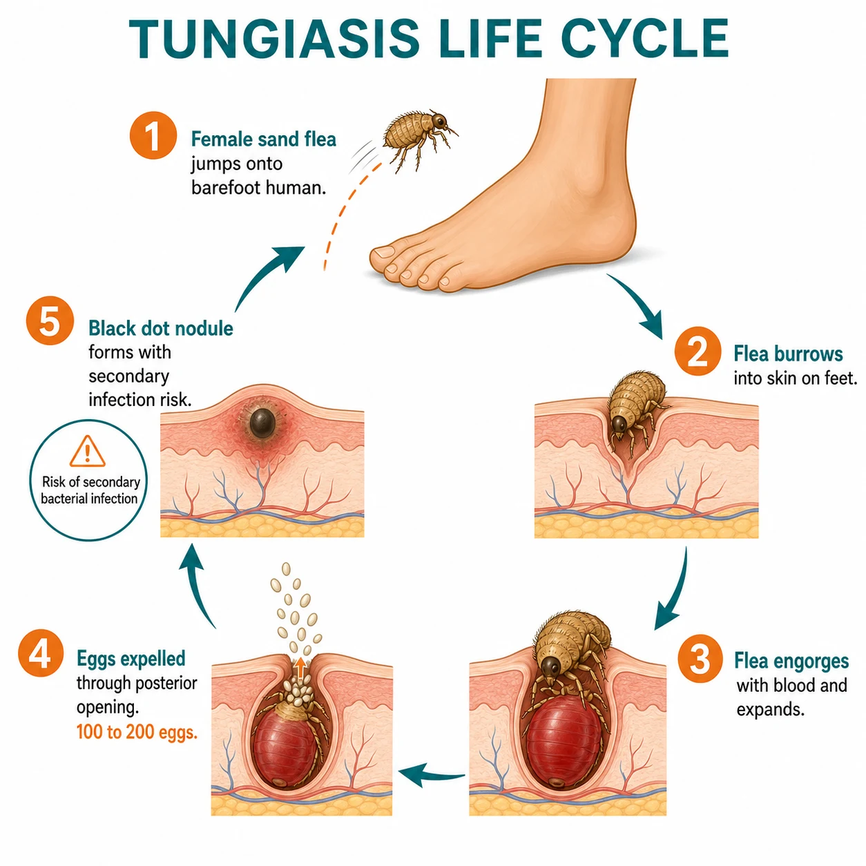
The gravid female T. penetrans (only 1 mm before infestation) jumps onto a barefoot host and rapidly burrows into the stratum corneum using its mouthparts, advancing until its abdomen is flush with the skin surface. Over the next 7 to 14 days the flea feeds on dermal blood and enlarges enormously — the abdomen swells to the size of a pea (hypertrophy of about 2000-fold) while the head remains buried deep in the dermis. The posterior cone — bearing the respiratory, excretory, and genital openings — is the only part that stays flush with the skin surface, and it is this dark ring of chitin that produces the pathognomonic black dot in the centre of the nodule.[3][10]
Over the next 7 to 10 days the flea expels 150 to 200 eggs through the posterior opening onto the ground, where they hatch into free-living larvae, pupate, and emerge as adult fleas to repeat the cycle. After egg expulsion the flea dies in situ and is either extruded or calcified in the dermis. The surrounding erythema, pain, and pruritus reflect a foreign-body and inflammatory reaction to the embedded arthropod and its faecal material; the risk of secondary bacterial infection is high because the lesion is an open portal to the dermis.[3]
Furuncular myiasis — the larva in a breathing pore
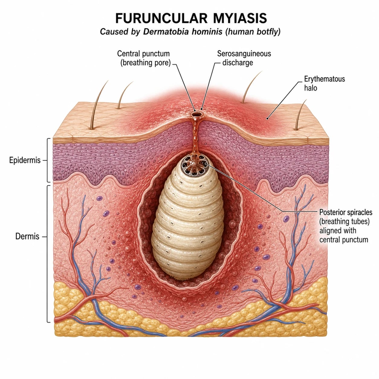
In Dermatobia hominis infestation, the first-instar larva hatches on the skin from a mosquito-borne egg and penetrates the dermis through the mosquito bite wound or a hair follicle. Inside the dermis the larva moults to the second and third instar, excavating a cavity (the furuncle) and anchoring itself with rows of cuticular spines that make manual extraction difficult and painful. Crucially, the larva's posterior spiracles (its respiratory openings) align with the central punctum on the skin surface, so it breathes atmospheric air through this opening. This anatomy is the key to treatment: occluding the punctum cuts off the oxygen supply and forces the larva to surface, after which it can be extracted with forceps.[4][5]
The surrounding inflammation produces the classical furuncle-like nodule with erythema, tenderness, and a serosanguineous or purulent discharge from the punctum; patients often report a sensation of movement or a sharp pricking pain within the lesion as the larva feeds. Rupturing the larva in situ provokes a severe foreign-body granulomatous reaction and secondary infection, so intact extraction is the aim.[5][6]
Wound myiasis — opportunistic colonisation
Facultative calliphorid flies are attracted to the odour of decaying tissue and exudate in chronic wounds, ulcers, and body orifices of debilitated patients. Eggs are laid in clusters; the larvae (screwworms or maggots) feed on necrotic tissue and serous exudate, and in obligate species such as Cochliomyia hominivorax they also invade living tissue, causing extensive destruction. Wound myiasis is therefore a marker of neglected nursing care as much as of parasitism.[4][9]
Cutaneous larva migrans — the lost hookworm
Animal hookworm larvae in dog or cat faeces hatch on warm, moist sand and penetrate the bare human skin of a beach-goer. In the natural canine or feline host they would migrate via the bloodstream to the intestine, but in the human abnormal host they cannot penetrate the basement membrane of the epidermis. They therefore wander laterally in the stratum granulosum/spinosum, advancing 1 to 2 cm per day and laying down the track of inflammation, pruritus, and serpiginous erythema that gives the disease its name. Because the larvae cannot complete their life cycle, they eventually die — but only after weeks to months of misery and a high risk of excoriation and secondary bacterial infection.[7][8]
Clinical Presentation
Tungiasis
The classic lesion is a solitary, firm, whitish-yellow nodule, 5 to 10 mm across, with a central dark punctum (the black dot), surrounded by a variable rim of erythema and scaling. The site is almost always a weight-bearing or contact area of the foot: the plantar surface, the toes, the periungual folds, the interdigital webs, and the heels. Lesions outside the feet (hands, elbows, buttocks, scrotum) occur when the host has been sitting or lying on infested ground. Pain, tenderness, and pruritus are the dominant symptoms; itch is especially prominent early and pain late as the flea enlarges and stretches the dermis.[1][3]
In hyperendemic communities the disease is polymorphic and recurrent: dozens of lesions, deformed and lost toenails, difficulty walking, fissuring, lymphangitis, and abscess formation. The most feared acute complications are secondary bacterial infection (Streptococcus pyogenes, Staphylococcus aureus), ascending lymphangitis, abscess, sepsis, tetanus, and even gas gangrene in unvaccinated children.[3][10]
Furuncular myiasis
The patient — often a returning traveller or a rural child in the neotropics — presents with one or more furuncle-like nodules on an exposed site (scalp in children; face, neck, shoulders, arms, or legs in adults) that have failed to respond to antibiotics and that are accompanied by a sharp, intermittent lancinating pain ("something moving"). Closer inspection reveals a central punctum from which serosanguineous fluid may weep, and occasionally the black or yellow posterior tip of the larva is visible at the pore, bobbing up to breathe. The surrounding skin is erythematous and oedematous; regional lymphadenopathy and low-grade fever may be present. Lesions are typically solitary with Dermatobia and multiple with Cordylobia (the tumbu fly), which may lay many eggs on soiled clothing.[5][6][9]
Wound myiasis
The presentation is a neglected, malodorous wound, ulcer, or body cavity teeming with maggots in a debilitated patient. Larvae may be visible in the wound bed or expressed on dressing changes. Surrounding tissue destruction, erythema, and a pungent odour are typical. The clinical emergency is to exclude invasion of deeper structures, the orbit, the middle ear, or the paranasal sinuses.[4][9]
Cutaneous larva migrans
The hallmark is an intensely pruritic, serpiginous (snake-like), erythematous, slightly raised track that migrates 1 to 2 cm per day as the larva advances through the epidermis. The track is tortuous and may be several centimetres long, with the leading edge (the larva) at the advancing tip and the trailing segment fading as it heals. Sites are determined by contact with contaminated sand: the feet (most common), buttocks ('beach bum'), thighs, and abdomen. Multiple tracks occur in heavy infestation. Bacterial superinfection from scratching, folliculitis, and (rarely) a Löffler-like syndrome of transient pulmonary infiltrates with eosinophilia have been reported.[7][8]
The incubation is short — pruritus usually begins within hours to a few days of beach exposure, which is why CLM is the single most common dermatological diagnosis in travellers presenting in the first one to two weeks after return. Untreated, the disease is self-limiting: the larva cannot complete its life cycle in the human dead-end host and dies after weeks to months, but the intense itch, sleep loss, and risk of impetiginisation make active treatment preferable to watchful waiting. Vesiculobullous and follicular variants are described but uncommon.[7][8]
Differential Diagnosis
The differentials overlap because all three conditions produce a nodule, a track, or a chronic wound. The discriminator is the morphology of the punctum and the movement, combined with travel and exposure history. [1]
- Furuncular myiasis|Painful furuncle-like nodule with a central breathing pore, movement sensation, serosanguineous discharge, travel to the Americas/Africa, no response to antibiotics
- Tungiasis|Solitary black-dot nodule on a weight-bearing area of the foot, periungual, barefoot exposure in an endemic community
- Bacterial furuncle / abscess|Acute, erythematous, fluctuant, painful, no central punctum to atmosphere, responds to antibiotics or drainage, purulent not serosanguineous
- Foreign-body granuloma|History of a splinter or thorn, firm nodule, no punctum, no movement
- Cutaneous leishmaniasis|Chronic slowly enlarging nodule or ulcer with a raised border and central crust, no movement, New or Old World travel, biopsy shows amastigotes
- Dracunculiasis (Guinea worm)|Blister on the lower leg that ruptures to expose the adult worm, not a furuncle; rural Africa
- Cutaneous larva migrans|Serpiginous wandering pruritic track, beach exposure, larva at the leading edge
- Larva currens (Strongyloides)|Fast-moving urticarial track (several cm per day, perianal, recurrent), not confined to the epidermis; positive Strongyloides serology
Two distinctions reward candidates. First, larva currens of strongyloidiasis moves much faster (5 to 10 cm per day, often perianal and recurrent from autoinfection) than CLM, and is urticarial rather than serpiginous-scaly. Second, migratory myiasis (from Gasterophilus or Hypoderma) produces a creeping eruption indistinguishable at sight from CLM, but the track is longer and less pruritic, the patient has animal (horse/cattle) exposure, and the larva is a fly larva — extractable rather than anthelmintic-responsive.[4][7]
Clinical & Bedside Assessment
Diagnosis is overwhelmingly clinical — the morphology plus the travel history is the test. Bedside steps that add value: [1]
- Inspect under good light and magnification (dermoscopy or a loupe). For furuncular myiasis, dermoscopy may show the white-yellow body of the larva with dark posterior spiracles (the "respiratory spikes") centred on the punctum; direct visualisation of rhythmic movement of the larval spiracles clinches the diagnosis.
- For tungiasis, the target appearance — a whitish nodule with a dark central punctum on a periungual toe — is pathognomonic. Note the number and depth of lesions, the presence of pus, lymphangitis, or systemic signs.
- For CLM, mark the leading edge with a skin-marking pen and review in 24 hours: measurable migration of the track confirms the diagnosis and excludes fixed mimics (tinea, granuloma annulare).
- Always take a full travel, occupational, animal, and footwear history, and check tetanus vaccination status in any patient with a skin infestation.[1][4][8]
Investigations
Investigations are usually unnecessary, but they have specific roles: [1]
Dermoscopy
- Furuncular myiasis: yellow-white larval segments with central dark posterior spiracles
- Confirms the larva in a furuncle-like nodule and guides extraction
- Non-invasive, rapid, and bedside
Skin biopsy / excision histopathology
- Tungiasis: cross-sections of the embedded flea with eggs, hypodermis, and exoskeleton in the dermis
- Resolves an atypical or excised lesion; not needed for the classic case
- A foreign-body granuloma around arthropod parts also supports the diagnosis
Bacterial swab and bloods
- Wound swab and blood cultures if secondary infection, lymphangitis, cellulitis, or sepsis
- FBC for eosinophilia (CLM may show mild eosinophilia; heavy infestation with superinfection shows leucocytosis)
- CRP and electrolytes if systemic involvement
Imaging
- Plain radiograph or ultrasound if deep tissue, bone (osteomyelitis), or foreign body is suspected in tungiasis
- CT/MRI for cavitary myiasis involving the orbit, ear, sinuses, or nasopharynx
Parasitology referral
- Identify the larva to species if the travel history is atypical or the public-health implication is significant (e.g., screw-worm)
- Larval posterior spiracle morphology is diagnostic
A normal eosinophil count does not exclude CLM or myiasis; eosinophilia, when present, is supportive but never the basis of the diagnosis.[4][7]
Management — Resuscitation
Most ectoparasite infestations are managed electively, but three situations demand immediate action: [1]
- Severe secondary bacterial infection (cellulitis, lymphangitis, abscess, sepsis) complicating tungiasis — take blood cultures, give empirical intravenous antibiotics against Streptococcus pyogenes and Staphylococcus aureus (e.g., intravenous flucloxacillin, or vancomycin if MRSA is suspected), and review tetanus status with tetanus toxoid and immunoglobulin as indicated.
- Cavitary myiasis involving the nasopharynx, ear, orbit, or a deep wound in a debilitated patient — this is a surgical and ENT emergency requiring removal of all larvae under anaesthesia, debridement of necrotic tissue, and systemic antibiotics; Cochliomyia hominivorax (New World screw-worm) invades living tissue and can cause catastrophic destruction.[9]
- Anaphylaxis to rupture of a larva is rare but reported during extraction of botfly larvae; have adrenaline and resuscitation equipment available when extracting deep or multiple lesions.[4]
Management — Definitive & Stepwise
Tungiasis: extract the flea en bloc
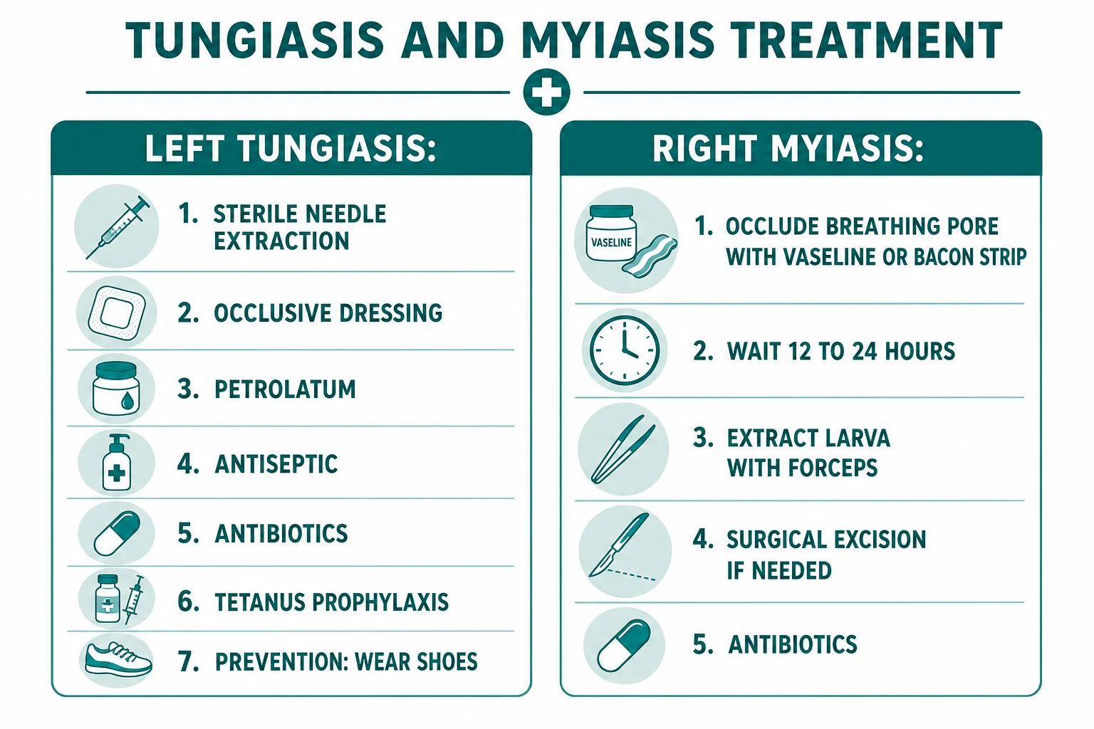
Clean the skin with antiseptic (chlorhexidine or povidone-iodine) and infiltrate local anaesthetic if the lesion is painful or deep.
Under sterile technique and good light, **curette or lift out the flea en bloc** with a sterile needle or the bevel of a scalpel; the entire flea, with eggs, must be removed — residual parts cause a foreign-body granuloma and infection.
Confirm complete extraction by inspecting the cavity; curette the edges to remove any retained eggs or chitin.
Irrigate, apply antiseptic, and dress; review in 24 to 48 hours.
Give **tetanus prophylaxis** if the patient is not fully immunised.
Prescribe **antistaphylococcal and antistreptococcal antibiotics** if there is any surrounding cellulitis or pus.
For heavy or recurrent infestation in endemic settings, **oral ivermectin 300 mcg/kg** (some sources use 200 mcg/kg) single or two doses reduces the parasite load and may be combined with topical permethrin; **topical tiabendazole 10%** or **metrifonate** have been used as suffocating agents.
The principle is mechanical completeness. A partially extracted flea continues to cause inflammation and superinfection, and retained chitin provokes a chronic granuloma. In endemic communities, mass drug administration with ivermectin and topical agents has been studied as a public-health measure, and environmental insecticide spraying, concrete floors, and footwear are the structural interventions that actually reduce transmission.[3][10]
Furuncular myiasis: asphyxiate then extract
The aim is to deprive the larva of oxygen so it migrates to the surface, after which it can be gripped and extracted intact. Rupturing the larva in situ must be avoided because the cuticular spines anchor it and the contents provoke a severe foreign-body reaction.[5][6]
Apply a thick layer of an **occlusive substance over the punctum**: petrolatum (Vaseline), petroleum jelly, bacon fat, pork fat, nail polish, or a chloroform-paraffin mixture. This blocks the larva's air supply.
Wait **3 to 24 hours** (commonly overnight); the larva emerges to breathe as its oxygen tension falls.
Gently express or grasp the larva with **mosquito forceps** at the punctum and extract it slowly and intact, taking care not to rupture it.
If extraction fails after two occlusion attempts, **surgically excise** the nodule under local anaesthetic through a small cruciate incision over the punctum.
Irrigate, apply antiseptic, and dress; prescribe antibiotics only if there is secondary infection.
Advise the patient that the wound will heal over 1 to 2 weeks with minimal scarring if the larva is removed intact.
Ivermectin (oral)
Ivermectin (oral)
Albendazole (oral)
Wound myiasis: debride and irrigate
Management is mechanical: expose the wound, remove all larvae with forceps under good lighting and irrigation (saline, dilute povidone-iodine, or ether/chloroform if larvae are deep), debride necrotic tissue, and dress the wound. Topical ivermectin and oral ivermectin have a role in extensive infestation. Address the underlying cause — neglected wound care, debility, incontinence, pressure relief — because recurrence signals a failure of basic nursing attention.[4][9]
Cutaneous larva migrans: anthelmintics, not extraction
CLM is not treated by extracting a larva — the larva is microscopic and cannot be grasped. Treatment is an oral anthelmintic: [1]
- Oral albendazole 400 mg daily for 3 to 5 days (first-line; safe in adults and children over 2 years).
- Oral ivermectin 200 mcg/kg single dose, repeated once after 7 to 14 days for extensive or relapsing disease.
- Topical thiabendazole 10 to 15% two to three times daily for 5 to 7 days — for localised, uncomplicated disease, pregnancy, or young children where oral therapy is avoided.
- Cryotherapy of the leading edge is less effective and risks ulceration; it is reserved for a single persistent track.[7][8]
Specific Subtypes & Scenarios
Dermatobia hominis (human botfly)
- Neotropics (Mexico to Argentina)
- Phoretic: eggs glued to a mosquito vector; larvae hatch on the human at the mosquito bite
- Solitary furuncular nodule; scalp in children
- Occlude the pore, extract intact
Cordylobia anthropophaga (tumbu fly)
- Sub-Saharan Africa
- Eggs laid on shaded sandy soil and soiled clothing drying on the ground; larvae penetrate warm skin on contact
- Often multiple furuncular nodules on covered areas (trunk, thighs)
- Same extraction principle; iron clothes to prevent
Cochliomyia hominivorax (New World screw-worm)
- Obligate parasite of living tissue; Central and South America
- Larvae infest wounds and body orifices and burrow into living tissue
- Cavitary and wound myiasis; tissue destruction; notifiable disease
- Surgical removal and debridement; public-health eradication programmes (sterile insect release)
Migratory (creeping) myiasis
- *Gasterophilus* (horse botfly), *Hypoderma* (cattle botfly)
- Larva migrates in the skin producing a creeping eruption
- Mimics CLM; animal exposure; track longer and less pruritic
- Extract the larva or give ivermectin
Nasopharyngeal / aural / ophthalmic myiasis
- Debilitated, elderly, or immunocompromised patients with chronic nasopharyngeal, aural, or ocular disease
- Larvae in the nasal cavity, external ear, or eye; tissue destruction
- ENT or ophthalmic emergency
- Removal under anaesthesia; topical and systemic ivermectin; antibiotics
Complications & Pitfalls
Disease complications
- Tungiasis: secondary bacterial infection (cellulitis, lymphangitis, abscess, sepsis), tetanus, gangrene, deformed or lost nails, disability, autoamputation in severe neglected disease
- Furuncular myiasis: secondary infection, foreign-body granuloma if the larva is ruptured, and (rarely) anaphylaxis during extraction
- Wound myiasis: extensive tissue destruction, especially with screw-worms; a marker of neglect
- CLM: impetiginisation from scratching, folliculitis, and rarely a Löffler-like pulmonary eosinophilic syndrome
Procedural pitfalls
- Partial extraction of Tunga with retained flea parts or eggs — causes granuloma and recurrent infection
- Incising a furuncular myiasis nodule like an abscess — releases the larva into the wound and fails to remove it
- Rupturing a botfly larva during extraction — severe foreign-body reaction and secondary infection
- Cryotherapy of CLM — painful, often ineffective, and risks ulceration; the larva is not at the visible end of the track
- Forgetting tetanus prophylaxis in any tungiasis or wound-myiasis case
Diagnostic pitfalls
- Treating furuncular myiasis with repeated courses of antibiotics because it 'looks like a boil' — always look for the breathing pore and movement
- Mislabelling larva currens (Strongyloides) as CLM — larva currens is faster, perianal, urticarial, and recurrent
- Overlooking wound myiasis in a debilitated patient with a malodorous ulcer
The single most common procedural error in exams and in practice is incising a furuncular myiasis lesion. A botfly nodule looks like a furuncle, but incising it releases the larva, contaminates the wound, and leaves the spined larva behind. The correct manoeuvre is occlusion then extraction.[5][6]
Prognosis & Disposition
With correct treatment all three conditions have an excellent prognosis. Simple tungiasis heals within days of complete extraction; furuncular myiasis heals in 1 to 2 weeks after intact larval removal; CLM resolves within days of an effective anthelmintic. The prognosis deteriorates in endemic poverty — recurrent tungiasis in resource-poor communities causes chronic disability, deformity, lost schooling, and (rarely) death from tetanus or sepsis — and in neglected wound myiasis in the debilitated, where the underlying frailty drives outcome.[3][10]
Most patients can be discharged after definitive treatment with safety-net advice: return if there is spreading erythema, increasing pain, fever, or pus. In returning travellers, screen for co-acquired tropical infections (malaria, dengue, Strongyloides, schistosomiasis) that may share the same exposure. [1]
Special Populations
Children
- Tungiasis: barefoot school-age children in endemic areas are the peak age group; polymorphic and recurrent disease
- Furuncular myiasis: scalp lesions predominate in children
- CLM: beach and sandbox exposure; dose albendazole and ivermectin by weight; favour topical thiabendazole in the very young
Pregnancy
- Topical permethrin and topical thiabendazole are preferred for localised ectoparasitosis in pregnancy
- Albendazole is avoided in the first trimester (animal teratogenicity); ivermectin is generally avoided in pregnancy where alternatives exist — weigh benefit versus risk and involve obstetrics
- Mechanical extraction of tungiasis and myiasis is safe and preferred
Elderly and debilitated
- Wound myiasis is the dominant concern in immobilised, diabetic, demented, or alcoholic patients with pressure ulcers and chronic wounds
- Multidisciplinary nursing, pressure-area care, and wound hygiene are as important as larval removal
- Screen for and treat the underlying cause of neglect
Immunocompromised
- Broader differential in the returning traveller (disseminated strongyloidiasis, cutaneous leishmaniasis, fungal mimics)
- Higher risk of secondary infection; lower threshold for systemic antibiotics and for specialist referral
Returning travellers
- Take a precise geographical and activity history (beach, jungle, farm, animal contact)
- Screen for co-acquired infections: malaria film if febrile, Strongyloides serology, STI screen
- Provide a written safety-net and travel-medicine follow-up
Evidence, Guidelines & Regional Differences
There is no single global guideline; practice draws on WHO public-health guidance for tungiasis, travel-medicine consensus for furuncular myiasis and CLM, and national dermatology guidance for management in non-endemic returning travellers. [1]
WHO / public health (Global)
- Recognises tungiasis as an important parasitic skin disease in endemic countries, emphasising structural prevention (footwear, concrete floors, sealing cracks, insecticide spraying) and ivermectin-based mass drug administration
- Screw-worm eradication by sterile insect release (Central America, Libya outbreak) is a landmark public-health intervention
PAHO / Americas
- Strong clinical guidance on Dermatobia hominis and Cochliomyia in Latin America
- Emphasises occlusion-extraction for furuncular myiasis and the public-health response to screw-worm
Europe / UK (travel medicine)
- Travel-medicine services (e.g., HPA, ECDC, Swiss TPH) provide returning-traveller management
- Ivermectin and albendazole are both accepted first-line for CLM; topical thiabendazole reserved for localised disease, pregnancy, and children
- Royal colleges include tropical ectoparasitoses in the MRCP SCE and FRCDerm curricula
IADVL / India
- Tungiasis reported from coastal and tribal India; IADVL guidance emphasises footwear and topical agents
- CLM (called 'creeping eruption') is managed with albendazole as first-line and ivermectin as second-line
Evidence base & controversies
- CLM: meta-analyses support oral ivermectin and albendazole over topical thiabendazole for efficacy, with no clear single best agent; one-dose ivermectin is convenient
- Tungiasis: evidence for oral ivermectin (300 mcg/kg) and topical metrifonate is from observational studies in endemic communities; no randomised trial of extraction technique
- Myiasis: evidence is almost entirely from case series; occlusion-extraction is consensus-based, not trial-based
The principal controversy is the drug choice for CLM: oral ivermectin (one dose, convenient) versus oral albendazole (3 to 5 days, well tolerated, globally available). Both are effective; the choice often depends on availability, pregnancy status, and patient age. For tungiasis, the structural interventions (footwear, concrete floors) are the only measures shown to reduce community burden.[3][8][10]
Scabies vs Other Ectoparasites — The Board Comparison
Because the assignment and the frontmatter link tungiasis and myiasis to the wider ectoparasite family, the examiner will frequently ask for a distinguishing table. The five ectoparasitoses a candidate must separate are scabies, pediculosis, tungiasis, myiasis, and CLM.[11]
- Organism|Sarcoptes scabiei (mite)|Tunga penetrans (gravid female sand flea)|Dermatobia hominis (botfly larva) or Cordylobia anthropophaga (tumbu fly)|Ancylostoma braziliense (dog/cat hookworm larva)
- Key lesion|Burrows in finger webs, wrists, waist, genitalia; nocturnal itch|Black-dot nodule on the foot (periungual)|Furuncle-like nodule with a breathing pore|Serpiginous pruritic migrating track
- Symptom|Intense nocturnal pruritus; family cluster|Pain, tenderness, itch; solitary or multiple|Pain, movement sensation, weeping|Intense itch; track advances 1 to 2 cm/day
- Site|Web spaces, wrists, waist, genitalia, axillae|Feet (soles, toes, periungual)|Scalp (children), face, exposed limbs|Feet, buttocks, thighs
- Endemic / exposure|Worldwide; institutional outbreaks|Tropical; poverty, barefoot, earthen floors|Neotropics (botfly) / sub-Saharan Africa (tumbu)|Tropical beaches; dog/cat faeces
- Diagnosis|Clinical; burrow scrapings with mite|Clinical; black-dot morphology|Clinical; dermoscopy shows larva|Clinical; migrating serpiginous track
- First-line treatment|Permethrin 5% two doses; oral ivermectin|Sterile needle/curette extraction; tetanus|Occlude pore then extract larva|Oral albendazole or ivermectin
- Key differentiator|Burrows + family itch + nocturnal|Black dot on a periungual toe|Breathing pore + movement|Serpiginous wandering track
The must-not-miss message: a painful furuncle on the scalp of a returning traveller from the Americas that has not responded to antibiotics is a botfly until proven otherwise; a black dot on a barefoot child's toe is tungiasis; a wandering itchy track after a beach holiday is CLM; and intense nocturnal itch shared by household contacts is scabies.[1][4][7][11]
Prevention
Tungiasis
- Closed footwear (shoes, not sandals) — the single most effective personal measure
- Concrete floors and sealed cracks in endemic housing; insecticide spraying of houses and animal resting sites
- Treat and separate domestic animals (dogs, pigs) that carry the flea
- Community mass treatment with ivermectin and topical agents in hyperendemic areas
- Tetanus vaccination in endemic communities
Furuncular myiasis
- Insect repellent (DEET) and protective clothing in the neotropics
- Iron clothes dried on the ground in sub-Saharan Africa (kills tumbu fly eggs)
- Vector (mosquito) control
- Inspect skin after rural travel
Wound myiasis
- Meticulous wound care and dressings in debilitated and immobilised patients
- Fly screens and environmental insect control in hospitals and care homes
- Pressure-area and continence care
Cutaneous larva migrans
- Wear footwear and sit on a mat on tropical beaches
- Deworm dogs and cats; cover sandboxes; control stray animals on tourist beaches
- Avoid lying directly on sand contaminated by animal faeces
Exam Pearls
FECT — the four tropical skin infestations to separate at sight
Red Flags
Exam application bank (NEET-PG / INICET)
One-line answer
Tungiasis: gravid female Tunga penetrans (sand flea) burrows into the stratum corneum of the feet → a white-yellow nodule with a central black dot (the flea's posterior respiratory cone); endemic Caribbean/sub-Saharan Africa/South America; treatment is sterile needle/curette extraction en bloc plus antiseptic and tetanus prophylaxis, with oral ivermectin 200 mcg/kg for heavy infestation. Myiasis: infestation of living tissue by dipteran fly larvae — furuncular (Dermatobia hominis botfly, Central/South America, painful nodule with a breathing pore; Cordylobia anthropophaga tumbu fly, sub-Saharan Africa), wound (Lucilia/Calliphora/Cochliomyia in neglected wounds), migratory, and nasopharyngeal; treatment is occlude the pore with petrolatum/bacon to asphyxiate the larva → extract with forceps, plus debridement for wound myiasis. Cutaneous larva migrans: dog/cat hookworm larvae ( [1]
Worked stems (answer without another resource)
Stem 1 — Classic presentation. Map symptoms to mechanism; name the first investigation and first treatment step with dose/route if drug therapy is standard. [1]
Stem 2 — Unstable / complicated. List red flags that force immediate resuscitation, theatre, ICU, antidote, or reperfusion — and what you do in the first 15 minutes. [1]
Stem 3 — Atypical group. Elderly, pregnancy, child, or immunocompromised: how presentation and thresholds change. [1]
Stem 4 — Differential trap. Name the three closest mimics and one discriminator for each. [1]
Stem 5 — Disposition. Who goes home with safety-netting, who is admitted, who needs HDU/ICU/theatre, and what follow-up is mandatory. [1]
Rapid viva checklist
- Definition + classification
- Pathophysiology chain
- Bedside signs / criteria
- Score with exact components (if any)
- Emergency bundle
- Definitive therapy with doses
- Complications of disease and of treatment
- Special populations
- Guideline/trial name if classic
- Three exam traps
Coverage self-check
If you cannot answer any stem above from this page alone, re-read the matching section — the page is intended to be self-sufficient for final-prof and NEET-PG/INICET questions on Tungiasis and myiasis.
References
- [1]Coates SJ, Thomas C, Chosidow O, et al. Ectoparasites: Pediculosis and tungiasis J Am Acad Dermatol, 2020.PMID 31306729
- [2]Cestari TF, Pessato S, Ramos-e-Silva M. Tungiasis and myiasis Clin Dermatol, 2007.PMID 17350494
- [3]Heukelbach J. Tungiasis Rev Inst Med Trop Sao Paulo, 2005.PMID 16553319
- [4]Francesconi F, Lupi O. Myiasis Clin Microbiol Rev, 2012.PMID 22232372
- [5]Solomon M, Lachish T, Schwartz E. Cutaneous Myiasis Curr Infect Dis Rep, 2016.PMID 27443558
- [6]McGraw TA, Turiansky GW. Cutaneous myiasis J Am Acad Dermatol, 2008.PMID 18485982
- [7]Leung AKC, Barankin B, Hon KLE. Cutaneous Larva Migrans Recent Pat Inflamm Allergy Drug Discov, 2017.PMID 28078983
- [8]Palaniappan V, Gopinath H, Karthikeyan K. Cutaneous larva migrans Clin Exp Dermatol, 2026.PMID 40795202
- [9]Calvopina M, Ortiz-Prado E, Castañeda B, et al. Human myiasis in Ecuador PLoS Negl Trop Dis, 2020.PMID 32084134
- [10]Heukelbach J, Feldmeier H. Ectoparasites--the underestimated realm Lancet, 2004.PMID 15032237
- [11]Galván-Casas C, Salavastru C, Ortiz-Álvarez J. Scabies and other ectoparasitoses Clin Exp Dermatol, 2026.PMID 40567146