Dermatology · Medicine
Urticaria (hives) and angioedema
Also known as Urticaria · Hives · Chronic spontaneous urticaria (CSU) · Chronic idiopathic urticaria (CIU) · Angioedema · Physical/inducible urticaria · Hereditary angioedema (HAE) · Urticarial vasculitis
Urticaria (hives) is a mast-cell-driven disease producing transient (<24h), intensely itchy, raised erythematous wheals with central pallor, caused by histamine and other mediators producing dermal oedema. Angioedema is the deeper counterpart (submucosal/subcutaneous swelling, non-pitting, especially lips, tongue, eyelids, genitals, airway). Classified: acute (<6 weeks) — often triggered by infection, drugs (NSAIDs, antibiotics), food, sting; chronic (6 weeks) — chronic spontaneous urticaria (CSU, most common; often autoimmune with anti-FcεRI or anti-IgE), inducible (cold, pressure, dermographism, cholinergic, solar, aquagenic). Hereditary angioedema (C1-inhibitor deficiency, bradykinin-mediated, NO urticaria, low C4) requires C1-INH/icatibant — NOT antihistamines. Urticarial vasculitis (lesions 24h, bruising, systemic symptoms) requires biopsy. Management: 2nd-gen H1 antihistamines (up-dose 4x) → H2 + montelukast → omalizumab → ciclosporin.
On this page & tools
Your progress
Saved locally on this device.
Exam tags
Red flags
Urticaria at a glance — the high-yield numbers
Pathophysiology & high-yield pearls
Urticaria quick numbers
UAS7 - urticaria activity score
0 none, 1 less than 20 wheals, 2 twenty to fifty, 3 more than fifty
Wheal score 0-3 + itch score 0-3 = 0-6 per day
Well controlled 0, mild 1-6, moderate 7-15, severe 16-42
UAS7 = weekly score, gold standard for CSU monitoring
Triggers of acute and chronic urticaria [1]
Triggers are clinically useful because they shape the history, inform provocation testing, and tell the patient what to avoid. The four core trigger categories are food, drug, physical, and contact triggers, with infection and autoimmune processes acting as upstream activators of chronic disease.[1][3]
| Trigger category | Specific examples | Mechanism | Notes |
|---|---|---|---|
| Food (IgE-mediated) | Cow's milk, hen's egg, peanut, tree nut, sesame, fish, shellfish, wheat, soya | Type I IgE-FcεRI mast-cell degranulation | True food allergy is rare in chronic urticaria; pseudoallergens (salicylates, amines, additives) more relevant in CSU |
| Food (pseudoallergen / non-IgE) | Strawberries, tomatoes, cheese, fermented foods, red wine, shellfish, artificial colourings/preservatives | Direct mast-cell activation or COX-1 inhibition | 3-week low-pseudoallergen diet may help selected CSU patients |
| Drugs (IgE-mediated) | Penicillins, cephalosporins, neuromuscular blockers, latex-hevein cross-reactive foods | Type I IgE | Acute urticaria ± anaphylaxis; can occur on first re-exposure |
| Drugs (non-IgE / COX-1) | Aspirin, NSAIDs (ibuprofen, naproxen, diclofenac), opiates (morphine, codeine), vancomycin, rifampicin, quinolones | COX-1 inhibition → leukotriene overproduction; direct mast-cell degranulation | NSAID-exacerbated cutaneous disease (NECD) in up to 30% of CSU; COX-2-selective safer |
| Drugs (ACE-inhibitors) | Captopril, enalapril, lisinopril, ramipril, perindopril | ↓ bradykinin degradation | Angioedema WITHOUT urticaria; onset can be weeks to years after starting |
| Physical | Cold, heat, pressure, UV/visible light, vibration, water, stroking (dermographism) | Mast-cell degranulation via specific cutaneous receptors | Each inducible type has a standardised provocation test with a defined threshold (TempTest, FricTest, pressure gauges) |
| Contact | Latex (gloves, catheters, balloons), chemicals (sodium benzoate, cinnamaldehyde, sorbic acid), cosmetics, plant saps, animal saliva | Percutaneous IgE-mediated or non-IgE histamine release | May be localised (contact urticaria) or generalised if absorption is high |
| Insect sting / bite | Honeybee, wasp, hornet, fire ant; mosquito, horsefly | Type I IgE venom allergy | Acute urticaria ± anaphylaxis; venom immunotherapy for repeated stings |
| Infection | Viral URTI (esp. children — rhinovirus, coronavirus, EBV, CMV), bacterial (H. pylori, Streptococcus, Staphylococcus), parasitic (Giardia, helminths), hepatitis B/C, herpes simplex | Immune-complex / molecular mimicry / chronic inflammation | Most common cause of acute urticaria in children; treat underlying infection |
| Autoimmune / autoimmune-association | Anti-FcεRIα / anti-IgE autoantibodies; thyroid autoimmunity (anti-TPO, anti-TG); coeliac disease; SLE; type 1 diabetes | Type IIb autoantibody; FcεRI cross-linking; chronic Th1/Th2 dysregulation | Strongest association: anti-TPO antibodies in CSU (~20–30%) |
Definition and overview
Urticaria (hives) is a mast-cell-driven disease of the skin and mucosa producing transient (<24h), pruritic, raised erythematous wheals with central pallor, caused by vasodilation and increased vascular permeability from mast-cell mediator release (primarily histamine). Angioedema is the deeper counterpart: submucosal/subcutaneous tissue swelling that is non-pitting, non-dependent, and not always itchy (may cause a burning/tight sensation), classically affecting the lips, tongue, eyelids, genitals, and larynx. Angioedema may accompany urticaria (mast-cell/histamine-mediated) or occur alone (bradykinin-mediated — hereditary angioedema, ACE-inhibitor angioedema).[1][3]
The lifetime prevalence of acute urticaria is ~20% of the population; chronic spontaneous urticaria (CSU) affects 0.5–1% of the population at any time. The key clinical skill is distinguishing benign acute urticaria (often post-viral in children) from CSU (autoimmune, needs stepwise therapy) from life-threatening angioedema/anaphylaxis (airway threat) from hereditary angioedema (C1-INH deficiency — NOT responding to antihistamines).[1][2]
Classification
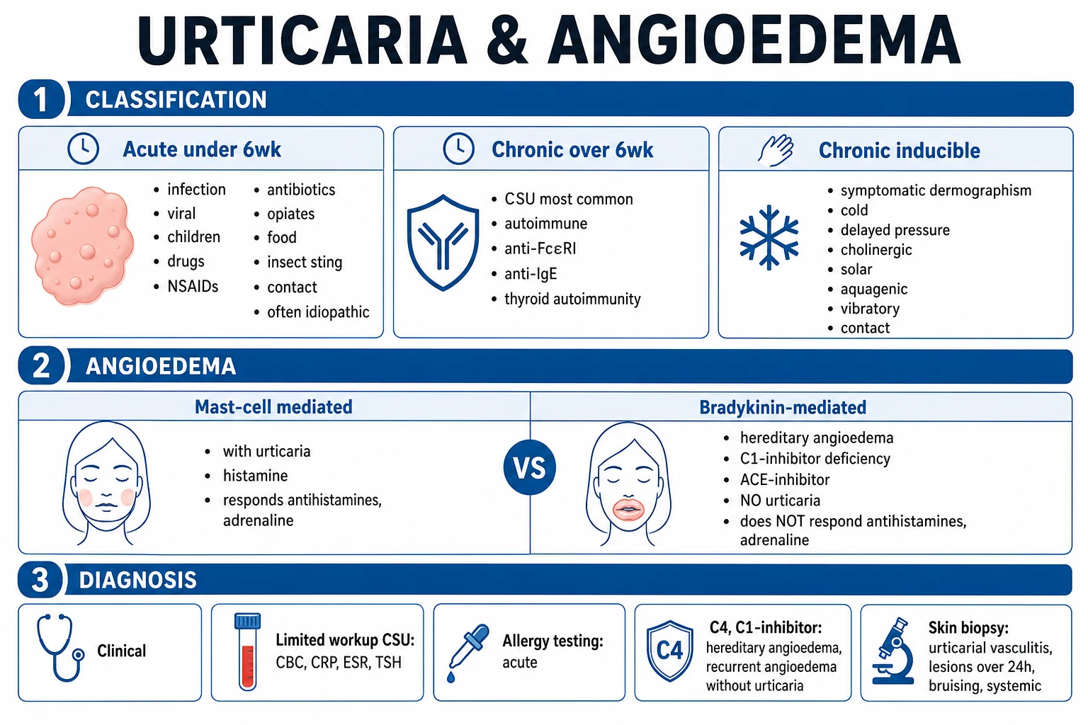
By duration
| Type | Duration | Common causes |
|---|---|---|
| Acute urticaria | <6 weeks | Infection (viral, bacterial), drugs (NSAIDs, antibiotics, opiates), food (shellfish, nuts), insect sting, contact allergen, idiopathic |
| Chronic urticaria | >6 weeks | See below |
Chronic urticaria subtypes
- Chronic spontaneous urticaria (CSU) — formerly chronic idiopathic urticaria (CIU); the commonest form; wheals and/or angioedema occur spontaneously without an identifiable external trigger. 30–50% are autoimmune (anti-FcεRIα or anti-IgE autoantibodies activating mast cells and basophils). Associated with thyroid autoimmunity (anti-TPO), Helicobacter pylori, and other autoimmune conditions.[4][6]
- Chronic inducible urticaria — wheals are triggered by a specific physical stimulus:
- Symptomatic dermographism — wheals from shearing force/stroking skin (most common inducible type)
- Cold contact urticaria — cold air, water, objects (check cold stimulation test; rule out cryoglobulinaemia)
- Delayed pressure urticaria — wheals 4–8h after sustained pressure (backpack straps, tight clothing, walking)
- Cholinergic urticaria — tiny pinpoint wheals triggered by heat, exercise, emotion (sweat-induced)
- Solar urticaria — UV or visible light exposure (rule out porphyria with porphyrin screen)
- Aquagenic urticaria — water of any temperature
- Vibratory angioedema/urticaria — vibration (vibratory tools, jogging)
- Heat contact urticaria — direct contact with warm objects
Angioedema classification (critical distinction)
| Type | Mechanism | Urticaria present? | Responds to antihistamines? |
|---|---|---|---|
| Mast-cell–mediated | Histamine | Yes (usually) | Yes |
| Hereditary angioedema (HAE) | Bradykinin (C1-INH deficiency) | No | No |
| Acquired angioedema (AAE) | Bradykinin (C1-INH consumption) | No | No |
| ACE-inhibitor angioedema | Bradykinin (↓ degradation) | No | No |
| Idiopathic histaminergic | Histamine | Variable | Yes |
Pathophysiology
Mast-cell/histamine pathway (urticaria and most angioedema)
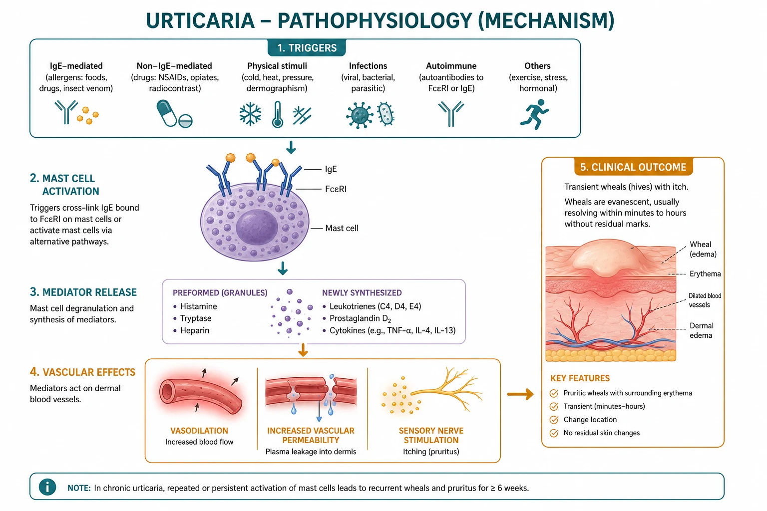
Mast cells in the dermis (and basophils in circulation) degranulate upon cross-linking of FcεRI (high-affinity IgE receptor) by allergen-IgE complexes, or upon activation by:
- Autoantibodies: anti-FcεRIα or anti-IgE (in CSU — functional autoantibodies directly activate mast cells without external allergen)[6]
- Complement: C5a (anaphylatoxin)
- Drugs: NSAIDs (cyclooxygenase inhibition → leukotriene shift), opiates, vancomycin (direct mast-cell degranulation)
- Physical stimuli: cold, pressure, heat (inducible urticarias)
Degranulation releases histamine (H1 receptor: vasodilation, pruritus, increased permeability; H2 receptor: gastric acid, also contributes to vasodilation), prostaglandin D2, leukotrienes (LTC4, LTD4, LTE4), tryptase, and cytokines (TNF-α, IL-4, IL-13). The net effect is dermal vasodilation and increased vascular permeability → wheal formation.[1][4]
Bradykinin pathway (hereditary angioedema, ACE-inhibitor angioedema)
Bradykinin is a potent vasoactive peptide that increases vascular permeability in the deep dermis and submucosa (producing angioedema without wheals). It is normally degraded by:
- Angiotensin-converting enzyme (ACE) — hence ACE inhibitors cause angioedema by blocking bradykinin degradation
- C1-inhibitor (C1-INH / SERPING1) — inhibits kallikrein (which generates bradykinin from high-molecular-weight kininogen) [1]
In hereditary angioedema (HAE), deficiency of C1-INH → uncontrolled kallikrein activity → excess bradykinin → angioedema without urticaria or itching. Critically, bradykinin-mediated angioedema does NOT respond to antihistamines, corticosteroids, or adrenaline.[9][10][11]
Clinical features
Urticaria (wheals)
- Morphology: raised, erythematous plaques with a pale centre (central pallor from vasoconstriction); sizes range from pinpoint (cholinergic) to large (>10 cm coalescent plaques); intensely pruritic.
- Duration: individual wheals resolve within <24 hours (the key distinguishing feature from urticarial vasculitis where lesions persist >24 hours); wheals migrate — new ones appear as old ones fade, and they resolve without residual marks (no bruising, no scaling, no pigment change).
- Distribution: any body site; commonly trunk, extremities, and areas of pressure (waistband, under bra straps). [1]
Angioedema
- Swellings of the deep dermis/subcutis/submucosa: lips, tongue, eyelids, face, hands, feet, genitals, and potentially the larynx (airway threat).
- Non-pitting, non-dependent (does not settle with gravity), skin-coloured or slightly erythematous; patients describe a tight or burning sensation rather than itch.
- Onset: minutes to hours; resolves over 24–72 hours. [1]
Clinical patterns by type
- Acute urticaria: sudden onset of widespread wheals ± angioedema; often follows infection (especially viral in children), drug ingestion, or food; resolves within days–weeks.
- CSU: daily or near-daily wheals ± angioedema for >6 weeks; course unpredictable (months to years); pruritus is most severe at night, disrupting sleep.[2][13]
- Dermographism: wheals appear within minutes of stroking or scratching the skin (positive "write on skin" test).
- Cold urticaria: wheals on cold-exposed skin; may be triggered by cold water, wind, or food (ice cream); risk of systemic anaphylaxis with whole-body cold exposure (swimming).[1]
- Cholinergic urticaria: pinpoint (1–3 mm) wheals surrounded by a large flare, triggered by exercise, hot showers, emotion, or spicy food; lesions are intensely pruritic.
- Delayed pressure urticaria: deep, tender swellings 4–8 hours after sustained pressure (e.g., on feet after walking, on shoulders from backpack); may be painful rather than itchy.
- Acute urticaria: sudden onset of widespread wheals ± angioedema; often follows a viral upper respiratory tract infection (especially in children), a drug, or a food; usually resolves within days to less than 6 weeks. NSAIDs (ibuprofen, aspirin), beta-lactam antibiotics, opiates (morphine, codeine), and insect stings are common precipitators.
- CSU: daily or near-daily wheals ± angioedema for more than 6 weeks; course is unpredictable (mean duration 1–5 years); pruritus is most severe at night, disrupting sleep; ~50% have angioedema and ~10% have angioedema alone.[2][13]
- Symptomatic dermographism (urticaria factitia): the most common chronic inducible urticaria; wheals appear within 1–5 minutes of stroking or scratching the skin (positive FricTest / dermographometer reading ≥3 on a 0–4 scale); triggered by towel-drying, scratching, tight clothing; resolves in 30 minutes.
- Cold contact urticaria: wheals on cold-exposed skin within minutes; confirmed by ice-cube test (5 min on forearm, observe for 10 min) or TempTest (graded 4–44 °C); threshold temperature guides prognosis (≥ −10 °C good; ≥25 °C — high risk of systemic reactions); cold food (ice cream) may trigger oropharyngeal angioedema; whole-body immersion (swimming) carries risk of anaphylaxis and drowning — advise against swimming, prescribe adrenaline auto-injector.[1][3]
- Cholinergic urticaria: pinpoint (1–3 mm) wheals surrounded by a large erythematous flare, triggered by any rise in core body temperature — exercise, hot showers, fever, emotional stress, spicy food; provoked by passive warming or a bicycle-ergometer test in clinic; intensely pruritic; can overlap with cold urticaria and exercise-induced anaphylaxis.
- Delayed pressure urticaria: deep, tender, often painful swellings 4–8 hours after sustained pressure (shoulder strap, buttocks on a hard chair, soles of feet after walking); confirmed by a calibrated pressure stimulator (2.5–7.5 kg over 15 min); typically refractory to antihistamines and may need NSAIDs, dapsone, or omalizumab.
- Solar urticaria: wheals within minutes of UV or visible-light exposure (wavelength-specific action spectrum tested with a monochromator); rule out erythropoietic protoporphyria and lupus with porphyrin screen and ANA; photoprotective clothing and broad-spectrum sunscreen first-line; omalizumab for refractory cases.
- Heat contact urticaria: rare; wheals at sites of direct contact with warm objects (more than 38–45 °C); confirmed by hot-arm bath or TempTest.
- Aquagenic urticaria: rare; wheals within minutes of contact with water of any temperature; tested by applying a wet compress at body temperature (37 °C); differentiated from cholinergic by absence of thermal trigger and from cold urticaria by tepid water.
- Vibratory angioedema/urticaria: familial or acquired; swellings within minutes of vibration (jogging, mountain biking, vibratory tools, motorbike handlebars); confirmed by a laboratory vortex test (4 min at 1380 rpm against the forearm); autosomal-dominant hereditary vibratory angioedema has been linked to ADGRE2 mutations.
- Contact urticaria: localised wheals within 30–60 minutes of skin contact with the offending substance (latex gloves, ammonium persulphate in hair bleach, cinnamaldehyde in toothpaste, sodium benzoate, balsam of Peru); tested by open application or prick testing; can be IgE-mediated (latex, peanut protein in cookware) or non-immunologic (sorbic acid, cinnamic acid).
Differential diagnosis
| Condition | Key distinguishing features |
|---|---|
| Urticarial vasculitis | Lesions persist >24h, are painful/burning (not just itchy), leave residual bruising/hyperpigmentation; may have systemic symptoms (fever, arthralgia, abdominal pain); biopsy shows leukocytoclastic vasculitis; associated with SLE, hepatitis B/C, hypocomplementaemia.[7] |
| Hereditary angioedema | Recurrent angioedema WITHOUT urticaria or itching; family history (AD); low C4 and C1-INH (antigen and function); triggered by trauma, dental work, stress, menstruation; NOT responsive to antihistamines/adrenaline.[9][10] |
| ACE-inhibitor angioedema | Onset weeks to years after starting ACE inhibitor; tongue/lip swelling without urticaria; more common in Black patients; stop the drug.[11] |
| Contact dermatitis | Localised, eczematous, at the site of allergen exposure; not transient wheals. |
| Erythema multiforme | Targetoid lesions; mucosal involvement; persists days (not <24h). |
| Atopic dermatitis | Chronic, lichenified, flexural distribution; not transient wheals. |
| Serum sickness | Urticaria-like rash + fever + arthralgia + lymphadenopathy; immune-complex mediated. |
| Mastocytosis (urticaria pigmentosa) | Red-brown macules that urticate on rubbing (Darier sign); persists (not transient); skin biopsy shows mast cell infiltrate; serum tryptase elevated. |
Investigations
Acute urticaria
- Diagnosis is clinical — no routine investigations needed.
- If an allergen is suspected: specific IgE (RAST) or skin prick testing (refer to allergy specialist); food diary if food allergy suspected. [1]
Chronic spontaneous urticaria
- Limited workup: the EAACI/GA²LEN guideline recommends no routine allergy testing for CSU (food allergy is rarely the cause of chronic urticaria).[3]
- FBC and CRP/ESR (rule out systemic inflammation/infection).
- Thyroid function (TSH) and anti-thyroid antibodies (anti-TPO) — autoimmune thyroid disease is the most common autoimmune comorbidity in CSU.[6]
- Helicobacter pylori stool antigen or breath test (controversial; treat if positive).
- Autologous serum skin test (ASST) — intradermal injection of the patient's own serum; a positive wheal suggests autoimmune urticaria (anti-FcεRIα or anti-IgE autoantibodies).[6]
- Basophil activation test (BAT) or anti-FcεRIα/anti-IgE assay (research/specialist).
Suspected urticarial vasculitis
- Skin biopsy (punch biopsy of a lesion >24h old): leukocytoclastic vasculitis (neutrophilic infiltrate, nuclear dust, fibrinoid necrosis of vessel walls).
- Complement levels: C3, C4, C1q (low in hypocomplementaemic UV — Schnitzler syndrome, UV associated with SLE).[7][8]
- ANA, anti-dsDNA (rule out SLE); hepatitis B/C serology; cryoglobulins; serum protein electrophoresis (Schnitzler = monoclonal IgM + chronic urticaria).
Suspected hereditary angioedema
- C4 level (low at baseline and during attacks — the best screening test).
- C1-inhibitor antigen (level) and C1-inhibitor function (functional assay is more sensitive than antigenic).
- C1q level (low in acquired C1-INH deficiency associated with lymphoproliferative disease; normal in HAE types I/II).[9][10]
- Genetic testing for SERPING1 mutations (HAE type I/II) and F12 mutations (HAE type III, oestrogen-related, normal C1-INH).
Disease activity and control scoring — UAS7 and UCT
Two validated patient-reported outcome measures drive CSU management decisions and define the goal of treatment (UAS7 ≤6, UCT ≥12) in the EAACI/GA²LEN/EuroGuiDerm 2022 guideline.[3][12]
Urticaria Activity Score (UAS7) is the gold-standard prospective daily diary used in trials and in motivated patients. Each day, the patient scores number of wheals (0 = none, 1 = less than 20, 2 = 20–50, 3 = more than 50) and intensity of pruritus (0 = none, 1 = mild, 2 = moderate, 3 = intense). The two daily subscores are added (range 0–6 per day) and summed over 7 consecutive days to give the UAS7 (range 0–42). Categories: [1]
| UAS7 score | Disease activity | Treatment implication |
|---|---|---|
| 0 | Urticaria-free | Treatment goal |
| 1–6 | Well-controlled | Continue current therapy |
| 7–15 | Mild activity | Step up if persistent |
| 16–27 | Moderate activity | Step up; consider omalizumab |
| 28–42 | Severe activity | Step up; refer for biologic |
Urticaria Control Test (UCT) is the validated retrospective 4-item questionnaire covering symptoms, quality of life, treatment effectiveness and overall control over the previous 4 weeks (range 0–16). It is the practical choice in clinic — quick to administer and reliable between visits. [1]
| UCT score | Disease control | Treatment implication |
|---|---|---|
| ≥12 | Well-controlled | Continue current therapy |
| ≤11 | Poorly-controlled | Step up treatment |
| Change ≥3 points | Minimal clinically important difference (MCID) | Therapeutic response confirmed |
| Change ≥6 points | Marked response | Continue / consider taper once sustained |
The treatment goal in CSU is complete control (UAS7 = 0; UCT ≥ 12) maintained for at least 1–3 months before any attempt to taper. Treat-to-target is now the recommended paradigm (Maurer 2025).[12]
Management
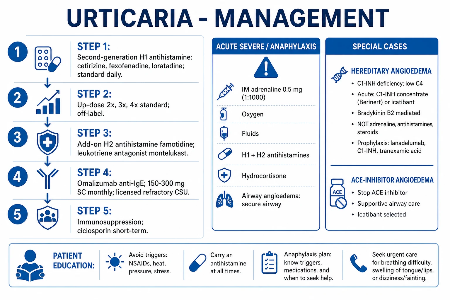
Step 1: Second-generation H1 antihistamine (first-line for all urticaria)
- Cetirizine 10 mg, levocetirizine 5 mg, fexofenadine 180 mg, loratadine 10 mg, desloratadine 5 mg, or bilastine 20 mg once daily.[3][12]
- Second-generation agents are non-sedating (do not cross the blood-brain barrier) and have minimal anticholinergic effects — preferred over first-generation (chlorphenamine, hydroxyzine, promethazine).
Step 2: Up-dose H1 antihistamine (up to 4× standard dose)
- Increase to 2×, 3×, or up to 4× the standard dose (e.g., cetirizine 40 mg daily).[3][12]
- Evidence-based and safe (off-label); the EAACI/GA²LEN guideline explicitly supports up-dosing to 4× before adding other agents.
Step 3: Add-on therapies
- H2 antihistamine: famotidine 20 mg BD (adds histamine H2-receptor blockade; some vasodilation is H2-mediated).
- Leukotriene receptor antagonist: montelukast 10 mg nocte (especially useful for NSAID-exacerbated CSU).
- First-generation H1 at night (hydroxyzine 25–50 mg) for sedation and nocturnal pruritus. [1]
Step 4: Omalizumab (anti-IgE monoclonal antibody)
- Omalizumab 150–300 mg subcutaneously every 4 weeks — licensed and effective for refractory CSU.[2][12]
- Mechanism: reduces free IgE → downregulates FcεRI on mast cells/basophils → reduced mast-cell activation.
- Response rate: ~65% of patients achieve complete or near-complete response; onset within 1–2 weeks; well-tolerated.
- Approved by NICE, FDA, and EMA for CSU refractory to H1 antihistamines.
Step 5: Immunosuppression (severe, refractory)
- Ciclosporin 3–4 mg/kg/day (short-term, 3–6 months) — inhibits T-cell activation and IL-2 (calcineurin pathway); effective but nephrotoxic; monitor blood pressure and renal function.
- Emerging biologics: dupilumab (anti-IL-4Rα), ligelizumab (anti-IgE, more potent than omalizumab), remibrutinib (BTK inhibitor, oral).[12]
Acute severe / anaphylaxis
- IM adrenaline (epinephrine) 0.5 mg (0.5 mL of 1:1000) into the anterolateral thigh — repeat after 5 minutes if no response.
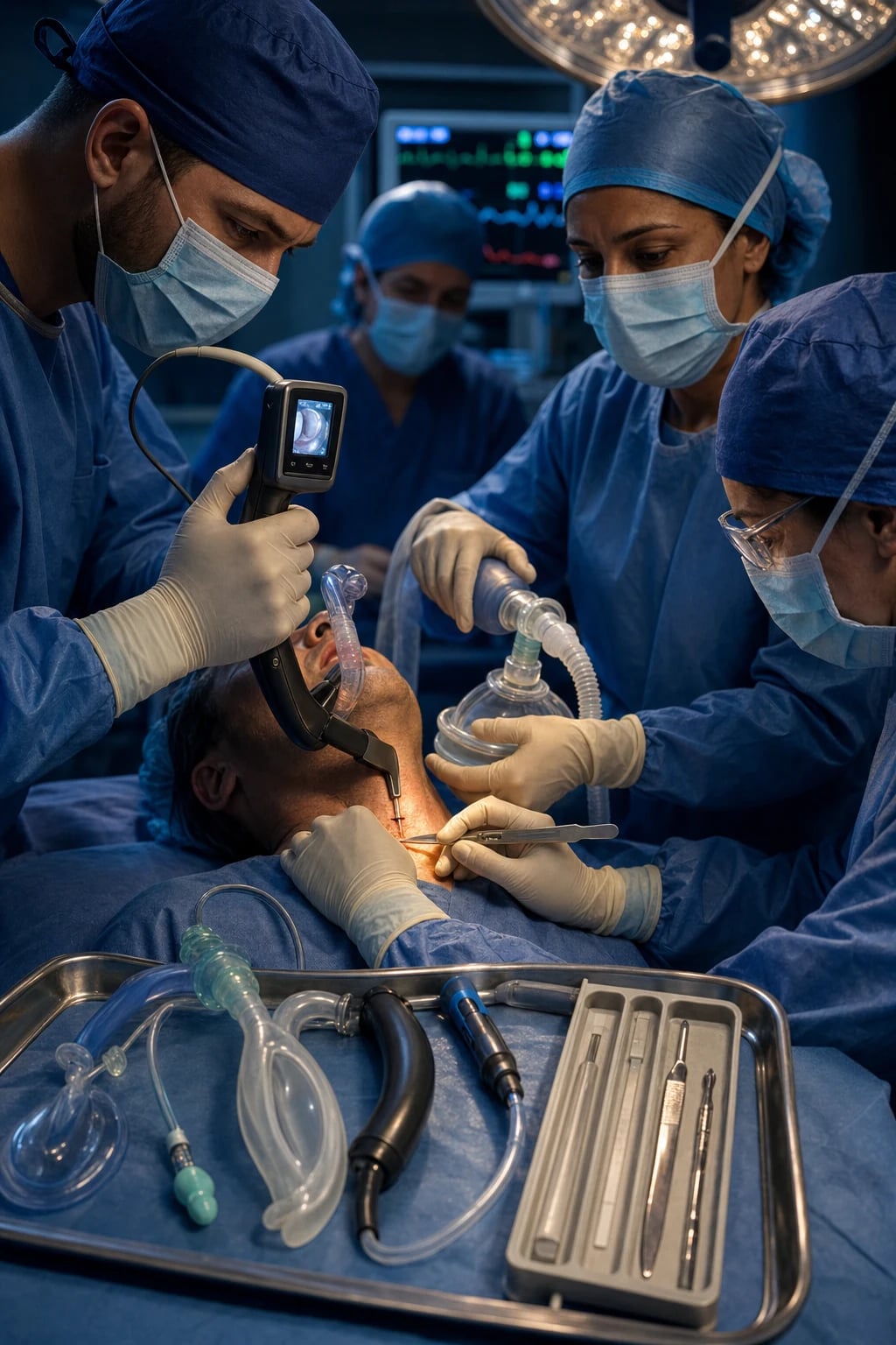
- Oxygen (high-flow), IV fluids (crystalloid), chlorphenamine 10 mg IV, hydrocortisone 200 mg IV ( adjunctive; onset slow).
- Recovery position, call for help, monitor airway; ICU admission if airway compromised. [1]
Hereditary angioedema (HAE)
- Autosomal dominant (SERPING1 gene → C1-INH deficiency); penetrance ~80%.
- Type I (85%): low C1-INH antigen AND function.
- Type II (15%): normal or elevated C1-INH antigen but dysfunctional protein (low function).
- Type III: normal C1-INH (F12 gene mutation; rare; oestrogen-sensitive; females only).
- Triggers: trauma, dental work, surgery, stress, infection, menstruation, ACE inhibitors, oestrogen-containing contraceptives.
- Clinical features: episodic angioedema (face, airway, GI tract → severe abdominal pain from bowel wall oedema) without urticaria or itching; attacks last 24–72 hours.[9][10]
- Acute treatment: C1-INH concentrate (berinert/ruconest) IV, icatibant (bradykinin B2 receptor antagonist) SC, or ecallantide (kallikrein inhibitor) SC. NOT antihistamines, corticosteroids, or adrenaline (ineffective for bradykinin-mediated angioedema).
- Prophylaxis: lanadelumab (anti-kallikreb mAb, SC every 2 weeks), berotralstat (oral plasma kallikrein inhibitor), C1-INH (IV/SC regularly), attenuated androgens (danazol — increases hepatic C1-INH synthesis; virilising side effects limit use).[9][10]
Hereditary angioedema — acute attack and prophylaxis timeline
Urticarial vasculitis
- Lesions persist >24 hours, are painful/burning (not just pruritic), and leave residual bruising or hyperpigmentation.[7]
- May have systemic features: fever, arthralgia, abdominal pain, renal disease (proteinuria, haematuria), and pulmonary involvement.
- Hypocomplementaemic UV (low C3, C4, C1q) is associated with SLE; Schnitzler syndrome = chronic urticaria + monoclonal IgM gammopathy + fever + bone pain (interleukin-1 driven; treat with anakinra/canakinumab).
- Biopsy is diagnostic: leukocytoclastic vasculitis (neutrophilic infiltrate, nuclear dust, fibrinoid necrosis of post-capillary venules).
- Treatment: antihistamines (often insufficient), NSAIDs/colchicine/dapsone for mild disease; systemic corticosteroids, hydroxychloroquine, mycophenolate mofetil, or rituximab for refractory/hypocomplementaemic disease.[8]
Special populations
- Children: acute urticaria is common (usually post-viral); weight-adjusted antihistamine dosing; reassure parents; omalizumab is approved for CSU in children ≥12 years.[5]
- Pregnancy: cetirizine and loratadine are safe (category B); avoid omalizumab if possible (limited data); manage triggers; avoid aspirin/NSAIDs.[5]
- HAE: screen family members (C4 level ± genetic testing); medic-alert bracelet; carry emergency treatment (C1-INH/icatibant); avoid ACE inhibitors and oestrogen.[10]
High-yield snapshots
Hereditary angioedema — three high-yield handles
The seven chronic inducible urticarias — one-line flashcards
Exam pearls
[1]Red flags
Exam application bank (NEET-PG / INICET)
One-line answer
Urticaria (hives) is a mast-cell-driven disease producing transient (<24h), intensely itchy, raised erythematous wheals with central pallor, caused by histamine and other mediators producing dermal oedema. Angioedema is the deeper counterpart (submucosal/subcutaneous swelling, non-pitting, especially lips, tongue, eyelids, genitals, airway). Classified: acute (<6 weeks) — often triggered by infection, drugs (NSAIDs, antibiotics), food, sting; chronic (>6 weeks) — chronic spontaneous urticaria (CSU, most common; often autoimmune with anti-FcεRI or anti-IgE), inducible (cold, pressure, dermographism, cholinergic, solar, aquagenic). Hereditary angioedema (C1-inhibitor deficiency, bradykinin-mediated, NO urticaria, low C4) requires C1-INH/icatibant — NOT antihistamines. Urticarial vasculitis (lesions >24h, bruising, systemic symptoms) requires biopsy. Management: 2nd-gen H1 antihistami
Worked stems (answer without another resource)
Stem 1 — Classic presentation. Map symptoms to mechanism; name the first investigation and first treatment step with dose/route if drug therapy is standard. [1]
Stem 2 — Unstable / complicated. List red flags that force immediate resuscitation, theatre, ICU, antidote, or reperfusion — and what you do in the first 15 minutes. [1]
Stem 3 — Atypical group. Elderly, pregnancy, child, or immunocompromised: how presentation and thresholds change. [1]
Stem 4 — Differential trap. Name the three closest mimics and one discriminator for each. [1]
Stem 5 — Disposition. Who goes home with safety-netting, who is admitted, who needs HDU/ICU/theatre, and what follow-up is mandatory. [1]
Rapid viva checklist
- Definition + classification
- Pathophysiology chain
- Bedside signs / criteria
- Score with exact components (if any)
- Emergency bundle
- Definitive therapy with doses
- Complications of disease and of treatment
- Special populations
- Guideline/trial name if classic
- Three exam traps
Coverage self-check
If you cannot answer any stem above from this page alone, re-read the matching section — the page is intended to be self-sufficient for final-prof and NEET-PG/INICET questions on Urticaria (hives) and angioedema.
[1]References
- [1]Sánchez-Borges M, Ansotegui IJ, Bielory L, et al. Urticaria Nat Rev Dis Primers, 2022.PMID 36109590
- [2]Saini SS, Kaplan AP. Chronic Spontaneous Urticaria: A Review JAMA, 2024.PMID 39325444
- [3]Zuberbier T, Abdul Latiff AH, Abuzakouk M, et al. The international EAACI/GA²LEN/EuroGuiDerm/APAAACI guideline for the definition, classification, diagnosis, and management of urticaria Allergy, 2022.PMID 34536239
- [4]Saini SS, Vasagar K, Lieberman JA. Chronic Spontaneous Urticaria: Etiology and Pathogenesis Immunol Allergy Clin North Am, 2024.PMID 38937007
- [5]Bernstein JA, Lang DM, Khan DA, et al. The diagnosis and management of acute and chronic urticaria: 2014 update J Allergy Clin Immunol, 2014.PMID 24766875
- [6]Kolkhir P, Giménez-Arnau AM, Altrichter S, et al. Autoimmune chronic spontaneous urticaria J Allergy Clin Immunol, 2022.PMID 35667749
- [7]Grotzinger J, Brown Whitehorn TF, Morgan G, et al. Urticarial Vasculitis Immunol Allergy Clin North Am, 2024.PMID 38937011
- [8]Russell TJ, DiShio M, Lohman LC, et al. Treatment of urticarial vasculitis: A systematic review J Allergy Clin Immunol, 2019.PMID 30268388
- [9]Banerji A, RESHEF A, FARKAS H, et al. Hereditary Angioedema: A Review of the Current and Evolving Treatment Landscape J Allergy Clin Immunol Pract, 2023.PMID 37116793
- [10]Betschel S, Badiou J, Binkley K, et al. The international WAO/EAACI guideline for the management of hereditary angioedema-The 2021 revision and update Allergy, 2022.PMID 35006617
- [11]Banerji A, Riedl MA, Bernstein JA, et al. Bradykinin Metabolism and Drug-Induced Angioedema Int J Mol Sci, 2023.PMID 37511409
- [12]Maurer M, Metz M, Gericke J, et al. Management of Chronic Spontaneous Urticaria Made Practical: What Every Clinician Should Know J Allergy Clin Immunol Pract, 2025.PMID 40815254
- [13]Greiner B, Nicks S, Adame M. Pathophysiology, Diagnosis, and Management of Chronic Spontaneous Urticaria: A Literature Review Clin Rev Allergy Immunol, 2022.PMID 36048326