Dermatology · Medicine
Urticaria / angioedema
Also known as Chronic spontaneous urticaria (CSU) · Chronic idiopathic urticaria · Inducible urticaria · Hereditary angioedema (HAE) · Acquired angioedema due to C1-INH deficiency
Urticaria and angioedema encompass mast-cell/histamine-driven wheals and swelling and, at the other end of the spectrum, bradykinin-mediated hereditary and acquired angioedema. Fellowship-level assessment demands mastery of the acute/chronic threshold, the inducible urticarias, autoimmune chronic spontaneous urticaria, validated activity scores (UAS7, UCT, AAS), the EAACI stepwise algorithm with second-generation H1-antihistamine up-dosing, omalizumab and cyclosporine, emerging Bruton tyrosine kinase and IL-4Rα/KIT biologics, and the full bradykinin pathway for hereditary angioedema (C1-INH replacement, icatibant, ecallantide, lanadelumab, berotralstat, donidalorsen) and ACE-inhibitor angioedema.
On this page & tools
Your progress
Saved locally on this device.
Exam tags
Red flags
Overview
Urticaria is characterised by transient wheals (erythematous, oedematous papules or plaques with central pallor) and/or angioedema (deep dermal and subcutaneous swelling), and angioedema occurring without wheals demands a fundamentally different mechanistic and therapeutic framework. Fellowship assessment requires precise classification (the six-week acute/chronic threshold, the chronic spontaneous and inducible subtypes, and the mast-cell-mediated versus bradykinin-mediated angioedema dichotomy), a structured diagnostic work-up that avoids over-investigation, validated activity and quality-of-life scores, the EAACI stepwise treatment algorithm with H1-antihistamine up-dosing, the landmark omalizumab trial data, emerging Bruton tyrosine kinase (BTK) and IL-4Rα/KIT biologics, and comprehensive command of hereditary angioedema (HAE) from C1-inhibitor (C1-INH) biochemistry through to lanadelumab, berotralstat and donidalorsen.[1][2][3]
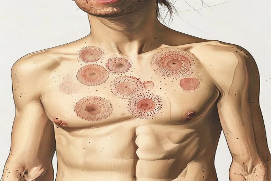
Definition and classification
A wheal has three hallmark features: (1) a central swelling of variable size surrounded by a reflex erythema, (2) an associated itching or sometimes burning sensation, and (3) a fleeting nature such that the skin returns to its normal appearance usually within 1–24 hours. Angioedema is a sudden, pronounced swelling of the lower dermis and subcutis (and frequently mucosa) that is more painful than itchy, pale rather than erythematous, and resolves over hours to (occasionally) up to 72 hours.[3]
Urticaria is classified by duration and by trigger pattern:[1][3]
- Acute urticaria — symptoms present for under 6 weeks. Often triggered by infections (especially viral in children), drugs (NSAIDs, antibiotics), or foods; frequently self-limiting.
- Chronic urticaria — symptoms persist for 6 weeks or longer, subdivided into:
- Chronic spontaneous urticaria (CSU) — wheals, angioedema, or both arising spontaneously without an identifiable external trigger (previously "chronic idiopathic" or "chronic autoimmune" urticaria).
- Inducible urticaria (CIndU) — symptoms elicited by a specific physical or environmental stimulus, confirmed by provocation testing. [1]
The inducible urticarias are heterogeneous in stimulus, latency, lesion morphology, and treatment response:[3]
- Symptomatic dermographism — whealing from shearing force (5 kg calibrated dermographometer); the commonest inducible form.
- Cold urticaria — cold air, water, or objects; carries a risk of systemic reactions on whole-body cold exposure (swimming).
- Delayed pressure urticaria — deep, painful swelling 4–8 hours after sustained pressure (backpack straps, walking); responds poorly to antihistamines and often to corticosteroids.
- Solar urticaria — UV or visible light within minutes.
- Heat urticaria, vibratory angioedema, cholinergic urticaria (rise in core body temperature), aquagenic urticaria, and contact urticaria. [1]
Epidemiology
The lifetime prevalence of any urticaria is roughly 20%, and chronic urticaria affects approximately 1% of the population at any time, with CSU accounting for around two-thirds of chronic cases. A global systematic review and meta-analysis reports a point prevalence of CSU of about 0.7% and a lifetime prevalence of approximately 2.3%, with a female predominance (about 2:1) and a peak onset between 20 and 40 years of age. Mean disease duration is 1–5 years, but 10–15% persist beyond 5 years and a subset for over a decade.[4][2]
Pathophysiology
Mast-cell/histamine axis
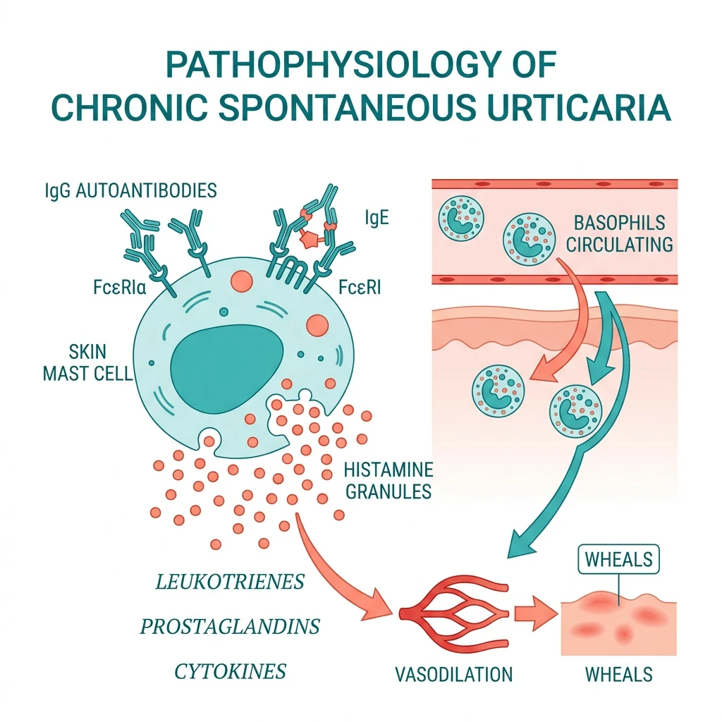
Wheal and flare in urticaria reflect activation of skin mast cells, predominantly around post-capillary venules, with release of histamine, platelet-activating factor, prostaglandin D2, leukotrienes (LTC4/D4/E4), and cytokines including tumour necrosis factor-α. Histamine acting on H1 receptors on sensory nerves produces itch, on vascular H1 and H2 receptors produces vasodilatation and increased permeability (wheal and flare), and on H3 receptors modulates further mediator release. The modern framework emphasises that CSU is not a pure histamine disease: co-existent activation of the kallikrein–kinin axis, basophil dysfunction, and a Th1/Th17 T-cell infiltrate all contribute.[5][8]
Autoimmune chronic spontaneous urticaria
A substantial fraction of CSU is autoimmune. Functional IgG autoantibodies directed against the high-affinity IgE receptor FcεRIα (or, less often, against IgE itself) cross-link receptor-bound IgE on mast cells and basophils, triggering degranulation. The autologous serum skin test (ASST) is a screening bioassay; confirmation traditionally required the basophil histamine release assay. Around 30–50% of CSU patients have evidence of this autoimmune subset, which is associated with thyroid autoimmunity, other autoimmune comorbidity, a more prolonged disease course, a higher disease burden, and a preferential response to immunomodulatory therapies. Biomarker work (IgG anti-FcεRIα, total IgE, C-reactive protein, D-dimer, basophil activation markers) continues to refine autoimmune identification.[6][7][9]
Co-factors and exacerbating triggers
Aspirin and other non-steroidal anti-inflammatory drugs (NSAIDs) worsen disease in 20–40% of CSU patients through cyclo-oxygenase-1 inhibition and shifts in arachidonic-acid metabolism toward leukotrienes. Infections (Helicobacter pylori, streptococcal, viral hepatitis), stress, hormonal factors (menstrual cycle exacerbation), alcohol, and opiates are commonly reported triggers but the evidence for causal eradication is variable; H. pylori eradication produces remission in a subset of infected patients.[3][8]
The bradykinin-mediated angioedema dichotomy
Angioedema is mechanistically mast-cell/histamine-driven (usually accompanied by urticaria and pruritus, responding to antihistamines, adrenaline, and corticosteroids) or bradykinin-driven (typically isolated, without urticaria or itch, and unresponsive to those agents). Hereditary angioedema types I and II, hereditary angioedema with normal C1-INH, acquired angioedema due to C1-INH deficiency, and ACE-inhibitor angioedema are bradykinin-mediated. Distinguishing these is critical because the wrong treatment — adrenaline, antihistamines, corticosteroids — is ineffective in bradykinin-mediated attacks and can delay life-saving on-target therapy.[18][19][20]
Histaminergic angioedema
Mast-cell / IgE driven
- Accompanies urticaria or pruritus (wheals present)
- Onset within minutes of exposure
- Resolves within hours
- Responds to H1-antihistamines, corticosteroids, adrenaline
- Examples: CSU, acute urticaria, anaphylaxis, NSAID-induced
Bradykinin-mediated angioedema
Contact-system / C1-INH driven
- Isolated swelling — no wheals, no itch, no flushing
- Onset 1–8 hours, persists 2–5 days
- **Does NOT** respond to antihistamines or adrenaline
- Treated with C1-INH, icatibant, ecallantide, lanadelumab
- Examples: HAE I/II, HAE-nC1-INH, AAE, ACE-inhibitor angioedema
Clinical rule of thumb
Bedside distinction
- Wheals + itch → give adrenaline pathway
- No wheals + no itch + slow resolution → screen for C1-INH
- Recurrent facial/airway swelling on an ACE inhibitor → bradykinin
- Family history + low C4 → hereditary; low C1q → acquired
Clinical features
UAS7 severity bands at a glance
In CSU, wheals occur daily or near-daily, appear at any site, are intensely pruritic, and individual lesions resolve within 24 hours. Nocturnal worsening is typical and sleep disturbance is common. Angioedema accompanies CSU in roughly 40% of patients, most often affecting the eyelids, lips, tongue, oropharynx, genitals, and extremities; when the larynx is involved the presentation becomes an airway emergency. Constitutional symptoms (fever, weight loss, arthralgia) are not features of uncomplicated CSU and their presence should prompt consideration of urticarial vasculitis or an underlying systemic process.[2][3]
Urticarial vasculitis should be suspected when individual lesions persist beyond 24 hours (often 24–72 hours), are painful or burning rather than pruritic, leave residual bruising or hyperpigmentation, and are associated with systemic features; skin biopsy confirms a leukocytoclastic small-vessel vasculitis.[2]
Investigations
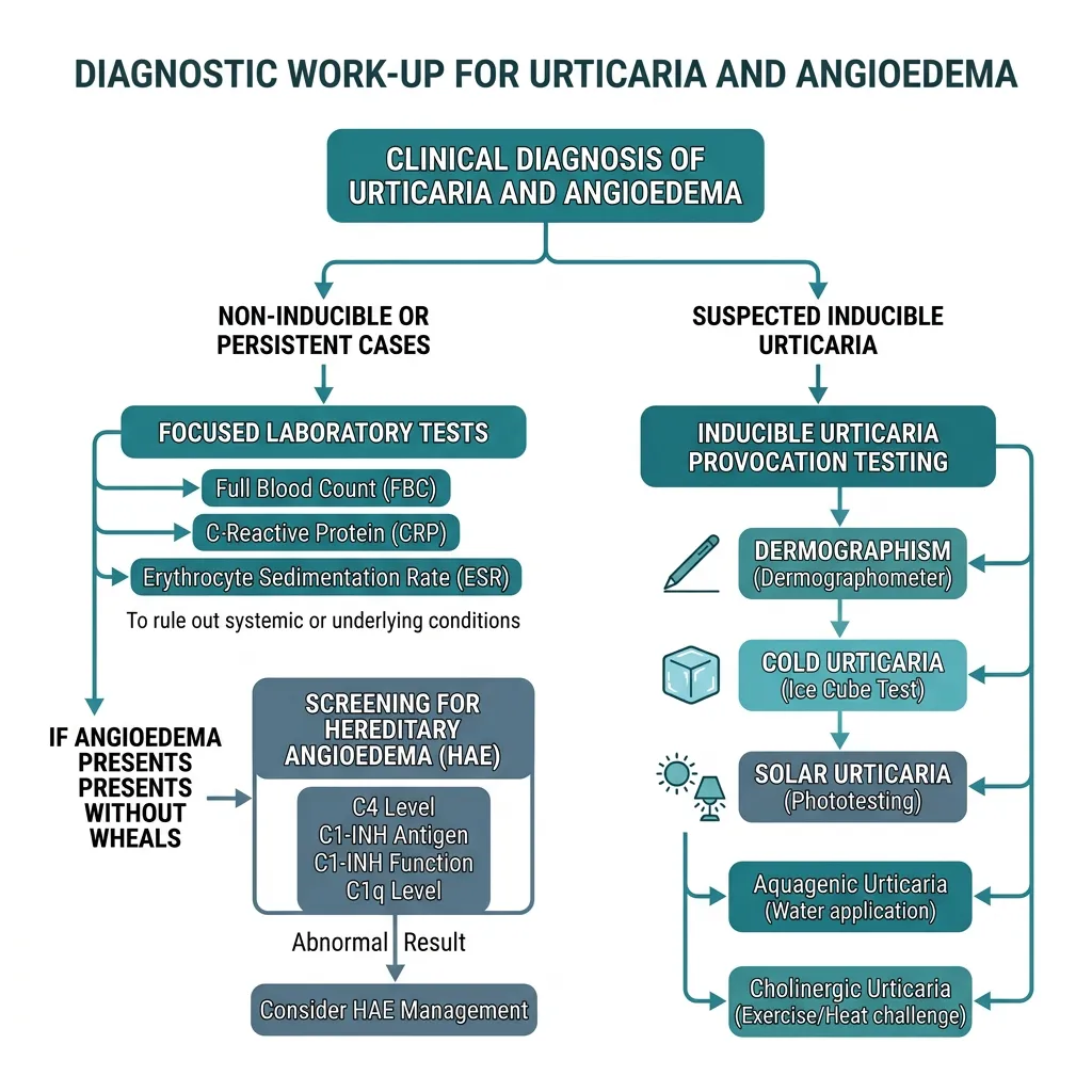
The diagnosis of urticaria is clinical. Routine "allergy panels" are not indicated in CSU and over-investigation is common. The EAACI guideline recommends a focused, stepwise evaluation:[3]
- All patients: detailed history (onset, frequency, duration, triggers, co-factors, drug and diet associations, infection, occupation, quality-of-life impact), physical examination, and differential blood count with CRP; for CSU also an ESR or CRP and consideration of C4 to screen for HAE.
- Screen for HAE when angioedema occurs without wheals: complement C4 (low in HAE types I and II and acquired C1-INH deficiency), C1-INH antigen and function, and C1q (low in acquired angioedema).
- Directed tests only for suspected inducible urticaria (provocation testing — dermographometer, ice cube test, cold-stimulation temp threshold, phototesting, pressure weights, aquagenic challenge, exercise).
- Extended work-up reserved for atypical features: thyroid autoantibodies and function, Helicobacter pylori testing, stool ova/cysts/parasites (especially with eosinophilia or travel), tryptase (to screen for clonal mast-cell disorder and to stratify anaphylaxis risk), hepatitis B/C and HIV if risk factors, autologous serum skin test or basophil activation marker as a research-grade autoimmune screen, and skin biopsy for suspected urticarial vasculitis. [1]
Activity and severity scoring
Validated instruments drive objective management decisions and are examinable:[3][2]
- Urticaria Activity Score over 7 days (UAS7) — the sum of daily wheal (0–3) and itch (0–3) scores recorded by the patient over a week (range 0–42). Mild 1–14, moderate 15–28, severe 29–42. UAS7 is the primary endpoint in most CSU clinical trials.
- Urticaria Control Test (UCT) — four-item recall over four weeks (0–16); a score of 12 or above indicates well-controlled disease, and a change of 3 points is clinically meaningful.
- Angioedema Activity Score (AAS) — patient-reported severity over 4 weeks for patients with angioedema.
- Dermatology Life Quality Index (DLQI) — generic dermatology quality-of-life measure (0–30); higher scores indicate greater impairment. [1]
Differential diagnosis
The differential includes urticarial vasculitis, autoinflammatory syndromes (cryopyrin-associated periodic syndromes, Schnitzler syndrome with monoclonal IgM gammopathy), serum sickness and serum-sickness-like reactions, mastocytosis, drug eruptions (especially morbilliform or acute generalised exanthematous pustulosis), erythema multiforme and Stevens–Johnson syndrome, bullous pemphigoid (urticarial pre-bullous phase), contact dermatitis, and the bradykinin-mediated angioedemas. Persistent fixed lesions, purpura, target lesions, bullae, mucosal erosion, or systemic inflammation redirect the diagnosis away from simple urticaria.[2]
Management of chronic spontaneous urticaria — the EAACI stepwise algorithm
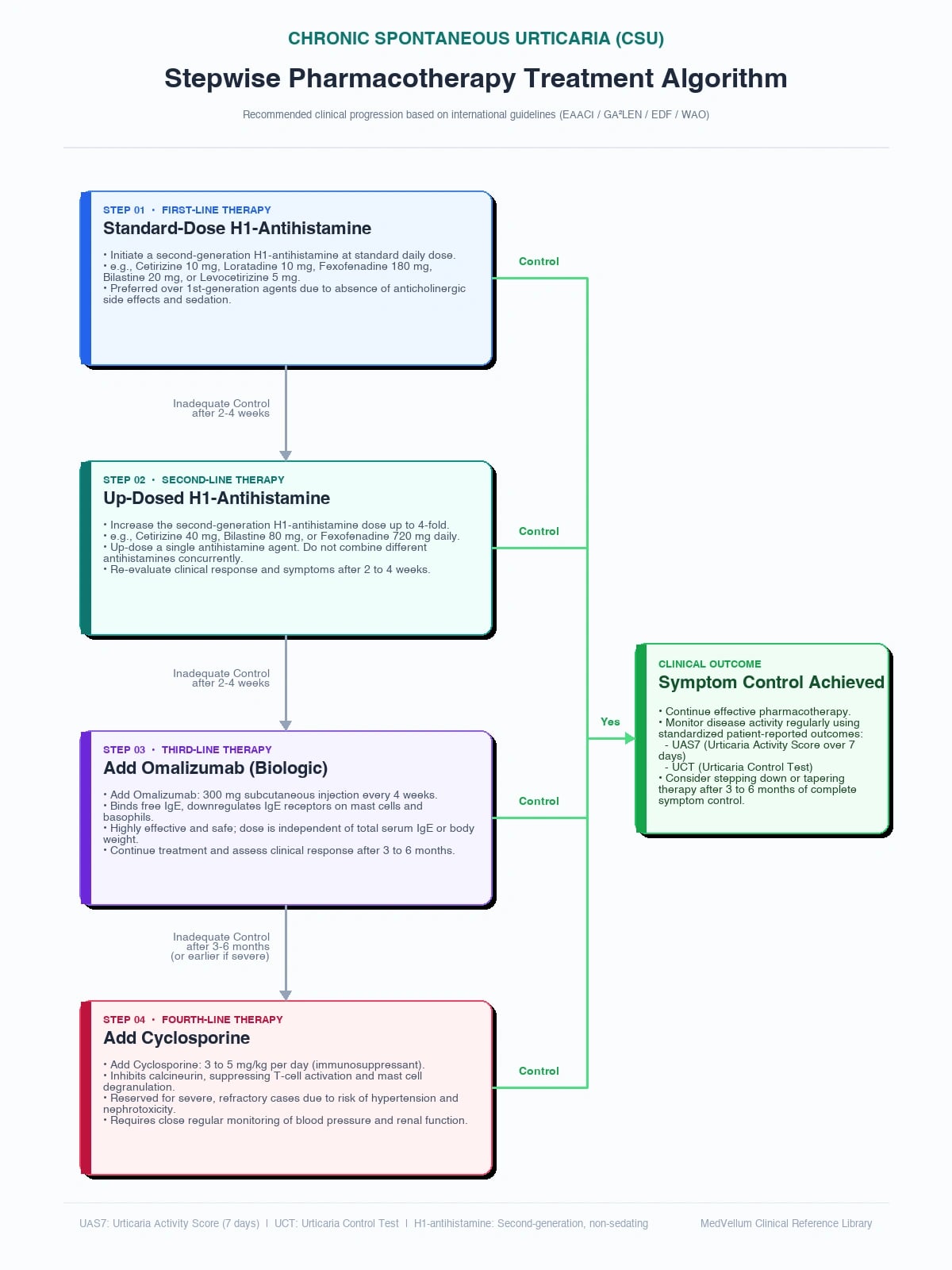
The international guideline endorses a four-step algorithm.[3]
Step 1 — Second-generation H1-antihistamines at standard dose. Bilastine, cetirizine, levocetirizine, desloratadine, fexofenadine, and loratadine are preferred over first-generation agents (which impair cognition, are anticholinergic, and disturb sleep architecture). Second-generation agents have established paediatric safety and tolerability profiles.[10]
Step 2 — Up-dosing up to four-fold. In patients uncontrolled on standard dose, the guideline recommends increasing the second-generation H1-antihistamine to two, then three, then four times the standard dose (i.e., up to 4×) before escalating to add-on therapy. The evidence base for up-dosing comes from observational data and post-hoc trial analyses; benefit is seen in a meaningful proportion, with an acceptable safety profile across the second-generation class. [1]
Step 3 — Add-on omalizumab. For patients refractory to up-dosed H1-antihistamines, omalizumab (anti-IgE monoclonal antibody), 300 mg subcutaneously every 4 weeks, is the recommended first add-on. The pivotal phase 3 trials established efficacy: ASTERIA II (Maurer et al., NEJM 2013) and ASTERIA I (Saini et al.) demonstrated dose-dependent increases in well-controlled weeks (itch and hive scores both minimal) versus placebo, with the 300 mg dose producing well-controlled activity in roughly one-third to one-half of patients. GLACIAL (Casale et al.) confirmed the 300 mg every-4-weeks regimen in patients uncontrolled on H1-antihistamines. Onset is usually within 1–2 weeks, response continues to improve over 3–6 months, and approximately two-thirds of treated patients achieve a complete response; failures are uncommon and often relate to undertreatment of angioedema or very low baseline IgE.[11][12][13][14]
Step 4 — Ciclosporin. In omalizumab non-responders or where omalizumab is unavailable, ciclosporin (2.5–4 mg/kg/day, cautiously titrated) is recommended for a limited period, with blood-pressure and renal-function monitoring, because of nephrotoxicity and hypertension. Additional immunomodulators (mycophenolate, tacrolimus, low-dose corticosteroid tapers for short flares) are used off-label in refractory disease. [1]
Emerging and pipeline biologics
- Dupilumab (anti-IL-4Rα) — the phase 3 LIBERTY-CSU CUPID trials showed significant reductions in itch and hive scores at 24 weeks versus placebo in patients uncontrolled on H1-antihistamines, and dupilumab is now approved for CSU in several jurisdictions, including an indication in children from a young age.[15]
- Remibrutinib — an oral covalent Bruton tyrosine kinase (BTK) inhibitor. A phase 3 trial (Metz et al., NEJM 2025) demonstrated rapid and substantial reductions in UAS7 versus placebo, addressing the mast-cell and basophil signalling axis upstream of histamine.[16]
- Barzolvolimab — an anti-KIT monoclonal antibody that depletes mast cells. Phase 2/3 data (Maurer et al.) show dose-dependent improvement, with the principal on-treatment adverse effect being transient hair colour change (grey repigmentation) attributable to mast-cell depletion at the hair bulb.[17]
Inducible urticaria — management specifics
Symptomatic dermographism, cold, cholinergic, and solar urticarias are managed with second-generation H1-antihistamines (often up-dosed); omalizumab is effective for several inducible forms. Solar urticaria requires photoprotection and may respond to graduated phototherapy or omalizumab; cold urticaria patients must be warned of the drowning risk with whole-body cold water immersion; delayed pressure urticaria is notoriously antihistamine-resistant and may require NSAID avoidance and short corticosteroid courses.[3]
Hereditary angioedema
Overview and pathophysiology
Hereditary angioedema (HAE) is an autosomal dominant disorder caused by deficiency or dysfunction of C1-inhibitor (C1-INH, encoded by SERPING1), with a prevalence of approximately 1 in 50,000. C1-INH normally inhibits activated factor XII, plasma kallikrein, and complement C1r/C1s; its absence permits uncontrolled generation of bradykinin via the contact (kallikrein–kinin) system, producing increased vascular permeability and non-pitting, non-pruritic swelling of subcutaneous and submucosal tissues. The swelling is not histamine-mediated and does not respond to adrenaline, antihistamines, or corticosteroids.[20][21]
Classification
- HAE type I (about 85%) — low antigenic and functional C1-INH levels.
- HAE type II (about 15%) — normal or raised antigenic C1-INH but dysfunctional protein (low function), due to missense mutations in the reactive-site loop.
- HAE with normal C1-INH — mutations in F12 (factor XII), plasminogen, angiopoietin-1, or kininogen; oestrogen-sensitive, predominantly affects women.
- Acquired angioedema due to C1-INH deficiency (AAE) — usually associated with B-cell lymphoproliferative disease or autoantibodies to C1-INH; onset in adulthood without family history, with low C4 and low C1q.[20][18]
Angioedema subtypes — the bradykinin versus histamine dichotomy
Angioedema is best understood as either mast-cell/histamine-mediated or bradykinin-mediated, and the distinction is the single most consequential step in acute management. Histaminergic angioedema almost always accompanies urticaria or pruritus, develops within minutes of exposure, and responds to H1-antihistamines, corticosteroids, and adrenaline. Bradykinin-mediated angioedema is characteristically isolated (no wheals, no itch), is slower in onset (often 1–8 hours), persists for 2–5 days, does not respond to antihistamines or adrenaline, and is treated by replacing or bypassing the deficient C1-INH/bradykinin pathway. The bradykinin group comprises hereditary angioedema types I, II, and HAE with normal C1-INH, acquired angioedema due to C1-INH deficiency, ACE-inhibitor angioedema, and idiopathic non-histaminergic acquired angioedema.[18][19][20]
- Hereditary angioedema (HAE types I and II) — autosomal dominant SERPING1 mutations causing quantitative or functional C1-INH deficiency; onset typically in childhood/adolescence, with attacks persisting throughout life. Family history is present in most cases, but de novo mutations occur in up to 25%.
- HAE with normal C1-INH (HAE-nC1-INH) — formerly HAE type III; clinically identical to HAE I/II but with normal C4, C1-INH antigen, and function. Driven by mutations in F12, PLG (plasminogen), ANGPT1 (angiopoietin-1), or KNG1 (kininogen). Predominantly affects women and is often oestrogen-sensitive (worsened by combined oral contraceptives, pregnancy).
- Acquired angioedema (AAE) — adult-onset (typically >40 years), no family history, low C4, low C1q, and low C1-INH antigen/function due to consumption by an underlying B-cell lymphoproliferative disorder (chronic lymphocytic leukaemia, non-Hodgkin lymphoma, monoclonal gammopathy) or to anti-C1-INH autoantibodies. Screen for lymphoproliferative disease at diagnosis.
- ACE-inhibitor angioedema — bradykinin accumulation from reduced degradation; 0.1–0.7% incidence; can occur at any time from days to years after starting therapy; higher risk in Black patients, women, and smokers.
- Idiopathic non-histaminergic acquired angioedema — recurrent angioedema without urticaria that fails H1-antihistamine therapy but has normal complement and no identifiable genetic or drug cause; a diagnosis of exclusion. [1]
This taxonomy is the framework for therapy: every patient with recurrent angioedema without urticaria must have C4 measured, and a low C4 mandates C1-INH antigen and function, with C1q to discriminate hereditary from acquired disease.[18][19][20][21]
Diagnosis
The screening test is complement C4, which is markedly and persistently reduced between and during attacks in HAE types I and II (and in AAE). Confirmation requires C1-INH antigen and functional assay (function is the most reliable single test). C1q is low in acquired disease, helping distinguish it from hereditary forms. Genetic testing of SERPING1 or F12 is performed when the phenotype is atypical or for family counselling. Because of autosomal dominant inheritance with incomplete penetrance, first-degree relatives should be screened.[20][21][22]
Clinical course and triggers
Attacks are episodic, last 2–5 days if untreated, and affect the skin (extremities, face, genitals, trunk), abdomen (visceral oedema producing severe pain, nausea, vomiting, and diarrhoea that can mimic a surgical abdomen), and the upper airway. Laryngeal attacks are the principal cause of death, with upper-airway oedema progressing rapidly to asphyxiation; historical mortality from laryngeal attacks approached one-third of untreated patients before modern therapy. Triggers include trauma (notably dental procedures), stress, infection, oestrogen-containing contraceptives, pregnancy, and ACE inhibitors.[23]
Acute attack treatment
Bradykinin-targeted on-demand therapy should be available to every diagnosed patient and given as early as possible — treatment within the first hour of symptom onset shortens attack duration and reduces progression to severe or laryngeal involvement. The WAO/EAACI 2021 guideline recommends that every HAE patient carry at least two doses of an approved on-demand agent, regardless of attack frequency, and that the agent be administered at the earliest symptom of an attack.[21][22]
- Plasma-derived C1-INH concentrate (Berinert, Cinryze) — intravenous; Berinert 20 units/kg for acute attacks (efficacy established in the I.M.P.A.C.T. trials, with onset of relief within 30–60 minutes); Cinryze 1000 units IV for acute attacks in the US (500 units in some regions); can be self-administered after training. Recombinant human C1-INH (Ruconest / conestat alfa) — 50 units/kg IV (maximum 4200 units) for acute attacks in adults and adolescents; an alternative for patients without plasma-derived exposure.
- Icatibant (Firazyr) — a selective bradykinin B2-receptor antagonist administered by subcutaneous injection, 30 mg (prefilled 3 mL syringe) into the abdomen, repeated once after 6 hours if symptoms persist (maximum 90 mg per 24 hours); onset of symptom relief typically within 30–90 minutes. Effective for self-administration and the most widely used on-demand agent worldwide. Local injection-site reactions (erythema, pain, pruritus) are common but transient.
- Ecallantide (Kalbitor) — a recombinant plasma kallikrein inhibitor administered subcutaneously, 30 mg (3 × 10 mg injections) for acute attacks; reduces attack severity and duration. Licensed in the United States; risk of anaphylaxis (~3%) mandates administration by a healthcare professional, not self-administration.
- Fresh frozen plasma (FFP) — 10–15 mL/kg IV as a contingency where specific therapy is unavailable; contains C1-INH but also kinin-generating substrates, so it may transiently worsen symptoms in some patients. Carries volume overload, transfusion reaction, and viral transmission risks.
- Tranexamic acid — oral 1 g four times daily (or 20–25 mg/kg four times daily in children); a less effective adjunct that inhibits plasmin-mediated C1-INH consumption; not first-line for acute attacks but used for short-term prophylaxis (e.g., before dental work) and in pregnancy.
- Supportive care — airway protection (the priority — early elective intubation in progressive laryngeal oedema rather than emergency tracheostomy), analgesia (opioids for severe abdominal attacks), antiemetics, and intravenous crystalloid resuscitation for abdominal attacks. Adrenaline, H1-antihistamines, and corticosteroids are ineffective for the bradykinin-mediated swelling and should not delay on-target therapy. [1]
Short-term (preprocedural) prophylaxis
Before dental work, endoscopy, intubation, or other known trigger events, short-term prophylaxis reduces attack incidence:[21][22]
- Plasma-derived C1-INH concentrate 1000 units IV 1–6 hours before the procedure (preferred).
- Attenuated androgens (danazol 2.5–10 mg/kg/day, maximum 600 mg/day, for 5–7 days before and 2–3 days after) — historically used where C1-INH concentrate is unavailable.
- Tranexamic acid 1 g four times daily for 5 days starting 2 days before the procedure — a weaker option, useful in pregnancy. [1]
Long-term prophylaxis
Long-term prophylaxis is indicated for patients with frequent attacks (typically ≥1 per week on average), severe attacks, prior laryngeal attacks, significant quality-of-life impairment, or limited access to emergency care. The modern strategy is to choose the agent based on patient age, comorbidity, pregnancy plans, route preference, and access — and to re-evaluate disease control every 3–6 months with the Hereditary Angioedema Activity Score (HAE-AS) or attack-frequency review, aiming for a ≥50% reduction and ideally complete control.[21][22]
- Lanadelumab (Takhzyro) — a fully human monoclonal antibody (IgG1κ) inhibiting plasma kallikrein. Dosed 300 mg subcutaneously every 2 weeks; if the patient is well controlled (e.g., attack-free for ≥6 months), the interval may be extended to 300 mg every 4 weeks. Self-administered after training. The phase 3 HELP study (Banerji et al., JAMA 2018) reduced attack rates by approximately 87% versus placebo at the 300 mg every-2-week dose, with a mean attack reduction of about 73% at the 150 mg every-4-week dose; the open-label HELP-extension confirmed durable efficacy and tolerability over ≥3 years, including in adolescents from age 12. Most common adverse events are injection-site reactions, which attenuate over time. Lanadelumab is the preferred first-line long-term prophylaxis in adults and adolescents ≥12 years in most international guidelines.[24]
- Berotralstat (Orladeyo) — an oral, once-daily, small-molecule plasma kallikrein inhibitor. Standard adult and adolescent dose: 150 mg orally once daily with food (capsule swallowed whole). The phase 3 APEX trial (Zuraw et al., JACI 2021) demonstrated a 44% reduction in attack rate versus placebo over 24 weeks at 150 mg daily, with reductions in attack severity and rescue-medication use. Common adverse events include abdominal pain, diarrhoea, vomiting, and back pain; rare QT prolongation has been noted. Avoid co-administration with strong CYP3A4 inhibitors (e.g., ketoconazole) and P-gp inhibitors; berotralstat is teratogenic in animal studies and not recommended in pregnancy.[25]
- Donidalorsen (Takhzyro-class agent) — a GalNAc-conjugated antisense oligonucleotide targeting prekallikrein messenger RNA in hepatocytes; 80 mg subcutaneously every 4 weeks, with an option to extend to every 8 weeks in well-controlled patients. Phase 3 data (Riedl et al., JACI-in-Practice 2025) demonstrated substantial attack reduction including in patients transitioning from other prophylactic regimens, with a favourable safety profile dominated by mild injection-site reactions.[26]
- Garadacimab (formerly CSL312) — a fully human IgG4 anti-factor XIIa monoclonal antibody (subcutaneous, monthly) — phase 3 efficacy has been reported and approval is expanding; another mechanism targeting the contact-system initiation step rather than downstream kallikrein.[22]
- Plasma-derived C1-INH concentrate for routine prophylaxis (Cinryze) — 1000 units IV every 3–4 days (some protocols use twice weekly); effective and the standard IV option where available. Recombinant human C1-INH (Ruconest) is also licensed for prophylaxis in some regions at 50 units/kg twice weekly.
- Attenuated androgens — danazol 200–600 mg/day orally (typical starting dose 400 mg/day, titrated down to the lowest effective dose) or stanozolol 2–6 mg/day; up-regulate hepatic C1-INH synthesis from the residual normal allele. Effective but limited by hepatotoxicity (raised transaminases, hepatic adenoma and peliosis), virilisation, dyslipidaemia, weight gain, mood change, and growth suppression in children. Contraindicated in pregnancy, breastfeeding, active liver disease, porphyria, and untreated malignancy; require baseline and 6-monthly liver function tests, lipid profile, and ultrasound surveillance where prolonged use is planned. They are now second- or third-line options given the availability of biologics.[21][22]
- Tranexamic acid — 1 g orally three to four times daily in adults (20–25 mg/kg four times daily in children); a weak option best reserved for pregnancy, short-term prophylaxis, or where modern therapies are unavailable; efficacy is modest and thrombotic risk warrants caution in patients with a history of thromboembolism or those on oestrogens.[21]
Hereditary angioedema in children and pregnancy
Paediatric HAE demands age-appropriate dosing of on-demand therapy, school emergency plans, and a cautious approach to long-term prophylaxis (attenuated androgens are avoided because of growth effects; lanadelumab and berotralstat have adolescent data). Pregnancy requires multidisciplinary planning: a C1-INH concentrate-based strategy is preferred for on-demand treatment and peripartum prophylaxis, attenuated androgens are contraindicated, and lanadelumab/berotralstat data in pregnancy remain limited. ACE inhibitors must be avoided.[28][22]
Acquired and drug-induced angioedema
Acquired angioedema (AAE) presents in adulthood without family history, with low C4 and low C1q and often an underlying B-cell lymphoproliferative disorder or autoantibody to C1-INH; management combines treatment of the associated condition with C1-INH concentrate or bradykinin-targeted agents for acute attacks.[18][19]
ACE-inhibitor angioedema results from accumulation of bradykinin (ACE degrades bradykinin, so its inhibition produces excess bradykinin), occurs in 0.1–0.7% of patients, may arise at any time after initiation (often years later), and disproportionately affects Black patients. It typically presents as isolated swelling of the lips, tongue, face, or upper airway without urticaria. Management is discontinuation of the ACE inhibitor (permanently), supportive airway care (the priority — early intubation if airway threatened), and consideration of icatibant, C1-INH concentrate, or ecallantide in severe or refractory cases, since conventional anaphylaxis therapy is ineffective. Patients should never be rechallenged with an ACE inhibitor and should avoid them thereafter.[27][19]
Anaphylaxis and the airway
Acute generalised urticaria with airway compromise, hypotension, persistent gastrointestinal symptoms, or wheeze meets criteria for anaphylaxis and requires immediate intramuscular adrenaline into the anterolateral thigh, repeatable every 5–15 minutes, airway support, high-flow oxygen, intravenous fluid resuscitation, supine positioning, and observation. Adjuncts (H1 and H2 antihistamines, corticosteroids) treat cutaneous symptoms and may reduce recurrence but do not substitute for adrenaline. Angioedema causing stridor, dysphonia, or drooling is an airway emergency irrespective of mechanism; in HAE the specific on-target agent (C1-INH concentrate or icatibant) must be given alongside airway protection.[23][21]
Clinical pearl
[1] [1]HAE-CATCH - 6 HAE features to remember
C1-INH deficiency (SERPING1); type I 85%, type II 15%; HAE-nC1-INH (normal level/function)
Non-pruritic, non-pitting, deeper; face/lips/tongue/extremities/genitals; NO wheals
Mimics acute abdomen; may have unnecessary surgery; ultrasound shows ascites and bowel wall thickening
Send C1-INH level AND function; low C4 is a sensitive screening test
Side effects: virilisation, hepatic; contraindicated in pregnancy; not for HAE-nC1-INH
Avoid ACEi in any patient with HAE; OCP and pregnancy can trigger
Berinert, Cinryze; recombinant Ruconest 50 U/kg; effective in 60-90 min
Bradykinin-mediated; specific therapies are icatibant, ecallantide, C1-INH; for prophylaxis lanadelumab, berotralstat
HAE / angioedema quick numbers
Red flags
Exam application bank (NEET-PG / INICET)
One-line answer
Urticaria and angioedema encompass mast-cell/histamine-driven wheals and swelling and, at the other end of the spectrum, bradykinin-mediated hereditary and acquired angioedema. Fellowship-level assessment demands mastery of the acute/chronic threshold, the inducible urticarias, autoimmune chronic spontaneous urticaria, validated activity scores (UAS7, UCT, AAS), the EAACI stepwise algorithm with second-generation H1-antihistamine up-dosing, omalizumab and cyclosporine, emerging Bruton tyrosine kinase and IL-4Rα/KIT biologics, and the full bradykinin pathway for hereditary angioedema (C1-INH replacement, icatibant, ecallantide, lanadelumab, berotralstat, donidalorsen) and ACE-inhibitor angioedema.
Worked stems (answer without another resource)
Stem 1 — Classic presentation. Map symptoms to mechanism; name the first investigation and first treatment step with dose/route if drug therapy is standard. [1]
Stem 2 — Unstable / complicated. List red flags that force immediate resuscitation, theatre, ICU, antidote, or reperfusion — and what you do in the first 15 minutes. [1]
Stem 3 — Atypical group. Elderly, pregnancy, child, or immunocompromised: how presentation and thresholds change. [1]
Stem 4 — Differential trap. Name the three closest mimics and one discriminator for each. [1]
Stem 5 — Disposition. Who goes home with safety-netting, who is admitted, who needs HDU/ICU/theatre, and what follow-up is mandatory. [1]
Rapid viva checklist
- Definition + classification
- Pathophysiology chain
- Bedside signs / criteria
- Score with exact components (if any)
- Emergency bundle
- Definitive therapy with doses
- Complications of disease and of treatment
- Special populations
- Guideline/trial name if classic
- Three exam traps
Coverage self-check
If you cannot answer any stem above from this page alone, re-read the matching section — the page is intended to be self-sufficient for final-prof and NEET-PG/INICET questions on Urticaria / angioedema.
[1]References
- [1]Kolkhir P, Giménez-Arnau AM, Kulthanan K, et al. Urticaria Nat Rev Dis Primers, 2022.PMID 36109590
- [2]Kolkhir P, Bonnekoh H, Metz M, et al. Chronic Spontaneous Urticaria: A Review JAMA, 2024.PMID 39325444
- [3]Zuberbier T, Abdul Latiff AH, Abuzakouk M, et al. The international EAACI/GA²LEN/EuroGuiDerm/APAAACI guideline for the definition, classification, diagnosis, and management of urticaria Allergy, 2022.PMID 34536239
- [4]Fricke J, Ávila G, Keller T, et al. Prevalence of chronic urticaria in children and adults across the globe: Systematic review with meta-analysis Allergy, 2020.PMID 31494963
- [5]Oliver ET, Saini SS. Chronic Spontaneous Urticaria: Etiology and Pathogenesis Immunol Allergy Clin North Am, 2024.PMID 38937007
- [6]Kolkhir P, Muñoz M, Asero R, et al. Autoimmune chronic spontaneous urticaria J Allergy Clin Immunol, 2022.PMID 35667749
- [7]Kaplan AP, Greaves M. Pathogenesis of chronic urticaria Clin Exp Allergy, 2009.PMID 19400905
- [8]Kaplan A, Lebwohl M, Giménez-Arnau AM, et al. Chronic spontaneous urticaria: Focus on pathophysiology to unlock treatment advances Allergy, 2023.PMID 36448493
- [9]Larenas-Linnemann D. Biomarkers of Autoimmune Chronic Spontaneous Urticaria Curr Allergy Asthma Rep, 2023.PMID 38064133
- [10]Papadopoulos NG, Zuberbier T. The safety and tolerability profile of bilastine for chronic urticaria in children Clin Transl Allergy, 2019.PMID 31660121
- [11]Maurer M, Rosén K, Hsieh HJ, et al. Omalizumab for the treatment of chronic idiopathic or spontaneous urticaria N Engl J Med, 2013.PMID 23432142
- [12]Saini SS, Bindslev-Jensen C, Maurer M, et al. Efficacy and safety of omalizumab in patients with chronic idiopathic/spontaneous urticaria who remain symptomatic on H1 antihistamines: a randomized, placebo-controlled study J Invest Dermatol, 2015.PMID 25046337
- [13]Casale TB, Bernstein JA, Maurer M, et al. Similar Efficacy with Omalizumab in Chronic Idiopathic/Spontaneous Urticaria Despite Different Background Therapy J Allergy Clin Immunol Pract, 2015.PMID 26054553
- [14]Kaplan A, Ferrer M, Bernstein JA, et al. Timing and duration of omalizumab response in patients with chronic idiopathic/spontaneous urticaria J Allergy Clin Immunol, 2016.PMID 26483177
- [15]Maurer M, Casale TB, Saini SS, et al. Dupilumab in patients with chronic spontaneous urticaria (LIBERTY-CSU CUPID): Two randomized, double-blind, placebo-controlled, phase 3 trials J Allergy Clin Immunol, 2024.PMID 38431226
- [16]Metz M, Giménez-Arnau A, Hide M, et al. Remibrutinib in Chronic Spontaneous Urticaria N Engl J Med, 2025.PMID 40043237
- [17]Maurer M, Metz M, Anderson J, et al. Anti-KIT Barzolvolimab for Chronic Spontaneous Urticaria Allergy, 2025.PMID 40415544
- [18]Kesh S, Bernstein JA. Isolated angioedema: A review of classification and update on management Ann Allergy Asthma Immunol, 2022.PMID 35988876
- [19]Young MC, Banerji A. Angioedema without urticaria: Diagnosis and management Allergy Asthma Proc, 2025.PMID 40380367
- [20]Busse PJ, Christiansen SC. Hereditary Angioedema N Engl J Med, 2020.PMID 32187470
- [21]Maurer M, Magerl M, Betschel S, et al. The international WAO/EAACI guideline for the management of hereditary angioedema-The 2021 revision and update Allergy, 2022.PMID 35006617
- [22]Busse PJ, Christiansen SC, Riedl MA, et al. US HAEA Medical Advisory Board 2020 Guidelines for the Management of Hereditary Angioedema J Allergy Clin Immunol Pract, 2021.PMID 32898710
- [23]Bork K, Hardt J, Witzke G. Fatal laryngeal attacks and mortality in hereditary angioedema due to C1-INH deficiency J Allergy Clin Immunol, 2012.PMID 22841766
- [24]Banerji A, Riedl MA, Bernstein JA, et al. Effect of Lanadelumab Compared With Placebo on Prevention of Hereditary Angioedema Attacks: A Randomized Clinical Trial JAMA, 2018.PMID 30480729
- [25]Zuraw B, Lumry WR, Johnston DT, et al. Oral once-daily berotralstat for the prevention of hereditary angioedema attacks: A randomized, double-blind, placebo-controlled phase 3 trial J Allergy Clin Immunol, 2021.PMID 33098856
- [26]Riedl MA, Bernstein JA, Jacobs JS, et al. Donidalorsen Treatment of Hereditary Angioedema in Patients Previously on Long-Term Prophylaxis J Allergy Clin Immunol Pract, 2025.PMID 40673861
- [27]Montinaro V, Cicardi M. ACE inhibitor-mediated angioedema Int Immunopharmacol, 2020.PMID 31835086
- [28]Pagnier A, Dermesropian A, Kevorkian-Verguet C, et al. Hereditary angioedema in children: Review and practical perspective for clinical management Pediatr Allergy Immunol, 2024.PMID 39655944