Dermatology · Medicine
Viral exanthems
Also known as Measles (rubeola, first disease) · Rubella (German measles, third disease) · Roseola infantum (exanthem subitum, sixth disease) · Erythema infectiosum (fifth disease, parvovirus B19) · Varicella (chickenpox) · Hand-foot-and-mouth disease · Mumps · Gianotti-Crosti syndrome (papular acrodermatitis of childhood)
Viral exanthems are cutaneous eruptions caused by systemic viral infections, classically the numbered childhood exanthems - measles (first), rubella (third), erythema infectiosum (fifth, parvovirus B19), roseola (sixth, HHV-6) - together with varicella, mumps, hand-foot-and-mouth disease, infectious mononucleosis, and Gianotti-Crosti syndrome. Each is identified by its causative virus, prodrome, morphology, and distribution: measles by the 3 Cs and Koplik spots, roseola by high fever in a well child that resolves as the rash appears, parvovirus B19 by the slapped cheek, varicella by vesicles in successive crops, and infectious mononucleosis by the amoxicillin-triggered morbilliform rash. Fellowship-level competence demands recognition of the at-risk populations (pregnancy, immunocompromise, haemolytic anaemia, neonate), the vaccine-preventable and notifiable infections, and the critical distinction from drug eruptions and Kawasaki disease.
On this page & tools
Your progress
Saved locally on this device.
Exam tags
Red flags
Overview & Definition
A viral exanthem is a widespread, usually symmetrical cutaneous eruption produced by a systemic viral infection, often accompanied by an enanthem (a mucosal eruption such as Koplik spots or oral vesicles). The term "exanthem" comes from the Greek exanthēma, meaning "to break out," and it is the pattern of that breaking out — its morphology, its distribution, and its timing relative to the fever and prodrome — that allows bedside identification of the causative virus.[1][9]
From the sixteenth to the twentieth century, clinicians numbered the rash illnesses of childhood that were then attributed to infection. The original six classic exanthems were: measles (first), scarlet fever (second), rubella (third), Filatov-Dukes disease (fourth, now historical and disputed), erythema infectiosum (fifth, parvovirus B19), and roseola infantum (sixth, HHV-6). Scarlet fever is bacterial (group A streptococcal erythrogenic toxin) and is covered in its own topic, but it is retained in the numbering because examiners still reference it. Today the working catalogue also includes the non-numbered viral exanthems — varicella, mumps, hand-foot-and-mouth disease (HFMD), infectious mononucleosis, and Gianotti-Crosti syndrome — because each is examinable on its own and each has a defining morphology, at-risk population, and public-health implication.[1][3]
The clinician's job at the bedside is three-fold. First, identify the virus from the morphology and tempo so that the right at-risk populations can be screened and the right public-health actions taken (notification, exclusion, contact tracing, vaccination). Second, recognise the complications — measles pneumonia, parvovirus aplastic crisis, varicella pneumonia, HFMD neurological disease — that transform a benign childhood illness into an emergency. Third, distinguish the viral exanthem from its dangerous mimics, above all the drug eruption, Kawasaki disease, and meningococcaemia.[9][10]
Classification
Viral exanthems are best classified by the causative virus and the clinical morphology, because these two axes together generate the diagnostic shortlist at the bedside. The figure below maps the numbered classic exanthems alongside the major non-numbered ones. [1]
Two structural principles organise recall. First, morphology tracks mechanism: maculopapular rashes reflect immune-complex and T-cell attack on virus-laden dermal endothelium, whereas true vesicles reflect direct viral cytopathic effect in the epidermis (varicella) or beneath it (enteroviruses). Second, the timing of the rash relative to the fever is diagnostic: in roseola the rash appears as the fever falls, in measles the rash appears after a rising fever and worsening coryza, and in varicella the fever and crops of vesicles evolve together.[5][12]
Epidemiology & Risk Factors
Viral exanthems are overwhelmingly diseases of childhood, but their age ranges, seasonal patterns, and transmission routes are distinct and examinable. Measles, rubella, mumps, varicella, and HFMD spread by the respiratory route (droplet and airborne); enteroviruses that cause HFMD also spread faeco-orally; EBV spreads in saliva ("kissing disease"); and parvovirus B19 spreads by the respiratory route and transplacentally, with a particular tropism for the fetal liver.[1][2][4]
Measles is among the most contagious infections known: with an R0 of 12 to 18, one case can infect an entire susceptible classroom, and the virus lingers in the air of a room for up to two hours after an infectious person has left.[1][13] Because of vaccine hesitancy and accumulating susceptibility cohorts, measles has resurged globally; the WHO declared the elimination status of several countries lost, and large outbreaks recur wherever vaccine coverage falls below the roughly 95% needed for herd immunity. Rubella and congenital rubella syndrome persist in regions without universal vaccination, and the WHO and many countries run measles-rubella (MR) or MMR campaigns to close immunity gaps.[2][17]
Pathophysiology
Most viral exanthems share a common pathogenetic cascade. The virus enters at a mucosal portal (respiratory, gastrointestinal, or oropharyngeal), undergoes primary replication at that site and in regional lymph nodes, and seeds a primary viraemia that distributes it to the reticuloendothelial system. After further replication, a larger secondary viraemia delivers virus to the skin and other target organs. The visible rash is largely a host immune response to viral antigen in the skin: cytotoxic T-cells and immune complexes attack virus-laden dermal endothelial cells and, in some infections, infected keratinocytes, producing the erythema, oedema, and epidermal change we recognise clinically.[1][9]
Three mechanistic patterns account for the different morphologies: [1]
The timing of the rash relative to the fever is the single most useful clinical consequence of this pathophysiology. Because the rash of roseola appears as the fever defervesces, a febrile infant who suddenly develops a rose-pink rash is, paradoxically, getting better — the rash is the immune response clearing the virus. In measles, by contrast, the child is at their most unwell when the rash appears, because the prodromal viraemia has been building for days. In varicella, fever and crops of vesicles wax and wane together over several days.[5][12]
Clinical Presentation
The clinical presentation is the heart of this topic. Each disease has a defining prodrome, morphology, distribution, and tempo, and examiners test these relentlessly. The comparative figure below is the single highest-yield visual; the sections that follow expand each disease to fellowship depth. [1]
The relationship between fever and rash is one of the highest-yield discriminator, summarised below: [1]
Fever and the 3 Cs worsen over 3 to 4 days; Koplik spots appear days 3 to 4. The maculopapular rash then erupts behind the ears and spreads cephalocaudally while the child is at their most unwell.
High fever for 3 to 5 days in a well-looking infant; the fever defervesces abruptly and the rose-pink rash appears simultaneously on the trunk. The rash is recovery.
Fever accompanies successive crops of vesicles over 3 to 5 days; papules, vesicles, pustules, and crusts coexist. Contagious until all lesions crusted.
Slapped cheek appears as the child defervesces; the reticulated rash then follows and may recur for weeks with heat and exercise. No longer infectious once rash appears.
Fever with painful oral ulcers and acral vesicles over 2 to 4 days; the child is most unwell from oral pain and dehydration rather than the rash itself.
Measles (rubeola, first disease) — measles virus (paramyxovirus)
Measles has a prodrome of three to four days of fever, cough, coryza, and conjunctivitis — the "3 Cs" — together with malaise and anorexia. The fever rises progressively and the child looks genuinely unwell. Koplik spots appear on the buccal mucosa in the prodrome, one to two days before the rash: they are pathognomonic bluish-white specks on an erythematous base, classically described as "grains of salt on a red background," found opposite the lower molars. The rash erupts on day 4, beginning behind the ears and at the hairline and spreading cephalocaudally over three days — face, then trunk, then limbs — becoming confluent, especially on the face and upper trunk, and then desquamating with fine branny scaling over the next week as the child improves.[1][13]
Rubella (German measles, third disease) — rubella virus (togavirus)
Rubella is a much milder illness. The prodrome is brief and low-grade — mild fever, malaise, and headache in older children and adults, often absent in young children. The defining feature is lymphadenopathy, especially postauricular, suboccipital, and cervical, which may precede the rash and persist for weeks. The rash is a fine pink maculopapular eruption that begins on the face and spreads cephalocaudally over one to three days, fading in the same order; it is less confluent than measles and the child is comparatively well. Forchheimer spots — small petechial or red macules on the soft palate — may be seen but are not pathognomonic. The clinical danger of rubella lies entirely in pregnancy: infection in the first trimester carries a high risk of congenital rubella syndrome.[2]
Erythema infectiosum (fifth disease) — parvovirus B19
The classic presentation is in two phases. After a non-specific prodrome of mild fever and headache, the first stage is the "slapped-cheek" appearance — bright red, confluent erythema of the cheeks with relative circumoral sparing, giving the face a windburned look. One to four days later, the second stage produces a reticulated (lacy), erythematous rash on the trunk and extensor surfaces of the limbs; this reticular pattern may recur for weeks with heat, sunlight, exercise, or bathing. By the time the rash appears the child is usually afebrile and well, and no longer infectious.[3][4]
Two adult and at-risk presentations dominate exam stems. Arthropathy — an acute symmetric small-joint polyarthritis of the hands, wrists, and knees — affects up to 60% of adult women (and a minority of men) and is the commonest presentation of parvovirus B19 in adults. In a patient with a chronic haemolytic anaemia (sickle cell disease, hereditary spherocytosis, thalassaemia), parvovirus B19 causes a transient aplastic crisis — sudden severe anaemia with profound reticulocytopenia — because the virus is tropic for erythroid progenitors. In immunocompromised patients who cannot mount the neutralising antibody response, infection becomes persistent, causing chronic pure red-cell aplasia. In pregnancy, transplacental infection in the second trimester can cause fetal hydrops and fetal loss through inhibition of fetal erythropoiesis and myocarditis.[3][4]
Roseola infantum (exanthem subitum, sixth disease) — human herpesvirus 6 (HHV-6)
Roseola is the prototypical "rash-after-fever" illness and the commonest cause of febrile seizures in infants under two years. The presentation is unmistakable when complete: three to five days of high fever (often 39 to 40 degrees C) in a child who looks remarkably well between fever spikes — the so-called "well-appearing febrile infant." The fever then abruptly defervesces, and as it falls, a rose-pink maculopapular rash appears on the trunk and spreads to the neck and extremities, sparing the face. The rash is often faint and lasts one to two days. The age range is characteristically six months to three years, reflecting the loss of maternal antibody; HHV-6 (and less often HHV-7) is the cause.[5][6]
Varicella (chickenpox) — varicella-zoster virus
Varicella begins with a mild prodrome of fever and malaise, followed within 24 hours by the characteristic itchy rash. The defining feature is vesicles in successive crops, so that lesions in different stages of evolution — papules, vesicles, pustules, and crusts — are present simultaneously. The rash begins on the face, scalp, and trunk and spreads centripetally to the limbs, also involving the mucosa (oral cavity, sometimes conjunctiva and genitalia). New crops appear for three to five days, and the child remains infectious until every lesion has crusted (typically around five to seven days after the last new vesicle). The incubation period is 14 to 21 days. Herpes zoster (shingles) is the reactivation of latent VZV in a dorsal root ganglion, occurring decades later, especially in older and immunocompromised people.[12]
Mumps — mumps virus (paramyxovirus)
Mumps presents with fever and painful swelling of the parotid glands (parotitis), usually bilateral but often beginning unilaterally, with the angle of the jaw pushed forward and the ear lobe elevated. The submandibular glands may also be involved. Orchitis affects roughly 20 to 30% of post-pubertal males and rarely causes infertility; oophoritis occurs in a small proportion of post-pubertal females. Aseptic meningitis is common (clinical meningitis in 1 to 10%, often mild and self-limiting), pancreatitis can occur (and may explain the rare documented association with diabetes), and sensorineural deafness is a classical, if uncommon, complication. Mumps is prevented by the MMR vaccine and the disease is notifiable.[14]
Hand-foot-and-mouth disease (HFMD) — coxsackievirus A16, enterovirus 71
HFMD predominantly affects children under ten and spreads readily in nurseries and schools, often in summer and autumn outbreaks. The presentation is painful oral ulcers or vesicles on the tongue, buccal mucosa, palate, and gums (often refusing food and drooling), together with vesicles on the hands, feet, and buttocks — typically oval, grey-white vesicles on an erythematous base, aligned with skin lines on the fingers and toes. Most cases are mild and self-limiting, but strains of enterovirus 71 are associated with neurological complications (aseptic meningitis, brainstem encephalitis, acute flaccid paralysis) and cardiopulmonary failure from neurogenic pulmonary oedema, particularly in young children in the Asia-Pacific region.[7][8]
Infectious mononucleosis — Epstein-Barr virus (EBV)
EBV infectious mononucleosis in adolescents and young adults presents with the triad of fever, pharyngitis, and lymphadenopathy (especially cervical), with splenomegaly in about half. A maculopapular rash occurs in a minority of untreated patients, but the exam-defining feature is that ampicillin or amoxicillin, given during the illness, triggers a dramatic morbilliform rash in roughly 90% of patients. This is not a true penicillin allergy — it reflects polyclonal T-cell activation by EBV — and patients generally tolerate penicillins subsequently. The classical heterophile antibody (Monospot) test supports the diagnosis, and patients must be counselled to avoid contact sports for three to four weeks because of the risk of splenic rupture.[16]
Gianotti-Crosti syndrome (papular acrodermatitis of childhood)
Gianotti-Crosti is a symmetrical, monomorphic, flat-topped, erythematous papular eruption on the face, buttocks, and extensor surfaces of the extremities, characteristically sparing the trunk. It affects children aged roughly six months to twelve years and lasts two to eight weeks. It is a paraviral reaction to many viruses — historically hepatitis B (in the original Italian descriptions), but in the vaccine era more commonly EBV, cytomegalovirus, coxsackievirus, parainfluenza, and respiratory syncytial virus — and may also follow vaccinations. It is self-limiting; the relevance to the exam is its distinctive distribution and its historical link to HBV, which in endemic regions still warrants a hepatitis screen.[15]
Differential Diagnosis
The differential of "a febrile child with a rash" is large, and the highest-yield exam task is to separate the benign self-limiting viral exanthems from the dangerous mimics. The single most common mimic is the drug eruption, and the single most dangerous is meningococcaemia; both must be actively excluded in every febrile child with a new rash.[9][10]
Several other conditions enter the broader differential. Staphylococcal scalded skin syndrome and toxic shock syndrome are toxin-mediated and present with erythroderma, skin tenderness, and superficial peeling (positive Nikolsky sign in SSSS). Pityriasis rosea begins with a herald patch and follows a Christmas-tree distribution on the trunk (and is thought to relate to HHV-6/7 reactivation). Erythema multiforme produces target lesions, typically triggered by HSV or Mycoplasma. The viral haemorrhagic fevers (dengue and others, depending on region) produce petechial and purpuric eruptions in a returned traveller and must not be missed.[11]
The central bedside distinction — viral exanthem versus drug eruption — deserves its own framework, because it is asked repeatedly. A drug eruption is more pruritic, lacks the characteristic viral prodrome, appears days after drug exposure, and frequently involves eosinophilia; a viral exanthem has a recognisable prodrome and morphology, is less itchy, and follows the characteristic course of the virus. Biopsy is rarely diagnostic but the exposure history is decisive, and a systematic comparison framework supports the distinction.[10]
Clinical & Bedside Assessment
The assessment is morphology-driven: a focused full-skin and mucosal examination usually yields the diagnosis before any test is sent. Examine the rash for morphology (macular, papular, vesicular, petechial), distribution (cephalocaudal vs centripetal vs acral), and evolution (successive crops vs a single wave). Examine the mucosa: Koplik spots point to measles, Forchheimer spots and palatal petechiae to rubella and scarlet fever respectively, oral vesicles and ulcers to HFMD and herpangina, and a strawberry tongue to scarlet fever and Kawasaki disease. Palpate the lymph nodes: postauricular and suboccipital enlargement suggests rubella, tender cervical adenopathy suggests EBV and GAS pharyngitis, and unilateral large cervical nodes are a Kawasaki criterion. [1]
The abdominal examination is directed at splenomegaly (EBV — counsel against contact sports) and hepatic tenderness. General examination assesses hydration (HFMD refusal to drink), work of breathing (measles and varicella pneumonia), and neurological status (HFMD enterovirus 71, with its risk of meningitis, encephalitis, and acute flaccid paralysis). Always take a drug history (to distinguish the drug eruption), a vaccination history (MMR and varicella status), a pregnancy status in any woman of reproductive age with a maculopapular rash, and a travel and contact history.[9]
Named signs and the morphology lexicon
Examiners reward precise use of named signs, and several of these are essentially pathognomonic or at least highly characteristic. The Koplik spot (measles) — a bluish-white speck on an erythematous buccal base opposite the lower molars, in the prodrome — is the single most reliable sign of measles. The Forchheimer spot (rubella) is a small petechial macule on the soft palate, supportive but not specific. The "slapped cheek" (parvovirus B19) and its reticulated (lacy) second stage are together highly suggestive of fifth disease. The strawberry tongue (scarlet fever, Kawasaki) evolves from a white coating through to a red, beefy, peeled surface with prominent papillae. Pastia lines (scarlet fever) are linear petechiae in the flexural creases. The herald patch of pityriasis rosea precedes the secondary Christmas-tree eruption by days. [1]
A practical morphology lexicon distinguishes macular (flat, blanchable), maculopapular / morbilliform (raised, measles-like, confluent), vesicular (clear fluid, under 5 mm), bullous (over 5 mm), pustular (pus), petechial / purpuric (non-blanching, haemorrhagic), urticarial (wheals), and targetoid (concentric rings of erythema multiforme). At the bedside, the distribution and progression are as informative as the morphology itself: a cephalocaudal march suggests measles or rubella; centripetal crops in different stages suggest varicella; an acral distribution suggests HFMD, erythema multiforme, or secondary syphilis; and a flexural predilection with Pastia lines suggests scarlet fever.[9][10]
Investigations
The diagnosis is clinical in classic, uncomplicated cases, and that is the expected board answer for most stems. Laboratory confirmation is reserved for the atypical case, the pregnant woman, the immunocompromised patient, the severe case, the outbreak requiring public-health action, and the notifiable disease.[1][3]
The notifiable diseases must be reported to public health without waiting for confirmation: measles, rubella (and congenital rubella syndrome), and mumps are notifiable in most jurisdictions, and public-health teams arrange confirmation, contact tracing, and post-exposure prophylaxis.[1][14]
Management — Resuscitation & General Principles
Most viral exanthems need no resuscitation at all — the expected answer to a well child with classic roseola or fifth disease is reassurance, antipyretics, and hydration. Escalation is reserved for the complications that transform a benign illness into an emergency: measles pneumonia or encephalitis, varicella pneumonia (especially in pregnancy and adults), parvovirus aplastic crisis, HFMD neurological complications, febrile seizures in roseola, and the dangerous mimics Kawasaki disease and meningococcaemia. [1]
Exam application bank (NEET-PG / INICET)
One-line answer
Viral exanthems are cutaneous eruptions caused by systemic viral infections, classically the numbered childhood exanthems - measles (first), rubella (third), erythema infectiosum (fifth, parvovirus B19), roseola (sixth, HHV-6) - together with varicella, mumps, hand-foot-and-mouth disease, infectious mononucleosis, and Gianotti-Crosti syndrome. Each is identified by its causative virus, prodrome, morphology, and distribution: measles by the 3 Cs and Koplik spots, roseola by high fever in a well child that resolves as the rash appears, parvovirus B19 by the slapped cheek, varicella by vesicles in successive crops, and infectious mononucleosis by the amoxicillin-triggered morbilliform rash. Fellowship-level competence demands recognition of the at-risk populations (pregnancy, immunocompromise, haemolytic anaemia, neonate), the vaccine-preventable and notifiable infections, and the critical
Worked stems (answer without another resource)
Stem 1 — Classic presentation. Map symptoms to mechanism; name the first investigation and first treatment step with dose/route if drug therapy is standard. [1]
Stem 2 — Unstable / complicated. List red flags that force immediate resuscitation, theatre, ICU, antidote, or reperfusion — and what you do in the first 15 minutes. [1]
Stem 3 — Atypical group. Elderly, pregnancy, child, or immunocompromised: how presentation and thresholds change. [1]
Stem 4 — Differential trap. Name the three closest mimics and one discriminator for each. [1]
Stem 5 — Disposition. Who goes home with safety-netting, who is admitted, who needs HDU/ICU/theatre, and what follow-up is mandatory. [1]
Rapid viva checklist
- Definition + classification
- Pathophysiology chain
- Bedside signs / criteria
- Score with exact components (if any)
- Emergency bundle
- Definitive therapy with doses
- Complications of disease and of treatment
- Special populations
- Guideline/trial name if classic
- Three exam traps
Coverage self-check
If you cannot answer any stem above from this page alone, re-read the matching section — the page is intended to be self-sufficient for final-prof and NEET-PG/INICET questions on Viral exanthems.
[1]Management — Definitive & Disease-Specific
Definitive management is disease-specific, and the treatments worth memorising are few but high-yield. The figure summarises the algorithm; the table that follows gives the agent, dose, route, and rationale for each. [1]
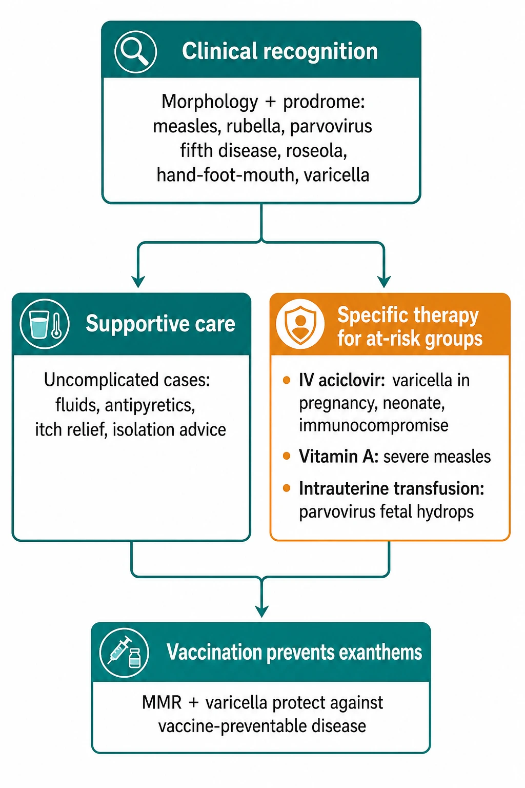
Two principles recur across the whole topic and are heavily tested. First, aspirin must be avoided in children with acute viral illness — especially varicella and influenza — because of the risk of Reye syndrome (hepatic encephalopathy with microvesicular fatty change). Use paracetamol or ibuprofen for fever instead. Second, vaccination prevents most of these diseases: the MMR vaccine (measles, mumps, rubella) given in two doses, and the varicella vaccine in countries that include it routinely, have transformed the epidemiology of childhood exanthems.[12][17]
Specific Subtypes & Scenarios
Certain scenarios are so examinable that they deserve isolated treatment. [1]
Congenital rubella syndrome
Maternal rubella infection in the first trimester carries the highest risk of congenital infection. The classic tetrad is cataracts (or other eye disease such as congenital glaucoma and pigmentary retinopathy), sensorineural deafness (the commonest single manifestation), cardiac defects (especially patent ductus arteriosus and peripheral pulmonary artery stenosis), and thrombocytopenia with a "blueberry muffin" rash of dermal extramedullary haematopoiesis. Prevention is universal MMR vaccination of all children plus antenatal rubella screening.[2]
Parvovirus B19 in pregnancy
Infection in pregnancy carries a roughly 5 to 10% risk of fetal hydrops when acquired in the first and second trimesters, through inhibition of fetal erythropoiesis and resultant high-output cardiac failure; the fetus is at greatest risk around 10 to 20 weeks' gestation. Management is maternal serology (IgM and IgG with avidity), serial fetal ultrasound for hydrops and middle cerebral artery Doppler for anaemia, and referral for intrauterine transfusion when fetal anaemia is confirmed.[4]
Varicella in pregnancy and the neonate
Maternal varicella in pregnancy risks maternal pneumonia (the leading cause of maternal death from varicella) and congenital or neonatal varicella. Congenital varicella syndrome (limb hypoplasia, cutaneous scarring, eye abnormalities, cortical atrophy) follows maternal infection in the first and second trimesters. Neonatal varicella, which can be severe and disseminated, follows maternal varicella in the peripartum period (from 5 days before to 2 days after delivery), when the baby receives the virus without protective antibody. Give VZIG to the neonate in this window and treat with IV aciclovir if disease develops.[12]
Transient aplastic crisis in haemolytic disease
In a child with sickle cell disease, hereditary spherocytosis, or another chronic haemolytic anaemia, parvovirus B19 produces a transient aplastic crisis — an abrupt drop in haemoglobin with profound reticulocytopenia (reticulocytes normally drive haemolysis compensation) and a sometimes life-threatening anaemia. The child is infectious (unlike the chronic anaemia of immunocompromise, which is persistent viraemia). Management is transfusion support until the crisis resolves and isolation from other haemolytic and pregnant patients.[4]
Complications & Pitfalls
Each exanthem has its classic complications, and examiners test the serious complications of common illnesses heavily. [1]
The commonest pitfalls are four. Missing the dangerous mimic — diagnosing "viral exanthem" in a child with early meningococcaemia or Kawasaki disease. Giving aspirin to a febrile child with varicella. Failing to screen pregnancy status in a woman with a maculopapular rash, missing congenital rubella or parvovirus hydrops. Mislabelling the amoxicillin-mono rash as penicillin allergy, depriving the patient of a first-line antibiotic class for life. Each of these is a recurring exam stem.[9][11]
Prognosis & Disposition
Most viral exanthems in immunocompetent children are self-limiting with full recovery, and the disposition for an uncomplicated case is home with safety-net advice (return if breathing difficulty, drowsiness, petechiae, dehydration, or persistent fever). Specific outcomes that matter: [1]
Disposition is home for uncomplicated cases, hospital for dehydration, secondary bacterial infection, pneumonia, encephalitis, aplastic crisis, and severe HFMD, and PICU/HDU for varicella pneumonia in pregnancy, EV-71 neurological disease, and measles pneumonia with respiratory failure. Pregnant contacts of measles, rubella, parvovirus B19, and varicella need urgent serological assessment and subspecialty input.[1][4]
Special Populations
Evidence, Guidelines & Regional Differences
MMR vaccination schedules are the backbone of prevention and differ in detail across regions. The US (CDC/ACIP) recommends two MMR doses at 12 to 15 months and 4 to 6 years, and routine two-dose varicella vaccination at the same visits. The UK (Green Book/JCVI) historically gave MMR at 12 to 13 months and a preschool booster at 3 years and 4 months, with varicella vaccine reserved for susceptible contacts and healthcare workers; the UK has more recently debated introducing routine varicella vaccination. The WHO drives global measles-rubella (MR) elimination campaigns, and India (ICMR/IAP) runs MR catch-up campaigns with a goal of measles and rubella elimination; the IAP schedule gives MMR at 9 months, 15 to 18 months, and 4 to 6 years. The MMR-autism controversy — originating in the retracted Wakefield study — has been refuted by large epidemiological studies and remains the single biggest cause of vaccine hesitancy and measles resurgence.[17]
The WHO recommends vitamin A for all children with measles in two age-appropriate doses, repeated the next day and at 4 weeks, on the basis of mortality reduction demonstrated in trials in vitamin-A-deficient populations. The evidence for post-exposure prophylaxis — MMR within 72 hours of measles exposure for susceptible contacts, HNIG within 6 days for susceptible pregnant and immunocompromised contacts, and VZIG for varicella-susceptible high-risk contacts — is guideline-based and regionally codified in the UK Green Book, CDC guidance, and national immunisation manuals.[1][12]
Exam Pearls
EXANTHEM
Self-test: a 10-month-old has had high fever for three days and looks well; today the fever resolved and a pink rash appeared on the trunk. What is the diagnosis and why?
This is roseola infantum (HHV-6, sixth disease). The diagnostic clue is the rash appearing AS the fever defervesces in a well-looking infant — the classic "fever-then-rash" sequence. Management is supportive: antipyretics as needed, and treat febrile seizures if they occur.
Self-test: a teenager with fever, sore throat, and cervical lymphadenopathy is given amoxicillin and develops a confluent rash. What is happening and what should you tell the patient?
This is the classic amoxicillin-triggered morbilliform rash of EBV infectious mononucleosis. It occurs in about 90% of patients given aminopenicillins during EBV and is not a true penicillin allergy — it reflects polyclonal T-cell activation by EBV. Stop the antibiotic, treat the EBV supportively, and counsel against contact sports for three to four weeks because of splenic rupture risk; reassure that penicillins can usually be tolerated in future.
References
- [1]Hübschen JM, Gouandjika-Vasilache I, Dina J. Measles Lancet, 2022.PMID 35093206
- [2]Winter AK, Moss WJ. Rubella Lancet, 2022.PMID 35367004
- [3]Leung AKC, Lam JM, Barankin B, et al. Erythema Infectiosum: A Narrative Review Curr Pediatr Rev, 2024.PMID 37132144
- [4]Heegaard ED, Brown KE. Human parvovirus B19 Clin Microbiol Rev, 2002.PMID 12097253
- [5]Leung AK, Lam JM, Barankin B, et al. Roseola Infantum: An Updated Review Curr Pediatr Rev, 2024.PMID 36411550
- [6]Agut H, Bonnafous P, Gautheret-Dejean A. Laboratory and clinical aspects of human herpesvirus 6 infections Clin Microbiol Rev, 2015.PMID 25762531
- [7]Leung AKC, Lam JM, Barankin B, et al. Hand, Foot, and Mouth Disease: A Narrative Review Recent Adv Inflamm Allergy Drug Discov, 2022.PMID 36284392
- [8]Saguil A, Kane SF, Lauters R, et al. Hand-Foot-and-Mouth Disease: Rapid Evidence Review Am Fam Physician, 2019.PMID 31573162
- [9]Haber JS, Cipriano SD, Oza VS. Morbilliform Eruptions in the Hospitalized Child Dermatol Clin, 2022.PMID 35366972
- [10]Khandpur S, Ahuja R. Drug-Induced vs. Viral Maculopapular Exanthem-Resolving the Dilemma Dermatopathology (Basel), 2022.PMID 35645232
- [11]Rife E, Gedalia A. Kawasaki Disease: an Update Curr Rheumatol Rep, 2020.PMID 32924089
- [12]Kennedy PGE, Gershon AA. Clinical Features of Varicella-Zoster Virus Infection Viruses, 2018.PMID 30400213
- [13]Moss WJ. Measles Lancet, 2017.PMID 28673424
- [14]Su SB, Chang HL, Chen AK. Current Status of Mumps Virus Infection: Epidemiology, Pathogenesis, and Vaccine Int J Environ Res Public Health, 2020.PMID 32150969
- [15]Leung AKC, Sergi CM, Lam JM, et al. Gianotti-Crosti syndrome (papular acrodermatitis of childhood) in the era of a viral recrudescence and vaccine opposition World J Pediatr, 2019.PMID 31134587
- [16]Mergoum AM. Amoxicillin Rash in Infectious Mononucleosis N Engl J Med, 2021.PMID 34496177
- [17]Lievano F, Galea SA, Thornton M, et al. Measles, mumps, and rubella virus vaccine (M-M-R™II): a review of 32 years of clinical and postmarketing experience Vaccine, 2012.PMID 22959986