Dermatology · General Medicine
Viral Warts (Verrucae)
Also known as Viral warts · Verrucae · Common warts · Plantar warts · Genital warts · Human papillomavirus · HPV · Condyloma acuminata
Viral warts (verrucae) are benign epidermal proliferations caused by human papillomavirus (HPV) infection of basal keratinocytes. Clinical subtypes: common (verruca vulgaris) — rough, hyperkeratotic papules on hands, fingers and knees (HPV 1, 2, 4, 27); plantar (verruca plantaris/myrmecia) — tender, inward-growing on soles with pathognomonic black dots that are thrombosed capillary loops (HPV 1); flat (verruca plana) — smooth, flat-topped papules on face and hands (HPV 3, 10, 28); filiform — finger-like projections on face and neck; genital (condyloma acuminata) — sexually transmitted cauliflower-like lesions (HPV 6, 11; oncogenic 16, 18, 31, 33). Most cutaneous warts resolve spontaneously in immunocompetent children (approximately 50 percent in 1 year, two-thirds in 2 years). First-line treatment is salicylic acid 12 to 40 percent for up to 12 weeks; cryotherapy with liquid nitrogen every 1 to 3 weeks is the main clinic alternative; imiquimod 5 percent or podophyllotoxin 0.5 percent for genital warts; intralesional bleomycin and immunotherapy for recalcitrant plantar warts. The 9-valent HPV vaccine (types 6, 11, 16, 18, 31, 33, 45, 52, 58) prevents genital warts and HPV-related cancer. Biopsy atypical, persistent or rapidly growing lesions to exclude squamous cell carcinoma or verrucous carcinoma.
On this page & tools
Your progress
Saved locally on this device.
Exam tags
Red flags
Overview & Definition
Viral warts (verrucae) are benign proliferations of the epidermis and of stratified squamous epithelium caused by infection of basal keratinocytes by the human papillomavirus (HPV). They are among the commonest of all human skin infections, affecting an estimated 7 to 12 percent of the general population and as many as 20 to 30 percent of school-aged children. Clinically they appear as rough, hyperkeratotic papules or plaques (the common wart), but the morphology varies enormously with anatomical site, HPV type and host immune status, giving rise to the recognised subtypes — common, plantar, flat, filiform, periungual, and genital.[1][4]
HPV is a small (approximately 55 nm), non-enveloped, icosahedral, double-stranded circular DNA virus of the family Papillomaviridae. More than 200 distinct HPV types have been characterised, of which at least 120 infect the human skin or mucosa. HPVs are grouped by tissue tropism (cutaneous versus mucosal) and by oncogenic risk: low-risk types (notably 6 and 11) cause benign genital warts and recurrent respiratory papillomatosis, whereas high-risk (oncogenic) types (notably 16 and 18, and also 31, 33, 45, 52 and 58) drive cervical, anal, vulvar, penile, vaginal and oropharyngeal cancer. The same virus that produces a trivial hand wart in one site can, in another site and another type, cause lethal cancer — the clinical skill is recognising which wart is benign and self-limiting, and which demands biopsy, screening and prevention.[9][4]
The clinical reasoning in viral warts rests on three questions. First, is it actually a wart? — corns, calluses, seborrhoeic keratoses, molluscum contagiosum, condyloma lata and skin cancers (squamous cell carcinoma, verrucous carcinoma, amelanotic melanoma) can all mimic warts. Second, where is it and which HPV type is likely? — a wart on the sole, the face, the periungual skin or the genitalia has a different natural history, different treatment and different cancer implication. Third, should I treat, reassure or biopsy? — most childhood cutaneous warts resolve spontaneously within two years and need only reassurance, but persistent, painful, cosmetically distressing, periungual, facial, genital or immunosuppression-associated warts require active treatment, and any atypical or refractory lesion demands biopsy to exclude malignancy.[1][2]
Classification
Viral warts are classified along three axes — anatomical/morphological subtype, causative HPV type, and oncogenic risk. All three matter clinically because they determine natural history, treatment choice and cancer implication. [1]
By clinical (morphological) subtype
| Subtype | HPV type(s) | Site | Morphology |
|---|---|---|---|
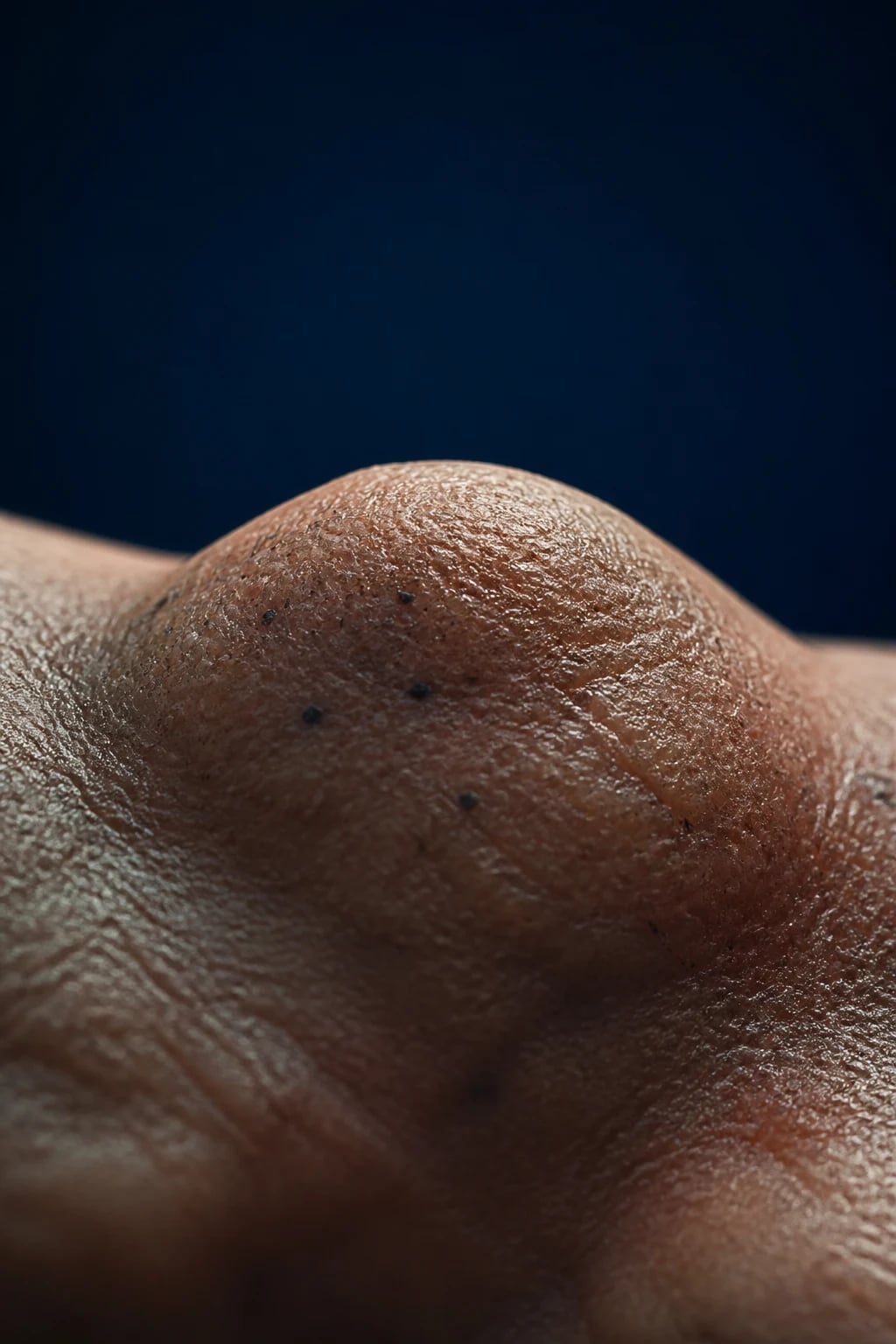
| Common wart (verruca vulgaris) | 1, 2, 4, 27, 57 | Fingers, hands, knees, periungual | Rough, hyperkeratotic, dome-shaped papule with papillomatous surface |
| Plantar wart (verruca plantaris / myrmecia) | 1 (deep myrmecia), 2, 27, 57 | Soles (pressure points) | Endophytic, tender, black dots; mosaic when confluent |
| Flat wart (verruca plana) | 3, 10, 28, 29, 41, 49 | Face, dorsum of hands, shins | Smooth, flat-topped, flesh-coloured or slightly hyperpigmented papules; linear (Koebner) |
| Filiform / digitate wart | 1, 2, 4, 27 | Lips, nostrils, eyelids, neck, beard | Finger-like or thread-like projections from a narrow base |
| Periungual / subungual wart | 1, 2, 4, 27 | Nail folds, under nail plate | Rough periungual hyperkeratosis; can distort or destroy the nail matrix |
| Genital wart (condyloma acuminata) | 6, 11 (low-risk); 16, 18, 31, 33, 45 (oncogenic) | Perineum, perianal, penis, vulva, vagina, cervix, urethra | Soft, fleshy, cauliflower-like (acuminate), pedunculated; may be macular/pigmented |
| Butcher's wart | 7 | Hands of meat/poultry/fish handlers | Large, cauliflower-like common warts on the hands |
| Epidermodysplasia verruciformis | 5, 8 (and 2, 3, 10, 12, 14, 15, 17, 19, 20, 21-25, 36-38, 47, 50) | Sun-exposed skin, face, dorsa of hands | Lifelong pityriasis-versicolor-like macules + flat-wart-like papules; premalignant |
By HPV type — the high-yield mapping
HPV type to disease — the mapping every candidate must know
By oncogenic risk
- Cutaneous, benign — HPV 1, 2, 4, 27, 57 (common), 3, 10, 28, 49 (flat), 7 (butcher's). These carry no malignant potential in immunocompetent hosts.
- Mucosal, low-risk — HPV 6, 11. Cause benign genital warts (condyloma acuminata) and juvenile-onset recurrent respiratory papillomatosis.
- Mucosal, high-risk (oncogenic) — HPV 16, 18, 31, 33, 35, 39, 45, 51, 52, 56, 58, 59, 68. Drive cervical intraepithelial neoplasia (CIN), cervical cancer, anal intraepithelial neoplasia, vulvar/vaginal/penile intraepithelial neoplasia, and oropharyngeal cancer. HPV 16 and 18 alone cause approximately 70 percent of cervical cancers.[9]
Epidemiology & Risk Factors
Cutaneous warts are ubiquitous. Lifetime prevalence is high, with a peak in children and adolescents (5 to 20 years) and a second smaller peak in young adults related to genital HPV acquisition. Warts are uncommon in infancy and decline steadily after adolescence as cell-mediated immunity matures. The strongest single risk factor is impaired skin barrier — HPV requires a micro-abrasion to reach the basal keratinocytes, so anything that breaks or macerates the stratum corneum increases risk.[1][9]
Risk factors for cutaneous warts: [1]
- Skin trauma and maceration — HPV enters through micro-abrasions; nail biting, picking at existing warts (autoinoculation), shaving of the face or legs (Koebner spread of flat warts).
- Moist environments and communal exposure — swimming pools, communal showers, shared footwear and towels; plantar warts classically acquired at swimming baths ("swimming-pool warts").
- Atopic dermatitis and an impaired epidermal barrier — atopic individuals have a higher wart load and are more refractory to treatment; disruption of the barrier by eczema facilitates viral entry.
- Immunosuppression — HIV/AIDS (especially with CD4 below 200), solid-organ transplant recipients (on ciclosporin, azathioprine, tacrolimus), haematological malignancy, and iatrogenic immunosuppression all produce warts that are more numerous, larger, more refractory, and more likely to harbour oncogenic HPV with malignant transformation.
- Occupational handlers of meat, poultry and fish — HPV 7 ("butcher's wart"), large cauliflower-like warts on the hands from chronic wet exposure and micro-trauma.[4]
- Genetic susceptibility — epidermodysplasia verruciformis (autosomal recessive, EVER1/TMC6 and EVER2/TMC8 genes), primary immunodeficiencies (Wiskott-Aldrich, DOCK8 deficiency, GATA2 deficiency, WHIM syndrome).[9]
Genital warts are the commonest sexually transmitted infection of viral origin. Peak incidence is in young adults aged 20 to 24 years. Risk factors are early sexual debut, multiple sexual partners, lack of condom use, smoking, and co-existing STIs (including HIV). Condoms are only partially protective because HPV infects the surrounding anogenital skin not covered by the condom. A single act of sexual intercourse with an infected partner transmits HPV in approximately 60 percent of cases, and most sexually active people acquire HPV at some point, though the large majority clear it subclinically. The introduction of quadrivalent and 9-valent HPV vaccination has produced dramatic, population-level declines in genital wart incidence in countries with high vaccine coverage (for example Australia), with measurable herd protection in unvaccinated males.[5][7]
Pathophysiology
Viral structure and cell entry
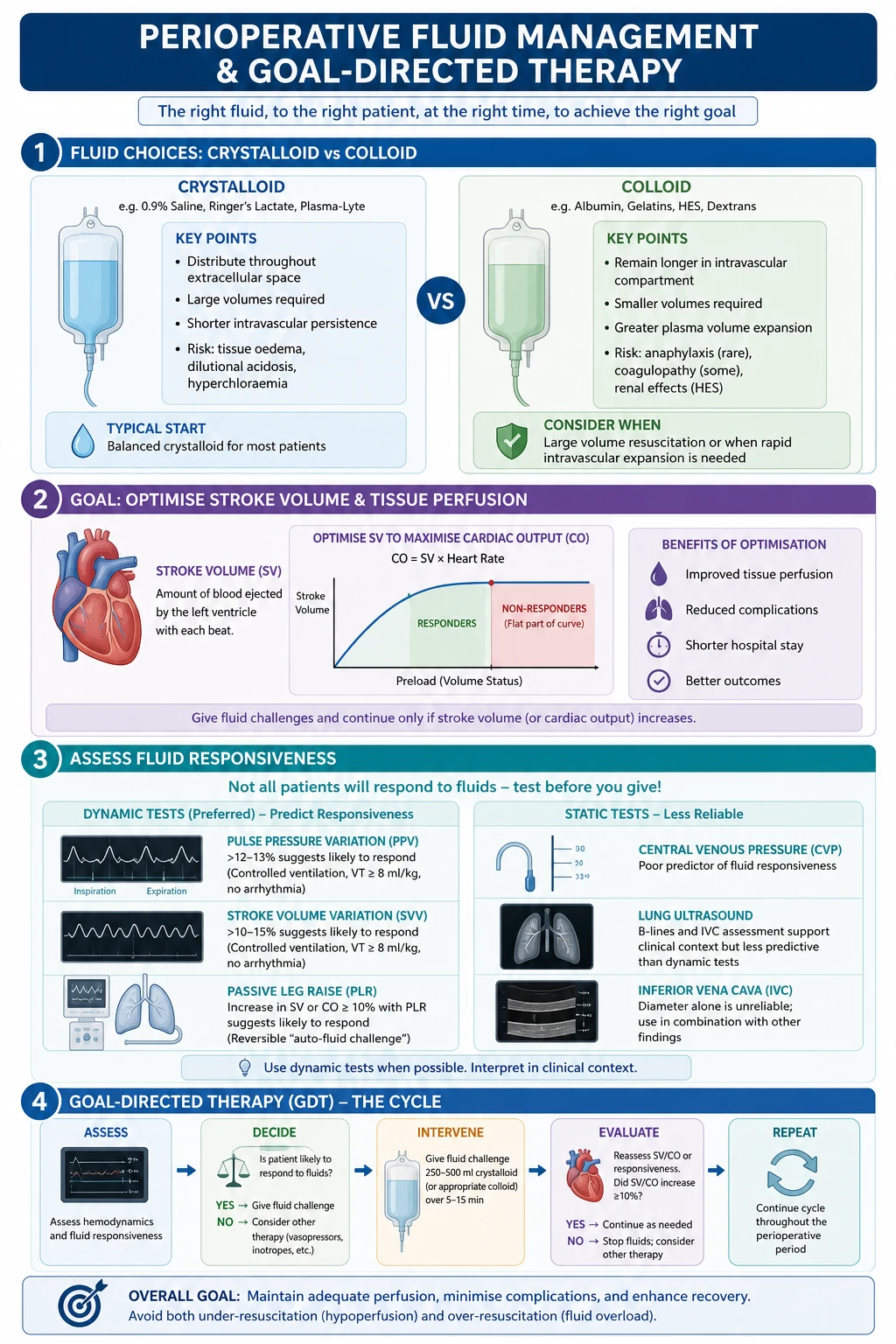
HPV is a non-enveloped, icosahedral (72 capsomeres), double-stranded circular DNA virus of approximately 7900 to 8000 base pairs. Its genome encodes eight early (E1–E8) and two late (L1, L2) proteins. L1 is the major capsid protein and the antigen used in all current HPV vaccines (which are virus-like particles assembled from L1). HPV is strictly epitheliotropic: it cannot traverse an intact stratum corneum and requires a micro-abrasion or mucosal micro-trauma to reach its target cell, the basal keratinocyte. The virus first binds to the basement membrane via heparan sulfate proteoglycans, undergoes a conformational change, and is then taken up by the basal keratinocyte through a process involving alpha-6 integrin and other receptors.[9][4]
Genome maintenance and the differentiation-dependent life cycle
Once inside the basal cell nucleus, HPV maintains its genome as a stable, low-copy episome (a circular extrachromosomal DNA) and divides passively with the basal cell. Productive viral replication — with amplification of the genome and expression of the late capsid proteins L1 and L2 — occurs only in the upper, differentiating spinous and granular layers, where keratinocyte differentiation activates the viral late promoter. This differentiation-dependent life cycle explains why HPV is poorly accessible to the immune system (the basal compartment is immunologically privileged) and why mature virions are shed in desquamating corneocytes without viraemia.[9]
The oncoproteins E6 and E7 — the molecular heart of the disease
The early proteins E6 and E7 are the effectors of epithelial proliferation and, in high-risk types, of malignant transformation: [1]
- E6 binds and degrades the tumour suppressor p53 via the E6-associated protein (E6AP) ubiquitin ligase, driving the proteasomal destruction of p53. Loss of p53 disables apoptosis and cell-cycle arrest in response to DNA damage. In high-risk HPV, E6 also activates telomerase (via hTERT), conferring replicative immortality.
- E7 binds and functionally inactivates the retinoblastoma protein (pRb), releasing the E2F transcription factor and driving the cell into S-phase (unscheduled cellular proliferation). E7 of high-risk types also disrupts cell-cycle checkpoints and genomic stability. [1]
In low-risk HPV (6, 11), E6 and E7 are expressed at low levels and bind p53/Rb weakly, producing benign, non-invasive proliferation. In high-risk HPV (16, 18), E6 and E7 are more potently expressed, driving progressive genomic instability, integration of viral DNA into the host genome (disrupting E2 repression of E6/E7), and ultimately malignant transformation — the molecular basis of cervical, anal and oropharyngeal cancer. This is the reason every genital wart deserves an HPV-aware assessment and the reason the HPV vaccine is also a cancer vaccine.[9]
Histopathology
The microscopic appearances explain the clinical morphology. The typical wart shows: [1]
- Hyperkeratosis with parakeratosis (nuclear retention in the stratum corneum, often with vertical columns over the papillomatous projections).
- Acanthosis (thickening of the spinous layer) and papillomatosis (elongation of the rete ridges, which in plantar warts are pushed inward by pressure, giving the endophytic growth).
- Koilocytes in the upper epidermis — pathognomonic cells with perinuclear vacuolation and a pyknotic, wrinkled "raisin-like" nucleus; these are keratinocytes actively producing HPV virions.
- Thrombosed capillary loops in the elongated dermal papillae — the histological correlate of the black dots seen clinically in plantar and common warts.
- Vacuolation of cells in the granular layer and clumping of keratohyalin granules.[1][4]
Host immunity and spontaneous resolution
Most warts are eventually cleared by cell-mediated (Th1) immunity — CD8 cytotoxic T cells recognise HPV antigens on infected keratinocytes, with macrophage and NK-cell help. The humoral response to L1 develops slowly and is protective against re-infection (the basis of vaccination) but does not clear existing lesions. This explains why warts may persist for years in an immunocompetent host and then vanish suddenly when cell-mediated immunity "switches on" — and why immunosuppressed patients (HIV, transplant) develop chronic, refractory, widespread warts. Approximately 50 percent of childhood warts clear within 1 year and two-thirds within 2 years without any treatment; spontaneous clearance in adults is slower and less frequent.[1][2]
Clinical Presentation
Common wart (verruca vulgaris)
The common wart presents as a firm, rough (verrucous), hyperkeratotic, dome-shaped papule, typically 1 to 10 mm in diameter, with a characteristic papillomatous (corrugated) surface bearing tiny finger-like projections, each capped by a thrombosed capillary that may appear as a black dot. The commonest sites are the fingers, the dorsum of the hands, around the nails (periungual), the knees, and the elbows. Warts are usually asymptomatic but may be tender if fissured or on a pressure point, and may cause cosmetic embarrassment. They are often multiple, may coalesce into a larger plaque, and are commonly distributed in areas of trauma (the Koebner/isomorphic phenomenon — warts appear at sites of scratching, biting, or shaving). Periungual and subungual warts may extend under the nail plate and produce nail dystrophy, fissuring and permanent matrix damage.[1]
Plantar wart (verruca plantaris / myrmecia)
A plantar wart arises on weight-bearing skin of the sole (the metatarsal heads, heel, and ball of the toe) and, because of constant pressure, grows endophytically (inward) rather than outward. The result is a tender, raised, yellowish, hyperkeratotic plaque with a disrupted skin-line pattern, which when pared with a scalpel reveals tiny black dots — the thrombosed capillary loops that are pathognomonic. Pain is characteristically elicited by lateral (side-to-side) pinching of the lesion (unlike a corn, which is tender on direct downward pressure). A single deep myrmecia (HPV 1) is sharply circumscribed and exquisitely tender; mosaic warts are shallow, coalesced plaques of many small warts that are often refractory to treatment. Plantar warts are the wart most likely to present clinically because of pain on walking.[1][2]
Flat wart (verruca plana)
Flat warts are smooth, flat-topped or slightly raised, flesh-coloured or lightly pigmented papules of 2 to 5 mm, often numerous, occurring on the face (especially the forehead, cheeks and chin), the dorsum of the hands, the wrists and the shins. They are commonest in children and young adults and show a striking linear (Koebner) distribution along the lines of scratching or shaving — a row of flat warts along a scratch mark is characteristic. They are usually asymptomatic but cosmetically troublesome on the face.[4]
Filiform and digitate warts
These are elongated, finger-like or thread-like projections arising from a narrow base, occurring on thin skin — the lips, the nostrils, the eyelids, the neck and the beard area. They are common in men in the beard area (where shaving disseminates them) and in children around the mouth and nose. They are usually solitary or few and are treated by snip excision or light cryotherapy.[1]
Genital wart (condyloma acuminata)
Genital warts are soft, fleshy, moist, pink or skin-coloured papules and plaques with a characteristic cauliflower-like (acuminate or condylomatous) surface. They may be pedunculated (on a stalk) or sessile, single or multiple, small or large enough to obstruct the vaginal introitus or anal canal. Sites include the labia, the vaginal introitus, the perineum, the perianal area, the penis (especially the frenulum, coronal sulcus, glans, and shaft), the scrotum, the cervix, the urethral meatus and the anus. Perianal warts are particularly common in men who have sex with men and in women with anal intercourse. Genital warts are usually asymptomatic but may cause itching, burning, pain, bleeding or mechanical obstruction. A pregnant woman may develop rapidly proliferating genital warts that can obstruct the birth canal and, rarely, transmit HPV 6/11 to the neonate, causing juvenile-onset recurrent respiratory papillomatosis.[5][7]
Atypical presentations
The Koebner phenomenon
Like psoriasis and lichen planus, viral warts show an isomorphic (Koebner) response: warts appear at sites of trauma. This is most striking in flat warts, which line up along scratch marks (linear distribution) and along the beard area in men who shave, and in periungual warts in nail-biters. Recognising Koebnerisation matters therapeutically — shaving, plucking, biting and picking must be addressed to stop autoinoculation.[1]
Differential Diagnosis
The differential diagnosis of a "wart" depends on the site and morphology. A mislabelled skin cancer is the cardinal pitfall — biopsy any lesion that is atypical, rapidly growing, ulcerated, fixed to deeper tissues, bleeding, or refractory to adequate treatment.[1]
| Condition | Distinguishing features from a wart |
|---|---|
| Corn (clavus) / callus | Hard, hyperkeratotic; skin lines preserved; no black dots; central translucent hard core (corn); tender on direct pressure (corn) vs lateral pressure (wart); callus is a diffuse yellow plaque at a pressure point. Paring a wart reveals black dots and disrupted skin lines; paring a corn reveals a clear cone with preserved lines. |
| Seborrhoeic keratosis | Stuck-on appearance, greasy surface, keratin-filled pits (horn cysts), brown/black, in older adults; anywhere but typical on trunk/face; not infectious. |
| Molluscum contagiosum | Dome-shaped, umbilicated papule with a central white core (expressed on curettage); caused by a poxvirus; common in children; genital forms in adults are STI. |
| Lichen planus (vs flat wart) | Pruritic, polygonal, purple, planar papules; Wickham striae on surface; oral mucosal involvement (reticular white plaques); flexor wrists; intensely itchy vs asymptomatic flat wart. |
| Acne / closed comedones (vs facial flat wart) | Comedones, papules, pustules; not flat-topped; respond to retinoids; polymorphic. |
| Condyloma lata (secondary syphilis) | Moist, flat, grey-white, hypertrophic plaques in anogenital region; teeming with spirochaetes; RPR/VDRL strongly positive; systemic features of secondary syphilis (rash, lymphadenopathy). |
| Pearly penile papules | 1 to 3 mm, smooth, dome-shaped, skin-coloured papules arranged in rows around the coronal margin; a normal anatomical variant; not infectious; asymptomatic. |
| Skin tag (acrochordon) (vs filiform wart) | Soft, fleshy, pedunculated, neck/axilla/groin; smooth surface vs the finger-like projections of a filiform wart. |
| Squamous cell carcinoma / verrucous carcinoma | Indurated, ulcerated, fixed or bleeding lesion in a sun-exposed or genital site in an older or immunosuppressed patient; endophytic or exophytic destructive growth; biopsy mandatory. |
| Amelanotic melanoma | Pink, vascular, nodular, rapidly growing, may bleed; anywhere; dermoscopy shows polymorphic vessels and residual pigment; biopsy mandatory — a missed amelanotic melanoma is a fatal error. |
| Bowenoid papulosis | Multiple pigmented reddish-brown papules/plaques on the anogenital skin caused by HPV 16/18; histologically SCC in situ; considered premalignant — distinguish clinically from benign condyloma. |
| Buschke-Lowenstein tumour (giant condyloma / verrucous carcinoma) | Large, cauliflower-like, locally destructive anogenital mass caused by HPV 6/11; histologically well-differentiated verrucous carcinoma; does not metastasise early but invades locally; treated by wide surgical excision. |
Clinical & Bedside Assessment
The diagnosis of a viral wart is clinical, made on morphology and the distribution of lesions, and confirmed at the bedside by two manoeuvres:[1]
- The paring (shave) test. Gently pare the surface of the lesion with a number-15 scalpel blade. A wart reveals disruption of the normal skin lines and tiny black dots or pinpoint bleeding points (thrombosed capillary loops). A corn reveals a central, hard, translucent, conical core with the surrounding skin lines preserved and arching over it. A callus is a diffuse yellow hyperkeratotic plaque with preserved skin lines and no black dots. This single test resolves the commonest diagnostic dilemma in plantar dermatology.
- Pain on pressure. A plantar wart is tender on lateral (side-to-side) pinching pressure, whereas a corn is tender on direct downward (perpendicular) pressure. [1]
Other bedside assessments: [1]
- Dermoscopy. A wart shows a papilliform surface with multiple small black or brown dots and short linear vessels representing the thrombosed capillaries. Corns show a central white-to-yellow round structure with a surrounding halo; molluscum shows a central polycyclic orifice.
- Acetic acid test. Application of 3 to 5 percent acetic acid to the anogenital skin for 3 to 5 minutes whitens HPV-infected epithelium — useful for detecting subclinical genital HPV infection (acetowhite macules/papules) but not specific (inflammation also whitens).
- Identify the host. Examine for stigmata of immunosuppression (oral candidiasis, oral hairy leukoplakia, generalised lymphadenopathy in HIV), examine the nail unit for periungual involvement, examine the anogenital region in any patient with a genital wart (look at the cervix, vagina, anal canal; examine the partner), and screen for co-existing STIs (syphilis serology, HIV, chlamydia, gonorrhoea, hepatitis B/C).
- Decide whether to biopsy. Indications for biopsy of a presumed wart: rapid growth, ulceration, bleeding, fixation to deeper structures, atypical pigmentation, immunosuppression, refractoriness to adequate treatment, or any genital lesion with suspicion of neoplasia.[1][2]
Investigations
The diagnosis is clinical in the overwhelming majority of cases. Investigation is reserved for atypical, refractory, immunosuppression-associated, or genital lesions, and for cervical screening.[1][4]
- Skin biopsy (shave or punch). Indicated whenever the diagnosis is in doubt, the lesion is atypical or refractory, or malignancy is suspected. Histopathology confirms the wart (hyperkeratosis, acanthosis, papillomatosis, koilocytes) and excludes squamous cell carcinoma, verrucous carcinoma, Bowenoid papulosis and melanoma.
- HPV DNA testing (PCR). Used for HPV typing in research and in selected clinical scenarios: atypical/recurrent genital warts (to identify high-risk types), cervical screening (HPV DNA testing with cytology triage), and investigation of recurrent respiratory papillomatosis. Not routinely needed for a typical cutaneous wart.
- Cervical cytology (Pap smear) and HPV co-testing. Recommended for all women with genital warts and per national cervical screening guidelines, regardless of wart presence, because genital warts are a marker of sexual risk and the high-risk HPV types co-circulate.
- Screening for immunosuppression. In a patient with extensive, refractory, atypical or genital warts, screen for HIV (with consent), diabetes, and a full blood count; in a transplant or chronically immunosuppressed patient, review the immunosuppressive regimen.
- Acetic acid application and colposcopy/anoscopy. For subclinical genital and anal HPV disease, especially in immunosuppressed patients and men who have sex with men.
- The koilocyte. On histology, the koilocyte — a keratinocyte with perinuclear vacuolation and a pyknotic, wrinkled "raisin-like" nucleus — is the cytopathic effect of HPV replication and confirms the viral aetiology.[4]
Management — Resuscitation
Viral warts are not a dermatological emergency in the usual sense, but a handful of situations demand prompt, structured action: [1]
- Suspected malignancy in a "wart". An atypical, rapidly growing, ulcerated, fixed or bleeding lesion — especially in a sun-exposed or genital site, in an older or immunosuppressed patient — is squamous cell carcinoma, verrucous carcinoma, Bowenoid papulosis or amelanotic melanoma until proven otherwise. First action: stop destructive treatment and obtain a biopsy (punch or incisional) before any further cryotherapy, cautery or excision. Destructing a cancer without histology is a catastrophic error.[1]
- Rapidly proliferating or giant genital warts. Especially in pregnancy or immunosuppression, giant condylomata can obstruct the vaginal introitus, urethra or anal canal, bleed, and predispose to secondary infection. Screen urgently for HIV and co-infections, exclude Buschke-Lowenstein verrucous carcinoma (biopsy), and arrange debulking (surgical excision, electrocautery or laser) plus systemic/input from gynaecology, urology or surgery as appropriate.
- Periungual warts and the nail matrix. Aggressive cryotherapy, electrosurgery or excision around the nail can produce permanent nail dystrophy, pterygium or nail loss. The principle is early, cautious, matrix-sparing treatment — favour topical (cantharidin, imiquimod, retinoid) over destructive modalities for periungual disease.[1]
- Genital warts in pregnancy. Podophyllin, podophyllotoxin and imiquimod are contraindicated (teratogenicity and systemic absorption). Safe options in pregnancy are cryotherapy, trichloroacetic acid 80 to 90 percent (provider-applied), and surgical excision / electrocautery. Plan delivery: caesarean section is not routinely indicated for genital warts alone, but may be considered for birth-canal obstruction or proliferation refractory to treatment, to reduce the risk of juvenile-onset recurrent respiratory papillomatosis.[5][7]
- Before any destructive treatment. Confirm the diagnosis clinically (or by biopsy if atypical), document the baseline number and size of lesions, photograph for monitoring, screen for and treat co-existing STIs in genital warts, give pregnancy testing where relevant, and counsel the patient that multiple treatments over weeks to months are usual, that scarring and pigmentary change may occur, and that recurrence is common.[2]
Management — Definitive & Stepwise
General principles
- Watchful waiting is a legitimate first option for asymptomatic cutaneous warts in immunocompetent children — most resolve within two years. The decision to treat is driven by symptoms (pain), cosmetic/psychological distress, persistence, periungual/facial location, immunosuppression, and the genital site.
- Match the treatment to the wart. Salicylic acid for common and plantar warts; cryotherapy for hand and facial filiform warts; cantharidin for children (painless); podophyllotoxin or imiquimod for genital warts; bleomycin or immunotherapy for recalcitrant plantar warts; biopsy any atypical lesion first.
- Treat the host. Address nail biting, shaving, and lip-licking (Koebner); treat co-existing atopic eczema; screen and treat co-existing STIs in genital warts; offer HPV vaccination; optimise HIV/immunosuppression.
- Counsel on expectations. Most treatments require multiple sessions over 4 to 12 weeks, may be painful, and carry a 30 to 50 percent recurrence rate — the goal is clearance, not cure, and the wart may return or new ones appear.[2][3]
Cutaneous (nongenital) warts
First-line — topical salicylic acid (keratolytic). [1]
- Agent: salicylic acid 12 to 40 percent (over-the-counter paints, gels, plasters and ointments; 40 percent plasters/occlusive dressings for plantar warts).
- How to use: soak the wart in warm water for 5 to 10 minutes, file the hyperkeratotic surface with an emery board or pumice stone, apply the salicylic acid to the wart only (protect the surrounding normal skin with petrolatum), allow to dry, and occlude if a plaster is used.
- Frequency and duration: once or twice daily for up to 12 weeks. Review at 12 weeks; if no response, step up.
- Rationale: salicylic acid dissolves keratin (keratolytic), de-bulks the wart, and may provoke a local immune response. It has the best evidence base of any topical agent and is recommended as first-line by the Cochrane review.[3]
- Adverse effects: local irritation, erythema, contact dermatitis, and (rarely, with extensive use in small children) salicylate toxicity (tinnitus, hyperventilation) — avoid in infants and on large body surface areas.
- Contraindications: not on the face or mucous membranes, not on moles/birthmarks, not in patients with peripheral neuropathy or poor circulation (diabetic foot) without supervision, not in pregnancy on large areas.
First-line alternative — cryotherapy with liquid nitrogen. [1]
- Agent: liquid nitrogen at minus 196 degrees Celsius, applied by cotton-tipped applicator (cotton bud) or spray.
- Technique: apply until an ice ball forms 1 to 2 mm beyond the wart edge, then allow to thaw; a second freeze-thaw cycle improves efficacy for thick warts. A blister typically forms over 24 to 48 hours.
- Frequency and duration: every 1 to 3 weeks for 4 to 6 sessions, occasionally more for plantar warts.
- Rationale: cryonecrosis of infected keratinocytes and induction of a local immune response.
- Adverse effects: pain (limiting in children), blistering, hypo- and hyper-pigmentation (a major concern in dark skin), scarring, nail dystrophy if periungual, and rarely neuropathy from freezing digital nerves.
- Evidence: the Cochrane review found cryotherapy effective for hand warts but not clearly superior to salicylic acid for common warts overall; aggressive (longer) freezing is not clearly better than gentle freezing for plantar warts and causes more side effects. Two- to three-weekly intervals are reasonable.[3]
Second-line and combination therapies: [1]
- Cantharidin 0.7 percent (with or without podophyllin and salicylic acid — "Cantharone Plus"). Applied by the clinician, covered with a non-occlusive tape, and washed off in 4 to 8 hours; produces a painless blister (ideal in children); repeated every 2 to 4 weeks. Avoid on the face, mucous membranes and genitalia; avoid occlusion that drives a deeper, painful blister.
- 5-Fluorouracil (5-FU) — topical 5 percent cream or intralesional; an antimetabolite that inhibits DNA synthesis in infected keratinocytes; used for refractory or genital warts.
- Topical retinoids (tretinoin, adapalene) — useful for flat warts by accelerating epidermal turnover; applied nightly.
- Formalin soaks — for multiple plantar warts; soak the foot in 3 percent formaldehyde for 10 to 15 minutes daily; hardens the wart and reduces pain; malodorous.
- Duct tape occlusion — cover the wart with silver duct tape for 6 days, remove, soak, file, leave open overnight, reapply; mixed evidence, harmless, and acceptable for children.[2]
Refractory / recalcitrant warts: [1]
- Intralesional bleomycin. A cytotoxic antibiotic injected directly into the wart (typically 0.25 to 1 unit per mL, 0.1 to 0.5 mL per wart, every 3 to 4 weeks). Highly effective for recalcitrant plantar and periungual warts. Painful (use local anaesthetic or refrigerant spray); may cause nail loss, Raynaud phenomenon, flagellate erythema. Contraindicated in pregnancy and in digital warts (risk of vascular compromise and digital necrosis).[8]
- Intralesional immunotherapy — Candida antigen, MMR vaccine, or BCG injected into a wart to induce a delayed-type hypersensitivity response that clears both the injected and distant warts; useful for multiple, refractory warts; relatively painless; randomised evidence is emerging.
- Pulsed-dye laser (585 to 595 nm) — selectively destroys the thrombosed capillary loops in the wart; useful for refractory, facial, and paediatric warts; multiple sessions; expensive.
- CO2 laser and electrosurgery (electrocautery) — destructive methods effective for filiform, periungual and refractory warts; risk of scarring and (with CO2 laser) HPV DNA in the laser plume (use smoke evacuator, mask).
- Intralesional / topical vitamin D3 analogues (calcipotriol, calcitriol) — emerging evidence as immunomodulatory adjuncts; comparative trials against bleomycin show comparable efficacy with fewer side effects.[8]
- Systemic options — oral cimetidine (immunomodulatory; high-dose, 30 to 40 mg/kg/day in divided doses; mixed evidence) and oral zinc sulphate (10 mg/kg/day, up to 600 mg/day; evidence in recalcitrant and immunosuppressed warts); both used off-label.[2]
Genital warts
Genital warts require a different approach: patient-applied and provider-applied modalities, exclusion of co-infection, partner management, and HPV vaccination. Salicylic acid and other cutaneous wart preparations must NOT be used on the genital mucosa.[5][6][7]
Patient-applied (first-line for uncomplicated external genital warts): [1]
- Podophyllotoxin 0.5 percent solution or gel. Self-applied twice daily for 3 consecutive days, followed by a 4-day treatment-free interval, repeated for up to 4 cycles. Mechanism: antimitotic (arrests mitosis in metaphase). Ideal for soft, non-keratotic warts. Adverse effects: local erosion, burning, pain. Contraindicated in pregnancy.
- Imiquimod 5 percent cream. Self-applied three times weekly (TIW), left on for 6 to 10 hours then washed off, for up to 16 weeks. Mechanism: immune response modifier; agonist of Toll-like receptor 7, inducing local interferon-alpha and cell-mediated immunity. Effective for external genital and perianal warts. Adverse effects: erythema, erosion, 'flu-like symptoms. Avoid in pregnancy (insufficient data; manufacturer advises caution). The HIPvac factorial RCT confirmed imiquimod and podophyllotoxin are both effective first-line patient-applied options, with comparable clearance.[7]
Provider-applied: [1]
- Cryotherapy with liquid nitrogen — every 1 to 2 weeks; safe in pregnancy; effective for external, perianal and vaginal warts. A systematic review confirmed efficacy in non-immunocompromised adults.[6]
- Trichloroacetic acid (TCA) 80 to 90 percent — provider-applied, weekly; chemically cauterises the wart; safe in pregnancy; useful for vaginal, cervical and anal warts where cryotherapy is impractical; protect surrounding skin with petrolatum.
- Surgical excision (scissors, curettage, electrocautery) — for large, pedunculated, refractory or biopsy-requiring warts; removes tissue for histology; local anaesthesia; risk of scarring.
- CO2 laser — for refractory, extensive, Buschke-Lowenstein-type warts, and warts in pregnancy that have failed cryotherapy/TCA; plume extraction mandatory.
Prevention — HPV vaccination. The 9-valent HPV vaccine (Gardasil 9) covers types 6, 11 (genital warts; recurrent respiratory papillomatosis) and 16, 18, 31, 33, 45, 52, 58 (cancer). It is prophylactic (L1 virus-like particles; prevents infection, does not treat existing disease) and most effective when given before sexual debut. Routine schedules: two doses for those aged 9 to 14 years (0 and 6 to 12 months), three doses for those aged 15 to 26 years and immunocompromised individuals. Vaccination is recommended for both sexes, and catch-up programmes extend into the mid-20s. HPV vaccination has produced dramatic declines in genital wart incidence and cervical pre-cancer in high-coverage countries (e.g. Australia).[7][9]
When to refer
Refer to dermatology, gynaecology/urology, or a sexual health clinic when the wart is recalcitrant to 3 months of first-line therapy, large or extensive, periungual or subungual (nail risk), facial near the eye, in pregnancy, immunosuppression-associated, genital and requiring expertise, or suspected to be malignant.[1][2]
Specific Subtypes & Scenarios
- Common hand wart. Salicylic acid 12 to 40 percent paint nightly for up to 12 weeks; if no response, cryotherapy every 2 to 3 weeks; cantharidin for children; address nail biting.
- Plantar wart (myrmecia). Pare down the hyperkeratosis, apply salicylic acid 40 percent plaster under occlusion nightly for up to 12 weeks, file between; if refractory, cryotherapy (a second freeze-thaw), then intralesional bleomycin or Candida antigen immunotherapy. Avoid over-aggressive cryotherapy or surgery on weight-bearing skin in diabetics — risk of ulceration and poor healing. Mosaic plantar warts are notoriously refractory; combination therapy and patience are needed.[2][8]
- Flat wart (verruca plana). Often resolve spontaneously. Topical retinoid (adapalene 0.1 percent or tretinoin 0.025 percent) nightly; mild salicylic acid; avoid trauma/shaving (Koebner). Extensive facial flat warts may respond to imiquimod or 5-FU. Reassure — spontaneous resolution is the rule in young patients.[4]
- Filiform wart. Snip excision under local anaesthesia (scissors and curette), or light cryotherapy, or electrocautery; cosmetically sensitive on the face — minimise scarring. Address shaving technique in beard involvement.
- Periungual / subungual wart. Treat early and cautiously — favour cantharidin, imiquimod, or topical retinoid; reserve cryotherapy for carefully selected cases; avoid aggressive cryotherapy, electrosurgery or excision that damages the nail matrix (permanent dystrophy, pterygium, nail loss). Refractory periungual warts may respond to intralesional bleomycin (avoid in digits with vascular disease).[1]
- Genital wart (condyloma acuminata). As above — podophyllotoxin or imiquimod for external warts; cryotherapy, TCA, or surgical excision; screen for and treat co-existing STIs; offer HPV vaccination (prevents re-infection with vaccine types and protects against cancer types); partner notification; cervical screening for women; anoscopy/high-resolution anoscopy surveillance in immunosuppressed men who have sex with men.[5][7]
- Epidermodysplasia verruciformis. A rare autosomal recessive disorder (EVER1/TMC6, EVER2/TMC8 mutations) with defective cell-mediated immunity to specific HPV types (5, 8 and others), presenting in childhood with lifelong pityriasis-versicolor-like macules and flat-wart-like papules on sun-exposed skin. Approximately 30 to 50 percent develop squamous cell carcinoma in sun-exposed areas in adulthood (driven by HPV 5 and 8 plus UV). Management: rigorous sun protection, regular dermatological surveillance, biopsy of any changing lesion, and treatment of premalignant/in situ lesions (5-FU, imiquimod, cryotherapy, excision).[4][9]
- Butcher's wart (HPV 7). Large cauliflower-like warts on the hands of meat, poultry and fish handlers from chronic wet micro-trauma; treat as for common warts, and address occupational protection (gloves).[4]
Complications & Pitfalls
Complications of the disease
- Malignant transformation of genital HPV. Persistent infection with high-risk HPV (16, 18, 31, 33, 45) drives cervical intraepithelial neoplasia (CIN) and cervical cancer, anal intraepithelial neoplasia (AIN) and anal cancer, vulvar/vaginal/penile intraepithelial neoplasia (VIN/VaIN/PIN), and oropharyngeal cancer (especially base of tongue and tonsil, rising in young men). HPV 16 and 18 cause approximately 70 percent of cervical cancers.[9]
- Bowenoid papulosis. Multiple pigmented, reddish-brown papules and plaques on the anogenital skin caused by HPV 16/18; histologically squamous cell carcinoma in situ; considered premalignant (low but real risk of invasive SCC). Distinguish clinically from benign condyloma — biopsy pigmented genital papules.[4]
- Buschke-Lowenstein tumour (giant condyloma acuminatum). A locally destructive, slow-growing, large cauliflower-like anogenital mass caused by HPV 6/11; histologically a well-differentiated verrucous carcinoma; does not metastasise early but invades locally into skin, subcutaneous tissue, bladder, rectum. Management: wide surgical excision; adjuvant radiotherapy/chemotherapy in selected cases.[4]
- Immunosuppression-related dissemination and malignancy. In HIV and transplant recipients, warts are extensive, refractory and prone to malignant change (especially HPV 16/18 periungual and anogenital SCC); surveillance and HPV vaccination (preferably pre-transplant) are essential.[9]
- Juvenile-onset recurrent respiratory papillomatosis. Vertical transmission of HPV 6/11 at birth causing recurrent papillomas of the larynx in children — hoarseness, stridor, airway obstruction; managed by laser/microdebrider debulking and antivirals; a rationale for treating maternal genital warts in pregnancy and for HPV vaccination.
- Permanent nail damage from periungual disease or over-aggressive treatment.[1]
Complications of treatment
- Cryotherapy — pain, blistering, hypo-/hyper-pigmentation (significant in dark skin), scarring, nail dystrophy, digital neuropathy, Koebnerisation.
- Salicylic acid — local irritation, contact dermatitis; rarely salicylate toxicity in infants or extensive use.
- Intralesional bleomycin — severe pain, nail loss, Raynaud, flagellate erythema, pigmentation; digit-threatening vascular compromise if used in digits; avoid in pregnancy.[8]
- Imiquimod — erosion, 'flu-like illness; avoid in pregnancy.
- CO2 laser / electrosurgery — scarring; HPV in the laser plume — use smoke evacuator and high-filtration mask.[2]
Diagnostic and therapeutic pitfalls
- Treating a squamous cell carcinoma or amelanotic melanoma as a "wart" — the cardinal, potentially fatal error. Biopsy any atypical, rapidly growing, ulcerated, fixed, bleeding or refractory lesion.
- Mislabelling condyloma lata (secondary syphilis) as genital warts — a missed syphilis diagnosis; check RPR/VDRL.
- Treating molluscum contagiosum or pearly penile papules as genital warts — unnecessary and stigmatising.
- Over-treating childhood warts that would resolve spontaneously — painful, scarring and unwarranted.
- Using salicylic acid or podophyllotoxin on the genital mucosa — chemical injury; podophyllotoxin is contraindicated in pregnancy.
- Failing to screen and vaccinate in genital warts — missed prevention of cervical cancer and reinfection. [1]
Prognosis & Disposition
The natural history is favourable in the immunocompetent and guarded in the immunosuppressed. Approximately 50 percent of cutaneous warts resolve spontaneously within 1 year and two-thirds within 2 years, especially in children; clearance is slower and less complete in adults and in the immunosuppressed. The recurrence rate after treatment is high — 30 to 50 percent after cryotherapy alone — because HPV persists in clinically normal surrounding skin, which is why combination therapy and patience are central.[1][3]
- Children — most clear within two years; reassure, treat only if symptomatic or cosmetic.
- Adults — slower clearance; active treatment more often needed.
- Immunosuppressed — chronic, refractory, recurrent warts with elevated cancer risk; lifelong surveillance.
- Genital warts — recurrence common; cancer risk determined by the HPV type (6/11 benign; 16/18 oncogenic); cervical screening and vaccination essential.
- Epidermodysplasia verruciformis — lifelong; approximately one-third to one-half develop SCC in sun-exposed skin.[4][9]
- Disposition — primary care manages most cutaneous warts; refer recalcitrant, periungual, facial, immunosuppressed, pregnancy-genital, or suspected-malignant lesions to dermatology or a sexual health clinic.[2]
Special Populations
- Children. Most warts resolve spontaneously within two years. Reassurance and watchful waiting are reasonable first options; when treatment is needed, salicylic acid (older children) or cantharidin (painless, paint-on) are preferred. Avoid painful cryotherapy in young infants; intralesional bleomycin is generally avoided in children.
- Pregnancy. Cutaneous warts behave normally; genital warts may proliferate rapidly and obstruct the birth canal. Podophyllin, podophyllotoxin and imiquimod are contraindicated (teratogenicity/systemic absorption). Safe options: cryotherapy, TCA 80 to 90 percent, surgical excision. Consider caesarean section only for birth-canal obstruction or refractory proliferation to reduce the risk of neonatal recurrent respiratory papillomatosis. Systemic retinoids are absolutely contraindicated.[5][7]
- Immunosuppressed (HIV, transplant). Warts are extensive, large, refractory and prone to malignant change — especially periungual and anogenital SCC driven by HPV 16/18. Screen for and optimise HIV (start/continue ART); review and minimise immunosuppression in transplant recipients; offer HPV vaccination (ideally pre-transplant); perform regular surveillance and biopsy of changing lesions; treat aggressively with combination therapy. Cervical and anal screening is intensified.[9]
- Diabetes mellitus / peripheral neuropathy / peripheral vascular disease. Avoid over-aggressive cryotherapy, electrosurgery or surgery on plantar warts — risk of ulceration, poor healing and secondary infection; involve podiatry; favour topical salicylic acid under occlusion.[1]
- Dark skin (Fitzpatrick IV to VI). Higher risk of post-inflammatory hypo- and hyper-pigmentation from cryotherapy and laser; discuss and minimise pigmentary sequelae; favour salicylic acid and cantharidin where possible.[2]
- Elderly / sun-damaged skin. Any apparent "wart" that is growing, ulcerated, fixed or bleeding on a sun-exposed site is squamous cell carcinoma or verrucous carcinoma until proven otherwise — biopsy before treatment.
Evidence, Guidelines & Regional Differences
- Cochrane review (Kwok 2012, PMID 22972052). Pooled the randomised evidence on topical treatments for cutaneous warts. Concluded that salicylic acid has a clear evidence base (cure rates around 50 to 70 percent at 12 weeks), that cryotherapy is effective but not clearly superior to salicylic acid for common warts overall, and that aggressive (longer) freezing does not clearly out-perform gentle freezing for plantar warts while causing more adverse effects. Evidence for many other topical agents (5-FU, formalin, duct tape) was of low or very low quality.[3]
- Systematic review of cryotherapy for anogenital warts (Bertolotti 2017, PMID 28651824). Confirmed cryotherapy as an effective provider-applied treatment for anogenital warts in non-immunocompromised adults, with clearance rates comparable to other modalities.[6]
- HIPvac factorial RCT (Gilson 2020, PMID 32975189). A UK randomised factorial trial comparing imiquimod versus podophyllotoxin, with and without therapeutic HPV vaccination, for anogenital warts. Both patient-applied treatments were effective; therapeutic HPV vaccination did not improve clearance of existing warts (reinforcing that the vaccine is prophylactic, not therapeutic).[7]
- Intralesional bleomycin versus vitamin D3 (Prathibha 2023, PMID 37554676). A comparative study showing both intralesional bleomycin and intralesional vitamin D3 are effective for difficult-to-heal palmoplantar warts, with vitamin D3 having a more favourable adverse-effect profile.[8]
- Comprehensive reviews of HPV and of recalcitrant warts (Magalhães 2021; Leerunyakul 2022; Cardoso & Calonje 2011; McLaughlin & Shafritz 2011) provide the pathophysiology, clinical spectrum and the treatment ladder for recalcitrant nongenital warts.[1][2][4][9]
- Guidelines. The European (IUSTI/WHO) guideline on the management of anogenital warts, the CDC STI treatment guidelines (US), and NICE CKS/NG196 (UK) broadly agree on a ladder of patient-applied (podophyllotoxin, imiquimod) and provider-applied (cryotherapy, TCA, surgery) options, with HPV vaccination as prevention.
- Regional differences. In India and other low- and middle-income settings, access to imiquimod, intralesional bleomycin and HPV vaccination is limited by cost; salicylic acid and cryotherapy dominate first-line, with podophyllin still used provider-applied where podophyllotoxin is unaffordable; national HPV vaccination programmes are expanding but coverage varies widely. Pigmentary sequelae of destructive therapy are a particular concern in darker skin phototypes.[7][9]
Exam application bank (NEET-PG / INICET)
One-line answer
Viral warts (verrucae) are benign epidermal proliferations caused by human papillomavirus (HPV) infection of basal keratinocytes. Clinical subtypes: common (verruca vulgaris) — rough, hyperkeratotic papules on hands, fingers and knees (HPV 1, 2, 4, 27); plantar (verruca plantaris/myrmecia) — tender, inward-growing on soles with pathognomonic black dots that are thrombosed capillary loops (HPV 1); flat (verruca plana) — smooth, flat-topped papules on face and hands (HPV 3, 10, 28); filiform — finger-like projections on face and neck; genital (condyloma acuminata) — sexually transmitted cauliflower-like lesions (HPV 6, 11; oncogenic 16, 18, 31, 33). Most cutaneous warts resolve spontaneously in immunocompetent children (approximately 50 percent in 1 year, two-thirds in 2 years). First-line treatment is salicylic acid 12 to 40 percent for up to 12 weeks; cryotherapy with liquid nitrogen eve
Worked stems (answer without another resource)
Stem 1 — Classic presentation. Map symptoms to mechanism; name the first investigation and first treatment step with dose/route if drug therapy is standard. [1]
Stem 2 — Unstable / complicated. List red flags that force immediate resuscitation, theatre, ICU, antidote, or reperfusion — and what you do in the first 15 minutes. [1]
Stem 3 — Atypical group. Elderly, pregnancy, child, or immunocompromised: how presentation and thresholds change. [1]
Stem 4 — Differential trap. Name the three closest mimics and one discriminator for each. [1]
Stem 5 — Disposition. Who goes home with safety-netting, who is admitted, who needs HDU/ICU/theatre, and what follow-up is mandatory. [1]
Rapid viva checklist
- Definition + classification
- Pathophysiology chain
- Bedside signs / criteria
- Score with exact components (if any)
- Emergency bundle
- Definitive therapy with doses
- Complications of disease and of treatment
- Special populations
- Guideline/trial name if classic
- Three exam traps
Coverage self-check
If you cannot answer any stem above from this page alone, re-read the matching section — the page is intended to be self-sufficient for final-prof and NEET-PG/INICET questions on Viral Warts (Verrucae).
[1]Exam Pearls
Viral warts — the high-yield core for NEET-PG / INICET
References
- [1]McLaughlin JS, Shafritz AB. Cutaneous warts J Hand Surg Am, 2011.PMID 21276900
- [2]Leerunyakul K, Thammarucha S, Suchonwanit P, Rutnin S. A comprehensive review of treatment options for recalcitrant nongenital cutaneous warts J Dermatolog Treat, 2022.PMID 32116076
- [3]Kwok CS, Gibbs S, Bennett C, Holland R, Eimmerman R, Hughes C, Newcombe R, Partlett C, Roderick P, Routen A, Thomas K, Barton R, Little P, Smith S, Ciu W. Topical treatments for cutaneous warts Cochrane Database Syst Rev, 2012.PMID 22972052
- [4]Cardoso JC, Calonje E. Cutaneous manifestations of human papillomaviruses: a review Acta Dermatovenerol Alp Pannonica Adriat, 2011.PMID 22131115
- [5]Lynde C, Vender R, Bourcier M, Bhatia N. Clinical features of external genital warts J Cutan Med Surg, 2013.PMID 24388559
- [6]Bertolotti A, Dupin N, Bouscarat F, Milpied B, Bagot M, Fouere S, Maruani A, Nguyen JM, Aubin F, Cabié A, Maciejewski-Ouellet E, Saiag P, Lok C, Aractingi S, Janier M, Dupic A, Jacquin-Porretaz L, Verdisse-Sivadjan F, Derancourt C, Dupouy-Camet J, et al. Cryotherapy to treat anogenital warts in nonimmunocompromised adults: Systematic review and meta-analysis J Am Acad Dermatol, 2017.PMID 28651824
- [7]Gilson R, Nugent D, Bennett K, Dore CJ, Jones L, Faulkner S, Tariq S, Mundle N, Landolfo S, Edwards S, Sayed H, Nathan M, Cingoz U, Portsmouth S, Wilkins E, Palfreeman A, Wilson J, Hopkins M, O'Farrell N, Glover G, et al. Imiquimod versus podophyllotoxin, with and without human papillomavirus vaccine, for anogenital warts: the HIPvac factorial RCT Health Technol Assess, 2020.PMID 32975189
- [8]Prathibha JP, Varghese N, Aithal VV. Intralesional Vitamin D3 Versus Bleomycin for Difficult-to-Heal Palmoplantar Warts: A Comparative Study J Cutan Aesthet Surg, 2023.PMID 37554676
- [9]Magalhães GM, Vieira ÉC, Garcia LC, De Carvalho-Leite MLR, de Aguiar LM, Ribeiro MA, Pfrimer M, do Carmo de Sá RB, do Lago CA, Carvalho AdeA. Update on human papilloma virus - part I: epidemiology, pathogenesis, and clinical spectrum An Bras Dermatol, 2021.PMID 33341319