Dermatology · Medicine
Vitiligo
Also known as Leucoderma · Acquired melanocytopenia · Non-segmental vitiligo · Segmental vitiligo
Vitiligo is an acquired, immune-mediated depigmenting disorder characterised by selective loss of melanocytes. Fellowship-level assessment requires mastery of classification (segmental vs non-segmental), the IFN-γ/JAK-STAT pathophysiological axis, clinical patterns and disease activity, validated assessment tools (VASI, VETF, DLQI), topical and phototherapy protocols, conventional systemic immunosuppressants, targeted JAK inhibition, surgical repigmentation, depigmentation for extensive disease, comorbidity screening, and special-population considerations.
On this page & tools
Your progress
Saved locally on this device.
Exam tags
Red flags
Treatment Doses at a Glance
Definition & Classification
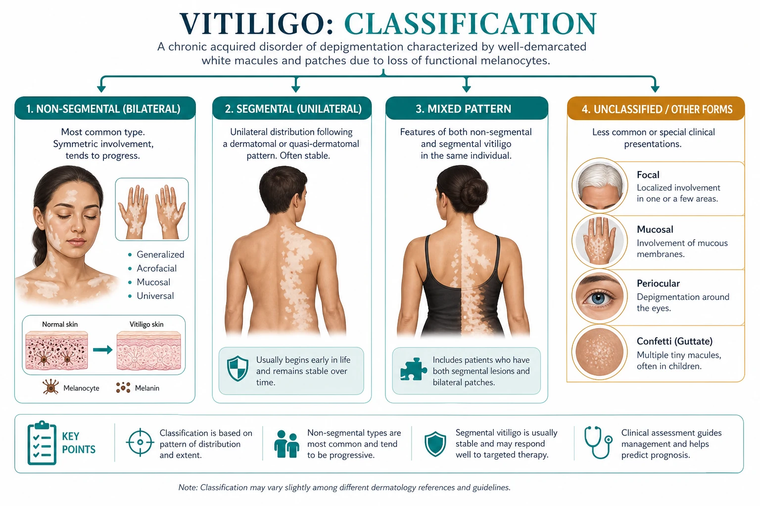
Vitiligo is an acquired, chronic, immune-mediated disorder characterised by selective loss of epidermal melanocytes and consequent depigmentation. The Vitiligo Global Issues Consensus Conference divides disease into two major categories:[3]
| Category | Characteristics | Notes |
|---|---|---|
| Non-segmental vitiligo (NSV) | Symmetrical, bilateral, progressive; often acrofacial, vulgaris, universalis | Most common; autoimmune associations; variable activity |
| Segmental vitiligo | Unilateral, dermatomal or Blaschkoid distribution; often early onset; rapid spread then stabilisation | Less common; thought to have neurogenic component; poorer response to medical therapy |
| Mixed vitiligo | Segmental plus non-segmental patches | Requires combined treatment approach |
| Unclassified/undefined | Mucosal, acrofacial, or early focal forms awaiting evolution | Reclassify over time |
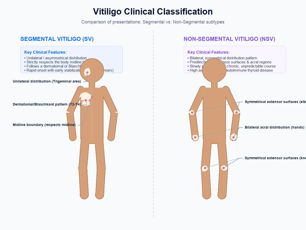
Clinical subtypes of non-segmental vitiligo [1]
- Generalised/vulgaris — widespread, often symmetrical patches.
- Acrofacial — distal digits and periorificial sites (lips, nostrils, eyes, genitalia).
- Mucosal — predominantly mucosal involvement.
- Universal — depigmentation involving most of the body surface area. [1]
Epidemiology & Risk Factors
Global prevalence is approximately 0.5-2%, with no clear sex predilection. Onset can occur at any age, but roughly half begin before age 20 and most before age 30. All races and skin phototypes are affected; the psychosocial impact is often greater in darker skin phototypes because of greater contrast.[1][4]
Genetic factors [1]
- Positive family history in roughly 20% of patients.
- Genome-wide association studies have linked vitiligo to immune-regulatory and melanocyte genes including NLRP1, XBP1, TYR, PMEL, and HLA loci.[5]
- NLRP1 variants promote inflammasome activation and IL-1β processing; XBP1 variants relate to endoplasmic-reticulum stress responses in melanocytes.[5]
Environmental triggers [1]
- Koebner phenomenon — new lesions at sites of trauma, friction, burns, or surgery.[7]
- Oxidative stress — reactive oxygen species may damage melanocytes and initiate autoimmunity.
- Infection, emotional stress, pregnancy, and phototherapy-related sunburn may precipitate or worsen disease.
- Chemical exposure — phenolic/catecholic compounds can induce occupational vitiligo-like leukoderma.
Pathophysiology
Vitiligo is a polygenic, multifactorial disorder converging on melanocyte destruction. [1]
The autoimmune hypothesis
The prevailing model involves cytotoxic CD8+ T-cell-mediated killing of melanocytes. Autoreactive T-cells recognise melanocyte antigens (e.g., tyrosinase, Melan-A/MART-1, gp100) and release interferon-gamma (IFN-γ). IFN-γ activates the JAK-STAT pathway in keratinocytes, driving expression of the chemokines CXCL9 and CXCL10, which recruit additional autoreactive T-cells and amplify the inflammatory cycle.[5]
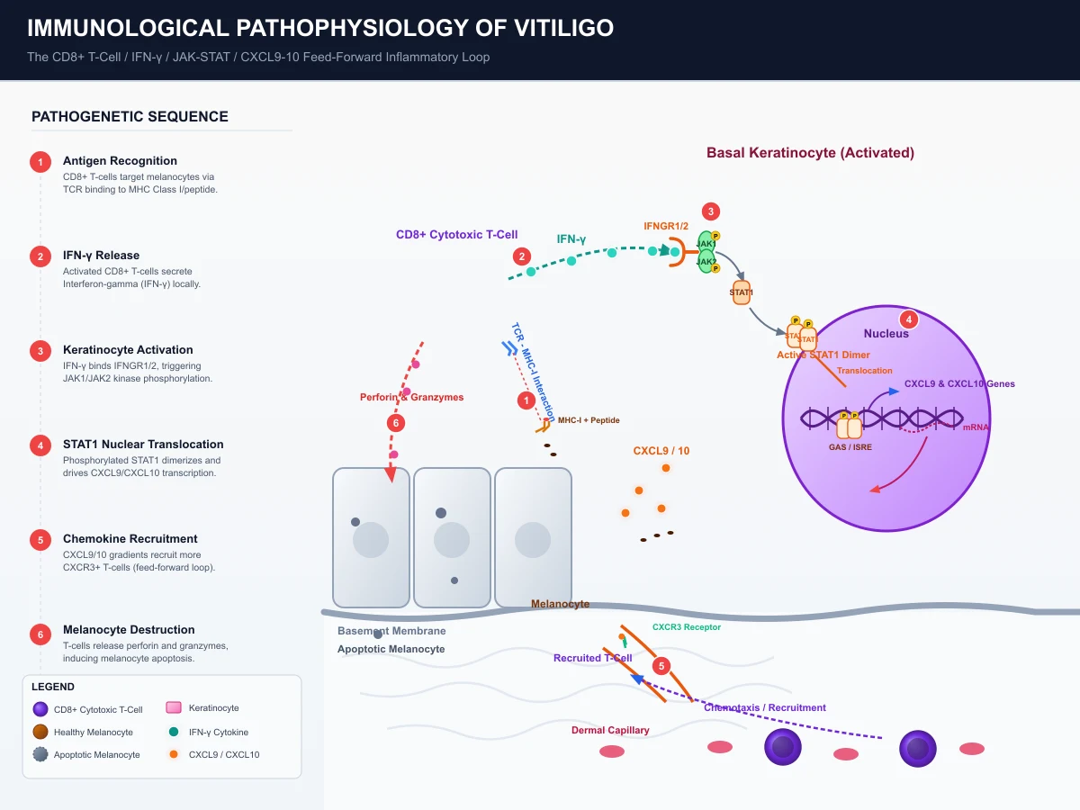
Oxidative stress and melanocyte intrinsic defects
Melanocytes in vitiligo skin show evidence of oxidative stress and impaired antioxidant defences. Reactive oxygen species may induce melanocyte senescence or death and unmask melanocyte antigens, initiating autoimmunity. Genetic variants affecting the unfolded protein response (XBP1) and inflammasome regulation (NLRP1) support a role for cellular stress in susceptibility.[5]
Melanocyte reservoirs and repigmentation
Repigmentation in vitiligo arises from melanocyte stem cells located in the hair follicle bulge. These stem cells migrate down the follicle, proliferate, and repopulate the epidermis. Treatments that remove inflammation (e.g., topical corticosteroids, calcineurin inhibitors, JAK inhibitors) and stimulate melanocyte proliferation (e.g., UVB) allow follicular melanocytes to recolonise depigmented skin. Hairy areas therefore repigment better than glabrous skin such as the lips, dorsal hands, and acral sites.[6]
Clinical Presentation
Cutaneous findings
- Chalk-white or milky-white, well-demarcated macules and patches with convex borders.
- Lesions may be round, oval, linear (segmental), or geographic.
- Trichrome vitiligo — three zones of colour (normal pigmentation, intermediate hypopigmentation, and depigmentation), implying active disease.
- Confetti-like lesions or perifollicular pigmentary changes suggest disease activity.
- Koebnerisation — lesions at sites of trauma, especially in active disease.[7]
Disease activity
A practical clinical distinction is stable versus active vitiligo: [1]
- Active disease — new lesions, enlargement of existing lesions, or confetti lesions within the past 3-6 months.
- Stable disease — no new lesions and no enlargement for at least 6-12 months.
- Stabilisation is a prerequisite for most surgical therapies.[2]
Common distributions
- Face and neck — periorificial (lips, eyelids), often early and cosmetically disabling.
- Hands, wrists, feet, ankles — acral disease, often resistant to therapy.
- Extensor surfaces — knees, elbows.
- Trunk — symmetrical patches.
- Genitalia and mucosae — common and distressing; requires gentle topical therapy. [1]
Differential Diagnosis
[1]Vitiligo vs ash-leaf macule vs piebaldism
Vitiligo
- Acquired (typically after birth); progressive; depigmented (chalk-white) macules
- AUTOIMMUNE — CD8+ T cells destroy melanocytes; positive family history ~20%
- Symmetric, bilateral (NSV) OR unilateral dermatomal/Blaschkoid (SV); periorificial and acral predilection
- Wood lamp: chalk-white / bright blue-white accentuation
- Strong association with autoimmune thyroid disease, T1DM, alopecia areata, pernicious anaemia
- TRICHOME lesions, confetti macules, Koebner phenomenon in active disease
- Treatment: topical steroids/calcineurin inhibitors/JAK inhibitors, NB-UVB, surgery for stable disease
Ash-leaf macule
- CONGENITAL (present at birth); hypopigmented (off-white, not chalk-white)
- Non-autoimmune — marker of TUBEROUS SCLEROSIS (TSC1/TSC2 mutation)
- Polygonal or 'ash-leaf' shape; typically trunk or limbs; usually solitary
- Wood lamp: enhances but remains off-white, NOT chalk-white
- Associated TSC features: angiofibromas, shagreen patch, periungual fibromas, epilepsy, autism, renal AMLs
- Stable throughout life; no progression or Koebner phenomenon
- No specific treatment needed; investigate for TSC if multiple lesions or features present
Piebaldism
- CONGENITAL (present at birth); stable depigmented patches
- Non-autoimmune — AUTOSOMAL DOMINANT; KIT gene mutation impairs melanocyte development/migration
- Symmetric depigmented patches on ventral trunk, mid-forehead, and limbs (knee/elbow sparing)
- Characteristic WHITE FORELOCK (poliosis) over the frontal scalp — present in 80-90%
- Wood lamp: chalk-white accentuation (true depigmentation, melanocytes absent)
- Hyperpigmented macules may be present within or at the border of depigmented patches
- No associated systemic disease (isolated cutaneous); no autoimmunity; no treatment required but photoprotection advised
Distinguishing congenital from acquired depigmentation is the single most important first step in the differential diagnosis. Congenital + stable = naevus depigmentosus, ash-leaf macule (TSC), or piebaldism. Acquired + progressive = vitiligo until proven otherwise. Wood lamp alone cannot separate these — the history of onset (birth vs later) and the family history and associated features are the discriminators. A biopsy showing complete absence of melanocytes (Melan-A negative) confirms true depigmentation and excludes hypopigmentation from post-inflammatory causes. In darker skin phototypes, hypopigmented patches are more conspicuous; in vitiligo the macules are depigmented (no melanin at all) whereas ash-leaf macules and naevus depigmentosus are hypopigmented (reduced but not absent melanin) and retain a faint tan under Wood lamp. [1]
Wood lamp examination enhances the contrast of depigmented lesions and helps delineate subclinical disease. It is useful for assessing extent and response to therapy.[4]
Histopathology
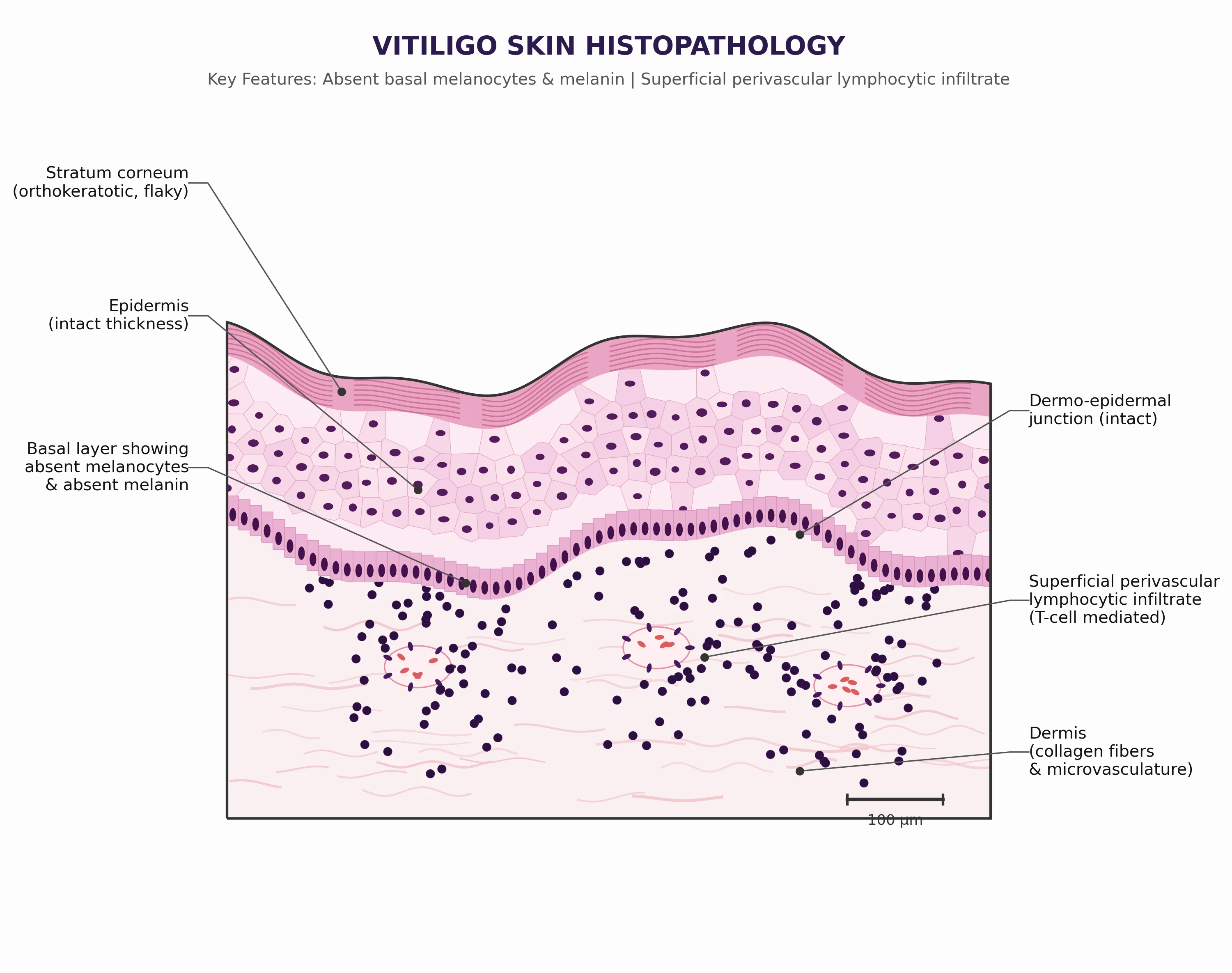
A skin biopsy is usually unnecessary if the clinical picture is classic, but is valuable when the diagnosis is uncertain. Histology shows absence or marked reduction of melanocytes and melanin in the basal layer, with a superficial perivascular lymphocytic infiltrate in early lesions. Special stains (e.g., Melan-A, S100, SOX10) confirm melanocyte loss.[4]
Clinical & Bedside Assessment
Assessment tools
| Tool | What it measures | Notes |
|---|---|---|
| VASI (Vitiligo Area Scoring Index) | Extent of depigmentation across body regions | Hand-unit estimates; 0-100 scale; used in trials |
| VETF (Vitiligo European Task Force) | Area, staging, and spreading score | Includes disease activity parameters |
| DLQI | Quality-of-life impact | Important for psychological burden and treatment response |
| Photography | Longitudinal comparison | Standardised lighting and backgrounds |
| Wood lamp | Subclinical lesion delineation | Useful in fair skin and for treatment monitoring |
Practical severity classification
- Limited/localised — under approximately 1% body surface area (BSA), few lesions.
- Generalised — 1-10% BSA.
- Extensive — more than 10% BSA or universal involvement.
- Special sites — face, hands, feet, genitalia, mucosae; often require targeted therapy. [1]
Investigations
Routine work-up
- FBC, LFT, U&E, creatinine — baseline before systemic therapy.
- Thyroid function tests (TSH) — autoimmune thyroid disease is the most common association.
- Fasting glucose or HbA1c — diabetes risk is increased.
- Vitamin B12 and folate — deficiencies may coexist.
- Antinuclear antibody (ANA) — screen if autoimmune disease is suspected. [1]
Before systemic immunosuppression
- Infection screen — hepatitis B/C, HIV per local protocol; tuberculosis screening where relevant.
- Pregnancy test — for women of childbearing potential before teratogenic drugs.
- Blood pressure, renal function, and fasting lipids — before ciclosporin. [1]
When to biopsy
- Atypical morphology or distribution.
- Lack of response to appropriate therapy.
- Suspicion for cutaneous T-cell lymphoma, sarcoidosis, or other hypopigmenting disorders.
- Need for histological confirmation before surgery or depigmentation.[4]
Management — Topical Therapy
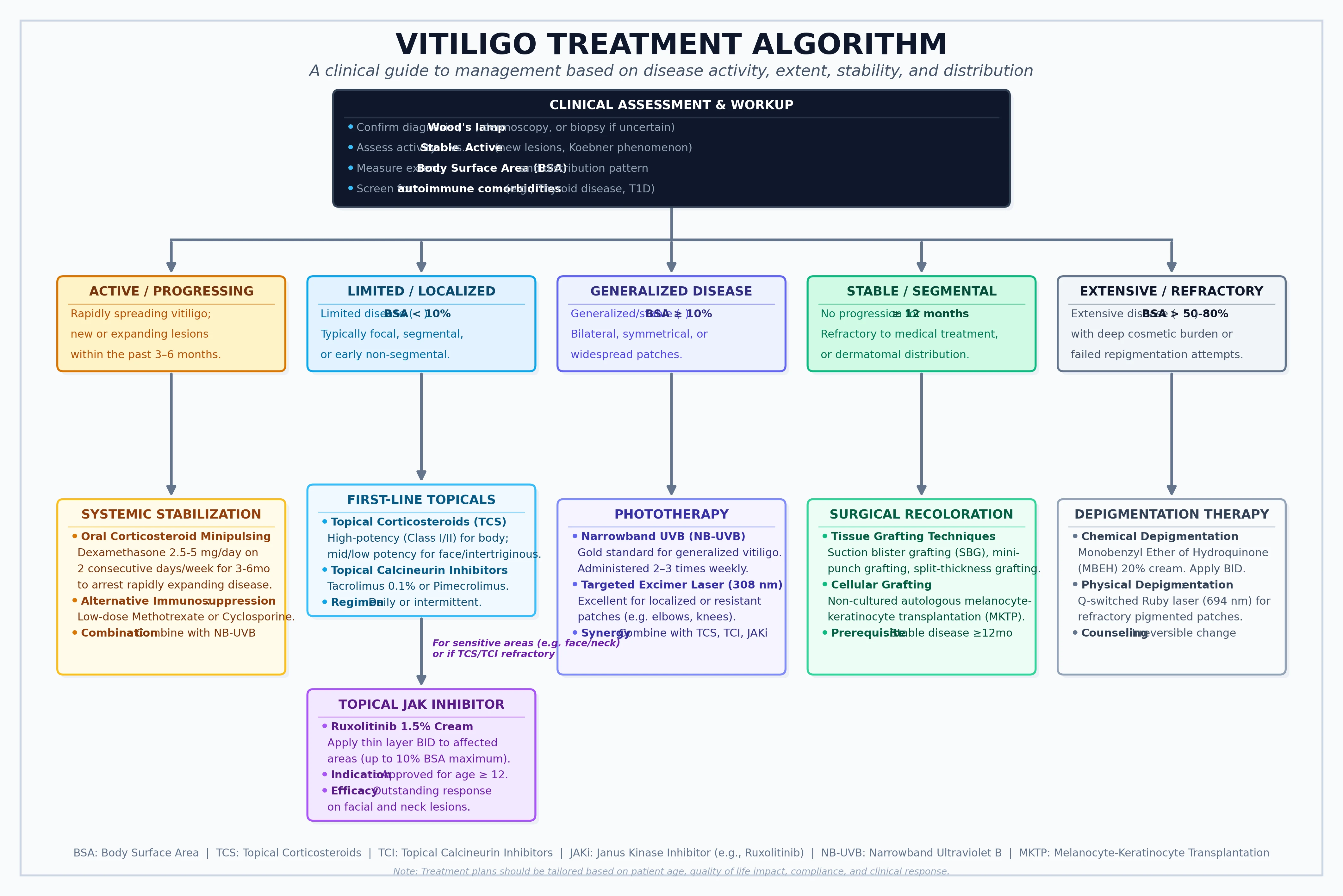
Topical therapy is first-line for limited, localised, or facial vitiligo, and an adjunct for more extensive disease. [1]
| Agent | Typical use | Notes |
|---|---|---|
| Topical corticosteroids | Trunk and limbs | Potent steroids for 2-3 months, then taper/rest; risk of atrophy and telangiectasia |
| Topical calcineurin inhibitors (tacrolimus 0.03-0.1%, pimecrolimus 1%) | Face, neck, eyelids, skin folds, genitalia | First-line for sensitive sites; no atrophy; combine with phototherapy |
| Topical ruxolitinib 1.5% | Non-segmental vitiligo in patients aged 12 years and older | JAK1/JAK2 inhibitor; FDA-approved; twice daily; monitor for application-site reactions and acne |
| Vitamin D analogues (calcipotriol) | Adjunct | Limited evidence; sometimes combined with phototherapy |
Ruxolitinib cream
Ruxolitinib 1.5% cream is a topical JAK1/JAK2 inhibitor approved for non-segmental vitiligo. Phase 2 and two phase 3 trials (TRuE-V1 and TRuE-V2) showed significantly greater facial and total-body repigmentation compared with vehicle at 24 weeks, with continued improvement to 52 weeks. It is applied twice daily to affected areas, with a maximum of approximately 20% BSA per label.[21][22]
Management — Phototherapy
Phototherapy is indicated for widespread or rapidly progressive non-segmental vitiligo and is often combined with topical therapy. [1]
Narrowband UVB (NB-UVB, 311 nm)
- First-line phototherapy for vitiligo.
- Administered 2-3 times weekly for at least 6-12 months.
- Response is slow; repigmentation often begins perifollicularly.
- Facial and trunk lesions respond better than acral lesions.
- Systematic review/meta-analysis shows NB-UVB is effective, with combination therapy generally outperforming monotherapy.[14]
PUVA
- Oral or topical psoralen plus UVA is effective but largely superseded by NB-UVB because of higher cumulative UVA dose and long-term photocarcinogenesis risk.
- May be considered for patients who cannot attend frequent NB-UVB sessions or for selected refractory cases.[15]
Excimer laser/light (308 nm)
- Useful for localised, stable lesions, particularly on the face and neck.
- Often combined with topical tacrolimus or corticosteroids for enhanced response.
- Network meta-analysis supports excimer laser combination therapies, especially for facial vitiligo.[27]
Combining phototherapy with topicals
Meta-analyses show that tacrolimus plus NB-UVB produces higher repigmentation rates than NB-UVB alone. Topical corticosteroids and calcineurin inhibitors are commonly used as adjuncts.[16]
Management — Systemic Therapy
Systemic therapy is used for rapidly progressive, extensive, or unstable vitiligo when topical/phototherapy is insufficient. [1]
Corticosteroids
- Oral mini-pulse dexamethasone (e.g., 2.5-5 mg on two consecutive days each week) is commonly used to stabilise active disease, with evidence from randomised trials showing it can halt progression.
- Oral prednisolone (e.g., 0.3-0.5 mg/kg/day, tapered) may be used short-term for rapidly spreading disease; monitor for metabolic and infectious adverse effects.
- Systemic steroids are often combined with phototherapy to enhance repigmentation.[17][18]
Ciclosporin
- Oral ciclosporin (e.g., 2-4 mg/kg/day) has shown efficacy in stabilising active vitiligo in a randomised controlled trial.
- Requires blood pressure and renal-function monitoring; avoid long-term use if possible.[19]
Methotrexate
- Low-dose methotrexate (e.g., 10-25 mg weekly) has been reported to stabilise progressive vitiligo, particularly in patients with extensive or refractory disease.
- Monitor FBC, LFT, and renal function; give folic acid.[20]
Other systemic agents
- Azathioprine, mycophenolate mofetil, and oral JAK inhibitors (e.g., ruxolitinib, tofacitinib) are used off-label in refractory cases, with emerging evidence supporting efficacy. Specialist oversight is required. [1]
Management — Surgery & Depigmentation
Surgical repigmentation
Surgical options are reserved for stable, localised vitiligo that has failed medical therapy: [1]
- Autologous non-cultured melanocyte-keratinocyte transplantation — epidermal cells harvested from a donor site and applied to the depigmented area; good for larger stable patches.
- Suction blister grafting — split-thickness epidermal grafts transferred by suction blister.
- Punch grafting — small full-thickness grafts; simpler but may produce cobblestoning.
- Cultured melanocyte transplantation — requires specialised laboratories. [1]
Stability for at least 6-12 months, absence of Koebner phenomenon, and realistic patient expectations are prerequisites.[24]
Depigmentation
For extensive or universal vitiligo (generally more than 50% BSA) that has failed or is unsuitable for repigmentation, depigmentation therapy with monobenzone can be offered. The goal is to achieve a uniform pale complexion by removing remaining pigment. It is permanent, requires lifelong sun protection, and is psychologically significant; counsel carefully.[25]
Special Populations
Children and adolescents
- Childhood vitiligo is frequently segmental or focal; non-segmental disease is also common.
- Topical calcineurin inhibitors are preferred for facial and flexural disease to avoid steroid atrophy.
- NB-UVB is safe and effective in children, although practical issues with attendance and photoprotection must be addressed.
- Psychological support is essential because of school-related stigma.[26]
Pregnancy and breastfeeding
- Most systemic agents are contraindicated; rely on topical corticosteroids and calcineurin inhibitors with limited exposure areas.
- Phototherapy may be continued with appropriate shielding of the abdomen and eyes.
- Avoid systemic immunosuppression, methotrexate, and ciclosporin in pregnancy unless specialist-directed. [1]
Comorbidities
Vitiligo is associated with a range of autoimmune and systemic disorders. A systematic review and meta-analysis found increased odds of: [1]
| Comorbidity | Notes |
|---|---|
| Autoimmune thyroid disease | Most common association; screen TSH periodically |
| Type 1 diabetes mellitus | Increased prevalence |
| Pernicious anaemia | Consider B12/folate assessment |
| Addison disease | Rare but important in appropriate clinical context |
| Alopecia areata | Shared autoimmune pathophysiology |
| Inflammatory bowel disease | Association reported |
| Psoriasis | Co-occurs more frequently than expected |
| Rheumatoid arthritis and SLE | Associations documented |
Prognosis & Follow-Up
Vitiligo is chronic and unpredictable. Treatment goals are: [1]
- Stabilise disease activity — stop new lesions and expansion.
- Achieve repigmentation — best on face, neck, and trunk; poorest on acral and mucosal sites.
- Maintain response — relapses are common, particularly after stopping therapy.
- Support quality of life — address psychological impact, camouflage, and sun protection. [1]
Follow-up intervals [1]
- Topical therapy: 8-12 weeks to assess response.
- Phototherapy: every 3-4 months, with photography.
- Systemic therapy: more frequent monitoring for adverse effects. [1]
Prognostic factors for repigmentation [1]
- Favourable: recent onset, facial involvement, darker skin phototype, presence of hairs within lesions.
- Unfavourable: long-standing disease, acral/mucosal lesions, segmental vitiligo (medical therapy), and prior extensive depigmentation. [1]
Evidence, Guidelines & Regional Differences
Major guidelines
- British Association of Dermatologists (BAD) 2021 guidelines — emphasise shared decision-making, early treatment of active disease, topical corticosteroids/calcineurin inhibitors for localised disease, NB-UVB for generalised vitiligo, and careful patient selection for surgery/depigmentation.[2]
- Cochrane review (2015) — high-level summary of interventions, highlighting limited high-quality trial data at the time and the need for larger RCTs.[13]
- JAMA clinical evidence review (2016) — graded summary of topical, phototherapy, and surgical options.[12]
Landmark trials
- Ruxolitinib phase 2 (Lancet 2020) — first rigorous evidence for topical JAK inhibition in vitiligo.[21]
- TRuE-V1 and TRuE-V2 (NEJM 2022) — phase 3 trials establishing ruxolitinib 1.5% cream for non-segmental vitiligo.[22]
- NB-UVB plus ruxolitinib (J Invest Dermatol 2022) — supports combination phototherapy and JAK inhibition.[23]
Exam Pearls
[1]VITILIGO — the seven clinical anchors
VITILIGO
Non-segmental (bilateral, symmetrical, autoimmune) vs segmental (unilateral, dermatomal/Blaschkoid, early onset, then stable)
CD8+ T cells → IFN-γ → JAK1/JAK2 → CXCL9/CXCL10 → melanocyte apoptosis; the target of ruxolitinib
Screen TSH/anti-TPO (Hashimoto/Graves ~20-30%); also T1DM, pernicious anaemia, alopecia areata, Addison disease
Topical corticosteroid (clobetasol 0.05% BID) OR calcineurin inhibitor (tacrolimus 0.1% BID) OR ruxolitinib 1.5% cream BID; add NB-UVB if widespread
Wood lamp (365 nm) accentuates vitiligo as chalk-white / bright blue-white; helps assess extent, activity, and treatment response
Inflammation must be controlled BEFORE repigmentation; repigmentation comes from hair follicle melanocyte stem cells (perifollicular pattern)
Acral sites (hands, feet, lips, genitalia) respond POORLY because they lack follicular melanocyte reservoirs
Stable for 6-12 months before surgical melanocyte transfer; relapse common; monobenzone depigmentation irreversible for extensive (>50% BSA) refractory disease
Autoimmune Associations & Comorbidity Screening
Vitiligo sits at the centre of a poly-autoimmune network and is one of the most consistent cutaneous markers of systemic autoimmunity. The 2023 systematic review and meta-analysis by Lee and colleagues confirmed increased prevalence of multiple autoimmune diseases in vitiligo patients compared with the general population. Autoimmune thyroid disease (Hashimoto thyroiditis and Graves disease) is the most common association, present in roughly 20-30% of vitiligo patients overall and up to 40-50% in adults over age 40. Type 1 diabetes mellitus, pernicious anaemia (with anti-intrinsic factor and anti-parietal cell antibodies), alopecia areata (sharing the same IFN-γ/JAK-STAT axis and CD8+ T-cell mechanism), Addison disease, systemic lupus erythematosus, inflammatory bowel disease, psoriasis, rheumatoid arthritis, dermatomyositis, and Sjögren syndrome have all been documented at increased frequency.[11]
The shared genetic architecture explains much of this clustering. GWAS have linked vitiligo to HLA class I and II loci (the strongest signal at HLA-DRB1 and HLA-A), PTPN22 (a lymphocyte signalling phosphatase also implicated in T1DM, RA, SLE), NLRP1 (inflammasome, also implicated in other autoimmune and inflammatory diseases), XBP1 (endoplasmic reticulum stress), TYR (tyrosinase, melanocyte autoantigen), PMEL (melanosomal protein), and several other immune-melanocyte genes. This shared susceptibility means that a patient with vitiligo has a higher pre-test probability of any autoimmune disease than the general population, justifying targeted screening even in the absence of symptoms. [1]
The minimum screening panel at diagnosis should include: [1]
- TSH ± anti-TPO antibodies — autoimmune thyroid disease is the most common association and the most likely to present insidiously with non-specific symptoms. Repeat TSH annually or with new symptoms (fatigue, weight change, cold intolerance, tremor).
- Fasting glucose or HbA1c — screen for type 1 diabetes and other glucose intolerance, particularly in patients with a family history or other autoimmune disease.
- Vitamin B12, folate, and red cell indices — screen for pernicious anaemia, especially in patients with macrocytosis, neurological symptoms, or a personal/family history of autoimmune disease.
- Antinuclear antibody (ANA) — screen if systemic autoimmune disease (SLE, Sjögren) is suspected based on symptoms.
- Morning cortisol / ACTH stimulation test — only if Addison disease is clinically suspected (fatigue, weight loss, hyperpigmentation of OTHER areas, hyponatraemia, hyperkalaemia).
- Celiac serology (anti-tissue transglutaminase IgA) — low threshold in patients with GI symptoms or family history.[11]
Beyond autoimmunity, vitiligo patients also have higher rates of psychiatric comorbidity: depression, anxiety, social phobia, sexual dysfunction, and reduced quality of life. The DLQI in vitiligo is comparable to that in psoriasis and atopic dermatitis. Screen with PHQ-9 (depression) and GAD-7 (anxiety) at diagnosis and during follow-up. Offer psychological support, cognitive-behavioural therapy, and referral to patient support organisations (Vitiligo Society, VR Foundation, American Vitiligo Research Foundation). Children with vitiligo require particular attention because of school-related stigma and bullying.[10]
Wood Lamp Examination — The Practical Technique
Wood lamp (also called Wood's light or UVA blacklight) emits long-wave ultraviolet A radiation at approximately 365 nm. It is inexpensive, portable, requires no special preparation, and is essential for accurate assessment of vitiligo in every patient. In depigmented skin (vitiligo, piebaldism), Wood lamp produces a striking chalk-white or bright blue-white fluorescence because there is no melanin to absorb the UV — the reflected light is enhanced. In hypopigmented skin (ash-leaf macule, pityriasis versicolor, post-inflammatory hypopigmentation), there is reduced but not absent melanin, and the fluorescence is off-white or tan rather than chalk-white. This contrast is the most useful clinical application.[4]
Practical technique: dim the room; hold the lamp 10-15 cm from the skin; examine for 30-60 seconds to allow dark adaptation of the examiner's eyes. The Wood lamp is particularly useful for: [1]
- Delineating the true extent of disease in patients with fair skin (Fitzpatrick I-II) where lesions may be subtle under normal lighting.
- Identifying subclinical lesions that are not yet visible to the naked eye — useful for documenting true disease extent and for monitoring treatment response.
- Distinguishing active from stable lesions — active lesions often show ill-defined, feathery borders under Wood lamp; stable lesions have sharper borders.
- Confirming depigmentation vs hypopigmentation — important for separating vitiligo (true depigmentation) from hypomelanotic disorders.
- Assessing repigmentation during treatment — perifollicular repigmentation is best visualised under Wood lamp. [1]
Wood lamp does NOT diagnose vitiligo definitively — many other conditions enhance with Wood lamp (erythrasma = coral-red, tinea capitis = green/yellow, porphyria = pink/red urine). The diagnosis of vitiligo is clinical, supported by Wood lamp and supplemented by biopsy in atypical cases.[4]
Surgical Repigmentation — When and How
For patients with stable, localised vitiligo that has failed an adequate trial of medical therapy (typically 12 months), surgical melanocyte transfer can restore pigment. The fundamental principle is that melanocytes (or melanocyte-keratinocyte suspensions) harvested from a normally pigmented donor site are transplanted into the depigmented recipient site, where they proliferate and repopulate the epidermis. The most important prerequisite is disease stability — defined as no new lesions and no enlargement of existing lesions for at least 6-12 months and absence of Koebner phenomenon. Transplanting into unstable disease leads to destruction of the transplanted melanocytes by the ongoing autoimmune process, with subsequent re-depigmentation.[24]
The main surgical options are: [1]
- Autologous non-cultured melanocyte-keratinocyte transplantation (MKT) — the most widely used technique. A thin split-thickness donor skin graft is harvested (often from the thigh or buttock), incubated in trypsin-EDTA to separate the epidermis, and the resulting melanocyte-keratinocyte suspension is applied to the dermabraded recipient site. Best for larger stable patches; good repigmentation rates (70-90%) on suitable sites. Limitation: requires a laboratory or experienced team.
- Suction blister grafting — negative-pressure suction is applied to the donor site to create blisters; the epidermal roof (containing melanocytes) is transferred to the dermabraded recipient. Good for small to moderate patches; cosmetically elegant; minimal donor-site morbidity.
- Punch grafting — small full-thickness punch biopsies from a donor site are transplanted into punch holes in the recipient area. Simple and inexpensive; main complication is cobblestoning (donor skin raised above the recipient level) and limited to small lesions.
- Cultured melanocyte transplantation — melanocytes from a small donor biopsy are expanded in a specialised cell-culture laboratory over weeks, then transplanted as a cell suspension or as a sheet. Useful for very large areas, but expensive and limited by regulatory requirements.
- Hair follicle grafting — single hair follicles harvested from the scalp (where melanocyte reservoirs are abundant) are transplanted into depigmented recipient sites. Particularly useful for repigmenting acral areas with no native follicles. [1]
Best responders: segmental vitiligo (SV), small stable lesions, face and neck lesions, adults with no Koebner. Poor responders: active disease, acral sites (lower density of follicular reservoirs), large areas in non-segmental vitiligo, patients with unrealistic expectations. Counselling: patients must understand that repigmentation takes months, that colour matching may not be perfect, and that there is a risk of re-depigmentation if disease reactivates.[24]
Red Flags
Exam application bank (NEET-PG / INICET)
One-line answer
Vitiligo is an acquired, immune-mediated depigmenting disorder characterised by selective loss of melanocytes. Fellowship-level assessment requires mastery of classification (segmental vs non-segmental), the IFN-γ/JAK-STAT pathophysiological axis, clinical patterns and disease activity, validated assessment tools (VASI, VETF, DLQI), topical and phototherapy protocols, conventional systemic immunosuppressants, targeted JAK inhibition, surgical repigmentation, depigmentation for extensive disease, comorbidity screening, and special-population considerations.
Worked stems (answer without another resource)
Stem 1 — Classic presentation. Map symptoms to mechanism; name the first investigation and first treatment step with dose/route if drug therapy is standard. [1]
Stem 2 — Unstable / complicated. List red flags that force immediate resuscitation, theatre, ICU, antidote, or reperfusion — and what you do in the first 15 minutes. [1]
Stem 3 — Atypical group. Elderly, pregnancy, child, or immunocompromised: how presentation and thresholds change. [1]
Stem 4 — Differential trap. Name the three closest mimics and one discriminator for each. [1]
Stem 5 — Disposition. Who goes home with safety-netting, who is admitted, who needs HDU/ICU/theatre, and what follow-up is mandatory. [1]
Rapid viva checklist
- Definition + classification
- Pathophysiology chain
- Bedside signs / criteria
- Score with exact components (if any)
- Emergency bundle
- Definitive therapy with doses
- Complications of disease and of treatment
- Special populations
- Guideline/trial name if classic
- Three exam traps
Coverage self-check
If you cannot answer any stem above from this page alone, re-read the matching section — the page is intended to be self-sufficient for final-prof and NEET-PG/INICET questions on Vitiligo.
Expanded exam teaching (depth pass)
Clinical reasoning
For Vitiligo, examiners test whether you can prioritise life threats, choose the right first test, and give specific therapy (agent, dose, route, timing). Generic phrases without numbers score poorly.
Mechanism → feature map
Build a short chain: cause → pathophysiologic intermediate → clinical feature → complication. Every major symptom in the classic vignette should sit on that chain.
Investigation strategy
- Bedside/first-line tests that change immediate management
- Confirmatory or staging tests
- What a normal result does not exclude
- When not to delay treatment for imaging (unstable patient)
Management ladder
- Resuscitation / ABC / sepsis or haemorrhage bundle as relevant
- Specific antidote / procedure / antimicrobial / reperfusion / surgery
- Supportive care and monitoring targets
- Definitive long-term therapy and secondary prevention
- Disposition and safety-net advice
Special populations
Always prepare one line each for children, pregnancy, elderly, renal/hepatic impairment, and immunocompromised patients when the topic allows.
Pitfalls that fail candidates
- Treating the number not the patient
- Missing pregnancy status when relevant
- Imaging before stabilisation
- Wrong empiric cover or wrong antidote timing
- Incomplete counselling on recurrence, adherence, or red-flag return
Vitiligo is an acquired, immune-mediated depigmenting disorder characterised by selective loss of melanocytes. Fellowship-level assessment requires mastery of classification (segmental vs non-segmental), the IFN-γ/JAK-STAT pathophysiological axis, clinical patterns and disease activity, validated assessment tools (VASI, VETF, DLQI), topical and phototherapy protocols, conventional systemic immunosuppressants, targeted JAK inhibition, surgical repigmentation, depigmentation for extensive disease, [1]
[1]References
- [1]Ezzedine K, Eleftheriadou V, Whitton M, van Geel N. Vitiligo Lancet, 2015.PMID 25596811
- [2]Eleftheriadou V, Atkar R, Batchelor J, et al. British Association of Dermatologists guidelines for the management of people with vitiligo 2021 Br J Dermatol, 2022.PMID 34160061
- [3]Ezzedine K, Lim HW, Suzuki T, Katayama I. Revised classification/nomenclature of vitiligo and related issues: the Vitiligo Global Issues Consensus Conference Pigment Cell Melanoma Res, 2012.PMID 22417114
- [4]Alikhan A, Felsten LM, Daly M, Petronic-Rosic V. Vitiligo: a comprehensive overview Part I. Introduction, epidemiology, quality of life, diagnosis, differential diagnosis, associations, histopathology, etiology, and work-up J Am Acad Dermatol, 2011.PMID 21839315
- [5]Mukhatayev Z, Le Poole IC. Vitiligo: advances in pathophysiology research and treatment development Trends Mol Med, 2024.PMID 38705825
- [6]Birlea SA, Costin GE, Roop DR, Norris DA. Trends in Regenerative Medicine: Repigmentation in Vitiligo Through Melanocyte Stem Cell Mobilization Med Res Rev, 2017.PMID 28029168
- [7]van Geel N, Speeckaert R, Taieb A, et al. Koebner's phenomenon in vitiligo: European position paper Pigment Cell Melanoma Res, 2011.PMID 21324101
- [8]Taïeb A, Picardo M, VETF Members. The definition and assessment of vitiligo: a consensus report of the Vitiligo European Task Force Pigment Cell Res, 2007.PMID 17250545
- [9]Ceresnie MS, Warbasse E, Gonzalez S, Jacob C. Implementation of the vitiligo area scoring index in clinical studies of patients with vitiligo: a scoping review Arch Dermatol Res, 2023.PMID 37029284
- [10]Morales-Sánchez MA, Vargas-Salinas M, Peralta-Pedrero ML, et al. Impact of Vitiligo on Quality of Life Actas Dermosifiliogr, 2017.PMID 28456327
- [11]Lee JH, Ju HJ, Seo JM, et al. Comorbidities in Patients with Vitiligo: A Systematic Review and Meta-Analysis J Invest Dermatol, 2023.PMID 36574529
- [12]Ezzedine K, Whitton M, Pinart M. Interventions for Vitiligo JAMA, 2016.PMID 27784078
- [13]Whitton ME, Pinart M, Batchelor J, et al. Interventions for vitiligo Cochrane Database Syst Rev, 2015.PMID 25710794
- [14]Bae JM, Jung HM, Hong BY, et al. Phototherapy for Vitiligo: A Systematic Review and Meta-analysis JAMA Dermatol, 2017.PMID 28355423
- [15]Shenoi SD, Prabhu S, Indian Association of Dermatologists, Venereologists and Leprologians. Photochemotherapy (PUVA) in psoriasis and vitiligo Indian J Dermatol Venereol Leprol, 2014.PMID 25382505
- [16]Dong Y, Yang Q, Guo B, et al. The effects of tacrolimus plus phototherapy in the treatment of vitiligo: a meta-analysis Arch Dermatol Res, 2021.PMID 32785837
- [17]El Mofty M, Essmat S, Youssef R, et al. The role of systemic steroids and phototherapy in the treatment of stable vitiligo: a randomized controlled trial Dermatol Ther, 2016.PMID 27528547
- [18]Singh A, Kanwar AJ, Parsad D, Kumar B. Randomized controlled study to evaluate the effectiveness of dexamethasone oral minipulse therapy versus oral minocycline in patients with active vitiligo vulgaris Indian J Dermatol Venereol Leprol, 2014.PMID 24448120
- [19]Mehta H, Kumar S, Parsad D, Kumaran MS. Oral cyclosporine is effective in stabilizing active vitiligo: Results of a randomized controlled trial Dermatol Ther, 2021.PMID 34151493
- [20]Tavitova A, Valle Y, Lomonosov K. Using methotrexate in the treatment of advanced vitiligo J Cosmet Dermatol, 2023.PMID 36409563
- [21]Rosmarin D, Pandya AG, Lebwohl M, et al. Ruxolitinib cream for treatment of vitiligo: a randomised, controlled, phase 2 trial Lancet, 2020.PMID 32653055
- [22]Rosmarin D, Passeron T, Pandya AG, et al. Two Phase 3, Randomized, Controlled Trials of Ruxolitinib Cream for Vitiligo N Engl J Med, 2022.PMID 36260792
- [23]Pandya AG, Harris JE, Lebwohl M, et al. Addition of Narrow-Band UVB Phototherapy to Ruxolitinib Cream in Patients With Vitiligo J Invest Dermatol, 2022.PMID 35787401
- [24]Bassiouny D, Esmat S. Autologous non-cultured melanocyte-keratinocyte transplantation in the treatment of vitiligo: patient selection and perspectives Clin Cosmet Investig Dermatol, 2018.PMID 30464567
- [25]Grimes PE, Nashawati R. Depigmentation Therapies for Vitiligo Dermatol Clin, 2017.PMID 28317530
- [26]Nicolaidou E, Mastraftsi S, Tzanetakou V, et al. Childhood Vitiligo Am J Clin Dermatol, 2019.PMID 30911977
- [27]Li C, Hu Y, Mu Z, Gao XH. Comparison of various excimer laser (EL) combination therapies for vitiligo: a systematic review and network meta-analysis J Dermatolog Treat, 2024.PMID 38230424