Dermatology · General Medicine
Vitiligo & Pigmentation Disorders
Also known as Vitiligo · Melasma · Chloasma · Post-inflammatory hyperpigmentation · Post-inflammatory hypopigmentation · Depigmentation · Oculocutaneous albinism · Drug-induced pigmentation
Vitiligo is an acquired autoimmune destruction of epidermal melanocytes producing well-demarcated, chalk-white (depigmented) macules and patches on the periorificial face, hands, extensor surfaces, genitalia and body folds; classified as segmental (unilateral, dermatomal, early onset, stabilises within 1 to 2 years) or non-segmental (bilateral, symmetrical, progressive). It is associated with autoimmune thyroid disease, type 1 diabetes, pernicious anaemia, Addison disease and alopecia areata, and carries a major psychological burden, especially in darker skin. Melasma (chloasma) is acquired symmetrical facial hyperpigmentation (forehead, malar cheeks, upper lip) triggered by sun, pregnancy, oral contraceptives and genetics. Post-inflammatory pigment change, oculocutaneous albinism, and drug-induced pigmentation (minocycline, amiodarone, chloroquine) complete the pigmentary differential. Diagnosis is clinical, confirmed with the Wood lamp; management combines topical corticosteroids, calcineurin inhibitors, JAK inhibitors, narrowband UVB phototherapy, excimer laser, surgery, camouflage and depigmentation for vitiligo, and strict sun protection with hydroquinone-based regimens for melasma.
On this page & tools
Your progress
Saved locally on this device.
Exam tags
Red flags
Overview & Definition
The pigmentary disorders sit at a fault line of dermatology: a single cell — the melanocyte — and a single pigment — melanin — explain almost all of them. When melanocytes are destroyed, skin turns chalk-white (vitiligo); when they are overactive or stimulated, skin turns brown (melasma, post-inflammatory hyperpigmentation); when they are genetically defective from birth, skin and eyes are globally under-pigmented (oculocutaneous albinism); and when drugs or metals deposit in the dermis, the colour is slate-grey or blue-black. The clinical skill is to recognise the pattern, distribution and tempo, then decide whether to screen for autoimmune disease (vitiligo), exclude Addison disease (generalised pigmentation), identify a drug culprit, or simply reassure. [1][5]
Vitiligo and melasma are the two commonest pigmentary disorders seen in MBBS practice. Both are chronic, both are driven by sunlight, and both carry a psychological burden out of all proportion to their physical seriousness — vitiligo because of the social stigma attached to visible depigmentation, especially in darker-skinned populations, and melasma because of its disfiguring effect on the face in young women. Neither is life-threatening, yet both can dominate a patient's life. The examiner rewards the candidate who treats these patients with empathy and a structured plan rather than dismissing them as cosmetic. [1][3]
The pigment spectrum is best learned as five clinical problems: (1) vitiligo and other causes of depigmentation/hypopigmentation; (2) melasma; (3) post-inflammatory pigment change; (4) oculocutaneous albinism; and (5) drug-induced and systemic pigmentation. This topic builds each one to a depth that answers any examiner question. [1]
Classification
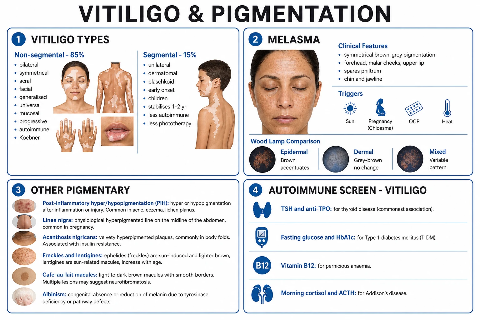
- Commonest form (~85%)
- Bilateral and symmetrical
- Progressive, relapsing
- Strong autoimmune association
- Koebner positive
- Responds to phototherapy and JAK inhibitors
- ~15% of cases
- Unilateral, dermatomal or Blaschkoid
- Onset in childhood/adolescence
- Spreads for 1 to 2 years then stabilises
- Leucotrichia common
- Weaker autoimmune link
- Surgery often needed; phototherapy less effective
- Acquired, symmetrical facial hyperpigmentation
- Sun-exposed sites: forehead, malar cheeks, upper lip, chin
- Triggered by UV, pregnancy, OCP, genetics
- Epidermal (brown) vs dermal (grey-brown) vs mixed
- Women 9:1; darker skin types
- Follows any skin injury or inflammation (acne, eczema, lichen planus, burns)
- Hyperpigmentation in melanin-rich skin
- Hypopigmentation when inflammation is severe
- May take months to years to fade
- Inherited (AR) tyrosinase-pathway defect
- Tyrosinase-negative vs tyrosinase-positive
- Absent/reduced melanin from birth
- Ocular signs: nystagmus, photophobia, foveal hypoplasia, iris transillumination
- High skin-cancer risk
The non-segmental subtypes are further divided by distribution: vulgaris/generalised (scattered bilateral patches, the commonest), acrofacial (distal extremities and periorificial face), universalis (over 80 percent body-surface-area depigmentation), mucosal (lips and genitalia), and mixed (segmental plus non-segmental). The 2011 international consensus formally separated segmental vitiligo from all other forms and reserved the unqualified term "vitiligo" for the non-segmental group — a distinction that matters because the two forms behave, associate, and respond to treatment differently. [1]
Epidemiology & Risk Factors
Vitiligo and melasma by the numbers
Vitiligo risk factors cluster around autoimmunity and skin trauma. A family history of vitiligo or autoimmune disease, the presence of another autoimmune disorder (thyroid disease is the commonest, in 15 to 30 percent), physical or chemical trauma to the skin (the Koebner phenomenon), emotional stress, sunburn, and exposure to phenolic or catechol chemicals (in industrial workers, hair dyes, and rubber) all increase the likelihood of onset or flare. Melasma is driven by the triad of sunlight, female sex hormones (pregnancy, oral contraceptives, hormone replacement), and a genetic predisposition commoner in darker-skinned (Fitzpatrick III to V) populations; it is rare in men, and essentially unheard of before puberty. [1][3]
Pathophysiology
Vitiligo is now unambiguously classified as an autoimmune disease — not a cosmetic problem. The dominant model is the IFN-gamma / CXCL9/10 chemokine axis: autoreactive CD8-positive cytotoxic T cells recognise melanocyte antigens and release interferon-gamma, which induces surrounding keratinocytes to secrete the chemokines CXCL9 and CXCL10; these chemokines recruit yet more CXCR3-bearing CD8 T cells to the skin in a self-amplifying positive-feedback loop that drives depigmentation. [2]
This discovery is not just academic — it explains why topical and systemic JAK inhibitors (which block IFN-gamma signalling) repigment vitiligo, and why disease relapses after treatment stops: tissue-resident memory T cells (TRM), seeded in lesional and peri-lesional skin during active disease, persist indefinitely and reignite the cascade when therapy is withdrawn. Newer strategies therefore aim to eliminate TRM (for example by targeting the IL-15 / CD122 pathway on which they depend) rather than merely suppressing them. [2]
The classical convergence (or convergence-combined) theory holds that several mechanisms act together: (1) autoimmunity (the dominant force, as above), (2) oxidative stress (accumulation of hydrogen peroxide in the epidermis overwhelms catalase and damages melanocytes), (3) neural (segmental vitiligo follows a dermatome, suggesting a neurochemical contribution), and (4) biochemical (intrinsic melanocyte defects with detached or deformed dendrites). Autoantibodies against melanocyte antigens (tyrosinase, gp100, MART-1) are detectable, but CD8 T cells, not antibodies, are the effector. [2]
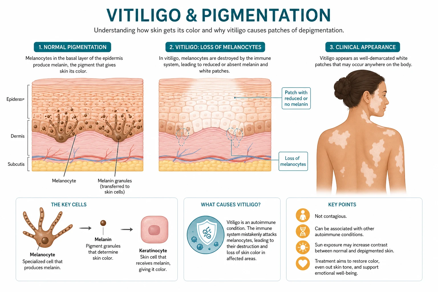
Melasma pathogenesis is multifactorial. Sunlight stimulates melanogenesis through alpha-MSH and the cAMP/MAPK pathway; oestrogen and progesterone upregulate tyrosinase and melanogenic enzymes (hence pregnancy and OCP). Histologically there is not only increased melanin in basal keratinocytes but also increased melanocyte number and dendricity, solar elastosis in the dermis, and increased vascularity. The disorder is best understood as a photoageing-related pigmentation rather than simple overproduction of melanin. [3]
Post-inflammatory hyperpigmentation (PIH) arises when inflammation (acne, eczema, lichen planus, burns, friction) stimulates melanocytes to overproduce and transfer melanosomes to basal keratinocytes (epidermal PIH — brown) and sometimes to drop melanin into the upper dermis where it is engulfed by macrophages (dermal PIH — grey-blue, slow to fade). In darker skin (Fitzpatrick IV to VI), even trivial inflammation produces striking PIH. The reverse — post-inflammatory hypopigmentation — occurs when inflammation is severe enough to deplete or stun melanocytes (after psoriasis, pityriasis alba, or burns), and usually recovers. [7]
Oculocutaneous albinism (OCA) is an autosomal recessive defect in the melanin-synthesis pathway. The pivotal enzyme is tyrosinase, which converts tyrosine to DOPA and then to melanin. In tyrosinase-negative OCA1A there is a complete loss of tyrosinase and no melanin is ever produced — the patient has white hair, white skin, and red-translucent irides from birth. In tyrosinase-positive forms (OCA1B, OCA2/P-gene, OCA3/TYRP1, OCA4/SLC45A2) some melanin accumulates over time, giving a milder phenotype. Because melanin is essential for normal foveal development and optic-tract routing, its absence produces the ocular hallmarks: nystagmus, photophobia, reduced visual acuity, foveal hypoplasia, iris transillumination, and a misrouted optic decussation (more fibres crossing at the chiasm, demonstrable on visual evoked potentials). [6]
Clinical Presentation
Vitiligo
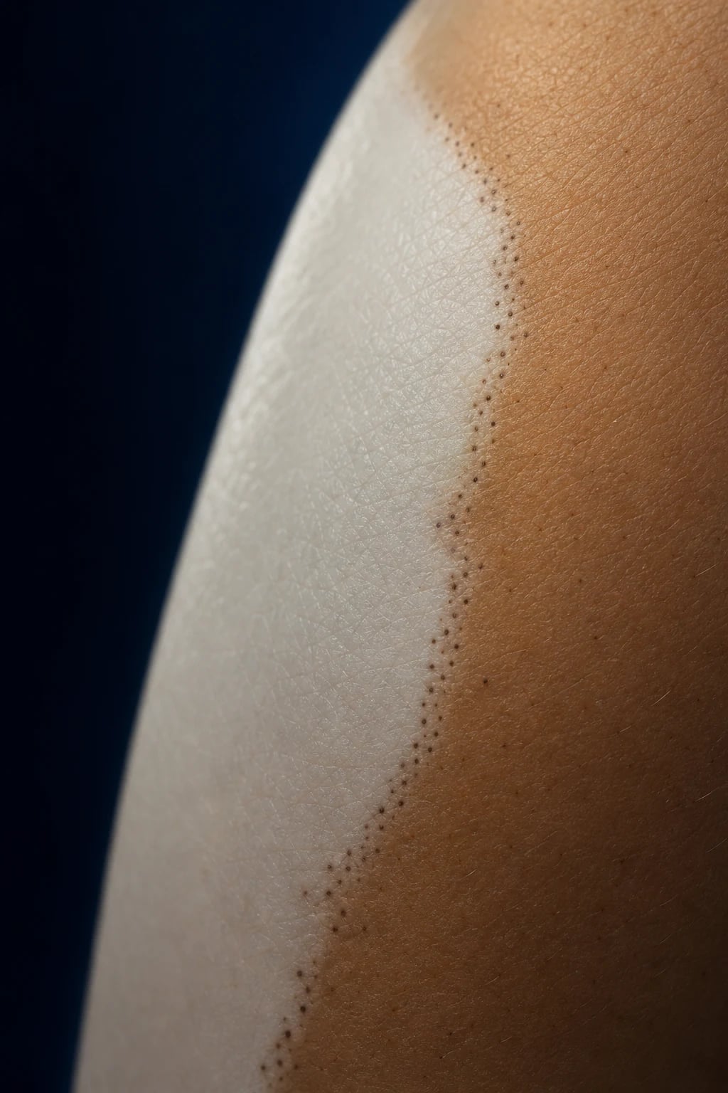
The vitiligo lesion is a completely depigmented, chalk-white, nonscaly macule or patch with sharply demarcated borders. There is no atrophy, no scale, no induration — only absence of pigment. The contrast with surrounding skin is dramatic in darker skin types and may be subtle in very fair skin, where the Wood lamp is essential. [1]
Characteristic sites are the periorificial face (around the eyes and mouth — the so-called lip-tip distribution, including the lips, fingertips, and genitalia), the dorsal hands and wrists, extensor forearms, knees, elbows, ankles, and the body folds (axillae, groins). Leucotrichia — depigmented (white) hairs within a patch — indicates that the hair-follicle melanocyte reservoir is also involved, which has treatment implications (follicular repigmentation is impossible where leucotrichia is present). The Koebner phenomenon (isomorphic response) produces new patches at sites of friction, pressure, cuts, or burns — the reason vitiligo appears under waistbands, bra straps, and watchstraps, and why patients must avoid trauma. [1]
Segmental vitiligo presents as a unilateral, dermatomal or Blaschkoid band of depigmentation, usually appearing in childhood or adolescence, spreading rapidly for 6 to 24 months and then arresting permanently. It frequently involves leucotrichia, has a weaker autoimmune association, and is the form most amenable to surgical (grafting) treatment once stable. [1]
Non-segmental vitiligo is bilateral and symmetrical, chronic and relapsing, and tends to progress over years with flare-ups often linked to stress, illness, or sunburn. Vitiligo universalis depigments nearly the entire skin surface. [1]
Atypical presentations the examiner tests include: onset during pregnancy (often flares post-partum), late-onset vitiligo in the elderly, vitiligo coexisting with melanoma (a paraneoplastic immune response that can herald tumour regression — but never assume a depigmented patch in an older patient is vitiligo without excluding amelanotic melanoma and halo naevus), and chemical leukoderma from exposure to phenols, catechols, or rubber chemicals in a pattern that mimics vitiligo but is exposure-related. [1]
Melasma
Melasma produces symmetrical, well-demarcated, light- to dark-brown patches on sun-exposed facial skin. Three patterns are described: centrofacial (forehead, nose, cheeks, upper lip, chin — about 63 percent), malar (cheeks and nose — about 21 percent), and mandibular (jawline — about 16 percent). The upper lip is typically involved but the philtrum is spared, and the patches darken in summer and during pregnancy or OCP use. There are no symptoms — only cosmetic concern. [3]
Post-inflammatory pigment change
PIH appears as brown, grey-brown, or blue-grey macules in the exact distribution of a preceding inflammatory dermatosis — typically acne (the commonest cause on the face and trunk), eczema, lichen planus (violaceous, wrist/ankle, Wickham striae), insect bites, friction, or cosmetic reactions. Post-inflammatory hypopigmentation, by contrast, is pale and poorly defined, follows more severe inflammation (psoriasis, pityriasis alba in atopic children, or burns), and is distinguished from vitiligo by incomplete pigment loss and a history of preceding rash. [1]
Oculocutaneous albinism
The infant presents at birth with white or yellow-white skin and hair (tyrosinase-negative) or cream-to-tan skin and hair that slowly darkens (tyrosinase-positive). The ocular signs are obligatory and diagnostic: congenital nystagmus, photophobia, strabismus, reduced best-corrected visual acuity (typically 6/60 to 6/18), iris translucency (the "pink-eye" red reflex on transillumination), foveal hypoplasia on fundoscopy, and a head thrust to compensate for the nystagmus. Ocular albinism (X-linked, ~1 in 50,000) affects the eye with only subtle skin findings and occurs almost exclusively in males. [6]
Drug-induced and systemic pigmentation
Drug-induced pigmentation is acquired, often symmetrical and sun-exposed, and the colour is typically slate-grey, blue-grey, blue-black, or yellow-brown — a clue that the pigment is not melanin alone but a drug-melanin complex, a direct drug deposit, or a post-inflammatory residue. The key drugs to know are listed in the differential section below. Systemic causes of generalised hyperpigmentation must always be considered when pigmentation is diffuse: Addison disease (generalised bronze pigmentation with accentuation of palmar creases, oral mucosa, scars, and sun-exposed sites — a must-not-miss association in vitiligo), haemochromatosis (bronze diabetes), primary biliary cholangitis, and ACTH/melanocyte-stimulating-hormone-producing tumours. [5]
Differential Diagnosis
[1] [1]For the drug-induced pigment patterns, the specific appearances are high-yield: [1]
Drug pigmentation — what colour, and where
Clinical & Bedside Assessment
Begin with lighting and pattern. Examine the skin under bright, preferably natural, light, and inspect the entire skin surface including the lips, genitalia, palms, soles, and scalp (part the hair to look for leucotrichia). Map the distribution: periorificial and acral (vitiligo), symmetrical sun-exposed face (melasma), a unilateral dermatomal band (segmental vitiligo), or following acne/eczema (PIH). Note the Koebner phenomenon (patches aligned along lines of friction, scars, or pressure) and any halo naevi. [1]
Sensation testing is mandatory for any hypopigmented patch in a leprosy-endemic area: test light touch with cotton wool and temperature. An anaesthetic or hypoesthetic patch with a thickened nearby nerve is leprosy until proven otherwise, never vitiligo. [1]
The Wood lamp (365 nm ultraviolet A)
The Wood lamp is the bedside investigation that anchors the pigmentary diagnosis. In a darkened room: [1]
- Vitiligo — depigmented patches glow bright porcelain-white or blue-white because there is no melanin to absorb the ultraviolet light. This accentuation distinguishes true depigmentation from hypopigmentation and defines the lesional border for treatment.
- Melasma — epidermal melasma enhances (darker brown, sharply demarcated, suggesting good response to topical therapy); dermal melasma does not enhance (indicating deeper pigment and a poorer prognosis).
- Pityriasis versicolor — pale yellow or coppery fluorescence.
- Erythrasma — coral-red fluorescence (corynebacterial).
- Ochronosis, ochronotic deposits — may show a yellowish hue. [1]
Severity and quality-of-life scoring
For vitiligo, quantify extent with the Vitiligo Area Scoring Index (VASI), the VES (Vitiligo European Score), or simply the body surface area involved (palm including fingers equals about one percent), and the body location (face and neck respond best, hands and feet worst). Always record a Dermatology Life Quality Index (DLQI) — vitiligo DLQI scores rival those of psoriasis and validate the impact the patient describes. For melasma, the mMASI (modified Melasma Area and Severity Index) is the standard outcome measure. [1]
Investigations
Vitiligo is a clinical diagnosis confirmed by the Wood lamp; no biopsy is needed for a typical case. The essential investigations are instead a screen for associated autoimmune disease, performed in every newly diagnosed patient and periodically thereafter: [1]
The vitiligo autoimmune screen — test every patient
When to biopsy: atypical depigmentation (a single patch in an older patient where amelanotic melanoma or a halo naevus must be excluded), a non-dermatomal distribution, or rapid change. Vitiligo histology shows a complete absence of melanocytes in the epidermal basal layer (confirmed with Melan-A, S100, or HMB-45 immunostains, which are negative in vitiligo and positive in normal skin). Melanoma, by contrast, retains atypical melanocytes. [1]
For melasma, the Wood lamp distinguishes epidermal from dermal pigment and a biopsy is rarely needed (it shows increased melanin in basal keratinocytes and occasionally in upper-dermal macrophages). For albinism, the diagnostic work-up is ophthalmological (visual acuity, iris transillumination, fundus for foveal hypoplasia, visual evoked potentials for optic misrouting) and genetic (sequencing of TYR, OCA2/P, TYRP1, SLC45A2 and, for ocular albinism, GPR143) — genetic counselling follows. [6]
Management — Principles & Stepwise Therapy
Pigmentary disorders are chronic. The aims are to halt progression (vitiligo), induce repigmentation (vitiligo) or reduce hyperpigmentation (melasma, PIH), protect from sunlight (all), and support the patient psychologically. Set realistic expectations: vitiligo repigmentation is partial and slow, especially on the hands and feet; melasma relapses with sun and pregnancy and is often lifelong. [1][3]
Vitiligo — the management ladder
1. Confirm and counsel
Clinical diagnosis plus Wood lamp; screen for autoimmune disease; explain chronicity, Koebner avoidance, sun protection; assess DLQI and psychological need.
2. Sun protection
Broad-spectrum SPF 50+ daily on depigmented AND normal skin (prevents sunburn of lesions, reduces contrast by tanning normal skin); protective clothing; avoid midday sun.
3. Topical therapy (localised)
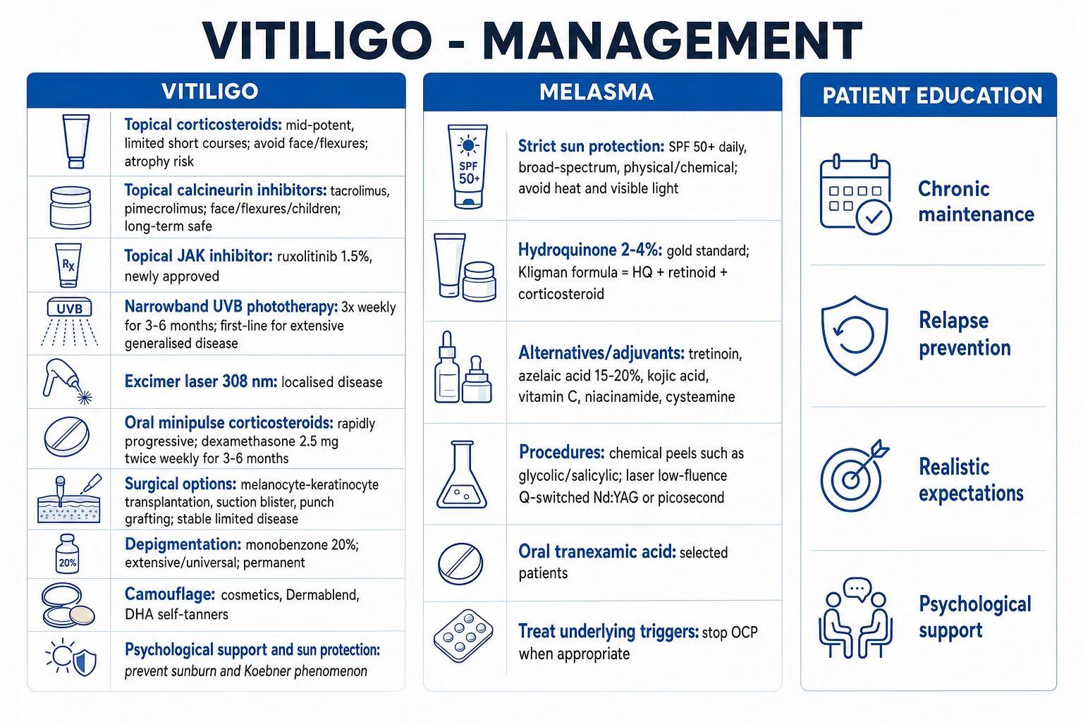
Topical corticosteroid (e.g. mometasone 0.1% OD, mid-potent) for limited body areas in short courses; or topical calcineurin inhibitor (tacrolimus 0.1% or pimecrolimus 1%) BD for face, flexures, genitals and children. Topical ruxolitinib 1.5% cream BD for non-segmental vitiligo.
4. Phototherapy (extensive)
Narrowband UVB 311 nm, 2 to 3 times weekly for 3 to 6 months — first-line for widespread disease. Excimer laser 308 nm for localised patches. Combine with topical TCI.
5. Systemic for rapidly progressive
Oral minipulse corticosteroid (dexamethasone 2.5 mg twice weekly or prednisolone low-dose on 2 consecutive days weekly) for 3 to 6 months to halt spread — NOT long-term.
6. Surgery (stable disease)
Only for patches stable for 6 to 12 months, Koebner-negative, segmental or refractory: melanocyte-keratinocyte transplantation, suction-blister or punch grafting, split-thickness graft.
7. Camouflage and depigmentation
Cosmetic camouflage (Dermablend), self-tanner (dihydroxyacetone). If over 50% BSA and patient accepts irreversible outcome: depigmentation with monobenzone 20% cream or Q-switched laser.
Topical corticosteroids are first-line for limited localised patches on the body. A mid-potent steroid (mometasone 0.1% or fluticasone) applied once daily for a course of several weeks to months is typical. They must never be used continuously on the face, eyelids, flexures, or genitals because of atrophy, telangiectasia, and striae — for these sites, the calcineurin inhibitors are used instead. [1]
Topical calcineurin inhibitors — tacrolimus 0.1% ointment and pimecrolimus 1% cream — are the preferred agents for the face, eyelids, lips, neck, flexures, and genitals, and are safe for long-term use and in children because, unlike corticosteroids, they do not cause atrophy. They are often combined with phototherapy. [1]
Topical JAK inhibitors are the major advance of the last decade. Ruxolitinib 1.5% cream applied twice daily is FDA-approved for non-segmental vitiligo in patients aged 12 years and older, based on the TRuE-V1 and TRuE-V2 phase 3 trials. In those trials, a 75 percent or greater improvement in the facial Vitiligo Area Scoring Index (F-VASI75) at 24 weeks was achieved by 29.8 percent and 30.9 percent of ruxolitinib-treated patients versus only 7.4 percent and 11.4 percent of vehicle-treated patients. The most common adverse events are application-site acne, nasopharyngitis, and application-site pruritus. [4]
Narrowband UVB phototherapy (311 nm) is the first-line treatment for extensive, generalised, or rapidly progressive non-segmental vitiligo. The regimen is two to three treatments per week for three to six months, with dose titration by skin type. A systematic review and meta-analysis confirmed that narrowband UVB achieves better repigmentation than psoralen plus UVA (PUVA) and is safer in children and pregnant women. Repigmentation comes from the hair-follicle melanocyte reservoir, appearing first as perifollicular macules — which is why hairy areas (face, neck, trunk) repigment well but the smooth, hairless skin of the hands, feet, lips, and genitals responds poorly (the "acral" sites lack follicles). [8]
The 308-nm excimer laser delivers targeted narrowband UVB to individual patches (useful for localised disease, especially the face and neck) two to three times weekly. PUVA (oral psoralen plus UVA) is now largely reserved for refractory cases because of its higher long-term skin-cancer risk and side-effect profile. [1]
Oral minipulse corticosteroids — dexamethasone 2.5 mg twice weekly (or low-dose prednisolone on two consecutive days weekly) for three to six months — are used to arrest rapidly progressive disease; they are not for long-term maintenance. The goal is to stop the spread, after which topical therapy and phototherapy induce repigmentation. [1]
Surgical treatments are reserved for stable disease (no new lesions and no Koebner for at least 6 to 12 months), and work especially well for segmental vitiligo. Options include autologous melanocyte-keratinocyte transplantation (a cell suspension applied to dermabraded recipient skin — the current gold standard for large areas), suction-blister epidermal grafting, punch (mini-) grafting, and split-thickness skin grafting. Depigmentation therapy with monobenzone ethyl ester 20% cream (or a Q-switched laser) is offered to patients with vitiligo universalis or over 50 percent body-surface involvement who accept that the result is permanent and irreversible — it removes the remaining normal pigment so the patient has a uniform, if depigmented, appearance. [1]
Camouflage and psychological support are not optional adjuncts but core therapy. Cosmetic camouflage (Dermablend, tinted foundations matched to skin tone), self-tanning agents containing dihydroxyacetone (which stain the depigmented skin without needing melanin), and psychological support or cognitive behavioural therapy address the visible stigma. Patient support organisations (the Vitiligo Society) are valuable. [1]
Melasma — the management ladder
Strict sun protection is foundational and non-negotiable. A broad-spectrum SPF 50+ sunscreen is applied every morning and reapplied — and because visible light (not just UV) also drives melasma, tinted sunscreens containing iron oxide are superior to non-tinted for facial melasma. Hats, protective clothing, and shade matter. Triggering factors (OCP, hormone replacement) are reviewed and changed where possible. [3]
Topical depigmenting agents form the pharmacological core: [1]
- Gold standard skin-lightener
- Inhibits tyrosinase
- Apply once nightly to pigmented areas only
- Use for 3 to 6 months then taper
- Risk of irritation and, rarely, ochronosis with long-term high-strength use in dark skin
- Retinoid; increases keratinocyte turnover
- Slows over months
- Additive with hydroquinone
- Teratogenic; avoid in pregnancy
- Photosensitising; use at night with sunscreen by day
- Inhibits tyrosinase; anti-proliferative on hyperactive melanocytes
- Pregnancy-safe (category B) — first-line in pregnancy
- Useful for acne-related PIH
- Mild irritation
- Adjuncts and alternatives
- Variable evidence
- Cysteamine and tranexamic acid gaining evidence
- Hydroquinone 4% + tretinoin 0.025% + hydrocortisone 1%
- Most effective single preparation (McKesey 2020)
- Use for up to 8 weeks then taper to avoid steroid effects and ochronosis
- Tretinoin prevents HQ oxidation; steroid reduces HQ irritation
Chemical peels (glycolic acid 20 to 70 percent, salicylic acid, mandelic acid, Jessner solution) and laser and light devices (low-fluence Q-switched Nd:YAG "laser toning", intense pulsed light, picosecond laser) are second-line for refractory melasma but carry a higher risk of post-inflammatory hyperpigmentation in darker skin, where they must be used cautiously at lower energies. Oral tranexamic acid 250 to 500 mg twice daily for several months is a promising systemic adjunct for moderate-to-severe recurrent melasma, thought to act by reducing UV-induced plasmin and prostaglandin-driven melanocyte stimulation; it is contraindicated in thromboembolic disease and pregnancy. [3]
Post-inflammatory hyperpigmentation and hypopigmentation
The first principle is to treat the underlying inflammatory condition aggressively — clear the acne, eczema, or lichen planus so that new pigment is not laid down. Add strict sun protection and, once the inflammation has settled, a topical lightening agent (hydroquinone 2 to 4 percent, azelaic acid 15 to 20 percent, or a retinoid) for several months. A systematic review of topical treatments for PIH found that hydroquinone, azelaic acid, retinoids, kojic acid, and ascorbic acid are all supported by varying levels of evidence; patience is essential, as epidermal PIH fades over months and dermal PIH over years. Post-inflammatory hypopigmentation usually recovers spontaneously as melanocytes recover; topical corticosteroid or calcineurin inhibitors are avoided (no active inflammation to suppress). [7]
Oculocutaneous albinism
There is no cure for albinism — the tyrosinase defect cannot be reversed. Management is entirely preventive and supportive: [1]
- Strict lifelong sun protection — broad-spectrum SPF 50+ sunscreen applied daily, opaque clothing, wide-brimmed hats, sunglasses, and shade. This is the single most important intervention because albinism carries a very high risk of actinic damage, squamous cell carcinoma, basal cell carcinoma, and melanoma, especially in sun-intense regions where it is a leading cause of skin-cancer death.
- Ophthalmological care — tinted lenses for photophobia, low-vision aids, refractive correction, management of strabismus and nystagmus, and regular review.
- Regular skin surveillance — full skin examination every 6 to 12 months; biopsy any changing or suspicious lesion early.
- Genetic counselling — for the family, with carrier testing and, where appropriate, prenatal diagnosis.
- Social and educational support — children may need educational accommodations for reduced vision; addressing stigma, especially in communities with superstition around albinism, is essential. [6]
Drug-induced pigmentation
Stop the offending drug wherever possible. Many drug-induced pigmentations fade slowly after cessation (amiodarone, antimalarials), but some are permanent (argyria, chrysiasis, minocycline dermal deposits). Sun protection helps limit phototoxic darkening. Q-switched laser therapy (ruby, alexandrite, or Nd:YAG) can remove dermal drug-melanin complexes or metal deposits in selected cases. [5]
Specific Subtypes & Scenarios
Segmental versus non-segmental vitiligo — why it matters
The segmental/non-segmental split is not academic. Segmental vitiligo onsets earlier, stabilises within 1 to 2 years, has a weaker autoimmune association, carries leucotrichia more often, and responds poorly to phototherapy — but is the form most amenable to surgical grafting once stable, because it does not Koebner and does not recur. Non-segmental vitiligo progresses, associates with autoimmune disease, responds to phototherapy and JAK inhibition, and is prone to relapse. Surgery is reserved for the stable disease in both forms. [1]
Melasma in pregnancy (chloasma gravidarum)
Melasma in pregnancy is driven by oestrogen, progesterone, and melanocyte-stimulating hormone. It usually fades post-partum but may persist, especially with sun exposure or subsequent pregnancy. Management in pregnancy is conservative: strict sun protection and azelaic acid 15 to 20 percent (pregnancy category B and the agent of choice in pregnancy). Hydroquinone, tretinoin, chemical peels, and lasers are avoided in pregnancy and resumed post-partum if needed. [3]
Extensive vitiligo and depigmentation
When vitiligo involves over 50 percent of the body surface and topical and phototherapeutic repigmentation has failed or is impractical, depigmentation therapy is offered. Monobenzone ethyl ester 20% cream is applied to the remaining normally pigmented skin over months to achieve a uniform depigmented appearance. This is irreversible, requires lifelong strict sun protection, and demands thorough counselling and patient consent. [1]
Complications & Pitfalls
The complications of pigmentary disease are largely psychological and social: depression, anxiety, social withdrawal, sexual dysfunction, and reduced quality of life with DLQI scores comparable to psoriasis. Vitiligo carries additional sunburn risk in depigmented skin and, paradoxically, can coexist with melanoma (immune-mediated melanocyte destruction may occasionally reflect an antitumour response). Albinism carries the gravest physical risk — skin cancer (SCC, BCC, melanoma) is a leading cause of death in untreated patients in sun-exposed populations. [1][6]
Treatment-related complications include topical steroid atrophy, telangiectasia, and striae on the face and flexures (avoid by using calcineurin inhibitors instead); phototherapy-related photodamage and skin-cancer risk with cumulative doses; application-site acne and pruritus with ruxolitinib; chronic Q-switched laser erythema; and, in dark skin treated for long periods with high-strength hydroquinone, exogenous ochronosis (paradoxical blue-black hyperpigmentation). [4]
Rapid self-test — a 45-year-old woman develops extensive vitiligo over six months. What is the single most important screening test, and why?
Measure TSH (with anti-TPO antibodies). Autoimmune thyroid disease is the commonest autoimmune association of vitiligo (occurring in 15 to 30 percent of patients), is frequently subclinical, and is treatable. Screen every vitiligo patient at diagnosis and periodically thereafter. [1]
The classic diagnostic pitfall is mislabelling a hypoesthetic, anaesthetic patch as vitiligo in an endemic region — it is leprosy until sensation is tested. Other pitfalls: confusing pityriasis versicolor (scaly, KOH positive) with vitiligo; mistaking pityriasis alba (atopic, scaly, ill-defined, paediatric cheek) for vitiligo; attributing drug-induced slate-grey pigmentation to Addison disease without a drug history; and missing Addison disease in a vitiligo patient with weight loss and fatigue. Always examine the palmar creases, oral mucosa, and recent scars for the hyperpigmentation of Addison disease. [1]
Prognosis & Disposition
Vitiligo runs a chronic, unpredictable course. Segmental vitiligo stabilises after 1 to 2 years and does not usually recur. Non-segmental vitiligo is progressive and relapsing, with periods of stability punctuated by flares often triggered by stress, illness, sunburn, or pregnancy. Repigmentation is partial and site-dependent: the face and neck respond best, the trunk moderately, and the hands, feet, lips, and genitals (acral, hairless sites) respond poorly because there is no follicular melanocyte reservoir. [1]
Melasma is chronic and relapsing, often lifelong, with each pregnancy, sun exposure, and OCP use darkening it; epidermal melasma responds to topical therapy while dermal melasma is refractory. Post-inflammatory pigmentation resolves over months (epidermal) to years (dermal). Albinism is a lifelong condition with normal life expectancy if skin protection is meticulous; without it, skin cancer shortens life dramatically. [1]
Most patients are managed in the outpatient dermatology setting. Indications for urgent referral include suspected Addison disease, atypical depigmentation requiring biopsy to exclude melanoma, and severe psychological distress requiring psychological or psychiatric input. [1]
Special Populations
Children with vitiligo — segmental vitiligo is commoner in children and is the form most likely to warrant surgery later. Topical calcineurin inhibitors are preferred on the face and flexures (no atrophy, safe long-term), and narrowband UVB is used from around age six for extensive disease. Psychological support is crucial because of bullying and stigma; school liaison may be needed. Ruxolitinib cream is approved from age 12. [1][4]
Pregnancy and melasma — sun protection and azelaic acid (pregnancy-safe); avoid hydroquinone, retinoids, and peels until post-partum (see above). [1]
Darker skin types (Fitzpatrick IV to VI) — vitiligo is more visible and stigmatising; melasma and post-inflammatory pigmentation are more severe and more resistant; phototherapy, chemical peels, and lasers must use lower energies to avoid post-inflammatory hyperpigmentation; and long-term high-strength hydroquinone risks ochronosis. Pigment changes (both loss and excess) are more striking and the psychological impact greater. [1][7]
The elderly — late-onset vitiligo warrants a careful autoimmune screen and a search for occult Addison disease or other autoimmune endocrinopathy; a solitary new depigmented patch must raise the question of amelanotic melanoma (biopsy if atypical, non-periorificial, or rapidly changing). Drug-induced pigmentation from polypharmacy (amiodarone, minocycline) is more common. [1]
Immunocompromised patients — vitiligo can coexist with autoimmune disease and HIV; pigmentation changes from drugs (antiretrovirals, ketoconazole) and from opportunistic infections must be distinguished. [1]
Evidence, Guidelines & Regional Differences
The understanding of vitiligo as an autoimmune disease driven by the IFN-gamma / CXCL10 chemokine axis (Frisoli et al. 2020) directly led to the development of JAK inhibitors: the TRuE-V1 and TRuE-V2 phase 3 trials (Rosmarin et al. 2022, NEJM) established ruxolitinib 1.5% cream as the first FDA-approved topical repigmenting therapy for non-segmental vitiligo in patients aged 12 and older. A systematic review and meta-analysis of phototherapy (Bae et al. 2017, JAMA Dermatology) confirmed narrowband UVB as superior to PUVA for repigmentation and safe in children and pregnancy. The Bergqvist and Ezzedine (2020) review in Dermatology remains the authoritative overview. [1][2][4][8]
For melasma, the McKesey et al. (2020) evidence-based review (American Journal of Clinical Dermatology, 113 randomised trials) established that triple-combination cream (hydroquinone, tretinoin, corticosteroid) and hydroquinone monotherapy are the most effective treatments, that peels and lasers are equal or inferior with more adverse effects, and that oral tranexamic acid is a promising systemic adjunct. For post-inflammatory hyperpigmentation, the Tan et al. (2022) systematic review supports hydroquinone, azelaic acid, retinoids, kojic acid, and ascorbic acid. For albinism, the GeneReviews overview (Thomas et al. 2023) is the definitive reference. [3][6][7]
TRuE-V1 and TRuE-V2 — Ruxolitinib cream in vitiligo (NEJM 2022)
Population: 674 patients aged 12+ with non-segmental vitiligo affecting 10% or less of body surface area
Key finding
F-VASI75 (75%+ improvement in facial Vitiligo Area Scoring Index) at 24 weeks: 29.8% (TRuE-V1) and 30.9% (TRuE-V2) with ruxolitinib vs 7.4% and 11.4% with vehicle (relative risk 4.0 and 2.7; P less than 0.001). Effect improved to 52 weeks. Most common adverse events: application-site acne, nasopharyngitis, application-site pruritus.
Practice change
Ruxolitinib 1.5% cream is the first FDA-approved topical JAK inhibitor for non-segmental vitiligo; it produces meaningful facial repigmentation beyond vehicle, with application-site acne and pruritus the main adverse events.
- United States (AAD) — ruxolitinib 1.5% cream is incorporated into AAD vitiligo guidance for non-segmental disease from age 12; narrowband UVB remains first-line for extensive disease; biologics access shapes melasma and PIH adjunct use.
- Europe (EADV, European Dermatology Forum) — JAK inhibitors increasingly adopted; broad agreement with AAD on phototherapy, topical corticosteroids, and calcineurin inhibitors; triple-combination cream widely used for melasma.
- United Kingdom (BAD, NICE) — cost-aware stepwise therapy; narrowband UVB the mainstay for extensive vitiligo; ruxolitinib via specialist commissioning; camouflage cosmetics available on the NHS.
- South Asia (IADVL / India) — vitiligo carries particular stigma; topical corticosteroids, calcineurin inhibitors, and narrowband UVB are the pragmatic mainstays; ruxolitinib access is limited by cost; leprosy must always be excluded before diagnosing vitiligo (test sensation in every hypopigmented patch); melasma is extremely common and triple-combination creams, kojic acid, and oral tranexamic acid are widely used; exogenous ochronosis from unregulated skin-lightening creams is an important regional complication.
- Sub-Saharan Africa — albinism is prevalent and associated with high skin-cancer mortality; sun-protection programmes and skin-cancer surveillance are public-health priorities; superstitious beliefs about albinism require community education.
Exam Pearls
VITILIGO — the high-yield exam one-liners
MELASMA — the high-yield exam one-liners
ALBINISM and DRUG-PIGMENT one-liners
Exam application bank (NEET-PG / INICET)
One-line answer
Vitiligo is an acquired autoimmune destruction of epidermal melanocytes producing well-demarcated, chalk-white (depigmented) macules and patches on the periorificial face, hands, extensor surfaces, genitalia and body folds; classified as segmental (unilateral, dermatomal, early onset, stabilises within 1 to 2 years) or non-segmental (bilateral, symmetrical, progressive). It is associated with autoimmune thyroid disease, type 1 diabetes, pernicious anaemia, Addison disease and alopecia areata, and carries a major psychological burden, especially in darker skin. Melasma (chloasma) is acquired symmetrical facial hyperpigmentation (forehead, malar cheeks, upper lip) triggered by sun, pregnancy, oral contraceptives and genetics. Post-inflammatory pigment change, oculocutaneous albinism, and drug-induced pigmentation (minocycline, amiodarone, chloroquine) complete the pigmentary differential.
Worked stems (answer without another resource)
Stem 1 — Classic presentation. Map symptoms to mechanism; name the first investigation and first treatment step with dose/route if drug therapy is standard. [1]
Stem 2 — Unstable / complicated. List red flags that force immediate resuscitation, theatre, ICU, antidote, or reperfusion — and what you do in the first 15 minutes. [1]
Stem 3 — Atypical group. Elderly, pregnancy, child, or immunocompromised: how presentation and thresholds change. [1]
Stem 4 — Differential trap. Name the three closest mimics and one discriminator for each. [1]
Stem 5 — Disposition. Who goes home with safety-netting, who is admitted, who needs HDU/ICU/theatre, and what follow-up is mandatory. [1]
Rapid viva checklist
- Definition + classification
- Pathophysiology chain
- Bedside signs / criteria
- Score with exact components (if any)
- Emergency bundle
- Definitive therapy with doses
- Complications of disease and of treatment
- Special populations
- Guideline/trial name if classic
- Three exam traps
Coverage self-check
If you cannot answer any stem above from this page alone, re-read the matching section — the page is intended to be self-sufficient for final-prof and NEET-PG/INICET questions on Vitiligo & Pigmentation Disorders.
[1]References
- [1]Bergqvist C, Ezzedine K. Vitiligo: A Review Dermatology, 2020.PMID 32155629
- [2]Frisoli ML, Essien K, Harris JE. Vitiligo: Mechanisms of Pathogenesis and Treatment Annu Rev Immunol, 2020.PMID 32017656
- [3]McKesey J, Tovar-Garza A, Pandya AG. Melasma Treatment: An Evidence-Based Review Am J Clin Dermatol, 2020.PMID 31802394
- [4]Rosmarin D, Passeron T, Pandya AG, Grimes P, Harris JE, Desai SR, Lebwohl M, Ruer-Mulard M, Seneschal J, Wolkerstorfer A, Kornacki D, Sun K, Butler K, Ezzedine K, TRuE-V Study Group. Two Phase 3, Randomized, Controlled Trials of Ruxolitinib Cream for Vitiligo N Engl J Med, 2022.PMID 36260792
- [5]Gimenez Garcia RM, Carrasco Molina S. Drug-Induced Hyperpigmentation: Review and Case Series J Am Board Fam Med, 2019.PMID 31300585
- [6]Ma EZ, Zhou AE, Hoegler KM, Khachemoune A. Oculocutaneous albinism: epidemiology, genetics, skin manifestation, and psychosocial issues Arch Dermatol Res, 2023.PMID 35217926
- [7]Tan MG, Kim WB, Jo CE, Nabieva K, Kirshen C, Ortiz AE. Topical treatment for postinflammatory hyperpigmentation: a systematic review J Dermatolog Treat, 2022.PMID 34525885
- [8]Bae JM, Jung HM, Hong BY, Lee JH, Choi WJ, Lee JH, Kim GM. Phototherapy for Vitiligo: A Systematic Review and Meta-analysis JAMA Dermatol, 2017.PMID 28355423