Dermatology · Medicine
Vulval dermatoses
Also known as Vulvar dermatoses · Vulval skin disease · Vulvar lichen sclerosus · Erosive vulval lichen planus · Vulvodynia / vestibulodynia · Kraurosis vulvae (historical)
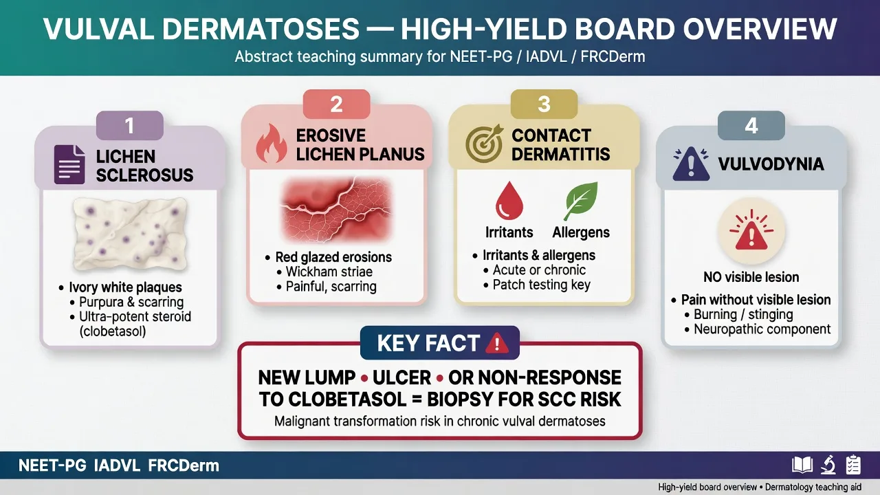
Special-site overview of vulval dermatoses for multi-board exams: pattern recognition (white plaque disease vs red erosive disease vs normal-looking painful vulva), lichen sclerosus as the high-stakes chronic inflammatory dermatosis with 2–5% SCC risk and clobetasol 0.05% ointment as gold-standard therapy, erosive lichen planus, infectious and contact mimics, differentiated VIN, and vulvodynia/provoked vestibulodynia as a pain syndrome without primary skin disease. Covers structured examination, biopsy thresholds, stepwise management, paediatric pitfalls, and regional guideline deltas (ACOG, EuroGuiderm/BAD, ISSVD).
On this page & tools
Your progress
Saved locally on this device.
Exam tags
Red flags
Overview & Definition
The vulva is a special site: thin epithelium, moisture, friction, microbiome, hormonal dependence, and sexual/urinary function mean the same disease (lichen planus, psoriasis, candida) looks different from extragenital skin and is often mislabelled as “chronic thrush.” ACOG frames vulvar skin disorders as a common reason for specialist referral and emphasises accurate diagnosis before long-term empirical therapy.[1]
This leaf is the clinic-level overview spanning inflammatory dermatoses (especially lichen sclerosus and erosive lichen planus), common infectious and contact mimics, premalignant/malignant disease, and vulvodynia. Deep monographs on isolated LS and LP live on their dedicated topics; here the examiner wants the integrated approach. [1]
Classification
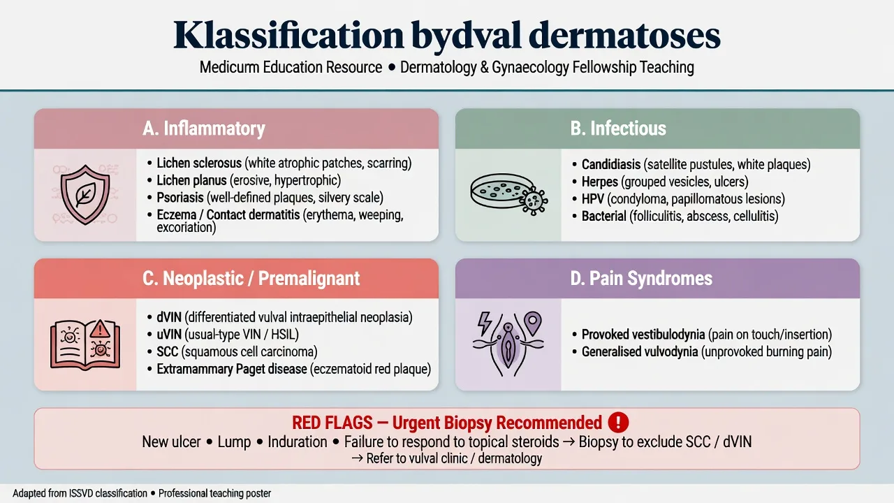
Inflammatory
Infectious
Neoplastic / premalignant
Pain syndromes
Hormone-related
Epidemiology & Risk Factors
Lichen sclerosus is uncommon but under-diagnosed; autoimmune comorbidity (thyroid disease, vitiligo, alopecia areata, pernicious anaemia) is enriched.[2][4] Erosive vulval LP peaks in middle-aged women and frequently coexists with oral or oesophageal disease.[8][9] Vulvodynia affects a substantial minority of women at some point; delayed diagnosis is the rule.[10] Risk amplifiers for inflammatory and infectious flares include wet wipes, fragranced products, tight synthetic underwear, uncontrolled diabetes, antibiotics, and oestrogen deficiency.[1]
Pathophysiology
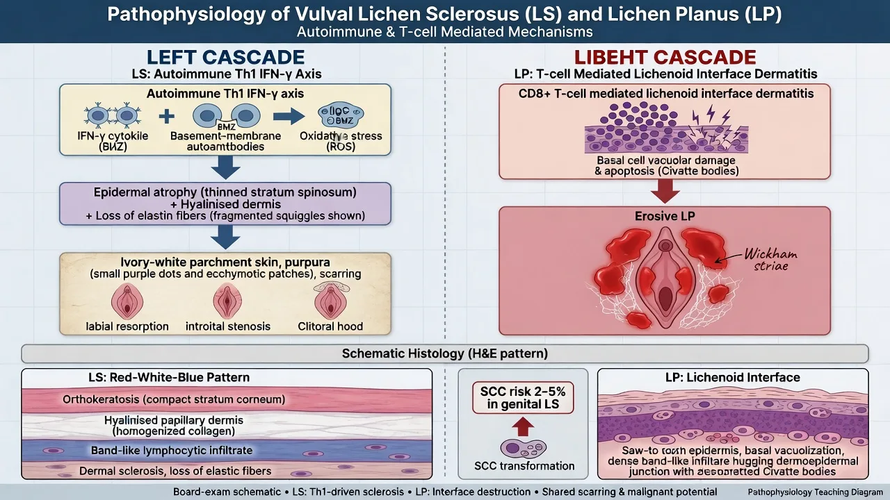
Lichen sclerosus is a chronic T-cell–mediated disease with a Th1/IFN-γ cytokine signature, oxidative stress, fibroblast dysfunction, and microvascular injury. Circulating antibodies to BP180/BP230 are reported in a subset and link LS to the basement-membrane antigen family. The end tissue result is epidermal atrophy, a hyalinised papillary dermis, and secondary scarring of vulvar architecture.[2][4]
Erosive lichen planus is a lichenoid interface dermatitis: cytotoxic T cells damage basal keratinocytes; on mucosa this produces painful erosions rather than classic polygonal papules.[6][7]
Provoked vestibulodynia is not a primary dermatosis. Peripheral sensitisation of vestibular nociceptors, pelvic floor muscle hypertonicity, and central pain amplification interact; psychosocial comorbidity is common and bidirectional.[10][11]
Clinical Presentation
Lichen sclerosus (highest stakes)
Ivory-white atrophic plaques with cigarette-paper wrinkling, intra-plaque purpura/ecchymoses (highly characteristic), fissures, and progressive architectural change (labial resorption, clitoral burying, introital narrowing). Distribution is often figure-of-eight (vulva + perianal). Leading symptoms: intractable itch, dyspareunia, dysuria, constipation in children.[2][4][12]
Erosive lichen planus
Glazed bright-red erosions of the vestibule and labia minora, lacy white (Wickham) network at margins, vaginal synechiae, and often concurrent oral/gingival disease. Pain and post-coital bleeding dominate over pure itch.[8][9]
Contact dermatitis and infection
Ill-defined erythema and burning after new products; candidiasis produces itch, curd-like discharge, satellite pustules, and thrives after antibiotics or hyperglycaemia.[1]
Vulvodynia / provoked vestibulodynia
Pain (burning, raw, knife-like) without primary skin disease. Cotton-swab mapping of the vestibule reproduces focal pain; the rest of the exam may be normal.[10]
Differential Diagnosis
| Pattern | Prefer | Against |
|---|---|---|
| White macule, normal texture | Vitiligo | No atrophy, no purpura, no scarring |
| White + atrophy + purpura + scarring | LS | — |
| Dry thin mucosa post-menopause | GSM | No true sclerosis/purpura |
| Glazed red erosions + oral LP | Erosive LP | — |
| Blisters, milia, positive DIF | Mucous membrane pemphigoid | DIF needed |
| Satellite pustules, diabetes | Candida | KOH/swab |
| Chronic single ulcer/lump | dVIN / SCC / Paget | Biopsy mandatory |
Paediatric LS must be distinguished from — and can coexist with — safeguarding concerns; do not force a false dichotomy.[12]
Clinical & Bedside Assessment
- History — itch vs pain, contactants, sexual function, urinary stream, bowel symptoms in children, autoimmune history, medications (fixed drug), diabetes.[1]
- Inspection — colour, texture, purpura, erosions, architecture, perianal extension.
- Extragenital survey — mouth, scalp, nails, flexural skin for LP/psoriasis.
- Q-tip test — map vestibular tenderness when exam otherwise normal.[10]
- Photography (with consent) for surveillance of LS fields.
Investigations
- Clinical diagnosis is often sufficient for classic LS when morphology is pathognomonic; biopsy if atypical, non-responsive, or to exclude neoplasia.[3][5]
- Biopsy technique — sample the active edge of white or erosive disease; avoid burnt-out scar alone. Histology of LS classically shows the layered atrophy/hyalinisation/lymphocytic band pattern (taught as red–white–blue).[2]
- Swabs — candida, bacterial, HSV PCR when vesicles/ulcers.
- STI screen when indicated by history.
- Patch testing for chronic contact dermatitis.
- DIF if bullous disease or pemphigoid spectrum suspected.
- Urgent biopsy for any suspicious focus in an LS field (dVIN pathway).[4]
Management — Resuscitation
Time-critical scenarios are uncommon but examinable: urinary retention from stenosis, rapidly progressive mucocutaneous erosions (consider SJS/TEN or severe erosive disease), and systemically unwell infection. Secure analgesia, exclude HSV/bacterial sepsis when appropriate, and obtain same-day specialist review.[1]
Management — Definitive & Stepwise
Shared foundations (all patients)
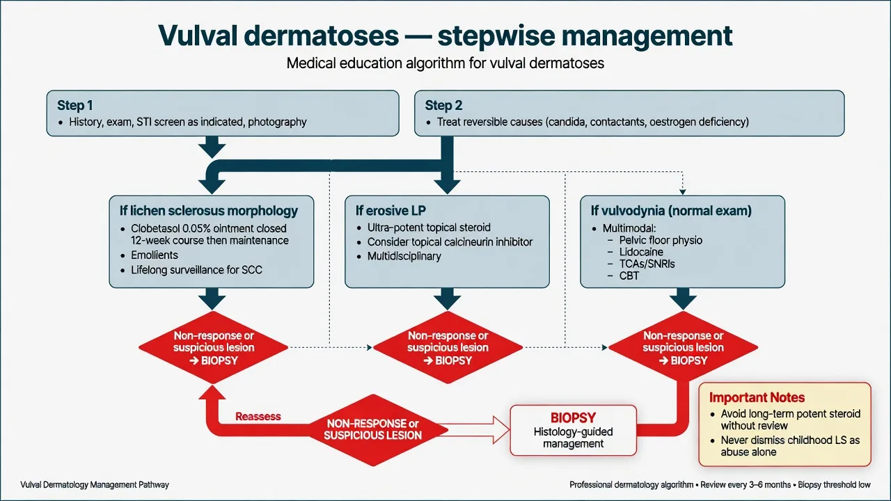
Soap substitutes, bland emollients, avoid fragranced products/wet wipes, loose cotton underwear, treat confirmed infection, optimise diabetes and oestrogen deficiency when present.[1]
Lichen sclerosus — gold standard
Clobetasol propionate 0.05% ointment (ultra-potent), typically a closed 12-week induction (often conceptualised as daily for 4 weeks → alternate nights 4 weeks → twice weekly 4 weeks, then twice-weekly lifelong maintenance for genital disease), plus emollients. Vehicle matters: ointment preferred on vulval skin. This is the evidence core of EuroGuiderm/BAD-aligned guidance.[3][5]
Second-line: topical calcineurin inhibitors (e.g. tacrolimus 0.1%) as steroid-sparing in selected patients under specialist care.[3] Surgery is for structural complications (stenosis), not routine excision of LS. Lifelong clinical surveillance for SCC is mandatory.[2][4]
Erosive lichen planus
Ultra-potent topical corticosteroid ointment is first-line; add topical calcineurin inhibitors; escalate to systemic agents (e.g. oral corticosteroids for flares, steroid-sparing immunosuppressants in refractory disease) in specialist clinics; multidisciplinary care for vaginal synechiae.[6][8][9]
Vulvodynia / provoked vestibulodynia
Stop unhelpful empirical antifungals. Multimodal care: pelvic floor physiotherapy, topical lidocaine before intercourse if provoked, oral neuromodulators (e.g. tricyclic antidepressants or SNRIs in selected adults), CBT/psychosexual therapy, and avoidance of irritants. Evidence supports multimodal packages more than any single magic drug.[10][11]
Specific Subtypes & Scenarios
Prepubertal LS presents with itch, bleeding, constipation, and white plaques; treat with age-appropriate potent topical steroid under specialist guidance; many improve around puberty but follow-up continues because residual and adult disease occur.[12]
Pregnancy — topical corticosteroids remain the mainstay for active LS; coordinate with obstetrics for delivery planning if stenosis is severe. [1]
LS–LP overlap — mixed morphology is recognised; treat the active inflammatory component and maintain SCC vigilance.[9]
Complications & Pitfalls
- Progressive scarring, sexual dysfunction, and urinary symptoms.
- dVIN → SCC if surveillance fails.[2][4]
- Years of “chronic thrush” without exam.
- Labelling childhood LS as abuse alone, or missing abuse because LS exists.
- Using mild hydrocortisone alone for genital LS (under-treatment).
- Operating on vulvodynia with laser/surgery without pain-clinic pathway.
Prognosis & Disposition
Genital LS is typically chronic and relapsing; maintenance therapy and surveillance continue for life even when asymptomatic.[3] Refer early to dermatology/vulval clinic for diagnostic uncertainty, treatment failure, paediatric disease, or any neoplastic concern. Psychosexual and pain-clinic referral for vulvodynia improves outcomes.[10]
Special Populations
Children need paediatric dermatology/gynaecology pathways and safeguarding literacy.[12] Postmenopausal women often have dual GSM + LS — treat both components. Immunosuppressed patients need lower thresholds for biopsy of non-healing lesions. In resource-limited settings, access to clobetasol ointment and timely biopsy pathways is the practical bottleneck — do not substitute with chronic weak steroid without review.
Evidence, Guidelines & Regional Differences
- ACOG Practice Bulletin 224 — structured diagnosis and management of vulvar skin disorders.[1]
- EuroGuiderm 2024 / earlier S3 LS guidance — ultra-potent topical corticosteroid as first-line treatment of anogenital LS.[3][5]
- European S1 lichen planus guideline — mucosal LP management framework.[6]
- Vulvodynia primers/systematic reviews — multimodal therapy; limited single-agent miracle data.[10][11]
Controversies include energy-based devices for LS (insufficient to replace steroids as first-line) and long-term calcineurin inhibitor safety signals — use specialist protocols. [1]
Exam Pearls
VULVA board map
- Purpura inside a white plaque is a gift for LS.[2]
- Erosive LP loves the mouth + vulva combo.[8]
- Potent steroid treats the atrophy of LS (paradox exam favourite).[3]
- Antibiotics/antifungals do not fix mechanical or autoimmune disease.
Exam application bank (NEET-PG / INICET)
One-line answer
Special-site overview of vulval dermatoses for multi-board exams: pattern recognition (white plaque disease vs red erosive disease vs normal-looking painful vulva), lichen sclerosus as the high-stakes chronic inflammatory dermatosis with 2–5% SCC risk and clobetasol 0.05% ointment as gold-standard therapy, erosive lichen planus, infectious and contact mimics, differentiated VIN, and vulvodynia/provoked vestibulodynia as a pain syndrome without primary skin disease. Covers structured examination, biopsy thresholds, stepwise management, paediatric pitfalls, and regional guideline deltas (ACOG, EuroGuiderm/BAD, ISSVD).
Worked stems (answer without another resource)
Stem 1 — Classic presentation. Map symptoms to mechanism; name the first investigation and first treatment step with dose/route if drug therapy is standard. [1]
Stem 2 — Unstable / complicated. List red flags that force immediate resuscitation, theatre, ICU, antidote, or reperfusion — and what you do in the first 15 minutes. [1]
Stem 3 — Atypical group. Elderly, pregnancy, child, or immunocompromised: how presentation and thresholds change. [1]
Stem 4 — Differential trap. Name the three closest mimics and one discriminator for each. [1]
Stem 5 — Disposition. Who goes home with safety-netting, who is admitted, who needs HDU/ICU/theatre, and what follow-up is mandatory. [1]
Rapid viva checklist
- Definition + classification
- Pathophysiology chain
- Bedside signs / criteria
- Score with exact components (if any)
- Emergency bundle
- Definitive therapy with doses
- Complications of disease and of treatment
- Special populations
- Guideline/trial name if classic
- Three exam traps
Coverage self-check
If you cannot answer any stem above from this page alone, re-read the matching section — the page is intended to be self-sufficient for final-prof and NEET-PG/INICET questions on Vulval dermatoses.
Expanded exam teaching (depth pass)
Clinical reasoning
For Vulval dermatoses, examiners test whether you can prioritise life threats, choose the right first test, and give specific therapy (agent, dose, route, timing). Generic phrases without numbers score poorly.
Mechanism → feature map
Build a short chain: cause → pathophysiologic intermediate → clinical feature → complication. Every major symptom in the classic vignette should sit on that chain.
Investigation strategy
- Bedside/first-line tests that change immediate management
- Confirmatory or staging tests
- What a normal result does not exclude
- When not to delay treatment for imaging (unstable patient)
Management ladder
- Resuscitation / ABC / sepsis or haemorrhage bundle as relevant
- Specific antidote / procedure / antimicrobial / reperfusion / surgery
- Supportive care and monitoring targets
- Definitive long-term therapy and secondary prevention
- Disposition and safety-net advice
Special populations
Always prepare one line each for children, pregnancy, elderly, renal/hepatic impairment, and immunocompromised patients when the topic allows.
Pitfalls that fail candidates
- Treating the number not the patient
- Missing pregnancy status when relevant
- Imaging before stabilisation
- Wrong empiric cover or wrong antidote timing
- Incomplete counselling on recurrence, adherence, or red-flag return
Special-site overview of vulval dermatoses for multi-board exams: pattern recognition (white plaque disease vs red erosive disease vs normal-looking painful vulva), lichen sclerosus as the high-stakes chronic inflammatory dermatosis with 2–5% SCC risk and clobetasol 0.05% ointment as gold-standard therapy, erosive lichen planus, infectious and contact mimics, differentiated VIN, and vulvodynia/provoked vestibulodynia as a pain syndrome without primary skin disease. Covers structured examination, [1]
Structured revision sheet
Must-know numbers and names
List every score, size threshold, dose, and time window from this topic on a blank page from memory, then check against the sections above.
Three classic MCQ angles
- Most likely diagnosis given a vignette
- Next best step in management
- Most appropriate investigation
Three classic SAQ angles
- Pathophysiology in five steps
- Management algorithm with doses
- Complications and prevention
Clinical station flow
Greet → focused history → targeted exam → investigations → explain diagnosis → emergency care → definitive plan → safety-net / follow-up → answer examiner questions on mechanism and pitfalls.
References
- [1]American College of Obstetricians and Gynecologists' Committee on Practice Bulletins—Gynecology. Diagnosis and Management of Vulvar Skin Disorders: ACOG Practice Bulletin, Number 224 Obstet Gynecol, 2020.PMID 32590724
- [2]De Luca DA, Papara C, Vorobyev A, et al. Lichen sclerosus: The 2023 update Front Med (Lausanne), 2023.PMID 36873861
- [3]Kirtschig G, Kinberger M, Kreuter A, et al. EuroGuiderm guideline on lichen sclerosus-Treatment of lichen sclerosus J Eur Acad Dermatol Venereol, 2024.PMID 38822598
- [4]Krapf JM, Mitchell L, Holton MA, et al. Vulvar Lichen Sclerosus: Current Perspectives Int J Womens Health, 2020.PMID 32021489
- [5]Kirtschig G, Becker K, Günthert A, et al. Evidence-based (S3) Guideline on (anogenital) Lichen sclerosus J Eur Acad Dermatol Venereol, 2015.PMID 26202852
- [6]Ioannides D, Vakirlis E, Kemeny L, et al. European S1 guidelines on the management of lichen planus: a cooperation of the European Dermatology Forum with the European Academy of Dermatology and Venereology J Eur Acad Dermatol Venereol, 2020.PMID 32678513
- [7]Le Cleach L, Chosidow O. Clinical practice. Lichen planus N Engl J Med, 2012.PMID 22356325
- [8]Mauskar M. Erosive Lichen Planus Obstet Gynecol Clin North Am, 2017.PMID 28778640
- [9]Lewis FM, Bogliatto F. Erosive vulval lichen planus--a diagnosis not to be missed: a clinical review Eur J Obstet Gynecol Reprod Biol, 2013.PMID 24183096
- [10]Bergeron S, Reed BD, Wesselmann U, Bohm-Starke N. Vulvodynia Nat Rev Dis Primers, 2020.PMID 32355269
- [11]Bohm-Starke N, Ramsay KW, Lytsy P, et al. Treatment of Provoked Vulvodynia: A Systematic Review J Sex Med, 2022.PMID 35331660
- [12]Orszulak D, Dulska A, Niziński K, et al. Pediatric Vulvar Lichen Sclerosus-A Review of the Literature Int J Environ Res Public Health, 2021.PMID 34281089