Dermatology · Medicine
Wood lamp / fluorescence examination
Also known as Wood's lamp · Wood's light · UVA fluorescence examination · Black light dermatology
Board-level module on Wood lamp examination: long-wave UVA (~320–400 nm, peak ~365 nm) bedside fluorescence and pigment contrast for erythrasma (coral-red), pityriasis versicolor (yellow-gold), Microsporum tinea capitis (green hair), vitiligo (bright blue-white), porphyria urine fluorescence, Pseudomonas cues, and selected surgical margin/suture uses. Covers dark-room technique, fluorophore biology, colour→diagnosis map, false positives/negatives, and when KOH, culture, or porphyria labs must confirm the pattern.
On this page & tools
Your progress
Saved locally on this device.
Exam tags
Red flags
Definition and clinical purpose
Wood lamp examination is a bedside optical test, not a laboratory assay. Robert Wood’s glass filter transmits long-wave UVA while blocking most visible light; modern hand-held devices and some UV-adapted dermatoscopes serve the same purpose.[1][2][4] It answers: Does this lesion fluoresce, and what colour? and Where are the true borders of depigmentation?
It is adjunctive. Fluorescence supports diagnoses such as erythrasma or Malassezia-related scale patterns, guides sampling of fluorescent hairs, and maps vitiligo extent, but negative examinations never exclude non-fluorescent pathogens (especially many Trichophyton scalp infections).[6][8]
Optical mechanism
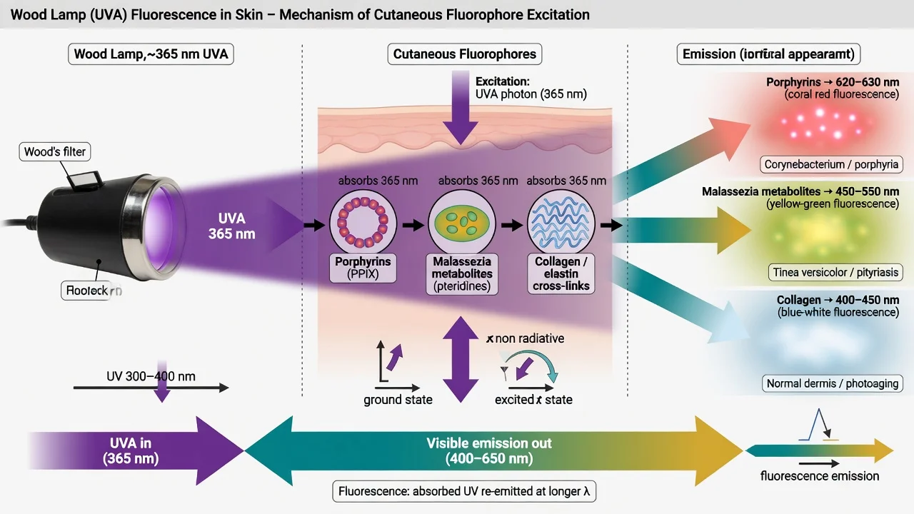
UVA photons excite tissue or microbial fluorophores; longer-wavelength visible light is emitted and seen as colour against a dark field. Important fluorophores include bacterial porphyrins (coral-red in Corynebacterium minutissimum erythrasma and some follicular flora), Malassezia-related tryptophan metabolites (yellow-gold scale fluorescence), fungal metabolites on hair shafts (Microsporum), and endogenous pigments/collagen that create bright contrast in fully depigmented vitiligo.[1][4][7]
Pigment disorders may show contrast without true microbial fluorescence: vitiligo appears strikingly white/blue-white because residual melanin is absent; ash-leaf macules and post-inflammatory hypopigmentation are usually less crisp.[1][3]
Technique (OSCE-ready)
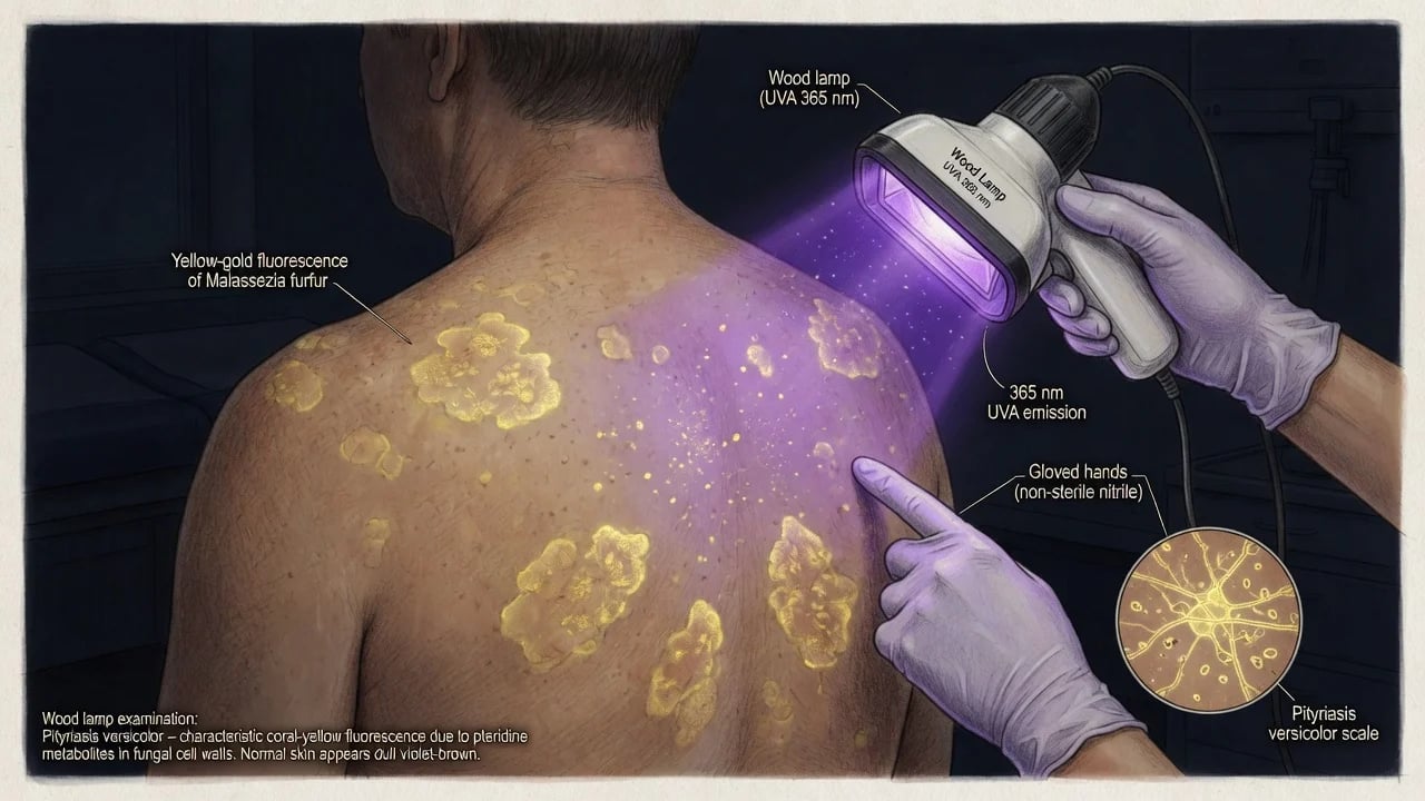
- Warm-up the lamp if required by the device; allow eyes to dark-adapt briefly.
- Dark room — ambient light is the commonest cause of a “negative” exam.[1]
- Distance roughly 10–15 cm; scan systematically (scalp hair shafts, flexures, trunk scale, nails, urine sample if indicated).
- Skin preparation — clean, dry, free of makeup, deodorant, topical medicines, and lint (false fluorescence).[1][2]
- Document colour, site, border sharpness, and whether hair/urine was tested.
- Eye protection and avoid prolonged unnecessary UVA exposure to examiner and patient.
Colour → diagnosis map (memorise)
| Colour / finding | Classic association | Next step |
|---|---|---|
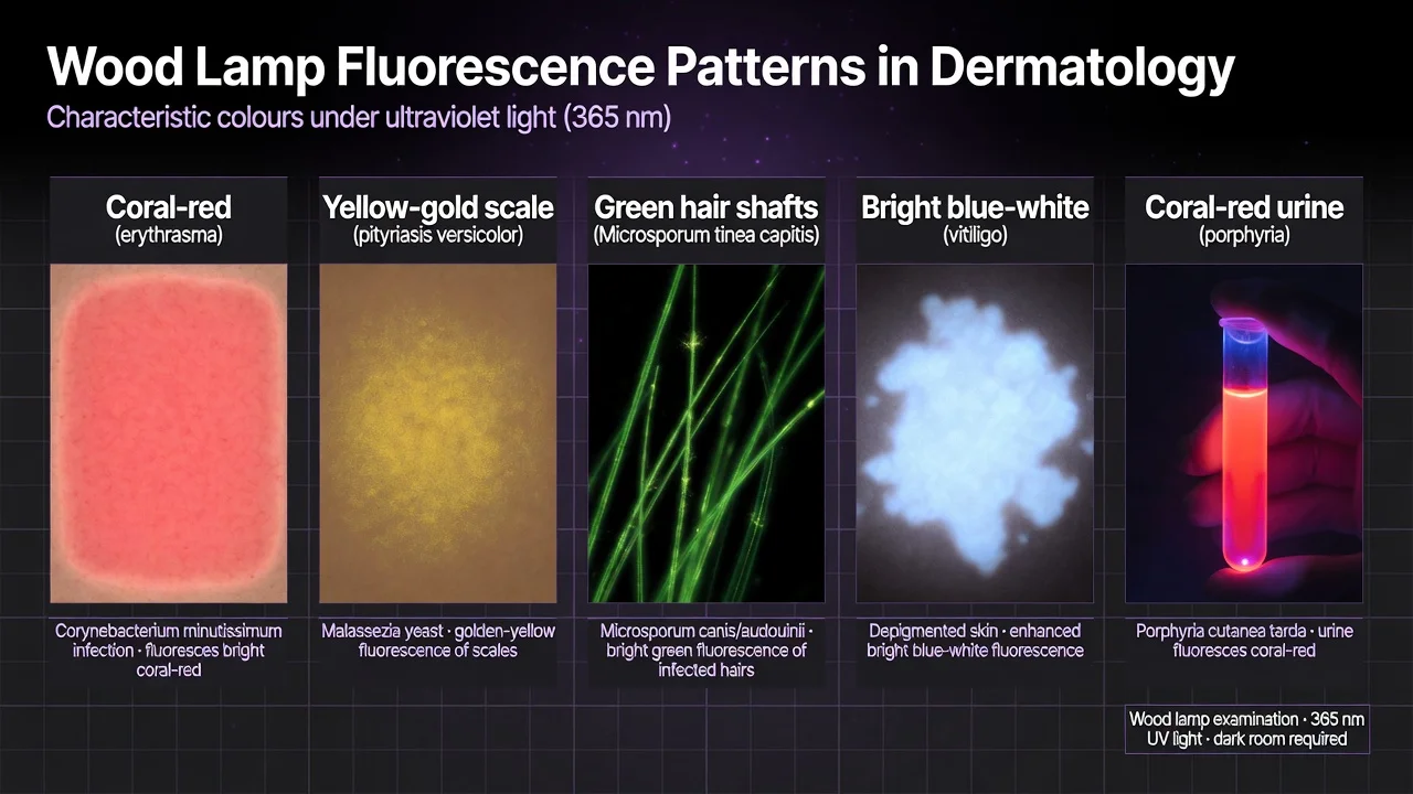
| Coral-red intertrigo (axilla, groin, toe webs) | Erythrasma (C. minutissimum) | Clinical Rx ± confirm; differentiate tinea/candida[5][10] |
| Yellow-gold follicular/scale glow | Pityriasis versicolor (Malassezia fluorochromes) | KOH; antifungal plan[7][3] |
| Green fluorescence of hair shafts | Microsporum tinea capitis (e.g. M. canis) | Mycology; note non-fluorescent Trichophyton[6][8] |
| Bright blue-white patches, sharp borders | Vitiligo extent mapping | Clinical/dermoscopy correlation[1][3] |
| Coral-red urine (acidified, under lamp) | Porphyrins (e.g. PCT context) | Formal porphyria labs — not lamp alone |
| Greenish nails/wounds (variable) | Pseudomonas clue | Culture as indicated |
| Enhanced pigment borders | Lentigo/melanoma margin adjunct (selected) | Not a substitute for histopathology[9] |
Exam-critical fluorescence patterns
Infection modules
Erythrasma
Presents as finely wrinkled brown-red intertriginous patches. Coral-red fluorescence is classic and supports diagnosis when morphology fits; treatment reviews emphasise topical and systemic antibacterial strategies after clinical diagnosis, with Wood lamp as a rapid bedside aid.[5][10][3]
Pityriasis versicolor
Malassezia produces fluorochromes (including pityrialactone-related chemistry in experimental work) that can yield yellow-gold scale fluorescence under UVA, complementing KOH of scrapings.[7][3]
Tinea capitis
Fluorescent Microsporum infections historically dominated school outbreaks; epidemiology shifted toward non-fluorescent anthropophilic Trichophyton in many regions — hence the enduring teaching point that Wood lamp cannot rule out tinea capitis.[8][6] When green hairs are present, they guide sampling.
Pigment, porphyria, and surgical adjuncts
- Vitiligo vs mimics: bright, sharp blue-white accentuation helps estimate extent and residual islands; ash-leaf macules of tuberous sclerosis and PIH are usually subtler.[1][3]
- Porphyria: urine (and sometimes teeth/stool in related disorders) may show coral-red fluorescence — always confirm biochemically.
- Oncologic adjuncts: selected practice uses Wood lamp to help demarcate pigmented margins (e.g. lentigo maligna/melanoma context) or locate buried dark sutures; this remains adjunctive to clinical, dermoscopic, and histologic margins.[9][3]
Differential diagnosis and confirmatory tests
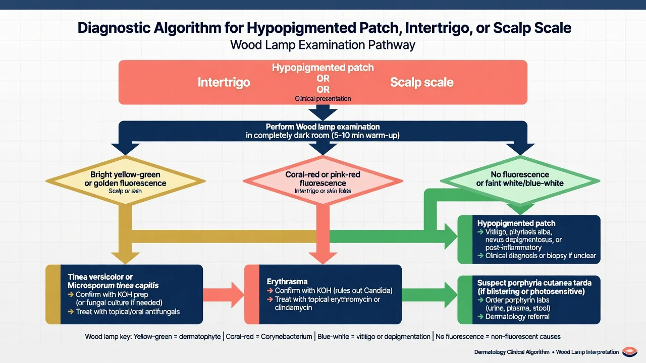
Always integrate morphology + dermoscopy + KOH/culture/PCR + biopsy when stakes are high. Coral-red flexures still need separation from dermatophyte and candidal intertrigo; yellow-gold scale needs KOH confirmation of yeast/hyphae; green hair needs mycology; non-fluorescent scalp scale still needs scrapings if tinea is possible.[3][6]
Pitfalls
- False positives: clothing fibres, topical tetracyclines or other medicines, soap, deodorant, residual dye, lint.[1]
- False negatives: light leakage, washed-off scale, non-fluorescent organisms, incomplete dark adaptation.
- Over-interpretation: treating every glow as infection; missing vitiligo extent because the room was not dark.
Special populations and regional notes
Paediatric clinics still use Wood lamp for rapid Microsporum screening in outbreaks, remembering local species mix.[6][8] In darker phototypes, vitiligo contrast under UVA is often especially helpful for border mapping.[3] IADVL/MRCP/FRCDerm viva expect the colour table and the Trichophyton non-fluorescence trap equally.
Evidence snapshot
Classic reviews (Asawanonda & Taylor; Klatte centenary) remain the conceptual backbone; newer pattern reviews expand uses in inflammatory disease and selected skin-cancer workflows, while infection-specific papers update erythrasma care and Microsporum detection.[1][2][3][5][6]
Clinical pearl
Red flags
Exam application bank (NEET-PG / INICET)
One-line answer
Board-level module on Wood lamp examination: long-wave UVA (~320–400 nm, peak ~365 nm) bedside fluorescence and pigment contrast for erythrasma (coral-red), pityriasis versicolor (yellow-gold), Microsporum tinea capitis (green hair), vitiligo (bright blue-white), porphyria urine fluorescence, Pseudomonas cues, and selected surgical margin/suture uses. Covers dark-room technique, fluorophore biology, colour→diagnosis map, false positives/negatives, and when KOH, culture, or porphyria labs must confirm the pattern.
Worked stems (answer without another resource)
Stem 1 — Classic presentation. Map symptoms to mechanism; name the first investigation and first treatment step with dose/route if drug therapy is standard. [1]
Stem 2 — Unstable / complicated. List red flags that force immediate resuscitation, theatre, ICU, antidote, or reperfusion — and what you do in the first 15 minutes. [1]
Stem 3 — Atypical group. Elderly, pregnancy, child, or immunocompromised: how presentation and thresholds change. [1]
Stem 4 — Differential trap. Name the three closest mimics and one discriminator for each. [1]
Stem 5 — Disposition. Who goes home with safety-netting, who is admitted, who needs HDU/ICU/theatre, and what follow-up is mandatory. [1]
Rapid viva checklist
- Definition + classification
- Pathophysiology chain
- Bedside signs / criteria
- Score with exact components (if any)
- Emergency bundle
- Definitive therapy with doses
- Complications of disease and of treatment
- Special populations
- Guideline/trial name if classic
- Three exam traps
Coverage self-check
If you cannot answer any stem above from this page alone, re-read the matching section — the page is intended to be self-sufficient for final-prof and NEET-PG/INICET questions on Wood lamp / fluorescence examination.
Expanded exam teaching (depth pass)
Clinical reasoning
For Wood lamp / fluorescence examination, examiners test whether you can prioritise life threats, choose the right first test, and give specific therapy (agent, dose, route, timing). Generic phrases without numbers score poorly.
Mechanism → feature map
Build a short chain: cause → pathophysiologic intermediate → clinical feature → complication. Every major symptom in the classic vignette should sit on that chain.
Investigation strategy
- Bedside/first-line tests that change immediate management
- Confirmatory or staging tests
- What a normal result does not exclude
- When not to delay treatment for imaging (unstable patient)
Management ladder
- Resuscitation / ABC / sepsis or haemorrhage bundle as relevant
- Specific antidote / procedure / antimicrobial / reperfusion / surgery
- Supportive care and monitoring targets
- Definitive long-term therapy and secondary prevention
- Disposition and safety-net advice
Special populations
Always prepare one line each for children, pregnancy, elderly, renal/hepatic impairment, and immunocompromised patients when the topic allows.
Pitfalls that fail candidates
- Treating the number not the patient
- Missing pregnancy status when relevant
- Imaging before stabilisation
- Wrong empiric cover or wrong antidote timing
- Incomplete counselling on recurrence, adherence, or red-flag return
Board-level module on Wood lamp examination: long-wave UVA (~320–400 nm, peak ~365 nm) bedside fluorescence and pigment contrast for erythrasma (coral-red), pityriasis versicolor (yellow-gold), Microsporum tinea capitis (green hair), vitiligo (bright blue-white), porphyria urine fluorescence, Pseudomonas cues, and selected surgical margin/suture uses. Covers dark-room technique, fluorophore biology, colour→diagnosis map, false positives/negatives, and when KOH, culture, or porphyria labs must co [1]
Structured revision sheet
Must-know numbers and names
List every score, size threshold, dose, and time window from this topic on a blank page from memory, then check against the sections above.
Three classic MCQ angles
- Most likely diagnosis given a vignette
- Next best step in management
- Most appropriate investigation
Three classic SAQ angles
- Pathophysiology in five steps
- Management algorithm with doses
- Complications and prevention
Clinical station flow
Greet → focused history → targeted exam → investigations → explain diagnosis → emergency care → definitive plan → safety-net / follow-up → answer examiner questions on mechanism and pitfalls.
Exam anchors
Exam triad
ACT
Define the problem and red flags
Highest-yield test or bedside clue
First-line management and follow-up
References
- [1]Asawanonda P, Taylor CR. Wood's light in dermatology Int J Dermatol, 1999.PMID 10583611
- [2]Klatte JL, van der Beek N, Kemperman PM. 100 years of Wood's lamp revised J Eur Acad Dermatol Venereol, 2015.PMID 25428804
- [3]Gomez-Martinez S, Ibaceta Ayala J, Morgado-Carrasco D. [Translated article] Wood's Light in Inflammatory and Autoimmune Dermatoses, Infections and Skin Cancer Actas Dermosifiliogr, 2025.PMID 39722344
- [4]Kwaśny M, Stachnio P, Bombalska A. Application of Wood's Lamp in Dermatological and Dental Photodiagnostics Sensors (Basel), 2025.PMID 40968799
- [5]Radhakrishnan S, Logamoorthy R, Karthikeyan K, et al. Erythrasma: a systematic review of interventions Clin Exp Dermatol, 2025.PMID 40635638
- [6]Sun D, Lu J, Liu T, et al. Wood's lamp for early detection of Microsporum Canis tinea capitis in children Photodiagnosis Photodyn Ther, 2025.PMID 39638219
- [7]Mayser P, Stapelkamp H, Krämer HJ, et al. Pityrialactone- a new fluorochrome from the tryptophan metabolism of Malassezia furfur Antonie Van Leeuwenhoek, 2003.PMID 14574113
- [8]Prevost E. The rise and fall of fluorescent tinea capitis Pediatr Dermatol, 1983.PMID 6680181
- [9]Nguyen K, Arora N, Ha O, et al. Utilizing Wood's Lamp to Demarcate Excisional Margins for Melanoma: A Retrospective Chart Review Dermatol Surg, 2025.PMID 41033300
- [10]Pan S, Ryan MP, Rapini RP. Erythrasma JAMA Dermatol, 2024.PMID 38231505