Dermatology · Medicine
Wound healing
Also known as Wound healing · Wound repair · Cutaneous healing · Tissue regeneration · Scar formation · Soft-tissue healing
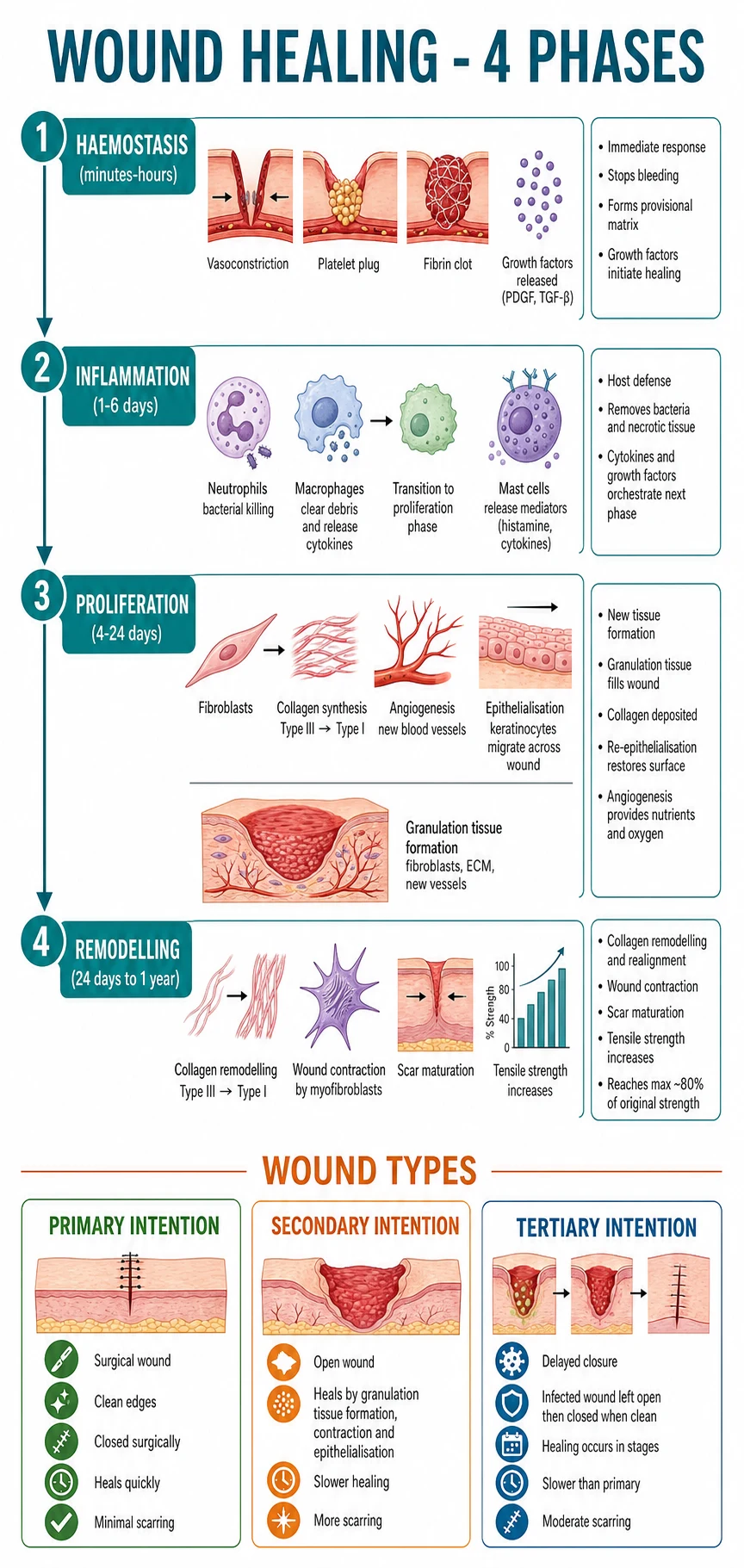
Wound healing is a coordinated, overlapping biological sequence of (1) Haemostasis (minutes–hours — platelet plug, fibrin, growth factors), (2) Inflammation (0–3 days — neutrophils then macrophages clear debris), (3) Proliferation (3 days–3 weeks — fibroblasts synthesise type III collagen, angiogenesis, epithelialisation, granulation tissue, wound contraction by myofibroblasts), and (4) Remodelling (3 weeks–12 months — type III → type I collagen, scar maturation, max ~80% tensile strength, never 100%). Healing by intention: primary (surgically approximated edges, fastest, minimal scar), secondary (open wound fills by granulation and contracts), tertiary (delayed primary closure after debridement). Growth factors central: PDGF, TGF-β1/2, VEGF, FGF-2, EGF. Chronic wounds (arterial, venous, diabetic, pressure, vasculitic, pyoderma gangrenosum, Marjolin) stall in inflammation. TIME wound-bed preparation (Tissue, Infection, Moisture, Edge). Modern dressings matched to exudate (hydrocolloid, hydrogel, foam, alginate, hydrofibre, silver/honey). Compression bandaging 40 mmHg class III for venous ulcer; offloading (total contact cast) for diabetic foot ulcer; revascularisation for arterial ulcer; pressure redistribution for pressure ulcer. Advanced therapies: NPWT (VAC), hyperbaric oxygen, Apligraf/Dermagraft, recombinant PDGF (becaplermin).
On this page & tools
Your progress
Saved locally on this device.
Exam tags
Red flags
Overview & Definition
A wound is any disruption of the normal anatomical continuity of skin or underlying soft tissue; wound healing is the highly conserved biological process that restores tissue continuity through a coordinated, overlapping sequence of cellular and molecular events. In adult mammalian skin, the end-result is repair with scar (not true regeneration). Understanding the phase-specific biology lets you diagnose a delayed wound, target the failing step, and choose the correct dressing, drug, or intervention.[1][2][4][9]
The four classical phases are: [1]
- Haemostasis — minutes to hours after injury.
- Inflammation — ~0 to 3 days (cells present up to day 5–7).
- Proliferation — 3 days to ~3 weeks (24 days by classic autopsy data).
- Remodelling — 3 weeks to 12 months (occasionally longer; the scar never reaches 100 % tensile strength). [1]
These phases are overlapping and continuous, not isolated — the macrophage at day 3 is the critical pivot between inflammation and proliferation, and its absence (in experimental models) causes the wound to stall in the inflammatory phase. [1]
The textbook distinction between healing by primary, secondary and tertiary intention is the cornerstone of acute wound management: [1]
- Primary intention — clean wound edges surgically approximated (suture, staple, glue, Steri-Strip); minimal tissue loss; fastest healing (7–14 days); least scar.
- Secondary intention — open wound left to granulate, contract, and re-epithelialise from the edges and adnexal remnants; greater tissue defect; slower healing (weeks to months); more scar and contraction.
- Tertiary (delayed primary) intention — contaminated, infected, or war wound left open initially, debrided/cleaned, then closed surgically 3–7 days later; balances infection control with timely closure. [1]
A related, often-tested distinction is between regeneration (true restoration of original tissue architecture, as in amphibian limb regrowth or early-gestation fetal skin) and repair (restoration of continuity with a fibrous scar, the rule in adult skin). [1]
Classification of wounds
Wounds are classified clinically along several axes — by cause, depth, contamination, and time to healing. The classification drives all downstream decisions: debridement, closure, dressing choice, antibiotic use, and prognosis.[2][4]
By cause
Insults that initiate the wound
- **Mechanical** — incisional (surgical), excisional, laceration, abrasion, contusion, crush, bite, degloving
- **Thermal** — cold (frostbite) or hot (burn, scald); depth depends on temperature + contact time
- **Chemical** — acid (coagulative necrosis), alkali (liquefactive necrosis), corrosive ingestion
- **Radiation** — ionising (stochastic late fibrosis) and UV (chronic photodamage, non-scarring)
- **Biological** — venom, allergen, infection (ulcer); pressure (decubitus); vasculopathy (auto-amputation)
- **Iatrogenic** — surgical, vascular access, drain, biopsy, injection site
By contamination
Drives primary-vs-secondary closure
- **Clean** — surgical incision under aseptic conditions; < 2% infection risk; close primarily
- **Clean-contaminated** — enters respiratory, GI, GU tract under controlled conditions; primary closure usually safe
- **Contaminated** — open traumatic wound (< 4 h old); inflamed tissue without pus; surgical debridement + careful closure
- **Dirty / infected** — old traumatic wound (>4 h) or with pus, devitalised tissue, or perforation; debridement + secondary closure or NPWT + tertiary closure
By time course
Drives escalation pathway
- **Acute wound** — recent, progressing through phases; should heal in 4–12 weeks
- **Chronic wound** — fails to complete healing within 12 weeks despite appropriate care; "stuck" in inflammation
- **Primary chronic** — never heals (continuous pathology — pressure, vasculitis, malignancy)
- **Recurrent** — healed but breaks down again (venous ulcer without life-long compression; pressure ulcer without continued offloading)
By healing mechanism
Intention
- **Primary** — clean edges surgically approximated; fastest; minimal scar
- **Secondary** — open; granulation + contraction + epithelialisation; slower; more scar
- **Tertiary (delayed primary)** — initially open then closed 3–7 days later; balances infection control vs timely closure
Epidemiology & Risk Factors
Cutaneous wound healing is a global health priority. The burden of chronic wounds is dominated by venous, arterial, diabetic, and pressure-related aetiologies, each with its own epidemiology and risk profile. Chronic wounds consume an estimated 2–3 % of the healthcare budget in industrialised countries, a figure that rises further when lost productivity, social care, and amputation are counted.[4][7][11]
Risk factors impairing wound healing can be grouped into local (in the wound) and systemic (in the patient). Diabetes is the single most consequential systemic factor, by virtue of microangiopathy, neuropathy, neutrophil dysfunction, and impaired fibroblast activity. Smoking, malnutrition (protein, vitamin C, zinc), corticosteroids, immunosuppression, age >80, anaemia, hypoxia, ionising radiation, and obesity all contribute additively. Local factors include infection, foreign body, haematoma, necrosis, tension, and ischaemia. [1]
Local factors — at the wound
Within surgeon control
- **Infection** — most common cause of delayed healing; bacterial burden invokes persistent neutrophil infiltration and MMP overexpression
- **Foreign body** — suture, glass, wood, dirt, hair; sustains inflammation
- **Ischaemia** — arterial disease, pressure, vasoconstriction (vasopressors, smoking)
- **Necrotic tissue** — slough or eschar acts as a barrier to granulation and a microbial substrate; debride before closure
- **Haematoma / seroma** — collection under the wound; impedes apposition and is a growth medium
- **Excessive tension** — wound under tension heals poorly with worse scar
- **Repeated trauma** — friction, pressure, shear (surgical line under clothing, prosthetic)
- **Radiation damage** — chronic irradiation impairs vascular supply and fibroblast function
- **Recurrence** — same site poorly offloaded (DFU) or unrelieved pressure
Systemic factors — in the patient
Patient-level risk
- **Diabetes** — microangiopathy, neuropathy, neutrophil dysfunction (chemotaxis, ROS generation), fibroblast dysfunction, AGE-collagen; HbA1c >9 % markedly impairs healing
- **Smoking** — nicotine vasoconstriction, CO reduces oxygen delivery, oxidative stress, impairs fibroblast migration and collagen synthesis
- **Malnutrition** — protein-energy malnutrition (PEM), deficiency of vitamin C (collagen cross-linking), zinc (epithelialisation), vitamin A (epithelial differentiation), iron (oxygen delivery), arginine/ glutamine (conditionally essential)
- **Corticosteroids** — impair all phases, especially collagen synthesis and wound contraction; vitamin A 25,000 IU/day counteracts
- **Immunosuppression** — chemotherapy, calcineurin inhibitors, azathioprine, biologics; opportunistic infection risk
- **Age >80** — reduced fibroblast proliferation, decreased collagen synthesis, immune senescence
- **Obesity** — increased wound tension; hypoperfusion in adipose tissue; higher infection rate
- **Anaemia and hypoxia** — impaired oxygen delivery (PaO₂ < 40 mmHg → collagen synthesis collapses)
- **Vasculopathy** — peripheral arterial disease, venous insufficiency; limits perfusion and oxygenation
- **Uraemia** — platelet dysfunction; calciphylaxis in dialysis
Pathophysiology
Wound healing is a coordinated, sequence-driven biological process. At every stage, there is a specific cellular cast, a specific signalling network, and a corresponding clinical and histological appearance. The molecule-by-molecule detail matters: most chronic wounds fail because one specific step is blocked, and identifying that step dictates the intervention.[1][2][4][6][9]

Phase 1 — Haemostasis (minutes to hours)
Within seconds of vessel injury, three overlapping processes are initiated: [1]
- Reflex vasoconstriction (endothelin, thromboxane A₂) — reduces blood loss.
- Platelet adhesion, activation, and aggregation (primary haemostasis) — platelets adhere to exposed subendothelial collagen and von Willebrand factor via GPIb-V-IX and GPVI receptors, then undergo shape change, release α-granule contents (PDGF, TGF-β, EGF, FGF, VEGF) and dense-granule contents (ADP, serotonin, calcium). ADP and thromboxane A₂ recruit more platelets, forming the platelet plug.
- Fibrin formation (secondary haemostasis) — exposure of tissue factor triggers the extrinsic pathway; contact with collagen triggers the intrinsic pathway; both converge on factor X activation → thrombin → fibrinogen → fibrin mesh that stabilises the plug and acts as a provisional extracellular matrix (ECM) scaffold for the migrating cells that arrive in the next phase. [1]
Crucially, the growth factors released in the first minutes are not just for stopping bleeding — they recruit neutrophils and macrophages within hours and orchestrate every later phase. The fibrin scaffold is also the road fibroblasts use to migrate into the wound from day 3 onwards.[1][2][6][9]
Phase 2 — Inflammation (0 to 3 days)
Inflammation is not just "rubor, calor, tumor, dolor" — it is the body's demolition and inspection crew at work. Neutrophils (polymorphonuclear leukocytes, PMNs) arrive within 6 hours, peak at 24–48 hours, and clear bacteria and devitalised tissue by phagocytosis, reactive oxygen species (NADPH oxidase–derived superoxide, hydrogen peroxide, hypochlorous acid via myeloperoxidase), and NETosis (release of neutrophil extracellular traps that bind pathogens). They undergo apoptosis after 24–48 hours and are cleared by incoming macrophages.[1][2]
Macrophages arrive on day 2–3 from circulating monocytes (recruited by MCP-1, PDGF, TGF-β), peak at day 3–5, and serve three critical roles: [1]
- Debridement — phagocytose apoptotic neutrophils and cellular debris.
- Antimicrobial defence — also kill bacteria via ROS and nitric oxide.
- Release of growth factors (TGF-β, VEGF, FGF, PDGF, IGF-1) that orchestrate the transition to proliferation: fibroblast activation, myofibroblast differentiation, angiogenesis, re-epithelialisation. [1]
The M1 → M2 phenotype switch by day 3–5 is the master timing signal. Prolonged M1 polarisation (from persistent infection, hypoxia, hyperglycaemia, or foreign body) is one of the central mechanisms by which chronic wounds get "stuck" in inflammation and fail to progress.[1][6][9]
T-lymphocytes arrive later (day 5–7) and modulate remodelling by releasing cytokines that influence collagen turnover and fibroblast activity. Mast cells and basophils contribute histamine-mediated vasodilation in the early phase. [1]
Phase 3 — Proliferation (3 days to 3 weeks)
Four overlapping subprocesses give the wound its tissue bulk:[1][2][4]
- Fibroplasia — platelet- and macrophage-derived TGF-β, PDGF, and FGF recruit fibroblasts into the wound. Fibroblasts migrate along the fibrin provisional matrix (using integrins and the fibronectin-rich scaffold), proliferate, and deposit type III collagen, fibronectin, glycosaminoglycans, and hyaluronic acid — the new extracellular matrix that replaces fibrin. This provisional matrix is mechanically weak but provides the template on which type I collagen will later be laid.
- Angiogenesis — VEGF, FGF-2, angiopoietin and PDGF drive endothelial cell migration and capillary sprouting into the wound. The result is granulation tissue — the bright-red, beefy, friable, vascularised connective tissue that fills the wound defect.
- Epithelialisation — within 24 hours of injury in a moist wound (longer in dry wounds), keratinocytes at the wound edge detach from the basement membrane, become migratory, glide over the provisional matrix (using integrin-α5β1 and fibronectin), proliferate behind the migration front, and eventually re-stratify to reform a stratified epidermis. Re-epithelialisation is dramatically faster in a moist wound (Winter 1962/63; Hinman 1963) — the reason for the modern occlusion/dressing approach.
- Wound contraction — under the influence of TGF-β, fibroblasts differentiate into myofibroblasts that express α-smooth-muscle actin and contract. Centripetal contraction can reduce an open wound by 0.5–1 mm per day, and accounts for most of the area reduction in secondary intention healing. [1]
The end of proliferation is marked by a confluent granulation tissue bed re-covered by advancing epithelium — the macroscopic appearance of an "active healing" wound. [1]
Phase 4 — Remodelling (3 weeks to 1+ year)
Remodelling is the longest phase and the one that determines final scar quality. Type III collagen (deposited in proliferation) is degraded by matrix metalloproteinases (MMP-1, MMP-8, MMP-13) and replaced by stronger type I collagen. New collagen fibres are reorganised along tension lines and cross-linked by lysyl oxidase (a copper-dependent enzyme). Myofibroblasts and fibroblasts are removed by Fas/FasL-mediated apoptosis. The scar becomes flatter, paler, and softer.[1][4]
Critically, tensile strength increases logarithmically: ~20 % at 3 weeks, ~30 % at 4 weeks, ~80 % at 3 months, then plateaus and never exceeds ~80 % of uninjured skin. The scar is acellular, avascular, and permanently weaker than unwounded skin. Long-term biological consequences include abnormal hair and sweat-gland function, loss of adnexal structures, and altered mechanical properties — the substrate for hypertrophic scarring, keloid formation, and contracture (covered in the keloid topic).[4]
Growth factors in wound healing — the master regulator cast
Wound healing is driven by growth factors released from platelets, macrophages, fibroblasts, endothelial cells, keratinocytes, and mast cells. Each has a specific receptor, a specific binding partner, and a specific cellular effect.[6][9]
Growth factors in wound healing — P.P.T.V.F.E (Platelet group / Vascular / TGF-β / EGF / FGF) on a single card
Platelet-vessel-TGF-FGF-EGF
Source: platelets α-granules, macrophages, endothelium, fibroblasts. Actions: chemotactic for neutrophils/macrophages/fibroblasts; mitogenic for fibroblasts and vascular smooth muscle; stimulates collagen synthesis. Recombinant form = becaplermin (see Management)
Source: platelets, macrophages, fibroblasts. Actions: TGF-β1/β2 drive fibroblast proliferation, collagen synthesis, myofibroblast differentiation. TGF-β3 is anti-fibrotic and is relatively deficient in keloids (the rationale for exogenous TGF-β3 research)
Source: keratinocytes, macrophages, fibroblasts. Actions: angiogenesis (endothelial proliferation and migration); vasodilation; early granulation tissue
Source: fibroblasts, endothelium, keratinocytes. Actions: FGF-2 (bFGF) drives angiogenesis, fibroblast and keratinocyte proliferation; applied topically in some advanced wound products
Source: platelets, macrophages, saliva. Actions: epithelialisation, keratinocyte migration and proliferation. KGF-1 and KGF-2 (keratinocyte growth factors 1 and 2) are epithelium-specific FGF family members
Other key mediators: [1]
- IGF-1 (insulin-like growth factor) — fibroblast proliferation.
- CSF / G-CSF / GM-CSF — haematopoietic recruitment.
- IL-1, IL-6, TNF-α — pro-inflammatory cytokines; orchestrate early inflammation but excess prolongs inflammation (causative in chronic wounds).
- Cyclooxygenase-2 (COX-2) → prostaglandin E₂ (PGE₂) — vasodilation, pain.
- Lipoxins, resolvins, protectins — termination of inflammation (resolution phase, increasingly studied).
- MMPs (matrix metalloproteinases) and TIMPs (tissue inhibitors of metalloproteinases) — balance of ECM synthesis and degradation; chronic wounds show MMP overexpression and TIMP downregulation, causing net collagen degradation. [1]
Moist wound healing — the Winter/Hinman principle and its rationale
G.D. Winter's 1962/63 and Hinman & Maibach's 1963 experiments changed surgical practice forever: occlusive and semi-occlusive dressings heal partial-thickness wounds 30–50 % faster than dry exposure because epithelial cells migrate ~2–3 times faster across a moist, warm surface than under a desiccating scab. Occlusion also enables autolytic debridement — phagocytes and matrix proteases digest devitalised tissue under the dressing with minimal surgical trauma. The corollary is that the old practice of letting wounds "form a good scab" is harmful. Modern dressings (hydrocolloid, hydrogel, foam, film, alginate, hydrofibre, antimicrobial) all aim to exploit this principle, with the dressing choice driven by exudate volume, depth, and infection status.[3]
Chronic wound pathophysiology — what makes a chronic wound chronic
Chronic wounds (venous, arterial, diabetic, pressure, vasculitic) are trapped in the inflammatory phase. The unifying pathophysiology is: [1]
- Tissue hypoxia (from vascular disease, oedema) → fails to support neutrophil oxidative burst, fibroblast proliferation, and collagen synthesis.
- Repetitive ischaemia-reperfusion → ROS, inflammation.
- Hyperglycaemia → advanced glycation end-products stiffen collagen; neutrophil dysfunction; reduce NO.
- Biofilm — chronic wounds harbour polymicrobial biofilms that resist antibiotics 100–1000× compared to planktonic bacteria; protect bacteria from leukocyte phagocytosis.
- MMP overexpression + TIMP reduction → net collagen degradation rather than remodelling.
- Growth factor sequestration or degradation — TGF-β, PDGF, KGF inactivated by excess protease activity.
- Cellular senescence — fibroblasts, keratinocytes, and endothelial cells from chronic wounds enter senescence and lose proliferative capacity; they secrete a SASP (senescence-associated secretory phenotype) of pro-inflammatory cytokines and proteases that perpetuates the non-healing state.
- Impaired M1→M2 macrophage polarisation — chronic wound macrophages remain pro-inflammatory. [1]
Understanding this pathophysiology is the basis for advanced therapies (NPWT, growth factor gels, skin substitutes, HBOT) and for the TIME framework below.[1][2][6][8]
Clinical Presentation
Acute wound
A normal acute wound progresses through the four phases with a predictable clinical pattern.[2]
Atypical acute-wound presentations — the examiner's favourites — include: [1]
- Diabetic patient: diminished inflammation (so neutrophil signs mute); a wound may be disproportionately infected despite modest erythema. Always investigate when in doubt.[10]
- Immunosuppressed: atypical organisms (Pseudomonas, fungal, mycobacterial), no pyrexia, atypical pain.
- Corticosteroid-dependent patient: minimal erythema, swelling, heat, or pain — the cardinal signs are suppressed. A wound that "doesn't hurt" in a steroid-dependent patient with diabetes is a wound to biopsy and culture.
- Pregnant woman: increased scarring tendency (keloid, hypertrophic scar); relative immune modulation.
- Elderly: muted inflammation, slower granulation, higher anaesthetic risk.
- Child: faster healing, but more prone to hypergranulation and exuberant scarring.
Chronic wound — and its look-alikes
A chronic wound fails to complete at least 50 % area reduction within 4 weeks, and to fully heal within 12 weeks, despite optimal aetiology-appropriate care. The wound is "stuck" in the inflammatory phase; the edge does not advance; the base is often pale, friable, hypergranular, sloughy, or exudative. Surrounding skin may be macerated, eczematous, or hyperpigmented (haemosiderin in venous disease; haematogenous pigmentation in chronic infection). Classic clinical hallmarks: persistent exudate, foul odour (biofilm or Pseudomonas), undermining, rolled and raised edges (think malignancy). [1]
Venous ulcer
GAITER area; painless
- **Site** — gaiter area; classically over the **medial malleolus** (great saphenous vein territory); lateral malleolus possible
- **Shape** — shallow, irregular, often with sloping edges
- **Base** — red, granular, moderately exudative
- **Surrounding skin** — **haemosiderin** (brown hyperpigmentation); lipodermatosclerosis ("inverted champagne bottle" leg); **atrophie blanche** (white, scarred, telangiectatic patches); venous eczema (itchy, weeping, sometimes weeping of contact allergy to dressings — think lanolin, framycetin); oedema
- **Pain** — usually absent or mild; pain relieved by leg elevation
- **History** — DVT, varicose veins, venous surgery, leg swelling, family history
- **ABPI** — typically 0.8-1.2 (unless mixed aetiology)
Arterial ulcer
TOES / lateral; PAINFUL
- **Site** — distal sites: **toes**, **lateral malleolus**, pressure points over bony prominences, dorsum of foot
- **Shape** — **"punched-out"**, well-demarcated; deep may be shallow
- **Base** — **pale, dry, necrotic**; minimal granulation
- **Surrounding skin** — cool, hairless, shiny, pale or **cyanosed**; **absent pulses**; atrophic nails
- **Pain** — **SEVERE**, especially at night; classically relieved by **dangling the leg over the bed** (gravity-assisted perfusion)
- **History** — claudication, rest pain, peripheral arterial disease, smoking, diabetes, hypertension, hyperlipidaemia
- **ABPI** — typically < 0.5 (severe); 0.5-0.8 (moderate)
Diabetic (neuropathic) ulcer
PLANTAR; painless
- **Site** — **plantar surface** over pressure points: metatarsal heads, great toe, heel, midfoot
- **Shape** — callus-surrounded (the callus is the foots at-risk indicator);**punched-out**
- **Base** — often pale, may probe to bone (suggests osteomyelitis)
- **Neuropathy** — loss of protective sensation (10 g monofilament); Charcot deformity, claw toes; painless ulcer
- **Ischaemia** — neuroischaemic ulcer = coexistence of PAD and neuropathy; more sinister
- **History** — peripheral neuropathy, prior foot ulcer, amputation
- **ABPI** — may be **falsely elevated >1.3** due to medial arterial calcification (Mönckeberg); use toe-brachial index (TBI < 0.7) or transcutaneous oxygen pressure (TcpO2)
Pressure ulcer (PI)
Over BONY prominences
- **Site** — over **bony prominences**: sacrum (most common), ischial tuberosity, greater trochanter, heels, occiput (paediatric), scapula, elbows
- **Shape** — mirror image of bony prominence
- **NPUAP/EPUAP/PPPIA 2016 staging** — Stage I non-blanchable erythema INTACT skin; Stage II partial-thickness loss (pink/red, no slough); Stage III full-thickness loss, subcutaneous fat visible; Stage IV full-thickness loss with tendon/muscle/bone visible; **Unstageable**: eschar/slough obscures base; **SDTI** (suspected deep tissue injury): persistent non-blanchable maroon/purple discolouration
- **Risk** — Braden Scale < 18 (use at admission and weekly); Norton / Waterlow alternatives
- **Cause** — pressure >32 mmHg sustained >2 h occludes capillary flow; shear, friction, moisture
Pyoderma gangrenosum (PG)
VIOLACEOUS undermined
- **Site** — usually lower leg; can occur anywhere
- **Appearance** — painful ulcer with **violaceous, undermined, irregular edges**; often necrotic base; perforating variant has cribriform margins
- **History** — preceding skin trauma at the site (**pathergy**; worsens with surgery); often associated systemic disease: **IBD** (25 %), **RA**, **haematological malignancy** (10 %, esp. MDS), monoclonal gammopathy
- **Course** — rapidly progressive without treatment
- **Diagnostic caution** — biopsy to exclude ulcerated SCC, vasculitis, atypical infection; histology shows **sterile neutrophilic infiltrate**
- **Avoid debridement** — pathergy worsens lesion
Vasculitic ulcer
PALPABLE PURPURA
- **Site** — over malleoli, tibia, dorsum of foot, often multiple in crops
- **Appearance** — punched-out, irregular; often **livedo reticularis**, palpable purpura, surrounding dusky discolouration
- **Symptoms** — pain, peripheral neuropathy, digital ischaemia, gangrene
- **Aetiologies** — ANCA-associated vasculitis (GPA, MPA, EGPA), cryoglobulinaemia, antiphospholipid, SLE, RA, Behçet, livedoid vasculopathy
- **Investigations** — ANCA, ANA, ENA, cryoglobulins, antiphospholipid antibodies, complement, hepatitis B/C, biopsy of the edge (leukocytoclastic or granulomatous vasculitis)
Marjolin ulcer (malignancy)
Raised, everted, friable
- **Site** — anywhere on a chronic wound, burn scar (latency 20–30 years), venous ulcer, vaccination scar, sinus, scar of any cause
- **Appearance** — **raised, everted, indurated edge**; exophytic friable granulation; bleeding on contact
- **Symptoms** — change in chronic wound (pain, discharge, smell, growth)
- **Course** — high-risk SCC (2-5 cm at presentation; prognosis worse than SCC in non-scarred skin); metastasis in 3-8 %
- **Action** — elliptical biopsy (NOT punch) from the indurated edge, sent BOTH for histology AND microbiology; *biopsy every chronic non-healing wound >3 months and every changing scar*
Martorell ulcer
ANTEROLATERAL calf; SEVERE pain
- **Site** — sudden, very painful ulcer on **anterolateral calf** in patients with poorly controlled hypertension
- **History** — long-standing, often severe hypertension
- **Histology** — pauci-immune thrombotic vasculopathy; subcutaneous arteriolar medial calcification and intimal hyperplasia; **NOT** vasculitis
- **Atypical** — sometimes autoimmune; some classified as limited forms of systemic sclerosis overlap
Differential Diagnosis

The differential of a chronic non-healing ulcer is wide, and every candidate must be evaluated. The mnemonic "VAMPiDRAVL" (Venous, Arterial, Marjolin/Martorell, Pyoderma gangrenosum/Martorell/Malignancy, Dermatitis artefacta, Rheumatologic, Atypical Mycobacteria, Vasculitis, Lymphoedema, Lymphoma) covers most; the working differential in any specific patient depends on site, age, history, and risk profile. [1]
Aetiology-based
Pathophysiology groups
- **Vascular** — venous insufficiency (60-70 %), arterial insufficiency (10-20 %), mixed
- **Neuropathic** — diabetic, alcohol, hereditary (HSAN), leprosy, tabes dorsalis, B12 deficiency
- **Pressure** — immobility, surgery, spinal cord injury, device-related
- **Vasculitis** — ANCA-associated (GPA, MPA, EGPA), SLE, antiphospholipid, cryoglobulin, livedoid vasculopathy
- **Neutrophilic dermatosis** — PG, Behçet, Sweet
- **Malignancy** — Marjolin SCC, BCC, amelanotic melanoma, DFSP, Kaposi sarcoma, cutaneous lymphoma, metastasis to scar
- **Infection** — atypical mycobacteria (M. ulcerans — Buruli; M. marinum; M. tuberculosis — tuberculosis cutis), deep fungi (chromoblastomycosis; eumycetoma; sporotrichoid spread), leishmaniasis, syphilitic gumma, ecthyma
- **Drug-induced** — hydroxyurea, nicorandil (oral/perianal ulcers), coumarin skin necrosis, methotrexate, doxycycline-induced oesophagitis/photodermatitis
- **Metabolic** — calciphylaxis (uraemic calcific arteriolopathy), necrobiosis lipoidica (commonly diabetic), gout, scleroderma digital ulcers
- **Haematological** — sickle cell leg ulcer, PG in MDS, thrombophilic ulcers (antiphospholipid)
Site-based
Anatomic clue
- **Medial gaiter** — venous; **lateral gaiter** — arterial more likely
- **Plantar foot** — neuropathic (diabetic), but consider foreign body insole and PVS
- **Pulp of toe** — arterial; **spaced ulcer on dorsum of foot** — early chondrodermatitis
- **Pretibial** — necrobiosis lipoidica, erythema nodosum, PG, vasculitis, diabetic dermopathy
- **Anterolateral calf** — Martorell
- **Sacrum / heel / occiput** — pressure
- **Perianal** — Crohn, PG, malignancy, nicorandil, SCC in situ (Bowen disease), hidradenitis suppurativa, sexually transmitted
- **Oral cavity** — aphthous, Behçet, PG, Crohn, SCC, lupus, pemphigoid
Acute wound differentials
Failure to progress
- Ongoing infection (subtle in immunocompromised/diabetic); foreign body (suture, glass, wood); haematoma; recurrent trauma; ischaemia; nutrition; corticosteroid; malignancy; pyoderma gangrenosum
- Always recalculate ABPI; check dressing regimen; consider X-ray for radio-opaque foreign body
Hypergranulation differential
Exuberant granulation tissue
- True hypergranulation (exuberant benign, friable, raised above wound surface) — usually over-moist, occlusive dressing; treat with caustic (silver nitrate), topical steroid, change to foam dressing
- **Pyogenic granuloma** — pedunculated nodular lesion; bleeds easily
- **Amelanotic melanoma** — pale or red nodule; biopsy if in doubt
- **Kaposi sarcoma** — violaceous nodule in HIV; biopsy
- **Capillary haemangioma** in adults; consider skin metastasis
Clinical & Bedside Assessment
Assessment of a wound is structured and systematic. The TIME framework (Schultz et al. 2003) is the international standard for chronic wound assessment; it captures the four clinical questions that drive management.[8]
TIME — the four questions to ask of any wound
TIME
Necrotic tissue (slough, eschar) is a barrier to granulation and a microbial substrate. Debride early with sharp/surgical (gold standard), enzymatic (streptokinase-streptodornase), autolytic (hydrocolloid/hydrogel), or larval (maggot Lucilia sericata). Healthy granulation: red, beefy, non-tender, non-friable.
Look for purulent discharge, >2 cm surrounding erythema, increasing pain, friable granulation, foul odour (Pseudomonas is sweet; anaerobes putrid), lymphangitis, fever, leucocytosis. Surface swabs unreliable in chronic wounds — Levine technique (rotate swab with pressure over 1 cm² after cleaning); tissue biopsy gold standard. Treat with antibiotics only if clinical infection is present, not for "colonisation".
Too wet → maceration; too dry → eschar. Match dressing to exudate volume. Aim for a clean, moist (NOT sloshy), well-granulating bed. Foam or hydrofibre or alginate for heavy exudate; hydrocolloid/film for light; hydrogel for dry wounds.
Advancing epithelial edge = good. Non-advancing, undermining, rolled/raised (think Marjolin), violaceous (think PG), macerated (moisture excess), hyperkeratotic (callus), or with adjacent lividity (think vasculitis) — all warrant reassessment; biopsy to exclude malignancy or vasculitis.
Comprehensive wound assessment (the structured bedside)
- Patient-level history — age, comorbidities (diabetes, vascular disease, RA, IBD), medications (corticosteroids, immunosuppressants, anticoagulants, chemotherapy), smoking, alcohol, nutrition, prior wounds, prior amputations, family history.
- Wound-specific history — onset, duration, precipitant, prior treatments, response, recurrence, frequency of dressing changes, pain (timing, character, modifiers), discharge (volume, colour, odour), impact on daily life.
- Wound examination — site (anatomic, clock-face), size (length × width × depth with ruler in centimetres), undermining/tunnelling (probe with cotton bud, document extent), base (% granulation, slough, eschar), exudate (type: serous/serosanguineous/purulent/haemorrhagic; volume: none/scant/moderate/heavy), edge (advancing/rolled/undermined/macerated), surrounding skin (cellulitis, eczema, maceration, callus, induration), odour, pain on dressing change.
- Peripheral limb examination — pulses (dorsalis pedis, posterior tibial, popliteal, femoral); capillary refill (< 3 sec normal); temperature gradient; oedema (pitting grade 0-3); trophic changes; foot deformity (Charcot, claw toes); footwear.
- Perfusion assessment — ABPI (see Investigations), toe-brachial index (TBI), transcutaneous oxygen (TcpO2) in selected cases.
- Sensory assessment (diabetic foot) — 10 g Semmes-Weinstein monofilament (≥4-6 plantar sites; loss = high ulcer risk); 128 Hz tuning fork at great toe; ankle reflexes (present/absent); pin-prick; proprioception.
- Risk assessment (pressure ulcer) — Braden Scale (most validated): six subscales scored 1-4 — Sensory perception, Moisture, Activity, Mobility, Nutrition, Friction/Shear; total score range 6-23; < 18 = at risk, < 13 = severe risk; Norton Scale (older, less discriminative), Waterlow Scale (UK, higher sensitivity), MUST (Malnutrition Universal Screening Tool, UK).
- Pressure ulcer staging (NPUAP/EPUAP/PPPIA 2016) — Stage I (non-blanchable erythema of INTACT skin), Stage II (partial-thickness loss; pink/red, no slough), Stage III (full-thickness loss, fat visible), Stage IV (full-thickness loss, tendon/muscle/bone visible), Unstageable (obscured by eschar/slough), Suspected Deep Tissue Injury (SDTI; persistent non-blanchable deep red/maroon/purple discolouration).
- Standardised serial photography — most practical objective monitoring tool. [1]
Bedside assessment scoring systems
Investigations
Investigations are not arbitrary; they must be aetiology-driven and used to alter management. Below is the structured investigation set, with named values, thresholds, and the evidence behind them.[7][11]
Perfusion (mandatory for any chronic lower-extremity ulcer)
Toe-brachial index (TBI) is preferred in diabetes (medial arterial calcification makes ABPI unreliable); a TBI of < 0.7 is abnormal. Transcutaneous oxygen pressure (TcpO2) on the dorsum of the foot reflects distal perfusion and is used in assessing amputation level (TcpO2 >30 mmHg suggests healing capacity). [1]
Vascular imaging
- Venous duplex ultrasound — first-line for suspected venous ulcer; identifies reflux (retrograde flow >0.5 sec, valve closure time) and obstruction in superficial (great saphenous, small saphenous) and deep (femoral, popliteal, tibial) systems. Drives the decision about endovenous ablation (foam sclerotherapy, endovenous laser, radiofrequency) of superficial reflux to improve healing.[7]
- Arterial duplex ultrasound — first-line non-invasive test for peripheral arterial disease; gives anatomic and haemodynamic information.
- CT angiography / MR angiography — when endovascular intervention is planned; provides anatomic detail of stenosis and run-off.
- Catheter digital subtraction angiography (DSA) — gold standard; reserved for when intervention is contemplated or as part of an endovascular procedure.
Metabolic and nutritional work-up
Microbiology
- Wound swab — standard for clinically infected wounds; gold standard is tissue biopsy in chronic non-healing wounds. Use the Levine technique: rotate a swab over a clean 1 cm² area with pressure; express fluid from the wound. In wounds with >10⁵ CFU/g of tissue, infection is considered present.
- MRSA screen — nasal + axillary + groin; pre-op if patient has history of MRSA.
- Pseudomonas screening — wet chronic wound with sweet/grape-like odour, blue-green exudate.
- AFB, fungal, atypical — for chronic non-healing with negative conventional cultures, especially with travel or tropical exposure (Buruli, chromoblastomycosis, leishmaniasis). [1]
Histopathology
- Biopsies are mandatory when: (a) a chronic wound has been present for >3 months without progression, (b) the wound shows any atypical feature (raised everted edge, exophytic growth, friable granulation, regional lymphadenopathy, or rapid change), (c) vasculitis is suspected (purpura, livedo, multi-system features), (d) infection with an unusual organism is suspected, (e) pyoderma gangrenosum is suspected.
- Technique: elliptical incisional biopsy from the wound edge (where pathology is most active), including both affected and adjacent normal skin. Send separately for histology AND microbiology. Multiple biopsies increase yield.
- Histology of healing: in early phases, granulation tissue with new capillaries and fibroblasts; later, dense collagen with reduced cellularity; in chronic wounds, persistent inflammatory infiltrate, fibrosis, fibrin deposition; in Marjolin, atypical keratinocytes with dermal invasion. [1]
Diagnostic criteria and named scores reproduced verbatim
- TIME framework — Tissue / Infection / Moisture / Edge (Schultz et al. 2003).[8]
- Braden Scale — six subscales: Sensory perception, Moisture, Activity, Mobility, Nutrition, Friction/Shear; score 6-23; < 18 at risk; < 13 severe risk.
- Norton Scale — five domains: physical condition, mental condition, activity, mobility, incontinence.
- Waterlow Scale — eleven items: sex, age, BMI, mobility, incontinence, skin type, weight loss, appetite, surgery, trauma, mental state.
- PUSH Score — three subscores: length × width (0-10), exudate amount (0-3), tissue type (0-4).
- Wagner grading (DFU) — 0 = no ulcer high-risk foot; 1 = superficial ulcer; 2 = deep ulcer to tendon/bone/capsule; 3 = deep ulcer with abscess or osteomyelitis; 4 = localised gangrene (forefoot); 5 = extensive gangrene (whole foot).
- Texas University — 4 grades (0, I, II, III) × 4 stages (A, B, C, D); depth increases with grade; B = infection, C = ischaemia, D = both.
- IWGDF risk categories — 0 no neuropathy; 1 neuropathy alone; 2 neuropathy + PAD or deformity; 3 neuropathy + PAD + deformity or prior ulcer/amputation.
- PEDIS — Perfusion, Extent/size, Depth/tissue loss, Infection, Sensation; primarily research use.
- S(AD)SAD — Size, Area, Depth, Sepsis, Arteriopathy, Neuropathy; >6 = poor healing.
- NPUAP/EPUAP/PPPIA 2016 pressure ulcer staging — Stage I-IV, Unstageable, Suspected Deep Tissue Injury.
- CEAP — Chronic venous disorders, Clinical (C0-6), Etiology (Ec = congenital, Ep = primary, Es = secondary), Anatomy (As = superficial, Ap = perforator, Ad = deep), Pathophysiology (Pr = reflux, Po = obstruction, Pr,o = both).
Management — Resuscitation
Severe wound complications require resuscitation-level interventions — wound sepsis, necrotising fasciitis, or wet gangrene. The bundle is ATLS-style, with a strong surgical-component bias.[2][4]
Resuscitation bundle for severe wound infection
A — Airway, B — Breathing
100 % high-flow oxygen; SpO₂ target ≥ 94 %; consider intubation if compromised (severe facial wound, inhalation injury, sepsis with encephalopathy).
C — Circulation
Two large-bore IV cannulae; send FBC, U&E, CRP, coagulation, lactate, blood gas; balanced crystalloid (Ringer lactate or Hartmann) 20 mL/kg bolus; cross-match (or in massive transfusion protocol); norepinephrine if MAP < 65 after fluid bolus; consider central line, arterial line, urinary catheter.
Empirical broad-spectrum IV antibiotics within 1 h
Piperacillin-tazobactam 4.5 g IV q8h + clindamycin 600 mg IV q6h (for toxin suppression in necrotising fasciitis); meropenem 1 g q8h if β-lactam-allergic; consider empiric teicoplanin/vancomycin for MRSA risk; re-dose as per renal function; narrow when sensitivities return.
Source control — URGENT surgical debridement
Within 1-2 h of diagnosis if necrotising fasciitis or wet gangrene — resection of all necrotic tissue to the level of healthy bleeding tissue; second look at 24-48 h; amputation if limb non-salvageable.
D — Disability, E — Exposure
Glasgow Coma Scale; pupils; blood glucose (exclude hypoglycaemia); tetanus status check — see below; temperature (active warming if < 36 °C); full examination and logroll.
Tetanus prophylaxis
Clean wound: tetanus toxoid 0.5 mL IM if last dose >10 years ago (or unknown); wound prone to tetanus (contaminated/soil/burn): tetanus toxoid 0.5 mL IM if last dose >5 years (or unknown) PLUS tetanus immunoglobulin 250 IU IM.
Analgesia, dressing, and disposition
IV morphine 5-10 mg PRN 4-hourly titrated; consider nerve block (femoral / sciatic / popliteal); document wound on serial photographs; discuss with critical care; transfer to burns / vascular surgery / dermatology as appropriate.
Management — Definitive & Stepwise
Acute wound management — the bedside ladder
- Stop bleeding — direct pressure, elevation, tourniquet (rare); surgical control if artery.
- Pain control — morphine 5-10 mg IV/SC/PO PRN; nerve block as appropriate; NSAIDs avoided in the first 48-72 h (delay inflammatory phase).
- Examination under aseptic conditions — depth, contamination, tendon/vessel/nerve/bone injury, joint penetration.
- Irrigation and debridement — copious normal saline (300-1000 mL) at low pressure (4-15 psi) with 20 mL syringe and 18-19 G catheter; reduce bacterial load by 10²-10⁵; avoid antiseptic solutions in clean wounds (toxic to fibroblasts); remove devitalised tissue (sharp/surgical best; enzymatic with streptokinase-streptodornase for selected cases; autolytic with hydrocolloid/hydrogel for thin slough; larval (maggot) therapy with sterile Lucilia sericata larvae for selected chronic wounds with MRSA or biofilms).
- Tetanus — see above.
- Antibiotics — only with clinical infection or heavily contaminated wound.
- Closure (within 6-8 h for clean wound, longer after careful debridement in contaminated) — primary suture (deep +/- superficial), skin closure strips, tissue adhesive (octyl/butyl cyanoacrylate, e.g. Dermabond); staples; consider delayed primary if heavy contamination.
- Dressing — matched to exudate and depth.
- Review — at 7-14 days; sutures removed when healed: face 5-7 d, scalp 7-10 d, trunk 10-14 d, limb 10-14 d, foot 14-21 d; absorbable sutures (Polydioxanone PDS, Monocryl) for deep tissue. [1]
Chronic wound management — the structured chronic-wound consult
The chronic wound consult has five mandatory components, in this order: [1]
- Establish aetiology — venous, arterial, diabetic/neuropathic, pressure, vasculitic, neoplastic, drug-induced, infective; mixed aetiologies are common.
- Treat systemic factors — HbA1c control, smoking cessation, nutrition (protein, vitamin C, zinc, vitamin A, iron), corticosteroid dose reduction, optimise cardiac output, anaemia correction.
- Wound bed preparation — TIME — see Mnemonic above.[8]
- Apply the correct specialist therapy — compression (venous), offloading (DFU), revascularisation (arterial), pressure redistribution (PU), immunosuppression (vasculitis/PG), wide excision (Marjolin).
- Reassess at 4 weeks — % area reduction — the single most useful early predictor. < 30 % area reduction at 4 weeks = unlikely to heal within 12 weeks; reconsider and intensify (NPWT, advanced dressing, growth factor, skin substitute, HBOT).[13]
Modern wound dressings — exudate-matched
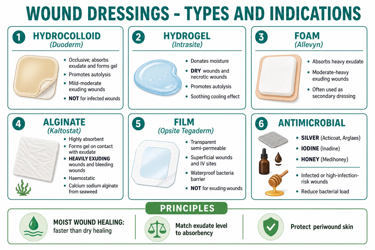
Modern dressings exploit the moist wound healing principle (Winter 1962). Dressing choice is driven by wound depth, exudate volume, infection status, patient comfort, and cost.[3]
Hydrocolloid
Occlusive; LIGHT-MODERATE exudate
- **Composition** — carboxymethylcellulose (CMC) particles embedded in adhesive matrix (e.g. Duoderm, Granuflex, Tegasorb)
- **Action** — on contact with exudate, CMC swells to form a gel that maintains a moist, warm environment; supports autolytic debridement
- **Indications** — mild-moderate exudate; superficial partial-thickness burns; pressure ulcer Stage II-III; shallow venous ulcer (under compression)
- **Cautions** — avoid in dry eschar (no moisture to activate gel); avoid in infected wounds (sealed bacterial medium); avoid over exposed tendon/bone
- **Change frequency** — every 3-5 days
Hydrogel
Donates moisture; DRY wounds
- **Composition** — polymer with 70-90 % water (e.g. Intrasite, Nu-Gel, AquaSite, Purilon)
- **Action** — DONATES water to wound; supports autolysis of dry eschar/slough
- **Indications** — dry wounds; necrotic wounds (rehydrates eschar for autolytic debridement); partial-thickness burns; radiation dermatitis
- **Cautions** — avoid in infected or heavily exuding wounds (provides moisture)
- **Change frequency** — every 1-3 days
Foam
MODERATE-HEAVY exudate
- **Composition** — hydrophilic polyurethane foam (e.g. Allevyn, Mepilex, Lyofoam)
- **Action** — absorbs exudate; thermal insulation; conforms to body contours; permeable to vapour (MVTR)
- **Indications** — moderate-heavy exudate; granulating wounds; under compression bandaging for venous ulcer; secondary dressing
- **Cautions** — avoid in dry wounds (dehydrates); avoid in third-degree burns (sticks); some require secondary dressing
- **Change frequency** — every 3-7 days
Alginate
HEAVY exudate + BLEEDING
- **Composition** — calcium/sodium alginate fibres from seaweed (e.g. Kaltostat, Sorbsan, Algisite)
- **Action** — highly absorbent (forms a gel on contact with fluid); haemostatic via Ca²⁺-mediated platelet activation
- **Indications** — **heavily exuding wounds**; bleeding wounds; cavity wounds; venous ulcer under compression; pressure ulcer
- **Cautions** — requires a secondary dressing (e.g. foam or film); not for dry wounds; some leaves fibres
- **Change frequency** — daily to every 3 days based on exudate
Hydrofibre
VERY HEAVY exudate
- **Composition** — carboxymethylcellulose fibres (e.g. Aquacel)
- **Action** — **absorbs 25× its weight**; vertical wicking reduces lateral spread and maceration; gel formation keeps moisture in place
- **Indications** — very heavy exudate; venous ulcer under compression; cavity wounds
- **Cautions** — requires secondary dressing
- **Change frequency** — daily to every 3 days
Film
Superficial; LIGHT exudate
- **Composition** — transparent, semi-permeable polyurethane (e.g. Opsite, Tegaderm, Mepitel Film)
- **Action** — protects from external contamination; maintains a moist surface; vapour-permeable
- **Indications** — superficial wounds; skin-graft donor sites; IV sites; second-intention healing as secondary dressing
- **Cautions** — does not absorb exudate (avoid exuding wounds)
Antimicrobial
INFECTED wounds / high risk
- **Silver** — Ag+ disrupts bacterial cell wall, broad-spectrum; **acts on biofilm**; reduce bacterial burden in moderate/heavily colonised or locally infected wounds (e.g. Acticoat, Aquacel Ag, Mepilex Ag); limit to 2-4 weeks to avoid resistance; discontinue when granulation healthy
- **Iodine (povidone-iodine or cadexomer iodine)** — broad-spectrum; cytotoxic to fibroblasts (avoid prolonged use); useful as a perioperative skin prep (povidone-iodine) or as cadexomer (slow-release) for infected venous ulcer
- **Medical-grade honey (Manuka)** — osmotic debridement; low pH; methylglyoxal; useful for sloughy or malodorous wounds (e.g. Medihoney, Revamil); evidence supports partial-thickness burns
- **PHMB (polyhexamethylene biguanide)** — broad-spectrum antiseptic; less cytotoxic than iodine
- **Sulfadiazine silver 1 % (Flamazine)** — burn dressings; contraindicated in pregnancy, neonates, porphyria, sulfa allergy
- **Topical metronidazole** — 0.75-1 % gel/cream 1-2 times daily for malodour / anaerobes
NPWT / VAC
Sub-atmospheric suction
- **Composition** — sealed foam dressing (PU or PV) + adhesive drape + suction pump; continuous 125 mmHg sub-atmospheric or intermittent -75 to -125 mmHg
- **Action** — removes exudate and inflammatory mediators (high MMPs); promotes granulation; increases perfusion; decreases oedema; contracts wound
- **Indications** — large, deep, exuding wounds (acute traumatic, post-debridement cavities, post-sternotomy mediastinitis, skin-graft bolster); DFU, pressure ulcer, leg ulcer with high exudate
- **Contraindications** — unexplored fistula, malignancy in the wound, untreated osteomyelitis, necrotic tissue with eschar (debride first), exposed vessels
- **Modern NPWTi-d** — timed instillation of saline/antibiotic/antiseptic; useful for infected wounds
- **Dressing change** — every 2-3 days; monitor infection; reassess at 7-14 days
A aetiology-specific ladders
Venous leg ulcer
The cornerstone of venous ulcer management is sustained compression that overcomes the venous hydrostatic pressure gradient (~80-100 mmHg in the leg). Without compression, healing rates are ~30 % at 12 weeks; with adequate compression 35-40 mmHg at the ankle, healing rates are 70-80 % at 12 weeks.[7][11]
Venous leg ulcer (VLU) management ladder
Establish venous aetiology
Clinical presentation in gaiter area; varicose veins, lipodermatosclerosis, haemosiderin, atrophie blanche; ABPI ≥0.8; venous duplex ultrasound to identify reflux/obstruction and to plan ablation of superficial reflux.
Sustained high compression 35–40 mmHg at the ankle (Class III)
Multilayer: 4-layer (Profore 40 mmHg; gold-standard RCT evidence); short-stretch (Actico / Comprilan; high working pressure, low resting pressure — better patient tolerability); 2-layer hosiery kits (class III; better patient adherence); Unna boot (zinc oxide paste bandage, semi-rigid, weekly change).
Adjunctive pentoxifylline 400 mg PO 3 × daily
Improves microcirculation; Cochrane review supports as adjunct to compression; reduces pain; contraindicated in recent cerebral/retinal haemorrhage, porphyria, intolerance.
Dressing under compression
Most dressings equally effective when used with compression. Simple non-adherent dressing often sufficient under 4-layer or short-stretch; choose by exudate (foam/alginate/hydrofibre if heavy).
Superficial venous reflux ablation
EVLT (endovenous laser), RFA (radiofrequency), or foam sclerotherapy — to ablate great/small saphenous vein reflux; ESCHAR and other trials show early ablation accelerates healing and reduces recurrence; in addition to compression.
Refractory venous ulcer (>12 weeks, < 30 % area reduction at 4 weeks)
Reassess aetiology; consider biopsy to exclude malignancy/Marjolin; consider punch-graft mesh split-skin graft (autograft taken from healthy skin, meshed, applied); consider NPWT; consider skin substitute (Apligraf); consider skin or venous surgery review for perforator ligation.
Lifelong maintenance
Compression hosiery class II-III lifelong once healed; reduces recurrence from ~30 % to < 10 %; review at 3 mo, 6 mo, then annually; patient education on lifelong compression.
Arterial ulcer
The cornerstone is revascularisation if feasible. Compression is contraindicated. Amputation may be necessary for non-revascularisable limb-threatening disease.[4]
Arterial ulcer management ladder
Confirm arterial aetiology
ABPI < 0.5 severe; 0.5-0.8 moderate; ABI elevations in DM (calcified vessels; use TBI < 0.7). Distal pulse assessment; toe pressure (< 30 mmHg = severe); TcpO2 (< 30 mmHg = poor healing).
Vascular surgery referral
For Fontaine III/IV or Rutherford 4-6 (claudication limiting life-style, rest pain, ulceration, gangrene) — urgent referral for imaging (CTA / MRA / DSA) and revascularisation (endovascular or open bypass).
Revascularisation (endovascular or bypass)
Angioplasty ± stent of the stenotic segment; bypass graft (e.g. femoro-popliteal). Distal bypass to tibial/pedal vessels even in critical limb ischaemia; consider "angiosome-targeted" tibial revascularisation for DFU.
Adjunctive conservative care
Suitable dressings (avoid compression); analgesia (often strong opiates needed, esp. nocturnal); ulcer hygiene; offloading; optimise cardiovascular risk factors.
If not revascularisable
Conservative wound care; be aware of progressive gangrene; minimise amputation level with TcpO2 (≥30 mmHg predicts healing). Major amputation (below-knee or above-knee) when infection, gangrene or pain not controllable; early proactive plan with patient/carers.
Risk-factor modification
Smoking cessation (most important); statin; antiplatelet (aspirin or clopidogrel); BP control (ACE inhibitor); glycaemic control; supervised exercise for claudication.
Diabetic foot ulcer (DFU)
The cornerstone is offloading to redistribute pressure away from the wound. Total contact cast (TCC) is the gold standard for non-infected, non-ischaemic plantar neuropathic DFU.[10]
Diabetic foot ulcer (DFU) management ladder
Confirm DFU + classify
Wagner 0-5 depth; Texas University stage (depth + infection/ischaemia); IWGDF risk 0-3. Probe-to-bone = osteomyelitis. ABPI may be falsely >1.3 (calcified vessels); use TBI < 0.7, transcutaneous oxygen, or skin perfusion pressure.
Offload (pressure redistribution)
Total contact cast (TCC) gold standard; removable cast walker made irremovable by bandaging (i.e., "instant TCC"); felted foam dressing for superficial plantar; footwear modification + orthotic for non-plantar; crutches or wheelchair for short term.
Debridement of callus and necrotic tissue
Sharp weekly debridement by podiatrist; remove callus to inspect deep tissue; autolytic or larval in selected cases.
Treat infection (if present)
Superficial cellulitis: oral antibiotic (flucloxacillin 500 mg q6h or co-amoxiclav 625 mg TDS 7-14 d); deep/limb-threatening: IV broad-spectrum (piperacillin-tazobactam ± metronidazole or daptomycin if MRSA); surgical drainage of abscess; bone biopsy and targeted therapy for osteomyelitis (4-6 wk).
Revascularise if ischaemic
ABPI < 0.9 or TBI < 0.7 → vascular surgery; CT/MR angiography; endovascular or open. Tibial angioplasty is increasingly used.
Adjunctive advanced dressings
Hydrocolloid / hydrogel for light exudate; alginate / hydrofibre for heavy; silver or honey dressings for infected. Consider Apligraf (living skin equivalent) or Dermagraft (neonatal fibroblast matrix) for DFU >6 weeks despite optimal care; becaplermin (recombinant PDGF 0.01 % gel) where available.
Optimise glycaemic and cardiovascular risk
HbA1c individualised target (7-8 %); statin; BP control (ACE inhibitor); smoking cessation; aspirin if not contraindicated.
Multidisciplinary foot team (MDT)
Podiatrist, vascular surgeon, orthotist, diabetes physician, wound-care nurse, orthopaedic surgeon. Drives 50-65 % healing at 12 wk and 50 % reduction in amputation.
Pressure ulcer
The cornerstone is pressure redistribution + repositioning + nutrition. [1]
Pressure ulcer management ladder
Pressure redistribution
Stage I/II: high-specification foam mattress or overlay; Stage III/IV: alternating-pressure mattress (APM) or low-air-loss; air-fluidised (Clinitron) for very severe. Reposition every 2 h (30° tilt, lifting devices).
Nutritional support
Protein-energy: 30-35 kcal/kg/day; protein 1.25-1.5 g/kg/day; oral nutritional supplements (high-protein, high-energy); monitor albumin, prealbumin, weight.
Wound care (matched to wound type)
Stage II: hydrocolloid / film / foam. Stage III/IV with cavity: cavity packing with alginate / hydrofibre; foam for moderate; topical haemostasis for bleeding. Debridement.
Infection control
Surface swabs unreliable; treat clinically if signs of infection; consider osteomyelitis if Stage IV over bony prominence. Antimicrobial dressings (silver) for biofilm-burden.
Surgical repair for selected Stage III/IV
Stage IV with large cavity or exposed tendon/bone may require musculocutaneous flap coverage — but only if patient is medically fit and will tolerate surgery.
Recurrence prevention
Continued pressure relief at home (AP mattress, wheelchair cushion); repositioning; nutrition; skincare (moisturise); patient and carer education.
Marjolin ulcer
Marjolin ulcer management ladder
Biopsy diagnosis
Elliptical biopsy from indurated edge; send for H&E and microbiology. Document size, depth, edge, regional lymphadenopathy.
Imaging staging for SCC
MRI for depth / invasion; ultrasound ± FNA for regional lymph node; CT/MRI ± biopsy for metastatic disease in high-risk SCC; chest X-ray as minimum.
Wide local excision
2-4 cm clinical margin to clinical normal skin; down to and including fascia; Mohs micrographic surgery for high-risk anatomic sites (face) or recurrences; sentinel lymph node biopsy for high-risk SCC (size >2 cm, depth >4 mm, recurrent, poorly differentiated, perineural, immunosuppressed).
Adjuvant radiotherapy for high-risk features
Incompletely excised / close margin; lymph node metastasis; perineural invasion; size >5 cm; depth >6 mm. Consider primary radiotherapy if surgery is not feasible.
Reconstruction
Split-skin graft (SSG); local/regional/musculocutaneous flap; delayed primary closure; reconstructive surgery is best planned WITH the resection.
Surveillance
3-6 monthly for 2 years (highest-risk recurrence period); then annually for life. Manage the underlying chronic wound / scar with continued care to prevent recurrence.
Pyoderma gangrenosum
The cornerstone is systemic immunomodulation with rigorous avoidance of surgical debridement (pathergy).[4]
Pyoderma gangrenosum management
Confirm diagnosis and exclude mimics
Biopsy to exclude infection, malignancy, vasculitis; sterile neutrophilic infiltrate on histology. Mandatory bloods: ANCA, ANA, IBD markers (calprotectin), haematology screen for MDS, monoclonal gammopathy screen.
Initial systemic immunosuppression
Prednisolone 0.5-1 mg/kg/day oral; pulse methylprednisolone 1 g/day IV × 3-5 days for severe / rapidly progressive; cyclosporin 3-5 mg/kg/day if steroid-sparing needed or steroid-refractory.
Second-line / steroid-sparing
Mycophenolate mofetil; methotrexate; azathioprine; biologics — anti-TNF (adalimumab, infliximab — now FDA-approved for PG); JAK inhibitors (ruxolitinib). Treat the underlying disease: IBD (anti-TNF), RA (DMARD), MDS/lymphoma (haematology-guided).
Wound care — DO NOT DEBRIDE surgically
Gentle non-adherent dressings; moisture balance; avoid any trauma to surrounding skin; pain management; biofilms screen.
Steroid tapering
Once stable and granulating, taper slowly over 3-6 months. Monitor for relapse.
Chronic wound management — comprehensive medication and procedural ladders
Antibiotics for clinically infected chronic wounds (only for clinical infection, NOT for colonisation): [1]
Topical metronidazole is an excellent adjunct for malodorous anaerobic ulcers (0.75 % gel or 1 % cream twice daily). [1]
Nutritional support for wound healing
Advanced wound therapies — when the wound is not healing
Advanced therapies ladder (refractory chronic wound)
NPWT (Negative Pressure Wound Therapy / VAC)
Continuous 125 mmHg sub-atmospheric or intermittent -75 to -125 mmHg; reduces oedema; improves perfusion; removes exudate and inhibitory MMPs; promotes granulation; useful for large, deep, exuding wounds (acute traumatic, post-debridement cavities, post-sternotomy mediastinitis, DFU, pressure ulcer). Change every 2-3 days.
Skin substitutes (biologic and biosynthetic)
Apligraf (bilayered living neonatal foreskin keratinocyte + fibroblast in bovine collagen; FDA-approved for venous and diabetic foot ulcers); Dermagraft (allogeneic neonatal foreskin fibroblast mesh; for chronic DFU >6 wk); Epifix/Grafix (dehydrated/cryopreserved human amnion/chorion; DFU); Biobrane (porcine collagen + silicone; partial-thickness burn); Integra (collagen + chondroitin + silicone; dermal regeneration template); Suprathel (synthetic copolymer; partial-thickness burn).
Hyperbaric oxygen therapy (HBOT)
100 % O₂ at 2.0-2.5 ATA for 90-120 min × 20-40 sessions; evidenced for DFU (modest reduction in major amputation; Cochrane), chronic refractory wounds, necrotising fasciitis, osteoradionecrosis, compromised skin grafts/flaps; modest effect size; limited availability; risk of middle-ear barotrauma, oxygen toxicity seizures.
Becaplermin (recombinant human PDGF 0.01 % gel)
Applied daily for chronic DFU >3 cm² with adequate perfusion, ≥standard-of-care adjunct; black-box warning for malignancy; avoid over malignant/suspected malignant tissue; not approved in Europe; in use in selected US centres.
Autologous platelet-rich plasma (PRP)
Point-of-care preparation; concentrated platelets release α-granule growth factors (PDGF, TGF-β, VEGF, EGF, FGF); RCTs mixed; widely used in wound care and sports medicine; not standard of care but low cost and low risk.
NPWTi-d (instillation dressing)
NPWT combined with timed installation (saline, antiseptics, antibiotics); for infected or cavitating wound that has not responded to conventional NPWT; still considered advanced but increasingly available.
Stem-cell therapies and amniotic membrane
Emerging; perinatal stem cells, adipose-derived mesenchymal cells; limited high-quality RCT evidence; research setting only for most.
Specific Subtypes & Scenarios
The clinical syndrome of "wound that won't heal" has many specific entities, each with its own pathophysiology, epidemiology, and management.[2][4]
Healing by primary, secondary, tertiary intention
Intention-based
- **Primary** — surgically approximated edges; **fastest** (7-14 d); least scar; e.g. clean surgical incision, clean laceration
- **Secondary** — open; fills by granulation; contracts; epithelium migrates from edges + adnexa; slowest; most scar
- **Tertiary (delayed primary)** — wound left open initially (≥24 h after debridement), closed 3-7 d later when clean (war wound, animal bite, heavily contaminated wound); balances infection control vs timely closure
Fetal wound healing
Scarless
- Before mid-gestation (~24 weeks), fetal mammalian skin heals **without scar** — true regeneration
- Amniotic fluid is rich in HA, TGF-β3, EGF, PDGF, **and low TGF-β1/2**; inflammatory cells are low in number and inflammatory response muted
- T-cell vs B-cell immaturity also contributes
- The corollary is that **antifibrotic strategies** in adult wound healing seek to mimic the fetal milieu — topical TGF-β3, anti-TGF-β1/2, HA-rich products, anti-LOX agents
Pathological scarring
Hypertrophic vs keloid
- **Hypertrophic scar** — **WITHIN** wound margin; develops 4-8 wks after injury; **may regress** over 12-24 months; **pressure, silicone, intralesional steroid**; histology: parallel myofibroblast/collagen nodules
- **Keloid** — **EXTENDS BEYOND** wound margin into normal skin; **does not regress**; recurs 50-100 % after excision; **skin of colour (Fitzpatrick IV-VI)** 15-fold higher risk; never excise without adjuvant
- Covered in depth in a separate keloid topic
Chronic wounds by aetiology
Six core aetiologies
- **Venous** (60-70 %) — medial gaiter; painless; **compression + pentoxifylline**
- **Arterial** (10-20 %) — toes/lateral; painful; **revascularise**
- **Diabetic/neuropathic** (15-25 %) — plantar; painless; **offload (TCC) + revascularise + multidisciplinary**
- **Pressure** (5-15 %) — over bony prominences; **pressure redistribution + repositioning + nutrition**
- **Vasculitic** (5-10 %) — palpable purpura; **immunosuppress (cyclophosphamide/azathioprine)**
- **Malignancy (Marjolin)** — chronic non-healing; **wide local excision + ± SLN + radiotherapy**
Atypical chronic wounds
Differential categories
- **Pyoderma gangrenosum** — neutrophilic dermatosis; violaceous undermined; AVOID surgery; immunosuppress
- **Martorell** — hypertensive ischaemic; anterolateral calf; severe pain; treat HTN; debride meticulously
- **Calciphylaxis** — uraemic calcific arteriolopathy; end-stage renal disease; non-healing violaceous livedoid → black eschar; very high mortality; urgent sodium thiosulphate, bisphosphonate, intensive wound care
- **Vasculitis** — ANCA-associated; palpable purpura, livedo; biopsy; immunosuppression
- **Malignancy/Marjolin** — SCC; need biopsy and wide excision
- **Drug-induced** — hydroxyurea (CML/Polycythaemia; malleolar ulcer), nicorandil (oral/perianal), coumarin-induced skin necrosis (rare, day 3-10 of warfarin; protein C deficiency)
- **Infective** — atypical mycobacteria (M. ulcerans — Buruli; M. marinum), deep fungi (chromoblastomycosis; mycetoma), leishmaniasis, ecthyma
- **Sickle cell leg ulcer** — common in HbSS; painful; hydroxyurea preventative
Granulation tissue variants and complications
- Hypergranulation (over-granulation, exuberant granulation tissue that prevents epithelialisation) — friable red tissue raised above the wound surface; often due to over-moist environment or occlusive dressing; treat with silver nitrate cautery, topical corticosteroid, change to foam dressing; biopsy if atypical (pyogenic granuloma, amelanotic melanoma).
- Undermining / tunnelling — cavity extends under edges; common in pressure ulcer Stage III/IV; document with probing; pack with alginate or hydrofibre for cavity fill.
- Wound contracture — excessive wound contraction, often across joints (e.g. after burn); functionally limiting; may require surgical release and skin graft/flap.
- Fistula — abnormal communication with another epithelial surface (e.g. enterocutaneous); requires management of the source (surgery, drainage) before the wound can heal. [1]
Complications & Pitfalls
Wound-related complications range from extension of infection to systemic sepsis, from cosmetic to life-threatening. Recognising them in time is the art.[2][4]
Infection-related
Spread and severity
- **Cellulitis / erysipelas** — spread; erysipelas is superficial dermal lymphangitis (group A Strep, well-demarcated, throbbing); cellulitis is deeper, ill-defined; treat with phenoxymethylpenicillin or flucloxacillin
- **Abscess** — collection of pus requiring incision and drainage + antibiotics; do not just rely on antibiotics
- **Necrotising fasciitis** — Type I (polymicrobial, elderly/diabetic/immunocompromised), Type II (monomicrobial group A Streptococcus; toxin-driven, affects healthy adults); pain out of proportion, dusky discolouration, crepitus (gas-forming organism), systemic toxicity, bullae/cutaneous gangrene; emergency surgical debridement + broad-spectrum IV antibiotics + ICU
- **Wet gangrene** — infection in ischaemic tissue; emergency; risk of sepsis and rapid progression
- **Tetanus** — unimmunised or wound heavily contaminated with soil; muscle spasms, trismus (lockjaw), opisthotonus, risus sardonicus; mortality 10-25 %; requires ICU
- **Gas gangrene** — Clostridium perfringens (α-toxin); Gram-positive anaerobic; crepitus; rapidly progressive; emergency surgical debridement + penicillin G
- **Streptococcal toxic shock syndrome** — group A Strep; high fever, shock, multiorgan failure; requires ICU, IVIG, source control
Healing complications
Local wound failure
- **Dehiscence** — wound edge separation; risk factors: infection, haematoma, tension, malnutrition (vitamin C deficiency → scurvy), corticosteroid, anaemia, hypoxia; if early (< 48 h, clean), consider re-suture; if infected or late, debride and let granulate
- **Hypertrophic scar / contracture** — excessive scarring within wound margin; may regress; contracture across joint limits function
- **Keloid** — extends beyond wound margin; does not regress
- **Marjolin ulcer** — SCC in chronic wound/scar/burn; 0.5-1 % of chronic venous ulcers; latency 20-30 yr
- **Recurrence** — venous 26-50 % at 1 yr without maintenance compression; DFU 30-50 % at 1 yr; pressure ulcer 30-40 % at 1 yr
- **Haematoma / seroma** — collection under wound; aspirate or evacuate
Amputation and mortality
Severe outcomes
- **Amputation** — DFU precedes 85 % of diabetic lower-limb amputation; minor (toe, ray, transmetatarsal) and major (below-knee, above-knee); 30-day mortality post-major amputation ~10-20 %
- **5-year mortality** post-DFU ~30 %; post-major amputation ~50-70 %; comparable to many cancers
- **Recurrence** — re-ulceration of the contralateral foot is common (50 % at 5 yr)
Specific pitfalls
Classic errors
- **Mistaking cellulitis for normal inflammation** in early post-op period (or vice versa) — clinical signs in diabetics / immunosuppressed are muted; high index of suspicion; document a baseline photograph
- **Mistaking PG for necrotic ulcer requiring debridement** — debridement worsens PG by pathergy; biopsy before surgery on atypical violaceous ulcer
- **Failure to biopsy a non-healing wound >3 months** — masks Marjolin
- **Compressing an arterial ulcer** — must NOT; check ABPI before any compression in leg ulcer
- **Failure to recognise vascular calcification in DM (ABPI >1.3)** — gives false reassurance; use TBI / TcpO₂
- **Inadequate offloading in DFU** — TCC gold standard, but expensive/uncomfortable; partial offloading fails
Prognosis & Disposition
Prognosis depends on wound aetiology, size, duration, comorbidities, and adherence to therapy.[13]
Disposition: [1]
- Outpatient wound-care clinic / community nursing — most chronic wounds.
- Multidisciplinary foot service — DFU (podiatrist, vascular, orthotist, diabetes, wound nurse).
- Multidisciplinary leg ulcer service — venous ulcer (vascular + wound nurse + dermatology).
- Hospital admission — severe wound infection, sepsis, NPWT initiation, IV antibiotics, post-operative flap care, frail patient unable to self-care, non-healing despite optimal outpatient care.
- Referral — vascular surgery (ABPI < 0.5), dermatology (vasculitis/PG workup), plastic surgery (Marjolin, large flaps), orthotics (DFU insoles, footwear), wound nurse (chronic ulcer), dietetics (malnutrition), pain team (chronic ulcer pain). [1]
Safety net and follow-up:[13]
- Discharge criteria for acute wound — re-epithelialised; sutures removed; no inflammation/infection.
- Follow-up of chronic wound — at 1, 2, 4, 6, 8, 12 weeks; clinic visits at minimum 3-monthly for the first year.
- Skin-graft / flap follow-up — daily for 5-7 d, then weekly for 6 wk.
- Document — standardised serial photography, PUSH score trend, dressing review. [1]
Quality of life of patients with chronic wounds is significantly impaired — equivalent to congestive heart failure or COPD. Chronic pain, malodour, sleep disturbance, and mobility restriction drive depression, social isolation, and loss of work productivity. Address the psychosocial as well as the physical. [1]
Special Populations
Different patient groups shift the risk profile and the management priorities.[2][4]
Children
Younger, but scar-prone
- **Wound healing is generally faster** in children (higher fibroblast activity, optimal growth factor environment); but EXUBERANT scar (hypertrophic, keloid-prone) is more common
- **Immunisation** — tetanus check; routine schedule complete
- **Congenital skin fragility** (EB, harlequin ichthyosis) requires specialist care
- **Paediatric pressure ulcer** — uncommon but under-recognised; device-related (CPAP, splint), occiput (infant) and sacrum (older child); staging uses NPUAP/EPUAP/PPPIA
- **Burns** — scald common, lentiform; depth assessment harder; circumferential burns risk compartment syndrome
- **Topical agents** — avoid silver sulfadiazine in < 2 mo (kernicterus); avoid iodine and tincture of benzoin in extensive use; paracetamol dose-weight-based; lignocaine dose-limit
Pregnancy and postpartum
Hormonal modulation
- **Wound healing exaggerated** — higher fibroblast activity; risk of keloid and hypertrophic scar; spider angiomata and striae
- **Drug safety** — avoid tetracycline (tooth discolouration, fetal growth restriction); avoid fluoroquinolones (cartilage); metronidazole avoid 1st trimester; vitamin A > 10,000 IU teratogenic; ACE inhibitors and ARBs teratogenic
- **Compression bandaging safe** for venous disease (cautiously exclude DVT if unilateral leg swelling)
- **Caesarean section wound** — surgeon-specific technique (corner stitch, subcuticular suture) reduces wound complications; prophylactic antibiotics reduce infection
Elderly
Slower, polymedicated
- **Healing slower** — decreased fibroblast/keratinocyte proliferation, reduced collagen synthesis, immune senescence; chronic disease more common (diabetes, vascular disease)
- **High risk of falls** and pressure ulcer; nocturia, incontinence, dementia
- **Nutritional optimisation** critical — protein-energy malnutrition in 30-60 % of elderly with chronic wound
- **Polypharmacy** — corticosteroids, anticoagulants, immunosuppressants add impairment
- **Renal function** (eGFR) for drug dosing
- **Capillary fragility** in elderly — easy bruising, often mistaken for trauma
Immunocompromised
Atypical and atypical
- **HIV / AIDS, transplant, chemotherapy, biologics, long-term corticosteroids**
- Delayed healing; **atypical infections**: atypical mycobacteria (M. ulcerans, M. marinum), deep fungi (chromoblastomycosis), viral
- **Atypical presentation** of common infections — neutropenic patient with ecthyma gangrenosum (Pseudomonas); HIV patient with Bacillary angiomatosis (Bartonella)
- **Liberal biopsy** for diagnostic uncertainty
- **Avoid live vaccines** in immunosuppressed; vaccinate if possible (influenza, pneumococcal, COVID)
Anticoagulated
Hematoma risk
- **Warfarin / DOAC / heparin** — continue if strong indication (mechanical valve, recent VTE, atrial fibrillation with high stroke risk); pre-op reversal may be appropriate for high-risk surgery
- **Risk of haematoma** (impedes healing) higher if INR >3; aspirin or DOACs also increase bleeding risk
- **Intramuscular injections** avoided if INR elevated; calf ulcer differential includes warfarin-induced skin necrosis (rare, protein C deficiency, day 3-10 of initiation) and heparin-induced skin necrosis
Diabetic patient
Impaired healing; amputation risk
- HbA1c >9 % severely impairs healing (fibroblast dysfunction, leukocyte dysfunction, microangiopathy, AGE-collagen)
- **SGLT2 inhibitors** — possible amputation signal (canagliflozin 2017); subsequent data mixed; benefit-risk favours continued use in most
- **Tight glycaemic control** improves wound outcomes (UKPDS, DCCT legacy data)
- Aggressive wound surveillance, regular podiatry, education, footwear
Sickle cell disease (SCD)
Vaso-occlusion + chronic venous
- **Leg ulcer** common in HbSS — malleolus; painful; recurrent; multi-aetiology: chronic venous + arteriolar vaso-occlusion + local hypoxia
- **Hydroxyurea** may prevent
- **Zinc, vitamin C, A supplements** supportive
- Avoid cold, smoking, prolonged standing; elevate legs during sleep
Renal failure (esp. end-stage / dialysis)
Platelet dysfunction; calciphylaxis
- **Uraemic platelet dysfunction** — bleeding risk
- **Calcific uraemic arteriolopathy (calciphylaxis)** — life-threatening; livedoid violaceous plaques → black eschar; very painful; treat urgently with sodium thiosulphate, bisphosphonate, supportive wound care in a specialist setting
- **Pruritus** in uraemia; gabapentin, pregabalin can help
Evidence, Guidelines & Regional Differences
Wound management evidence is broad but heterogeneous — dominated by small RCTs, observational series, and expert consensus. The landmark papers and current guidelines are:[4][7][8][10][11]
Landmark studies and foundational documents: [1]
- Winter GD 1962/1963 — first demonstration of moist wound healing; occlusive dressings 30-50 % faster than dry exposure in partial-thickness wounds.
- Hinman CD, Maibach H 1963 — independently confirmed moist wound healing in humans.
- Schultz GS, Sibbald RG, Falanga V et al. 2003 — TIME framework (Wound Repair Regen 2003; 11 Suppl 1); systematic, scientific approach to chronic wound care.[8]
- Steed DL 1995 (J Am Coll Surg) and 2006 — pivotal RCTs of recombinant human PDGF (becaplermin) in chronic DFU; first growth factor therapy approved.
- Marston WA et al. 2016 — Wound Healing Society 2015 update on guidelines for venous ulcers; key recommendations on compression >30 mmHg, pentoxifylline, endovenous ablation, graft.[7]
- Nelson EA, Adderley U 2011 — BMJ Clinical Evidence review of compression for venous ulcer; multi-layer >40 mmHg class III > short-stretch > class II; 4-layer > short-stretch in trials.[11]
- IWGDF 2015, 2019, 2023 — international guidelines on DFU prevention and management; offloading (TCC gold standard), classification, infection, Charcot, peripheral arterial disease.
- NICE NG19 2015 (UK) — Diabetic foot problems: prevention and management; integrated foot care, urgent referral, antibiotic choices, offloading.
- EWMA (European Wound Management Association) 2017 onward — TIME framework implementation document; NPWT, antimicrobial, and biofilms position documents.
- Game FL et al. 2012 / 2016 (Cochrane) — HBOT for chronic DFU modestly reduces major amputation; not routine.
- Huang W et al. 2014/2016 (Cochrane) — Apligraf + standard care increases healing of venous ulcer and chronic DFU vs standard care alone; Dermagraft supported for DFU >6 wk.[12]
- Dai et al. 2016 — comparative analysis of skin substitutes used for DFU; Apligraf / Dermagraft evidence base; amniotic membrane and bilayered emerging.[12]
- Margolis DJ et al. 2000 (Br J Dermatol) — venous leg ulcer multicentre study of % area change as a prognostic index at 24 weeks — foundational for the 4-week healing-predictor rule.[13]
- Steed DL et al. 1996 and subsequent PDGF / becaplermin trials (Wound Rep Reg; J Amer Acad Derm).
- Greer N et al. 2013 (VA Evidence-based Synthesis Program) — NPWT for chronic wounds; evidence mixed.
- **Vuorisalo S, Brull DJ, Mohammadieh HA — venous ulcer recurrence prevention; compression hosiery lifelong reduces recurrence 50 %+.
Regional differences: [1]
Areas of controversy: [1]
- HBOT in DFU — Cochrane-positive for major amputation but trials small; cost and limited availability; selection of patients (Wagner grade not consistent across trials).
- Becaplermin (rhPDGF) — black-box warning for malignancy; not approved in Europe; clinical use declining.
- PRP (autologous platelet-rich plasma) — meta-analyses mixed; trial heterogeneity; not standard of care.
- Maggot (larval) therapy — RCTs show equivalence with conventional debridement (Sherman 2002; Dumville 2009); patient acceptance limits use.
- SGLT2 inhibitors and amputation — canagliflozin amputation-risk signal 2017; subsequent studies mixed; regulatory advisory relaxed; benefit-risk favours continued use in most.
- Vitamin C mega-dose for wound healing — limited evidence beyond repletion; mega-dose IV in burns and critically ill inconsistent.
- Manuka honey — Cochrane Jull et al. 2015/2017 supports honey for partial-thickness burns but not for chronic leg ulcer; commercial availability drives off-label use. [1]
Exam Pearls
A high-yield summary of the must-remember facts for any dermatology, surgery, or general medicine exam.[1][2][4]
FOUR PHASES — HIPReM
H.I.P.R.
Platelet plug + fibrin mesh; release of PDGF, TGF-β, EGF, VEGF from α-granules. Vascular endothelial cell–platelet interaction + coagulation cascade.
Neutrophils (24-48 h) → macrophages (2-5 d). Macrophage is the master regulator — clears debris, releases TGF-β, PDGF, VEGF, FGF, IGF to switch to proliferation. M1 (pro-inflammatory) → M2 (pro-repair) switch.
Fibroblasts + type III collagen; angiogenesis (granulation tissue); epithelialisation; myofibroblast contraction.
Type III → I collagen; cross-linking by lysyl oxidase; myofibroblast apoptosis; final ~80 % tensile strength, NEVER 100 %.
T-I-M-E for chronic wound assessment
TIME
Necrotic slough/eschar: debride (sharp, autolytic, enzymatic, larval). Healthy granulation: red, beefy, non-tender.
Clinical infection (purulence, erythema >2 cm, increasing pain, friable granulation, odour, systemic signs). Levine swab or tissue biopsy. Treat only with clinical infection, not colonisation.
Wet → maceration; dry → eschar. Match dressing to exudate: hydrogel (dry) → hydrocolloid/film (light) → foam (moderate) → alginate/hydrofibre (heavy).
Advancing pink = healing; non-advancing, rolled, violaceous, hyperkeratotic, undermined = reassess cause. Rolled everted edge = Marjolin until histology excludes SCC.
Six aetiologies of chronic leg ulcer
VAMPiDRA
Gaiter area (medial malleolus); painless; lipodermatosclerosis, haemosiderin, atrophie blanche; ABPI 0.8-1.3; → 4-layer compression + pentoxifylline
Toes / lateral malleolus; SEVERE pain (relieved by dangling); pale, hairless, cold; ABPI < 0.5; → revascularise (no compression)
Anterolateral calf; SEVERE pain; hypertension; pauci-immune thrombotic vasculopathy on histology
Violaceous undermined edges; pathergy; IBD, RA, MDS association; HIGH-DOSE STEROID (NO surgery)
Plantar over pressure points; painless; callus; probe-to-bone = osteomyelitis; ABPI may be falsely >1.3 (use TBI); → TCC offloading
Palpable purpura, livedo, multi-system; ANCA, ANA, cryoglobulins, antiphospholipid; → immunosuppression
Raised everted edge in chronic wound/scar/burn; biopsy-defining SCC; SLN biopsy for high-risk → wide local excision + SLN + radiotherapy
Exam-favourite MCQ stems: [1]
- Which cell is the master regulator of wound healing? — Macrophage (releases TGF-β to switch from inflammation to proliferation; debridement of apoptotic neutrophils).
- Which growth factor is anti-fibrotic? — TGF-β3 (TGF-β1 and β2 are pro-fibrotic).
- Which is true about scar maturation? — Final tensile strength ~80 % of uninjured skin, plateau; never reaches 100 %.
- Vitamin deficiency with impaired wound healing? — Vitamin C deficiency = scurvy = wound dehiscence (cofactor for prolyl/lysyl hydroxylase, collagen cross-linking).
- Element essential for epithelialisation? — Zinc (zinc deficiency impairs leukocyte function and epithelial cell proliferation).
- Which dressing for dry necrotic wound? — Hydrogel (donates moisture; supports autolysis).
- Which dressing for heavily exuding bleeding wound? — Alginate (Ca²⁺-mediated haemostasis; high absorbency).
- Which dressing for infected wound? — Antimicrobial: silver, iodine, honey, PHMB (NB: iodine cytotoxicity, silver for 2-4 weeks max).
- First-line for venous leg ulcer? — High-compression bandaging (40 mmHg at ankle) — Class III (+/- pentoxifylline 400 mg TDS); 4-layer > short-stretch; Unna's boot for tolerability.
- ABPI < 0.5 = ? — Severe PAD; compression CONTRAINDICATED; refer for revascularisation.
- ABPI >1.3 = ? — Non-compressible (calcified, often DM); use toe-brachial index (TBI < 0.7).
- Diabetic foot ulcer: which dressing is wrong if ABPI suggests calcification? — Use TBI; high ABPI often falsely normal in DM.
- What is the gold standard offloading for plantar neuropathic DFU? — Total contact cast (TCC).
- Pyoderma gangrenosum — what to AVOID? — Surgical debridement (pathergy worsens lesion); treat with high-dose corticosteroids or cyclosporin.
- Marjolin ulcer — most important action? — Biopsy ANY non-healing wound >3 months and ANY changing scar (elliptical biopsy of edge).
- Marjolin ulcer treatment? — Wide local excision 2-4 cm with SLN biopsy ± adjuvant radiotherapy.
- Classic dressing principles? — Moist wound healing (Winter 1962/63); match dressing to exudate; TIME framework.
- Pressure ulcer staging Stage I vs II? — Stage I = non-blanchable erythema, INTACT skin; Stage II = partial-thickness loss.
- Braden Scale components? — Sensory perception, Moisture, Activity, Mobility, Nutrition, Friction/Shear.
- Smoking in wound healing? — Nicotine vasoconstriction + CO reduces oxygen delivery + impairs fibroblast migration; smoking cessation 4 wk pre-elective halves wound complication.
- What reverses corticosteroid-induced healing impairment? — Vitamin A 25,000 IU/day.
- BECAPLERMIN contraindicated in? — Malignancy (black-box warning) — recombinant PDGF gel.
- Wet wound with foul odour — likely organism? — Anaerobes (or Pseudomonas in wet ulcer; sweet/grape-like smell); treat with topical / systemic metronidazole.
- Compression class for maintenance after venous ulcer healing? — Class II-III elastic hosiery lifelong.
- Pentoxifylline for VLU? — 400 mg PO TDS (Cochrane-supported as adjunct to compression); contraindicated in recent cerebral/retinal bleed. [1]
Exam application bank (NEET-PG / INICET)
One-line answer
Wound healing is a coordinated, overlapping biological sequence of (1) Haemostasis (minutes–hours — platelet plug, fibrin, growth factors), (2) Inflammation (0–3 days — neutrophils then macrophages clear debris), (3) Proliferation (3 days–3 weeks — fibroblasts synthesise type III collagen, angiogenesis, epithelialisation, granulation tissue, wound contraction by myofibroblasts), and (4) Remodelling (3 weeks–12 months — type III → type I collagen, scar maturation, max ~80% tensile strength, never 100%). Healing by intention: primary (surgically approximated edges, fastest, minimal scar), secondary (open wound fills by granulation and contracts), tertiary (delayed primary closure after debridement). Growth factors central: PDGF, TGF-β1/2, VEGF, FGF-2, EGF. Chronic wounds (arterial, venous, diabetic, pressure, vasculitic, pyoderma gangrenosum, Marjolin) stall in inflammation. TIME wound-bed
Worked stems (answer without another resource)
Stem 1 — Classic presentation. Map symptoms to mechanism; name the first investigation and first treatment step with dose/route if drug therapy is standard. [1]
Stem 2 — Unstable / complicated. List red flags that force immediate resuscitation, theatre, ICU, antidote, or reperfusion — and what you do in the first 15 minutes. [1]
Stem 3 — Atypical group. Elderly, pregnancy, child, or immunocompromised: how presentation and thresholds change. [1]
Stem 4 — Differential trap. Name the three closest mimics and one discriminator for each. [1]
Stem 5 — Disposition. Who goes home with safety-netting, who is admitted, who needs HDU/ICU/theatre, and what follow-up is mandatory. [1]
Rapid viva checklist
- Definition + classification
- Pathophysiology chain
- Bedside signs / criteria
- Score with exact components (if any)
- Emergency bundle
- Definitive therapy with doses
- Complications of disease and of treatment
- Special populations
- Guideline/trial name if classic
- Three exam traps
Coverage self-check
If you cannot answer any stem above from this page alone, re-read the matching section — the page is intended to be self-sufficient for final-prof and NEET-PG/INICET questions on Wound healing.
[1]References
- [1]Wilkinson HN, Hardman MJ. Wound healing: cellular mechanisms and pathological outcomes Open Biol, 2020.PMID 32993416
- [2]Wang PH, Huang BS, Horng HC, et al. Wound healing J Chin Med Assoc, 2018.PMID 29169897
- [3]Liang Y, He J, Guo B. Functional Hydrogels as Wound Dressing to Enhance Wound Healing ACS Nano, 2021.PMID 34374515
- [4]Almadani YH, Vorstenbosch J, Davison PG, et al. Wound Healing: A Comprehensive Review Semin Plast Surg, 2021.PMID 34526860
- [5]Ghaly P, Iliopoulos J, Ahmad M. The role of nutrition in wound healing: an overview Br J Nurs, 2021.PMID 33733851
- [6]Werner S, Grose R. Regulation of wound healing by growth factors and cytokines Physiol Rev, 2003.PMID 12843410
- [7]Marston WA, Carl HM, Vowden K, et al. Wound Healing Society 2015 update on guidelines for venous ulcers Wound Repair Regen, 2016.PMID 26663616
- [8]Schultz GS, Sibbald RG, Falanga V, et al. Preparing the wound bed 2003: focus on infection and inflammation Ostomy Wound Manage, 2003.PMID 14652411
- [9]Demidova-Rice TN, Hamblin MR, Herman IM. Acute and impaired wound healing: pathophysiology and current methods for drug delivery, part 2: role of growth factors in normal and pathological wound healing: therapeutic potential and methods of delivery Adv Skin Wound Care, 2012.PMID 22820962
- [10]Singh N, Armstrong DG, Lipsky BA. Preventing foot ulcers in patients with diabetes JAMA, 2005.PMID 15644549
- [11]Nelson EA, Adderley U. Venous leg ulcers BMJ Clin Evid, 2011.PMID 22189344
- [12]Dai C, Shih S, Khachemoune A. A comparative analysis of skin substitutes used in the management of diabetic foot ulcers J Wound Care, 2016.PMID 27681811
- [13]Margolis DJ, Berlin JA, Strom BL. A multicentre study of percentage change in venous leg ulcer area as a prognostic index of healing at 24 weeks Br J Dermatol, 2000.PMID 10809855