Dermatology · Medicine
Xanthoma and xanthelasma
Also known as Xanthoma · Xanthelasma palpebrarum · Tendinous (tendon) xanthoma · Eruptive xanthoma · Tuberous xanthoma · Plane xanthoma · Xanthoma striatum palmaris
Xanthomas are cutaneous, tendinous, and subcutaneous deposits of lipid-laden macrophages (foam cells) that act as visible markers of disordered lipoprotein metabolism. Six clinical types are recognised, each mapping to a specific lipoprotein defect: xanthelasma palpebrarum (eyelid plaques, about half normolipidaemic), tendinous xanthoma (Achilles and extensor tendons, pathognomonic for familial hypercholesterolaemia), tuberous xanthoma (elbows and knees, hypercholesterolaemia), eruptive xanthoma (crops of yellow papules in severe hypertriglyceridaemia, with pancreatitis risk), plane xanthoma (palmar creases in type III dysbetalipoproteinaemia), and diffuse plane xanthomatosis (widespread, signalling paraproteinaemia). Histology shows dermal foam cells with or without Touton giant cells. Management is dual: investigate and treat the underlying dyslipidaemia to prevent atherosclerotic cardiovascular disease and pancreatitis, and offer cosmetic removal (excision, laser, trichloroacetic acid) for xanthelasma, which recurs in roughly 40 per cent.
On this page & tools
Your progress
Saved locally on this device.
Exam tags
Red flags
Overview and definition
A xanthoma (Greek xanthos, yellow) is a focal accumulation of lipid-laden macrophages — so-called foam cells — within the dermis, subcutis, or tendon. Because each clinical type mirrors a particular lipoprotein abnormality, a xanthoma is best understood not as a single disease but as a visible signpost of disordered lipid metabolism. The same lesion that prompts a patient to attend for cosmetic reasons may be the first clue to familial hypercholesterolaemia, uncontrolled diabetes, or even an occult paraproteinaemia.[1]
The unifying histological finding across all types is a dermal infiltrate of foam cells: large macrophages stuffed with lipid that is dissolved out during tissue processing, leaving a clear, finely vacuolated cytoplasm. In tuberous and tendinous lesions, Touton giant cells appear — multinucleated giants with a peripheral wreath of nuclei surrounded by foamy cytoplasm — a pattern the examiner rewards. What changes between types is where the lipid comes from, how much accumulates, and how fast it appears, and it is this variation that drives the clinical classification.[1]
The practical message for the bedside is that every patient with a xanthoma of any type deserves a fasting lipid profile and a search for secondary causes of dyslipidaemia. The lesion is the marker; atherosclerotic cardiovascular disease, acute pancreatitis, and occult haematological malignancy are the threats that justify the investigation. [1]
Classification
Xanthomas are classified by morphology and anatomic site, and each type maps onto the Fredrickson (World Health Organization) hyperlipoproteinaemia phenotype, which names the offending lipoprotein class. Holding both systems in mind at once is the key to the topic. [1]
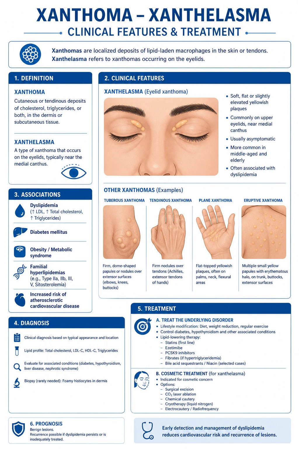
Xanthelasma palpebrarum
Tendinous xanthoma
Tuberous xanthoma
Eruptive xanthoma
Plane xanthoma
Diffuse plane xanthomatosis
Mapping type to lipoprotein: the Fredrickson phenotypes
The Fredrickson (WHO) phenotyping system names the elevated lipoprotein class, and pairing it with the xanthoma type is the single most reliable way to predict the metabolic defect at the bedside. The phenotypes relevant to cutaneous xanthoma are types IIa, IIb, III, IV and V; type I (pure chylomicronaemia, lipoprotein lipase deficiency) causes eruptive xanthomas and lipaemia retinalis in infancy but is rare. [1]
Type IIa
Type IIb
Type III (dysbetalipoproteinaemia)
Type IV
Type V
The examinable pattern is that IIa and III give the firm, indolent nodules (tendon and tuberous), while IV and V give the explosive eruptive crops, and III alone gives the palmar crease sign. Reading the lipid panel for which class is raised lets you name the phenotype and predict the complication — coronary disease for IIa and III, pancreatitis for V. [1]
Three further entities are worth separating because they are not classic lipid xanthomas yet appear in the differential: xanthoma disseminatum (a non-Langerhans cell histiocytosis of flexural skin and mucosa, classically with diabetes insipidus, and with normal lipids); verruciform xanthoma (a solitary, wart-like lesion of the oral or genital mucosa, also normolipidaemic); and juvenile xanthogranuloma (a benign self-limiting histiocytic lesion of infancy). Mentioning these in a viva demonstrates that you understand the boundary between metabolic and histiocytic lesions. [1]
Epidemiology and risk factors
Xanthelasma palpebrarum is by far the commonest type and affects roughly 1 per cent of adults, with a predilection for middle-aged women. Its epidemiology is dominated by one counter-intuitive fact: only about half of xanthelasma patients have a measurable dyslipidaemia, which is why a single normal lipid panel never closes the file — cardiovascular risk must still be assessed.[1][8]
Tendinous and tuberous xanthomas point to familial hypercholesterolaemia (FH), an autosomal dominant disorder of LDL clearance carried by about 1 in 250 people in its heterozygous form. Tendon xanthomas are essentially absent before the age of ten years, appear progressively thereafter, and are among the strongest single clinical markers of premature coronary disease that a dermatologist can find.[2]
Eruptive xanthomas arise when triglyceride-rich lipoproteins accumulate rapidly. The principal precipitants are uncontrolled diabetes mellitus, alcohol excess, obesity, exogenous oestrogens, systemic retinoids, glucocorticoids, bile-acid sequestrants, and the familial hypertriglyceridaemias. Recognising the precipitant is therapeutic, because removing it can resolve the lesions within weeks.[6]
Secondary causes of dyslipidaemia must always be excluded regardless of xanthoma type, because they are reversible and they change management. The search list is short and worth memorising. [1]
OFTEN
Because excluding a secondary cause changes management, it is worth holding the commonest culprits with the lipid pattern each produces. [1]
Hypothyroidism
Nephrotic syndrome
Cholestasis / PBC
Chronic kidney disease
Uncontrolled diabetes
Anorexia nervosa / refeeding
Drugs
The clinical rule that follows is that a xanthoma with a clear secondary cause is managed by treating the cause first, and only adding lipid-lowering therapy if the lipid abnormality persists. A patient whose eruptive xanthoma is driven by alcohol will often see the lesions resolve with abstinence and a fibrate; a patient whose plane xanthoma reflects biliary obstruction needs the obstruction addressed, not merely a statin. [1]
Pathophysiology
The shared mechanism is the conversion of a tissue macrophage into a foam cell by uptake of lipid that it cannot off-load. The cascade begins when circulating lipoproteins — LDL, VLDL remnants, chylomicron remnants, or lipoprotein-X — extravasate through dermal or tendinous capillaries. Once in the tissue, the lipid is engulfed by resident macrophages through scavenger receptors (notably SR-A and CD36). Critically, unlike the regulated LDL receptor, scavenger receptor uptake is not feedback-inhibited: the macrophage keeps ingesting lipid long after it should stop, and the accumulated cholesterol esters distend its cytoplasm into the clear, vacuolated foam cell.[1]
The type of lipoprotein in excess determines the morphology and tempo. In hypercholesterolaemia, insoluble LDL accumulates slowly and persistently in tendons and extensor skin, producing the firm, deep, indolent nodules of tendinous and tuberous xanthoma. In severe hypertriglyceridaemia, the much larger triglyceride-rich chylomicrons and VLDL leak rapidly through capillary walls and flood the dermis, generating the explosive, erythematous-haloed crops of eruptive xanthoma within days — a different speed and a different biology. In type III dysbetalipoproteinaemia, the defective remnant clearance (driven by the APOE E2/E2 genotype) deposits remnant particles preferentially in the palmar creases and tuberous sites.[2]
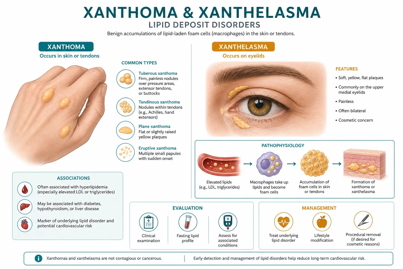
In diffuse plane xanthomatosis the mechanism is different again: a circulating monoclonal immunoglobulin complexes with lipoproteins, and the resulting immune complex deposits in the dermis, generating widespread flat plaques in a patient who may have entirely normal baseline lipids. This is why the lesion mandates a search for a paraprotein rather than a lipid panel alone. [1]
A useful clinical corollary is that the foam cell is the final common pathway, but the upstream defect determines everything else — the site, the speed, the associated systemic risk, and, crucially, whether treating the lipids will make the lesion disappear. Eruptive and tuberous lesions tend to regress with lipid control; tendinous xanthomas shrink only slowly; and xanthelasma, once formed, frequently persist despite statins and require cosmetic removal. [1]
Clinical presentation
Because each type has a distinct morphology, site and tempo, the clinical presentation is best taken type by type. The general principles are that xanthomas are painless (except eruptive lesions, which can be tender), non-pruritic, and yellow-orange because of their lipid content, and that their distribution is the single most useful diagnostic clue. [1]
Xanthelasma palpebrarum presents as soft, yellowish, flat or slightly raised plaques on the eyelids, most often beginning at the medial canthus and becoming bilateral over time. They grow slowly, cause no symptoms beyond cosmetic distress, and are the commonest reason a patient presents. Their importance is systemic: roughly half of affected patients have a dyslipidaemia, and even the normolipidaemic half carry a small but real increase in cardiovascular risk that must be addressed.[1][8]
Tendinous xanthoma presents as firm, deep, subcutaneous nodules that are firmly tethered to the tendon but the overlying skin moves freely. The Achilles tendon is the classic site, followed by the extensor tendons of the hands and feet and the patellar tendon. They are painless, grow over years, and their presence is pathognomonic for familial hypercholesterolaemia; an Achilles tendon that feels thickened, irregular or nodular in a patient under fifty should trigger a lipid profile and genetic consideration.[2]
Tuberous xanthoma presents as firm, yellow-orange nodules on the extensor surfaces — elbows, knees, buttocks, and the backs of the hands. They are slower and more superficial than tendinous lesions and are associated with hypercholesterolaemia (Fredrickson IIa) and, occasionally, type III dysbetalipoproteinaemia. [1]
Eruptive xanthoma presents with the most dramatic tempo: sudden crops of small (2 to 5 mm), firm, yellow papules surrounded by an erythematous halo, appearing over days on the buttocks, extensor arms, thighs and back. The lesions may be tender or pruritic and classically resolve rapidly once the triglycerides are controlled, sometimes leaving a small hyperpigmented macule. Their appearance is a warning of severe hypertriglyceridaemia with a real risk of acute pancreatitis, so a patient with eruptive xanthoma and abdominal pain is a medical emergency.[6][7]
Plane xanthoma of the palmar creases (xanthoma striatum palmaris) produces a characteristic orange-yellow discolouration of the palmar creases that is pathognomonic for type III dysbetalipoproteinaemia (dysbetalipoproteinaemia, APOE E2/E2), in which both cholesterol and triglycerides are elevated. Plane xanthomas also occur in biliary obstruction — particularly primary biliary cholangitis — where lipoprotein-X accumulates, and in this setting the palms are typically spared. [1]
Diffuse plane xanthomatosis presents as widespread, flat yellow-orange plaques over the face, neck, trunk and intertriginous flexures. The patient may have normal lipids, because the deposition is driven by a circulating paraprotein complexing with lipoproteins; the lesion is therefore a dermatological red flag for multiple myeloma or a monoclonal gammopathy. [1]
Atypical presentations
A handful of inherited and syndromic presentations are deliberately tested because they sit at the boundary of dermatology and metabolic medicine. In cerebrotendinous xanthomatosis, a defect in bile-acid synthesis causes cholestanol (not cholesterol) to accumulate, producing tendon xanthomas together with progressive ataxia, dementia, peripheral neuropathy, cataracts and premature atherosclerosis — and the specific treatment is chenodeoxycholic acid, which the examiner will expect you to name. In sitosterolaemia (ABCG5/ABCG8 transporter defect), plant sterols accumulate and mimic FH with tendon and tuberous xanthomas, but with the added clue of haemolytic anaemia and characteristic stomatocytes; the treatment is ezetimibe plus a low-plant-sterol diet.[9] In severe hypertriglyceridaemia, lipaemia retinalis — a milky, salmon-pink appearance of retinal vessels — is a striking bedside clue visible on fundoscopy.
Differential diagnosis
The differential depends on the lesion type, and each must be distinguished on morphology, site, tempo and — most decisively — the lipid profile. [1]
Xanthelasma vs mimics
Tendon xanthoma vs mimics
Tuberous xanthoma vs mimics
Eruptive xanthoma vs mimics
The decisive discriminator in every case is the fasting lipid profile, supported by histology where doubt remains. A lipid panel that is entirely normal should prompt the paraproteinaemia work-up for diffuse plane xanthoma and the consideration of xanthoma disseminatum or verruciform xanthoma for the mucosal and flexural forms. [1]
Clinical and bedside assessment
The focused examination of a patient with a suspected xanthoma is short but systematic, and it deliberately hunts for the systemic associations that determine risk. Begin with the skin, then move to the tendons, the eyes, and the cardiovascular system. [1]
Inspect the eyelids (medial canthus especially), the extensor surfaces (elbows, knees, knuckles), the buttocks and thighs (for eruptive crops), the palmar creases (for the orange line of xanthoma striatum palmaris), and the intertriginous flexures. Palpate the Achilles tendon and the extensor tendons of the hands and feet for nodularity, thickening and symmetry: an Achilles tendon that feels irregular and thickened is the single most reliable bedside sign of FH. Where available, Achilles tendon thickness can be measured by ultrasound or lateral x-ray; a thickness over 9 mm in an adult is strongly suggestive of FH, with normal values generally under 5.6 mm.[2]
Examine the eyes for corneal arcus — when present in a patient under forty-five it supports a diagnosis of dyslipidaemia, although an arcus senilis in the elderly is incidental — and perform fundoscopy to look for lipaemia retinalis in suspected severe hypertriglyceridaemia. Complete the assessment with a focused cardiovascular and metabolic examination: blood pressure, waist circumference, body-mass index, signs of chronic liver disease and hypothyroidism, and a search for peripheral arterial disease. A careful family history of premature coronary disease or sudden death is part of the examination, not an optional extra, because it drives cascade screening in FH. [1]
[1]Investigations
Investigation has two goals: to characterise the lipid disorder and to find any secondary cause. The fasting lipid panel is the cornerstone and is mandatory in every patient with a xanthoma of any type; the remaining tests are selected by the clinical type and the differential. [1]
Mandatory in all patients
A fasting lipid profile measuring total cholesterol, LDL-C, HDL-C and triglycerides is the single most informative test. The pattern of abnormality points to the underlying lipoprotein defect: isolated high LDL suggests familial hypercholesterolaemia (IIa); a combined rise of cholesterol and triglycerides suggests type IIb or, especially with palmar involvement, type III; and an isolated, often very high triglyceride level suggests types IV and V. Fasting glucose and HbA1c screen for diabetes, which is the commonest driver of eruptive xanthoma. Thyroid-stimulating hormone (TSH) excludes hypothyroidism, a classic and reversible cause of secondary hypercholesterolaemia. Urea, electrolytes and eGFR assess renal function, while urinalysis for protein screens for the nephrotic syndrome — another secondary cause of marked hyperlipidaemia. Liver function tests exclude cholestasis and the lipoprotein-X of primary biliary cholangitis.[1]
Targeted to the clinical type
For suspected familial hypercholesterolaemia, apply a validated set of clinical criteria — the Dutch Lipid Clinic Network Criteria internationally, or the Simon Broome criteria in the United Kingdom — and arrange genetic testing for pathogenic variants in LDLR, APOB and PCSK9. Lipoprotein(a) should be measured, as it is an independent cardiovascular risk enhancer and can run with FH. For type III dysbetalipoproteinaemia, APOE genotyping confirms the E2/E2 homozygosity, and lipoprotein electrophoresis shows the broad beta band of remnant particles. For diffuse plane xanthomatosis, a serum protein electrophoresis (SPEP), serum free light chains, urine Bence-Jones protein, serum calcium and renal function are mandatory to exclude a monoclonal gammopathy or multiple myeloma. For eruptive xanthoma with abdominal pain, check serum amylase and lipase to detect acute pancreatitis.[6]
Named diagnostic criteria for familial hypercholesterolaemia
Because FH is common, under-diagnosed, and amenable to cascade screening, two named clinical scoring systems are used to convert a lipid panel plus clinical signs into a diagnostic probability. Both reward the tendon xanthoma heavily, which is why a dermatologist's finding is pivotal to the diagnosis. [1]
Dutch Lipid Clinic Network Criteria (DLCNC) scores family history, personal history of premature vascular disease, physical signs and untreated LDL-C, with a bonus for a confirmed causative mutation. The weighted points combine into a diagnostic category: [1]
- Family history: first-degree relative with premature (men under 55, women under 60) coronary or vascular disease, or known high LDL-C — add 1 point; first-degree relative with tendon xanthoma or arcus, or a child under 18 with high LDL-C — add 2 points (family total capped).
- Clinical history: patient with premature coronary disease — add 2; premature cerebral or peripheral vascular disease — add 1.
- Examination: tendon xanthoma — add 6; arcus cornealis before age 45 — add 4.
- Untreated LDL-C: at or above 8.5 mmol/L — add 8; 6.5 to 8.4 — add 5; 5.0 to 6.4 — add 3; 4.0 to 4.9 — add 1.
- Genetic: a functional mutation in LDLR, APOB or PCSK9 — add 8. [1]
The interpretation the examiner expects: a total above 8 is "definite" FH, 6 to 8 is "probable", 3 to 5 is "possible", and under 3 is "unlikely". The tendon xanthoma (6 points) plus a high LDL-C can therefore secure a "probable" diagnosis even before genetic testing, which is why the bedside finding matters. [1]
Simon Broome (UK) distinguishes definite from possible FH. Definite FH requires a raised lipid level — total cholesterol over 7.5 mmol/L or LDL-C over 4.9 mmol/L in an adult (over 6.7 / over 4.0 in a child under 16) — plus either tendon xanthoma in the patient or a relative, or a confirmed DNA mutation. Possible FH meets the same lipid threshold plus one of: a family history of myocardial infarction under 50 (men) or under 60 (women) in a second-degree relative, a family history of total cholesterol over 7.5 mmol/L, or a child under 16 with total cholesterol over 6.7 mmol/L. In both systems, identifying one affected individual obliges cascade screening of the family, which is the public-health payoff of an accurate clinical diagnosis.[2][5]
Adjuncts
Achilles tendon ultrasound or lateral radiograph documents tendon xanthomas objectively and supports a DLCNC diagnosis when genetic testing is unavailable. Lipoprotein electrophoresis or ultracentrifugation resolves ambiguous mixed dyslipidaemias. Skin biopsy is reserved for cases where the diagnosis is genuinely in doubt: the finding of dermal foam cells confirms a xanthoma, but histology cannot distinguish the lipid types and so it does not replace the lipid panel. [1]
Management — the time-critical scenario
The only genuinely time-critical situation in the xanthoma family is eruptive xanthoma complicated by acute pancreatitis from chylomicronaemia, which is a medical emergency. The skin lesions are the warning sign; the threat is the inflamed pancreas behind them. [1]
Recognise the danger: eruptive xanthoma plus severe abdominal pain in a hypertriglyceridaemic, often diabetic, patient — check serum amylase, lipase and a fasting triglyceride urgently.
Admit and resuscitate: nil by mouth, aggressive intravenous crystalloid, opioid analgesia, and correction of any electrolyte or glucose disturbance; manage in a high-dependency setting if severe.
Lower triglycerides fast: an intravenous insulin infusion drives lipoprotein lipase and accelerates triglyceride clearance, especially when hyperglycaemia coexists; continue until the triglyceride level falls and the patient tolerates oral intake.
Consider apheresis: therapeutic plasma exchange is reserved for refractory severe hypertriglyceridaemic pancreatitis, typically with triglycerides over 40 mmol/L and failing maximal medical therapy, in a centre with the capability.
Remove triggers and plan transition: stop retinoids, oestrogens and unnecessary glucocorticoids; enforce alcohol abstinence; achieve glycaemic control; and start oral lipid-lowering therapy (fibrate plus omega-3) for the long term.
The skin lesions themselves need no acute treatment — they will resolve as the triglycerides fall, usually within weeks.[7]
Management — definitive and stepwise
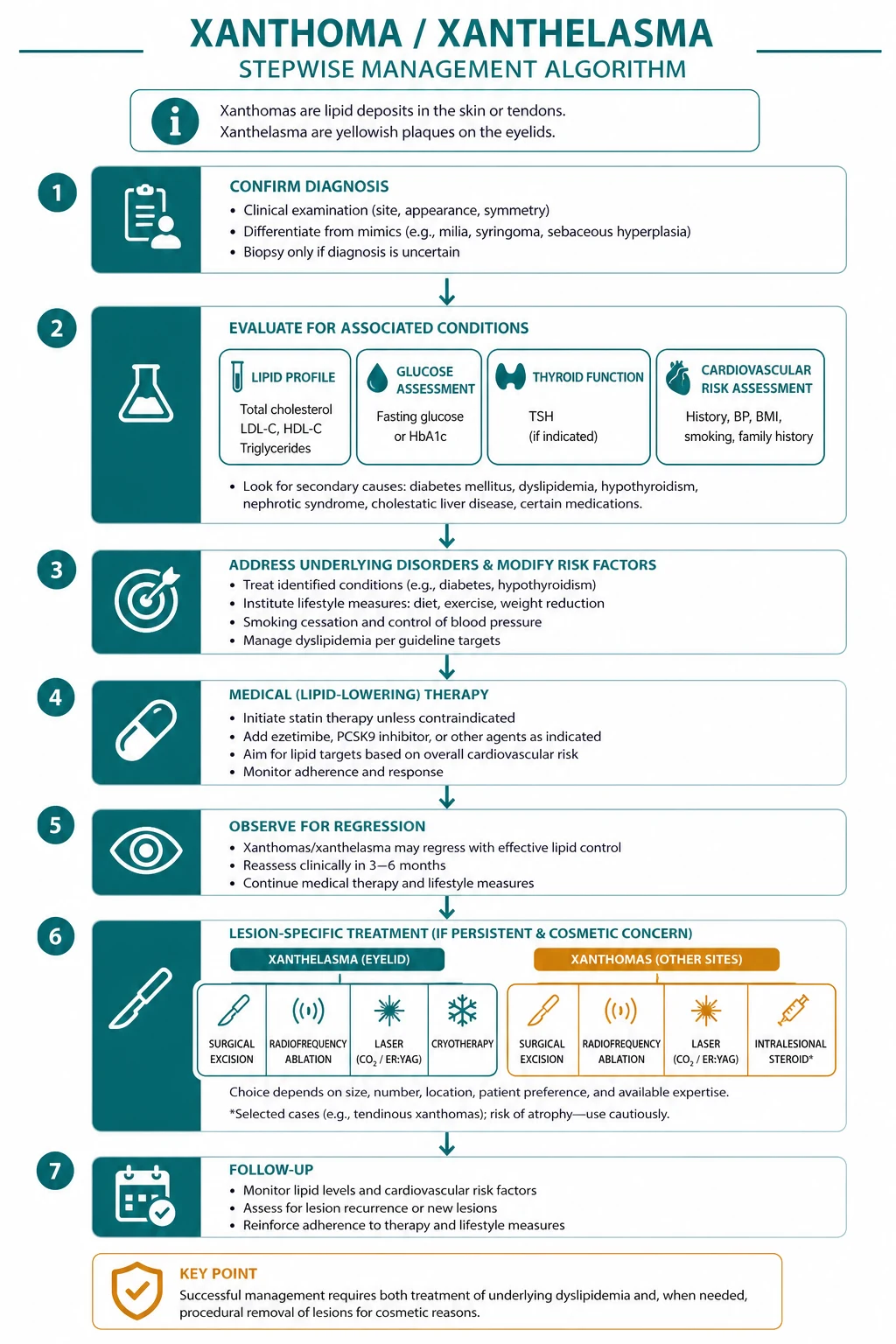
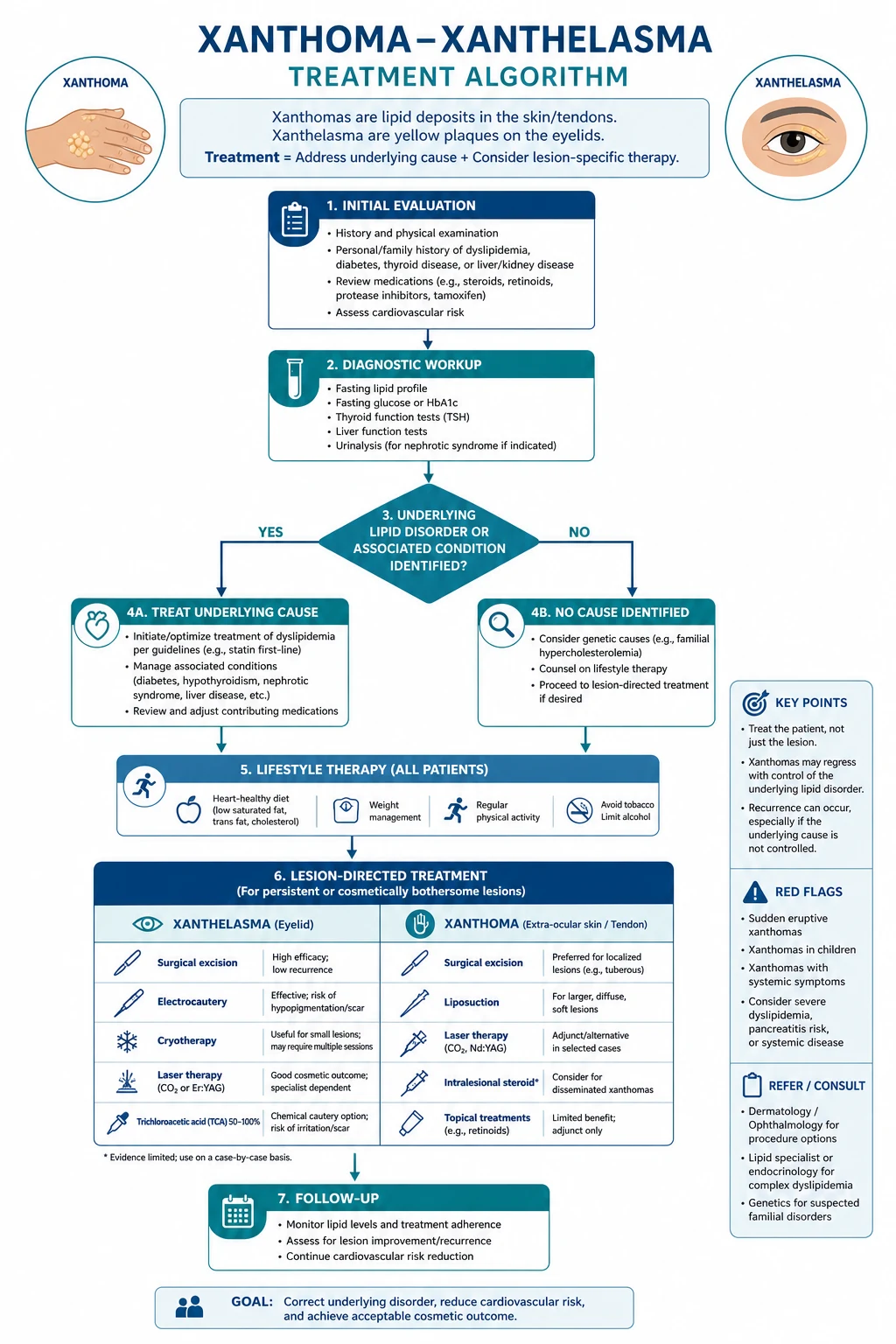
Definitive management has two parallel strands that must both be pursued: treat the underlying dyslipidaemia to prevent cardiovascular disease and pancreatitis, and remove or ablate the lesion cosmetically when it causes distress (principally xanthelasma). The cosmetic strand is never a substitute for the metabolic strand — a patient whose xanthelasma is excised but whose familial hypercholesterolaemia is left untreated has been failed. [1]
Treating the underlying dyslipidaemia
For heterozygous familial hypercholesterolaemia, the backbone is a high-intensity statin — atorvastatin 80 mg once daily, or rosuvastatin 20 to 40 mg once daily, by mouth — titrated to achieve a large LDL-C reduction, combined with ezetimibe 10 mg once daily. If the LDL-C remains above target, a PCSK9 inhibitor (alirocumab or evolocumab, subcutaneous) is added. Crucially, the diagnosis of FH obliges cascade screening of first-degree relatives, because each affected first-degree relative has a one in two chance of carrying the same mutation and benefits from early treatment.[5][10]
For homozygous FH, which presents in childhood with cutaneous and tendon xanthomas and aggressive aortic and coronary disease, management is specialist and intensive: a high-dose statin plus ezetimibe, a PCSK9 inhibitor, the microsomal triglyceride transfer protein inhibitor lomitapide, and in selected patients LDL apheresis; liver transplantation is considered for the most refractory cases.[5]
For severe hypertriglyceridaemia, the lipid-lowering ladder is a fibrate (fenofibrate 200 mg once daily, or gemfibrozil) with omega-3 ethyl esters 2 to 4 g daily, supplemented by nicotinic acid where tolerated, and — most importantly — removal of the precipitant: strict glycaemic control, alcohol cessation, a very-low-fat diet, and withdrawal of oestrogens or retinoids. Type III dysbetalipoproteinaemia responds well to a statin-fibrate combination plus lifestyle measures. [1]
Cosmetic treatment for xanthelasma
Xanthelasma, once established, frequently persists despite statins and is removed for cosmetic reasons. No single modality is clearly superior, and the recurrence rate after any treatment is roughly 40 per cent — a figure the examiner rewards and the patient must be warned about.[1][4]
Surgical excision
Laser ablation
Trichloroacetic acid (TCA) peeling
Expectant / lipid-lowering
A practical point of technique: treat the underlying dyslipidaemia before or alongside cosmetic removal, because untreated hyperlipidaemia is a contributor to recurrence, and the patient's overall cardiovascular risk is the more important outcome. A 2024 study and comprehensive review confirm that while multiple modalities are effective, none eliminates the substantial recurrence rate, and combination approaches are increasingly advocated for extensive disease.[3][4]
Specific subtypes and scenarios
A viva will often probe the rarer xanthomatous disorders that mimic the classic lipid types but have distinct mechanisms and management. Holding them in a separate mental drawer prevents mismanagement. [1]
Cerebrotendinous xanthomatosis (CTX) is an autosomal recessive defect in mitochondrial 27-hydroxylase (CYP27A1) that blocks bile-acid synthesis and causes cholestanol to accumulate. The patient develops tendon xanthomas together with progressive neurological disease (ataxia, spasticity, dementia, peripheral neuropathy), juvenile cataracts, and premature atherosclerosis. The specific, disease-modifying treatment is chenodeoxycholic acid, which must be started early to halt neurological decline — a fact that distinguishes it sharply from ordinary FH, where the answer is a statin. [1]
Sitosterolaemia results from mutations in the ABCG5 or ABCG8 transporters, causing intestinal hyperabsorption and impaired biliary excretion of plant sterols. Clinically it mimics FH with tendon and tuberous xanthomas and a high LDL-C, but the telling clues are haemolytic anaemia (with stomatocytes), characteristic xanthomas in childhood, and a strong response to dietary plant-sterol restriction. The cornerstone treatment is ezetimibe, which blocks sterol absorption, plus a low-plant-sterol diet; statins are relatively ineffective.[9]
Xanthoma disseminatum is a rare non-Langerhans cell histiocytosis of children and young adults that produces yellow-brown papules and plaques in the flexures, eyelids and mucosa, frequently with diabetes insipidus from pituitary infiltration, and with normal lipids. It is not a metabolic xanthoma at all, and it must be distinguished from the lipid types because its management is entirely different (observation, systemic therapy for aggressive disease). [1]
Verruciform xanthoma is a solitary, wart-like, normolipidaemic lesion of the oral or genital mucosa, treated by simple surgical excision and with no implication for the lipid profile. Normolipidaemic xanthelasma is a common practical scenario: even with normal lipids, the lesion should prompt a formal cardiovascular risk assessment, because subclinical atherogenic dyslipidaemia is common and the lesion itself carries a small independent cardiovascular signal.[8]
Complications and pitfalls
[1]The classic pitfalls are four, and each represents a recurring failure mode worth naming. First, treating the cosmetic xanthelasma without investigating the lipids — the patient leaves cosmetically improved but metabolically untreated. Second, recognising FH clinically but failing to arrange cascade screening of relatives, so that affected family members who would benefit from early statin therapy remain undiagnosed. Third, mistaking a diffuse plane xanthoma for a lipid lesion and missing the underlying paraproteinaemia. Fourth, attributing eruptive xanthoma to a primary skin disease and missing the pancreatitis that lies behind it. Each pitfall is avoided by the same discipline: every xanthoma begins with a fasting lipid profile and a search for secondary causes, and the dermatologist who follows that rule will not miss the systemic disease. [1]
Prognosis and disposition
The prognosis of a xanthoma is the prognosis of the disorder behind it, and the two strands must be explained to the patient separately. The skin lesions themselves behave predictably by type: eruptive xanthomas resolve within weeks to months once the triglycerides are controlled; tuberous xanthomas regress slowly over months to years with lipid lowering; tendinous xanthomas shrink only slowly and may persist; and xanthelasma, once formed, usually persist and recur in roughly 40 per cent after cosmetic removal. The patient should be told that clearing the lipids will improve the eruptive and tuberous lesions but may not clear the xanthelasma, which will likely need a separate cosmetic procedure. [1]
The systemic prognosis is determined by the cardiovascular risk. A patient with familial hypercholesterolaemia and tendon xanthomas has a markedly elevated lifetime risk of premature coronary disease that is substantially reduced — though never normalised — by early, sustained, high-intensity statin therapy. A patient with severe hypertriglyceridaemia carries the dual threat of pancreatitis and accelerated atherosclerosis. Diffuse plane xanthomatosis carries the prognosis of its underlying haematological disease. [1]
Disposition follows from this: most patients are managed in the outpatient setting with the dermatologist coordinating care alongside the primary physician and, where relevant, a lipidologist or endocrinologist. Admission is reserved for eruptive xanthoma with pancreatitis or refractory severe hypertriglyceridaemia. Genetic referral is appropriate for confirmed FH to organise cascade testing of the family, and haematology referral is essential for any patient with a paraproteinaemia-driven diffuse plane xanthoma. [1]
Follow-up and monitoring
Once the underlying disorder is identified, follow-up is structured around lipid targets and lesion behaviour. For familial hypercholesterolaemia, the LDL-C should be rechecked at 4 to 12 weeks after any change in therapy and titrated toward the regional goal (a greater than 50 per cent reduction in the UK; an LDL-C under 1.4 mmol/L in very-high-risk patients under European guidance). Lipid-lowering therapy is lifelong, and adherence — together with attention to all modifiable cardiovascular risk factors (blood pressure, smoking, glycaemic control, weight) — matters more than the precise drug chosen. For severe hypertriglyceridaemia, triglycerides should be monitored until they fall below the pancreatitis-risk zone (generally below 10 mmol/L), with reinforcement of alcohol abstinence and dietary fat restriction. The skin lesions themselves are a useful biofeedback signal: eruptive and tuberous lesions that recur or fail to regress indicate poor lipid control, which should prompt a review of adherence and secondary causes. [1]
The safety-net for every patient with a xanthoma is the cardiovascular system. A baseline 12-lead ECG, consideration of coronary artery calcium scoring in selected intermediate-risk patients, and structured cardiovascular risk assessment are appropriate at diagnosis. Patients with FH should carry the diagnosis forward into every future clinical encounter, because it changes the threshold for investigating cardiac symptoms and the aggressiveness of perioperative and antihypertensive management. The dermatologist who diagnoses a xanthoma has not merely described a skin lesion — they have opened a lifelong cardiovascular risk-reduction pathway. [1]
Special populations
In children, the appearance of a xanthoma is an alarm bell. Familial hypercholesterolaemia can be screened from as early as two years of age when a parent is affected, and a statin is typically initiated from age 8 to 10 under specialist guidance. Cerebrotendinous xanthomatosis and sitosterolaemia both present in childhood with tendon xanthomas and a high LDL-C, and both demand a specific (non-statin-led) diagnosis because their management differs fundamentally from ordinary FH. Any child with a xanthoma therefore warrants specialist metabolic referral rather than simple cosmetic reassurance. [1]
In pregnancy, the management of any pre-existing dyslipidaemia is complicated by the fact that statins are contraindicated because of teratogenic concern. Pregnancy itself, with its physiological hypertriglyceridaemia amplified by oestrogen, can precipitate eruptive xanthoma and even pancreatitis, particularly in the third trimester in a patient with baseline hypertriglyceridaemia or gestational diabetes; management is conservative, with diet and glycaemic control, and the lesions typically resolve post partum. [1]
In the elderly, xanthelasma is common and often isolated; the priority is to reassess cardiovascular risk and to balance the benefit of cosmetic treatment against the risk of lid complications and scarring. In the immunocompromised or any patient with diffuse plane xanthomatosis, the work-up for an occult monoclonal gammopathy is non-negotiable, because the lesion may precede the diagnosis of myeloma by months or years. [1]
Evidence, guidelines and regional differences
The evidence base for the management of xanthoma-associating dyslipidaemias is mature, and the regional guidelines agree on principles while differing on thresholds and tactics. [1]
European Society of Cardiology / European Atherosclerosis Society (2019) sets explicit LDL-C goals: under 1.4 mmol/L for very-high-risk and under 1.8 mmol/L for high-risk patients, with a greater than 50 per cent reduction from baseline. The EAS Consensus Panel position paper on homozygous FH (2014) drove earlier detection and the systematic use of combination lipid-lowering therapy, LDL apheresis, and liver transplantation in severe disease.[5]
The clearest regional deltas are three. First, the LDL-C target philosophy differs: Europe and the UK specify explicit values or percentage reductions, while the United States traditionally favours intensity-based therapy. Second, the strength of trichloroacetic acid used for xanthelasma ranges from the conventional 50 to 70 per cent up to 80 per cent reported as effective in a 2024 study, with practice varying by operator and setting.[3] Third, the age at which paediatric statin therapy is initiated for FH varies modestly between guidelines, typically from 8 to 10 years, and is deferred to specialist judgement. Where the evidence is weak — for example, the optimal cosmetic modality for xanthelasma and the role of combination apheresis in pancreatitis — the clinician should acknowledge the uncertainty and individualise treatment.
Exam pearls and high-yield minutiae
[1]Quick self-test: which type fits each clue?
- Yellow plaques on the medial canthus of the eyelids in a normolipidaemic woman — xanthelasma palpebrarum.
- Nodular thickening of the Achilles tendon in a man with LDL-C 6.5 mmol/L and a father who had an MI at 42 — tendinous xanthoma / familial hypercholesterolaemia.
- Sudden crops of tender yellow papules on the buttocks of a poorly controlled diabetic with epigastric pain — eruptive xanthoma; exclude pancreatitis.
- Orange discolouration of the palmar creases with raised cholesterol and triglycerides — plane xanthoma / type III dysbetalipoproteinaemia (APOE E2/E2).
- Widespread flat yellow plaques on the face and trunk with a normal lipid panel — diffuse plane xanthomatosis; screen for a paraprotein.
AXES
Red flags and when to escalate
Exam application bank (NEET-PG / INICET)
One-line answer
Xanthomas are cutaneous, tendinous, and subcutaneous deposits of lipid-laden macrophages (foam cells) that act as visible markers of disordered lipoprotein metabolism. Six clinical types are recognised, each mapping to a specific lipoprotein defect: xanthelasma palpebrarum (eyelid plaques, about half normolipidaemic), tendinous xanthoma (Achilles and extensor tendons, pathognomonic for familial hypercholesterolaemia), tuberous xanthoma (elbows and knees, hypercholesterolaemia), eruptive xanthoma (crops of yellow papules in severe hypertriglyceridaemia, with pancreatitis risk), plane xanthoma (palmar creases in type III dysbetalipoproteinaemia), and diffuse plane xanthomatosis (widespread, signalling paraproteinaemia). Histology shows dermal foam cells with or without Touton giant cells. Management is dual: investigate and treat the underlying dyslipidaemia to prevent atherosclerotic card
Worked stems (answer without another resource)
Stem 1 — Classic presentation. Map symptoms to mechanism; name the first investigation and first treatment step with dose/route if drug therapy is standard. [1]
Stem 2 — Unstable / complicated. List red flags that force immediate resuscitation, theatre, ICU, antidote, or reperfusion — and what you do in the first 15 minutes. [1]
Stem 3 — Atypical group. Elderly, pregnancy, child, or immunocompromised: how presentation and thresholds change. [1]
Stem 4 — Differential trap. Name the three closest mimics and one discriminator for each. [1]
Stem 5 — Disposition. Who goes home with safety-netting, who is admitted, who needs HDU/ICU/theatre, and what follow-up is mandatory. [1]
Rapid viva checklist
- Definition + classification
- Pathophysiology chain
- Bedside signs / criteria
- Score with exact components (if any)
- Emergency bundle
- Definitive therapy with doses
- Complications of disease and of treatment
- Special populations
- Guideline/trial name if classic
- Three exam traps
Coverage self-check
If you cannot answer any stem above from this page alone, re-read the matching section — the page is intended to be self-sufficient for final-prof and NEET-PG/INICET questions on Xanthoma and xanthelasma.
[1]The discipline that runs through the whole topic is simple and worth carrying into the exam: every xanthoma is a metabolic signal first and a cosmetic problem second. Read the site for the lipoprotein, confirm with the lipid panel, treat the underlying disorder to prevent cardiovascular disease and pancreatitis, and only then turn to cosmetic removal — accepting that the xanthelasma, at least, will probably recur. [1]
References
- [1]Nair PA, Singhal R Xanthelasma palpebrarum - a brief review Clin Cosmet Investig Dermatol, 2018.PMID 29296091
- [2]Tada H, Nohara A, Kawashiri MA, et al. Impact of Achilles tendon on diagnosis and phenotypes of familial hypercholesterolemia Curr Opin Lipidol, 2025.PMID 40277332
- [3]Sapra S, Tran JV, Gurm H, et al. Treatment of Xanthelasma Palpebrarum Using Trichloroacetic Acid 80 J Clin Aesthet Dermatol, 2024.PMID 39758222
- [4]Yee DA, Zhou AE, Khachemoune A Examining treatment strategies for xanthelasma palpebrarum: a comprehensive literature review of contemporary modalities Arch Dermatol Res, 2024.PMID 38724802
- [5]Cuchel M, Bruckert E, Ginsberg HN, et al. Homozygous familial hypercholesterolaemia: new insights and guidance for clinicians to improve detection and clinical management. A position paper from the Consensus Panel on Familial Hypercholesterolaemia of the European Atherosclerosis Society Eur Heart J, 2014.PMID 25053660
- [6]Shrestha A, Bam PK, Pandit A, et al. Eruptive xanthoma as a warning sign of uncontrolled hypertriglyceridemia presenting with acute pancreatitis and uncontrolled type II diabetes mellitus: A case report Clin Case Rep, 2024.PMID 38799544
- [7]Stark M, Stuart J Eruptive xanthoma in the setting of hypertriglyceridemia and pancreatitis Am J Emerg Med, 2018.PMID 29716801
- [8]Al Aboud AM, Shah SS, Blair K, et al. Xanthelasma Palpebrarum 2026.PMID 30285396
- [9]Tada H, Kojima N, Takamura M, et al. Sitosterolemia Adv Clin Chem, 2022.PMID 36210074
- [10]Nohara A, Tada H, Ogura M, et al. Homozygous Familial Hypercholesterolemia J Atheroscler Thromb, 2021.PMID 33867421