Dermatology · Medicine
Xeroderma pigmentosum
Also known as XP · Xeroderma pigmentosum variant · DeSanctis-Cacchione syndrome · Nucleotide excision repair deficiency
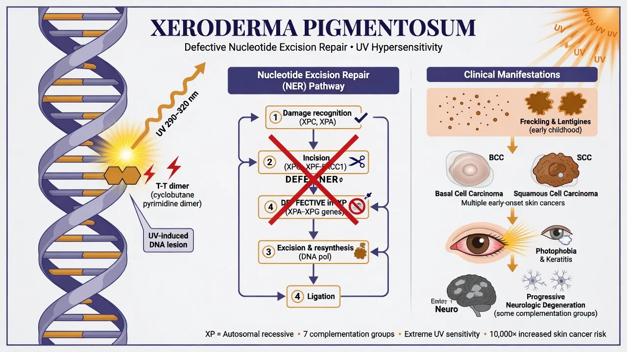
Xeroderma pigmentosum (XP) is an autosomal recessive disorder of nucleotide excision repair (complementation groups XPA–XPG) or of translesion synthesis polymerase eta (XP variant, POLH). Defective repair of UV-induced DNA photoproducts produces extreme photosensitivity, progressive freckling and poikiloderma on exposed skin, ocular surface disease, and skin cancers (BCC, SCC, melanoma) often in the first decade without protection. Selected groups develop progressive neurodegeneration. Management is lifelong rigorous photoprotection, frequent skin surveillance with early cancer surgery, ophthalmology and neurology follow-up, and genetic counselling. Early diagnosis is disease-modifying.
On this page & tools
Your progress
Saved locally on this device.
Exam tags
Red flags
Xeroderma pigmentosum (XP) is the archetype of a DNA-repair genodermatosis and a high-yield board topic because it links molecular pathway knowledge to a concrete, life-saving clinical programme. Without photoprotection, children develop the cutaneous sun damage of elderly outdoor workers within a few years and accumulate basal cell carcinoma, squamous cell carcinoma, and melanoma at rates orders of magnitude above the general population.[1][5] With early diagnosis and rigorous UV avoidance, that trajectory can be transformed.
Classification
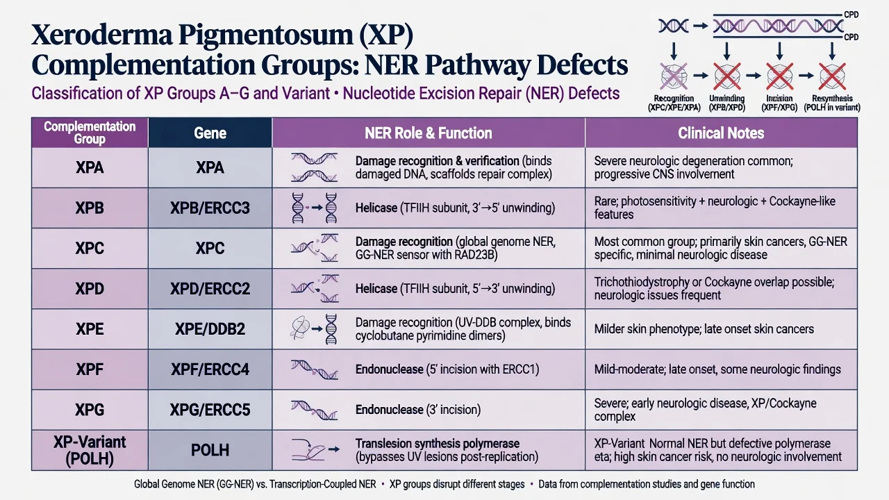
Classic XP is divided into complementation groups XPA through XPG, each corresponding to a gene product in nucleotide excision repair (NER). XP-variant (XP-V) is different: NER is intact, but DNA polymerase eta (POLH) fails at error-free bypass of UV lesions after replication.[1][2][8]
High-yield group tendencies
XPA
XPC
XPD / XPB
XPF / XPG
XPE
XP-V
Some patients meet criteria for XP/Cockayne complex phenotypes; distinguish pure Cockayne syndrome, which classically lacks the freckling-cancer picture of XP.[2][3]
Epidemiology
Incidence estimates are roughly 1 in 250,000 to 1 in 1,000,000 in Europe and the United States, higher in Japan and in some North African and Middle Eastern communities with founder mutations and consanguinity.[1][2][3] Indian series document clinically recognised cohorts with molecular confirmation where testing is available.[10] Sex incidence is equal (autosomal recessive). Ambient UV intensity and adherence to protection dominate modifiable outcome.[5][7]
Pathophysiology
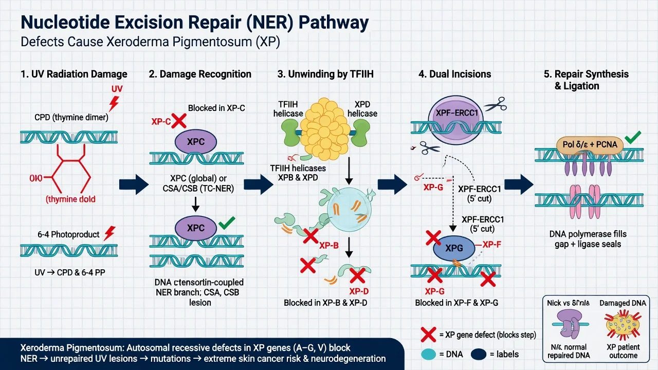
Ultraviolet B radiation generates cyclobutane pyrimidine dimers (CPDs) and 6-4 photoproducts. NER removes these bulky lesions through: [1]
- Damage recognition — global genome NER via XPC (with RAD23B) and UV-DDB (XPE/DDB2); transcription-coupled NER uses CSA/CSB.
- Unwinding — TFIIH helicases XPB and XPD.
- Dual incision — XPF–ERCC1 and XPG.
- Resynthesis and ligation — DNA polymerases and ligase fill the gap.[2][8][9]
When any step fails, photoproducts persist, replication errors accumulate, and the UV mutation signature drives carcinogenesis — now detailed in genomic studies of XP skin cancers.[6] In XP-V, POLH deficiency forces error-prone bypass of unrepaired CPDs. Neurologic degeneration in some groups is attributed to defective repair of endogenous oxidative lesions in post-mitotic neurons rather than UV alone.[5]
Clinical presentation
Cutaneous
Infants may show severe sunburn after minutes of sun. Progressive freckling and solar lentigines appear on exposed skin, often by age two, with xerosis and later poikiloderma (atrophy, telangiectasia, mottled pigment). The contrast between exposed and covered skin is striking.[1][3][4]
Without protection, basal cell carcinoma, squamous cell carcinoma, and melanoma arise in childhood or adolescence — decades earlier than in the general population.[5][6]
Ocular
Photophobia, conjunctivitis, keratitis, lid freckling and tumours, and corneal scarring threaten vision and require routine ophthalmology.[1][3]
Neurologic
A subset — enriched in XPA, XPD, XPG — develops progressive sensorineural deafness, ataxia, areflexia, cognitive decline, historically including DeSanctis-Cacchione-type severe disease. Neurologic progression is not prevented by photoprotection alone.[1][5]
Differential diagnosis
- Cockayne syndrome — photosensitivity, cachectic short stature, neurodegeneration; typically without freckling-associated early skin cancers of XP type.[2]
- Trichothiodystrophy — brittle sulfur-deficient hair, photosensitivity; may share XPB/XPD genetics.
- Bloom syndrome, Rothmund-Thomson — different genomic instability signatures and associated cancers.
- Porphyrias and drug phototoxicity — biochemical or drug clues; lack progressive XP freckling pattern.
- Albinism — pigment synthesis defect with ocular tetrad, not NER failure.[1]
- Mild XP-V may present later with multiple skin cancers and a subtler acute burn history.[2]
Assessment and investigations
Document UV reaction history, freckling map, full skin cancer survey, eye findings, hearing and neuro screen, pedigree and consanguinity.[1][3]
Molecular testing with a multi-gene NER/POLH panel is the modern diagnostic standard. Historical unscheduled DNA synthesis assays are largely research or specialised now.[1][2] Biopsy every suspicious neoplasm. Baseline and serial ophthalmology are mandatory; audiology and neurology follow group-specific risk.[5]
Management
Photoprotection (disease-modifying)
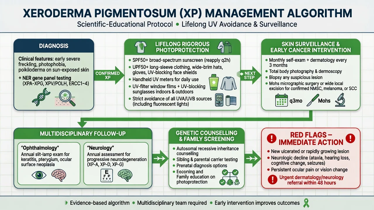
- Broad-spectrum SPF 50+ sunscreen to all exposed skin, reapplied every two hours and after swimming.
- UPF clothing, wide-brim hats, gloves, face shields as needed.
- UV-blocking eyewear indoors and outdoors when fluorescent or daylight UV is relevant.
- Window films, shade planning, and handheld UV meters.
- Avoid peak sun; rethink school and outdoor work exposure.
- Monitor vitamin D because strict avoidance can lower levels.[1][3][7]
Personalised adherence support improves real-world photoprotection behaviour in adults with XP.[7]
Surveillance and cancer treatment
Frequent dermatology review (often every 3 months or denser in high UV or active disease), total-body photography/dermoscopy, and low threshold for biopsy. Treat NMSC and melanoma with standard oncologic principles emphasising early complete excision or Mohs where anatomy demands tissue sparing.[3][4][5]
Multi-disciplinary follow-up
Ophthalmology, neurology (if group risk or symptoms), clinical genetics, psychology, and education/advocacy support. There is no routine approved systemic cure; experimental repair or antioxidant strategies remain investigational.[1][9]
Genetic counselling
Autosomal recessive recurrence risk is 25 percent for each sibling when both parents are carriers. Offer cascade testing, carrier testing for relatives, and prenatal/preimplantation options once variants are known.[1][10]
Complications and prognosis
Multiple primary skin cancers, metastatic disease, ocular surface failure, progressive neurodegeneration, vitamin D deficiency, and psychosocial isolation are the main burdens.[5][9] Historical untreated cohorts had high premature mortality from skin cancer; modern protected cohorts do substantially better on cutaneous endpoints, while neurologic forms still progress.[1][5]
Regional notes
High-UV tropical practice (including India) magnifies cancer risk when protection is incomplete; consanguinity increases case clusters; molecular access varies, but clothing, shade, and sunscreen education must not wait for a gene report.[10][3]
Exam pearls
XP core
- Groups A–G = NER proteins; V = POLH.[2]
- Freckling by age two on exposed skin is a classic clue.[1]
- Distinguish Cockayne (usually no XP-type freckling cancers).[2]
- Photoprotection is not cosmetic — it is the therapy.[7]
- Neurologic XP needs neurology even if skin is perfectly protected.[5]
Exam application bank (NEET-PG / INICET)
One-line answer
Xeroderma pigmentosum (XP) is an autosomal recessive disorder of nucleotide excision repair (complementation groups XPA–XPG) or of translesion synthesis polymerase eta (XP variant, POLH). Defective repair of UV-induced DNA photoproducts produces extreme photosensitivity, progressive freckling and poikiloderma on exposed skin, ocular surface disease, and skin cancers (BCC, SCC, melanoma) often in the first decade without protection. Selected groups develop progressive neurodegeneration. Management is lifelong rigorous photoprotection, frequent skin surveillance with early cancer surgery, ophthalmology and neurology follow-up, and genetic counselling. Early diagnosis is disease-modifying.
Worked stems (answer without another resource)
Stem 1 — Classic presentation. Map symptoms to mechanism; name the first investigation and first treatment step with dose/route if drug therapy is standard. [1]
Stem 2 — Unstable / complicated. List red flags that force immediate resuscitation, theatre, ICU, antidote, or reperfusion — and what you do in the first 15 minutes. [1]
Stem 3 — Atypical group. Elderly, pregnancy, child, or immunocompromised: how presentation and thresholds change. [1]
Stem 4 — Differential trap. Name the three closest mimics and one discriminator for each. [1]
Stem 5 — Disposition. Who goes home with safety-netting, who is admitted, who needs HDU/ICU/theatre, and what follow-up is mandatory. [1]
Rapid viva checklist
- Definition + classification
- Pathophysiology chain
- Bedside signs / criteria
- Score with exact components (if any)
- Emergency bundle
- Definitive therapy with doses
- Complications of disease and of treatment
- Special populations
- Guideline/trial name if classic
- Three exam traps
Coverage self-check
If you cannot answer any stem above from this page alone, re-read the matching section — the page is intended to be self-sufficient for final-prof and NEET-PG/INICET questions on Xeroderma pigmentosum.
Expanded exam teaching (depth pass)
Clinical reasoning
For Xeroderma pigmentosum, examiners test whether you can prioritise life threats, choose the right first test, and give specific therapy (agent, dose, route, timing). Generic phrases without numbers score poorly.
Mechanism → feature map
Build a short chain: cause → pathophysiologic intermediate → clinical feature → complication. Every major symptom in the classic vignette should sit on that chain.
Investigation strategy
- Bedside/first-line tests that change immediate management
- Confirmatory or staging tests
- What a normal result does not exclude
- When not to delay treatment for imaging (unstable patient)
Management ladder
- Resuscitation / ABC / sepsis or haemorrhage bundle as relevant
- Specific antidote / procedure / antimicrobial / reperfusion / surgery
- Supportive care and monitoring targets
- Definitive long-term therapy and secondary prevention
- Disposition and safety-net advice
Special populations
Always prepare one line each for children, pregnancy, elderly, renal/hepatic impairment, and immunocompromised patients when the topic allows.
Pitfalls that fail candidates
- Treating the number not the patient
- Missing pregnancy status when relevant
- Imaging before stabilisation
- Wrong empiric cover or wrong antidote timing
- Incomplete counselling on recurrence, adherence, or red-flag return
Xeroderma pigmentosum (XP) is an autosomal recessive disorder of nucleotide excision repair (complementation groups XPA–XPG) or of translesion synthesis polymerase eta (XP variant, POLH). Defective repair of UV-induced DNA photoproducts produces extreme photosensitivity, progressive freckling and poikiloderma on exposed skin, ocular surface disease, and skin cancers (BCC, SCC, melanoma) often in the first decade without protection. Selected groups develop progressive neurodegeneration. Managemen [1]
Structured revision sheet
Must-know numbers and names
List every score, size threshold, dose, and time window from this topic on a blank page from memory, then check against the sections above.
Three classic MCQ angles
- Most likely diagnosis given a vignette
- Next best step in management
- Most appropriate investigation
Three classic SAQ angles
- Pathophysiology in five steps
- Management algorithm with doses
- Complications and prevention
Clinical station flow
Greet → focused history → targeted exam → investigations → explain diagnosis → emergency care → definitive plan → safety-net / follow-up → answer examiner questions on mechanism and pitfalls.
References
- [1]Adam MP, Bick S, Mirzaa GM, et al. Xeroderma Pigmentosum 1993.PMID 20301571
- [2]Lehmann AR, McGibbon D, Stefanini M. Xeroderma pigmentosum Orphanet J Rare Dis, 2011.PMID 22044607
- [3]Leung AK, Barankin B, Lam JM, et al. Xeroderma pigmentosum: an updated review Drugs Context, 2022.PMID 35520754
- [4]Black JO. Xeroderma Pigmentosum Head Neck Pathol, 2016.PMID 26975629
- [5]Bradford PT, Goldstein AM, Tamura D, et al. Cancer and neurologic degeneration in xeroderma pigmentosum: long term follow-up characterises the role of DNA repair J Med Genet, 2011.PMID 21097776
- [6]Yurchenko AA, Rajabi F, Braz-Petta T, et al. Genomic mutation landscape of skin cancers from DNA repair-deficient xeroderma pigmentosum patients Nat Commun, 2023.PMID 37142601
- [7]Walburn J, Sarkany R, Norton S, et al. A personalized and systematically designed adherence intervention improves photoprotection in adults with xeroderma pigmentosum (XP): results of the XPAND randomized controlled trial Br J Dermatol, 2025.PMID 39401796
- [8]Feltes BC, Bonatto D. Every protagonist has a sidekick: Structural aspects of human xeroderma pigmentosum-binding proteins in nucleotide excision repair Protein Sci, 2021.PMID 34420242
- [9]Lehmann J, Seebode C, Martens MC, et al. Xeroderma Pigmentosum - Facts and Perspectives Anticancer Res, 2018.PMID 29374753
- [10]Tamhankar PM, Iyer SV, Sanghavi S, et al. Clinical profile and mutation analysis of xeroderma pigmentosum in Indian patients Indian J Dermatol Venereol Leprol, 2015.PMID 25566891