emergency-toxicology · emergency-toxicology
Anaphylaxis
Also known as Anaphylactic shock · Anaphylactoid reaction · Allergic emergency · Severe allergic reaction · Adrenaline-requiring allergy · Acute systemic allergic reaction
Anaphylaxis is a severe, life-threatening systemic hypersensitivity reaction that is rapid in onset (minutes to hours) and may cause death. It is a CLINICAL diagnosis — treat on suspicion. FIRST-LINE treatment is INTRAMUSCULAR ADRENALINE 0.5 mg (adult) into the anterolateral thigh, repeat after 5 minutes. Position the patient SUPINE with legs elevated (never sit/stand). Adjuncts (antihistamine, corticosteroid, oxygen, IV fluid) come AFTER adrenaline. Observe for 6 to 12 hours because a BIPHASIC reaction recurs in up to 15 percent of cases. Discharge with TWO adrenaline autoinjectors, an action plan, and an allergy referral.
On this page & tools
Your progress
Saved locally on this device.
Practise this topic
Exam tags
Red flags

Overview & Definition
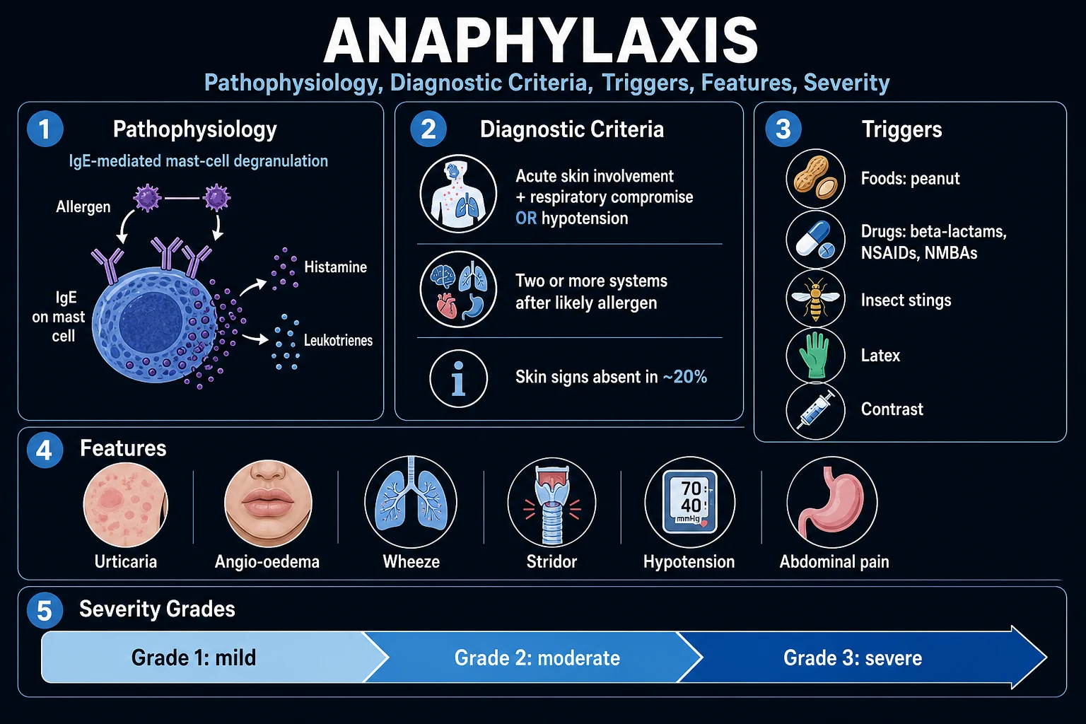
Anaphylaxis is defined by the World Allergy Organization (WAO) and the international consensus (NIAID/FAAN, 2006; EAACI 2021; WAO 2020) as a serious, generalised or systemic hypersensitivity reaction that is rapid in onset (minutes to a few hours) and may cause death — typically by airway obstruction, bronchospasm, or distributive (vasodilatory) shock.[1][3][4]
Three features separate anaphylaxis from a "simple" allergic reaction and decide when to give adrenaline:[4]
- Multisystem — simultaneous involvement of two or more organ systems (skin plus respiratory and/or cardiovascular is the classic pattern).
- Acute onset and rapid progression — typically minutes after exposure, occasionally up to an hour or more for oral triggers.
- Life-threatening — airway compromise, breathing difficulty, or haemodynamic collapse. [1]
The clinical task at the bedside is binary: does this meet the diagnostic criteria? If yes — give IM adrenaline now, do not wait for tests, do not wait for the rash to "develop", do not wait for the doctor. Anaphylaxis is the one diagnosis in medicine where a single IM injection given within seconds saves a life; delay by minutes is the strongest predictor of death.[3]
Two pathophysiological routes to the same syndrome
Historically two overlapping categories were taught: [1]
- Immunologic (IgE-mediated) — the classic "anaphylactic" reaction. Re-exposure to an allergen (food, drug, venom, latex) cross-links allergen-specific IgE on the high-affinity FcεRI receptor of mast cells and basophils, triggering degranulation. Penicillin, peanut, bee venom are archetypes.
- Non-immunologic (non-allergic, formerly "anaphylactoid") — direct mast-cell activation without IgE. Examples: radiocontrast media, opioids, vancomycin (rapid infusion), cold, heat, exercise, physical triggers. Clinically identical; management is identical. The distinction has been dropped from modern guidelines because treatment is the same — call both "anaphylaxis".[3][6]
A third category, immunologic non-IgE (e.g. IgG- or immune-complex-mediated, complement activation by blood products), produces an identical clinical picture.[2]
The single trap that kills patients is delayed adrenaline: the median time to cardiorespiratory arrest from food-induced anaphylaxis is 25 to 35 minutes, from insect venom 10 to 15 minutes, and from intravenous drugs under 5 minutes — minutes that are entirely preventable.[6]
Classification
Anaphylaxis is classified along three axes — mechanism, temporal pattern, and trigger — each of which changes management.[3][6]
By mechanism
- IgE-mediated (immunologic) — food, drug, venom, latex; requires prior sensitisation; reproducible on re-exposure
- Non-immunologic (direct mast-cell activation) — contrast, opioid, vancomycin, exercise, cold/heat; no prior sensitisation
- Idiopathic — no trigger identified after full workup (~20 percent); exclude mastocytosis
By temporal pattern
- Uniphasic — single episode resolving with adrenaline (most cases)
- Biphasic — recurrence 1 to 72 h (typically within 10 h) after apparent recovery, WITHOUT re-exposure (up to 15 percent)
- Protracted / refractory — symptoms persist or recur despite two or more doses of IM adrenaline; needs IV adrenaline infusion and ICU
By trigger
- Drug-induced (beta-lactam, NSAID, neuromuscular blocker, radiocontrast, chemo)
- Food-induced (peanut, tree nut, shellfish, milk, egg, sesame, wheat)
- Venom (bee, wasp, hornet, fire ant)
- Latex; perioperative (multi-trigger); exercise; cofactor-augmented (food + exercise/NSAID/alcohol)
Epidemiology & Risk Factors
Anaphylaxis is common and becoming commoner. Lifetime prevalence is now estimated at 1.6 to 5 percent of the population in high-income countries, with an annual incidence around 50 to 112 per 100 000 person-years and rising, particularly in children and young adults.[3][6] Despite its frequency, case-fatality is low — around 0.03 to 0.3 percent of episodes are fatal (about 0.3 deaths per million population per year) — because most episodes resolve with prompt adrenaline.[6]
Risk factors for a SEVERE or FATAL reaction
The single most useful bedside question is not "is this anaphylaxis?" but "what makes this patient at risk of dying from it?" Three categories determine severity.[1][3]
Risk factors for fatal anaphylaxis
Other contributors: adolescent and young-adult age (risk-taking, delayed autoinjector use), previous severe reaction (severity tends to escalate on re-exposure), allergen dose and route (intravenous triggers are the fastest and most dangerous), exercise and other cofactors (NSAIDs, alcohol, heat, infection, menstruation), aerosolised allergen in closed spaces (e.g. airplane peanut exposure), delayed adrenaline administration, geographic isolation from healthcare, older age and frailty, and limited physiological reserve.[1]
Cofactors ("augmentation factors")
Up to 30 percent of episodes involve a cofactor that converts a tolerable exposure into a life-threatening reaction. The classic triad is food PLUS exercise PLUS an NSAID. Cofactors include exercise, NSAIDs (aspirin, ibuprofen), alcohol, heat or cold, fever or infection, psychological stress, and menstruation. Recognising a cofactor matters because allergen avoidance alone may be insufficient — the patient must also avoid the cofactor.[3]
Pathophysiology
The IgE-mediated cascade
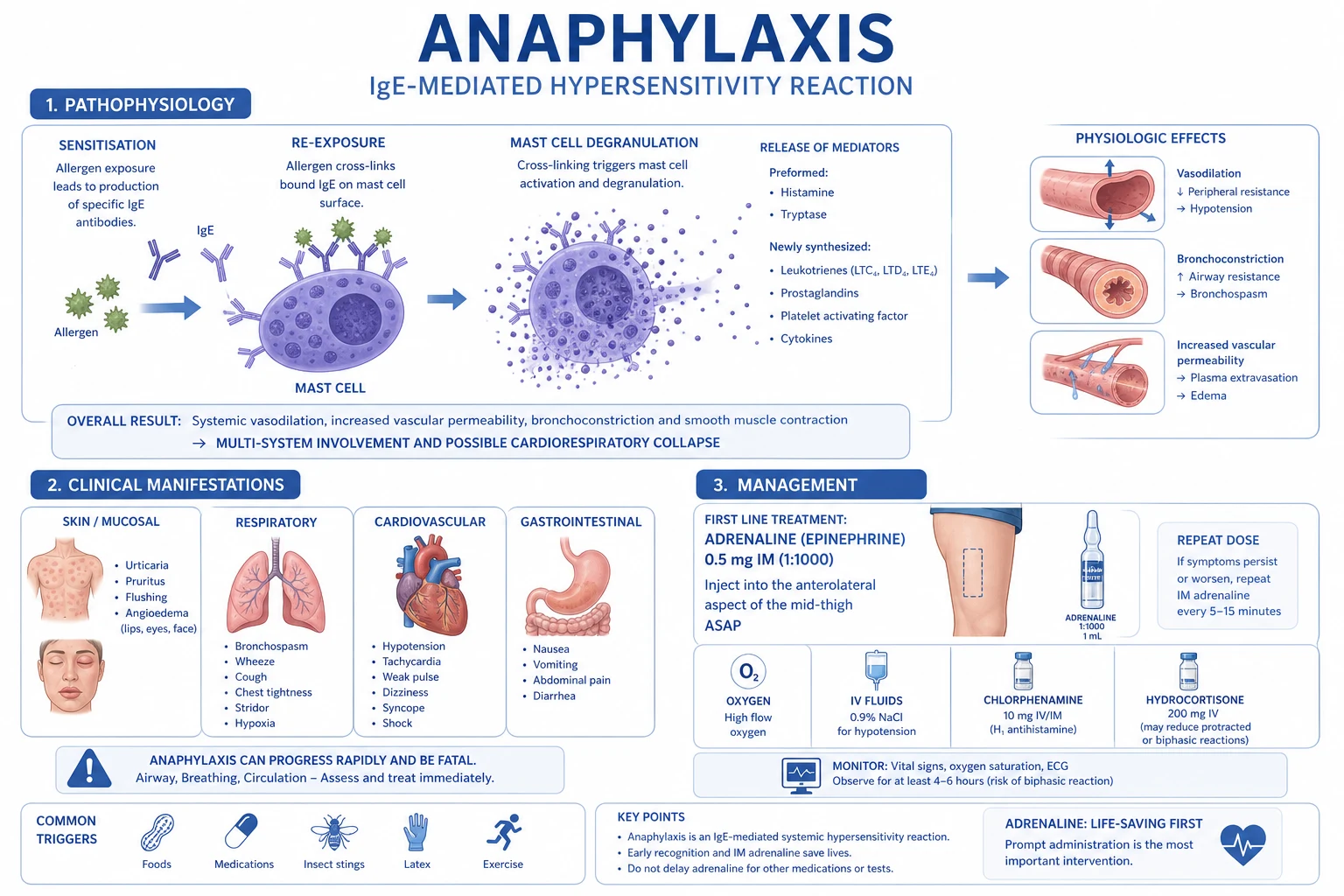
In sensitised individuals, the first exposure to an allergen generates allergen-specific IgE via B-cell class-switching driven by T-helper-2 (Th2) cytokines (IL-4, IL-13). This IgE binds the high-affinity FcεRI receptor on the surface of mast cells (in tissues) and basophils (in circulation), arming the cell. On re-exposure, the multivalent allergen cross-links adjacent IgE-FcεRI complexes, triggering the mast cell to degranulate within seconds and to synthesise de novo lipid mediators over minutes to hours.[1][2]
The signalling cascade is: [1]
- Allergen cross-links IgE on FcεRI → the immunoreceptor tyrosine-based activation motif (ITAM) of the beta and gamma chains is phosphorylated by Lyn (Src-family kinase).
- Syk tyrosine kinase is recruited and activated → phosphorylates LAT and PLCγ.
- PLCγ cleaves PIP2 → IP3 (releases intracellular calcium from ER) + diacylglycerol (activates PKC).
- Calcium and PKC trigger SNARE-mediated fusion of secretory granules with the cell membrane → immediate release of preformed mediators (exocytosis within minutes).
- Activation of cytosolic phospholipase A2 (cPLA2) liberates arachidonic acid from membrane phospholipids → 5-lipoxygenase forms leukotrienes (LTC4, LTD4, LTE4) and cyclooxygenase forms prostaglandin D2 (PGD2). These lipid mediators are synthesised de novo, peaking at 5 to 45 minutes and accounting for the protracted and recurrent features.[2][6]
Mediators and what they do
Preformed granule mediators (immediate)
- HISTAMINE — H1 receptor: vasodilation, increased vascular permeability, bronchoconstriction, pruritus, tachycardia; H2: gastric acid, further vasodilation, tachycardia
- TRYPTASE (beta) — marker of mast-cell activation; rises 1 to 2 h, baseline by 6 to 8 h; not from basophils (mast-cell-specific)
- HEPARIN — inhibits clotting; contributes to the prolonged bleeding time and hypocoagulability seen in severe cases
- CHYMASE, TRYPTASE, CARBOXYPEPTIDASE A3 — local tissue proteolysis, angioedema
Newly synthesised lipid mediators (minutes to hours)
- PLATELET-ACTIVATING FACTOR (PAF) — vasodilation, increased vascular permeability, platelet aggregation, bronchoconstriction; PAF level correlates with severity
- LEUKOTRIENES LTC4/LTD4/LTE4 — slow-reacting substance of anaphylaxis; potent sustained bronchoconstriction, mucosal oedema
- PROSTAGLANDIN D2 (PGD2) — bronchoconstriction, vasodilation, neutrophil chemotaxis
- CYTOKINES (TNF-alpha, IL-6) — late-phase inflammation, fever, contributes to the biphasic reaction
Clinical end-organ effects
- AIRWAY — laryngeal and pharyngeal oedema (stridor, drooling), bronchospasm (wheeze), increased mucus secretion
- CIRCULATION — vasodilation + capillary leak → distributive shock; tachycardia, hypotension, collapse; myocardial ischaemia and arrhythmia in susceptible patients
- GUT — cramping abdominal pain, vomiting, diarrhoea (especially food-induced)
- SKIN — urticaria, angioedema, flushing, pruritus
Why does the patient collapse? — the cardiovascular pathophysiology
The cardiovascular collapse of anaphylaxis is a distributive (vasodilatory) shock compounded by relative hypovolaemia:[2]
- Histamine and PAF cause widespread arteriolar vasodilation (drop in systemic vascular resistance) — the same peripheral vasodilation that causes flushing.
- Increased vascular permeability (capillary leak) shifts up to 35 percent of intravascular volume into the interstitium within minutes, producing relative and absolute hypovolaemia.
- The venous capacitance bed dilates — venous return to the heart falls ("relative empty vena cava syndrome").
- The result is reduced preload, reduced cardiac output, hypotension, and cerebral hypoperfusion → syncope. [1]
This is why positioning matters so much: a patient who suddenly sits up or stands during anaphylaxis can lose the residual venous return, empty the right heart, and arrest within seconds — the "empty vena cava / empty heart syndrome". Lie them flat with legs raised.[3]
The biphasic and protracted reactions
A biphasic reaction is a recurrence of symptoms 1 to 72 hours (typically within 10 hours) after apparent resolution, WITHOUT further allergen exposure. Incidence is up to 5 to 15 percent of episodes (a 2015 systematic review/meta-analysis reported an unweighted pooled rate of 4.6 percent overall but much higher in selected cohorts).[5] Predictors of a biphasic reaction include a severe initial reaction (need for two or more doses of adrenaline, hypotension), delayed or sub-therapeutic first-dose adrenaline, oral drug or unknown trigger, a wide pulse pressure, and a high initial tryptase.[5]
The mechanism is incompletely understood but is thought to involve late-phase recruitment of eosinophils, basophils and neutrophils by cytokines (TNF-alpha, IL-6) released from mast cells, sustained leukotriene and PAF production, and continued antigen absorption from the gut. The biphasic reaction can be milder, equal to, or more severe than the index event.[5]
Coagulation and the bradykinin contribution
Anaphylaxis activates coagulation (prothrombin fragments, D-dimer rise) and fibrinolysis, which can produce a transient procoagulant state and, in severe cases, disseminated intravascular coagulation contributing to collapse. In patients taking an ACE inhibitor, blockade of angiotensin-converting enzyme (which also degrades bradykinin) allows bradykinin accumulation, augmenting vasodilation and angioedema and rendering anaphylaxis more severe and more adrenaline-resistant.[2]
Clinical Presentation
The clinical picture reflects multisystem mediator release. Onset is typically within minutes of exposure (food, drug, venom) but may be delayed by hours after an oral drug or, rarely, exercise-induced episodes. The faster the onset, the more severe the reaction.[1][6]
By system and frequency
| System | Feature | Approximate frequency |
|---|---|---|
| Skin / mucosa | Urticaria, angioedema, flushing, pruritus | 80 to 90 percent |
| Respiratory | Wheeze, stridor, dyspnoea, hoarseness, cough, "lump in throat", hypoxia | 50 to 70 percent |
| Cardiovascular | Hypotension, tachycardia, collapse, syncope, chest tightness, arrhythmia | 30 to 40 percent |
| GI | Cramping abdominal pain, vomiting, diarrhoea (especially food) | 30 to 45 percent |
| Neurological | Sense of impending doom, dizziness, confusion, syncope, headache | common prodrome |
The skin is the commonest organ involved but is NOT a target for adrenaline — its presence or absence during treatment does not indicate whether adrenaline is working. Anaphylaxis can occur WITHOUT skin signs in 10 to 20 percent of cases (especially drug-induced, perioperative, or in children with food allergy) — never wait for a rash.[1][3]
The patient's own description
A sense of impending doom ("I am going to die"), a metallic taste, tingling of the lips or palms, warmth spreading from the chest, palpitations, and itching of the palms, soles, scalp, or genitalia are classical prodromes that precede overt organ involvement by seconds to minutes.[1]
Atypical presentations
- Elderly — comorbidity and medication (beta-blocker, ACE inhibitor, digoxin) attenuate or distort signs; tachycardia may be absent; the only sign may be sudden hypotension or collapse. A myocardial infarction can be precipitated by the catecholamine surge — distinguish from a primary cardiac event.[2]
- Pregnant — uterine cramps and vaginal discharge may herald anaphylaxis; fetal distress manifests as fetal bradycardia; eclampsia and amniotic-fluid embolism are differentials.[3]
- Beta-blocked patient — refractory hypotension and bradycardia; may not respond to adrenaline; give glucagon.[4]
- Mastocytosis (systemic mast-cell disorder) — reactions with a sting or a drug that are severe, recurrent, and often lack skin signs; baseline tryptase is persistently elevated.[3]
- Children — atypical features include sudden behavioural change (becoming clingy, quiet, or irritable), drooling, cyanosis, and collapse; skin features may be missed because children cannot describe itching.[6]
- Idiopathic — no trigger; recurring episodes, often flushing and cardiovascular features; diagnosis of exclusion after full workup including mastocytosis.[3]
Differential Diagnosis
The diagnosis is clinical and based on the NIAID/FAAN criteria, but several conditions mimic anaphylaxis and require different treatment. All should be considered in the collapsed or distressed patient.[1][3]
Vasovagal syncope
- Pallor, diaphoresis, nausea, brief loss of consciousness on standing or with pain/procedure
- Distinguishing: bradycardia (vs tachycardia in anaphylaxis), no urticaria/angioedema, no dyspnoea, normal BP on lying flat, rapid spontaneous recovery
- A common mimic; the pulse and skin usually decide it
Panic attack / vocal cord dysfunction
- Sense of choking, breathlessness, tingling fingers (carpopedal spasm from hyperventilation), normal SpO2, no rash
- Distinguishing: stridor localised to the throat, ability to speak in full sentences between breaths, normal BP/pulse, no angioedema on inspection
Acute severe asthma
- Wheeze, dyspnoea, hypoxia; may coexist with or be triggered by anaphylaxis
- Distinguishing: no urticaria, no angioedema, no hypotension early, peak flow reduced; if BOTH skin and airway involvement treat as anaphylaxis first
Hereditary / acquired angioedema (C1-inhibitor deficiency)
- Angioedema WITHOUT urticaria and WITHOUT hypotension; recurrent; family history; bradykinin-mediated (NOT histamine) — adrenaline does NOT work
- Treat with C1-inhibitor concentrate, icatibant, or fresh-frozen plasma; ACE-inhibitor angioedema is similar — stop the drug
Scombroid poisoning / monosodium glutamate / 'restaurant syndromes'
- Scombroid: histamine from spoiled dark-meat fish (tuna, mackerel) → flushing, palpitations, headache, diarrhoea within minutes of eating; resembles anaphylaxis
- Distinguishing: NO airway compromise; responds to antihistamines; multiple diners affected
Aspiration / foreign body / choking
- Sudden cough, choking, stridor, collapse; often witnessed during eating
- Distinguishing: no rash, no hypotension, normal mast-cell tryptase
Cardiogenic shock / pulmonary embolism / arrhythmia / MI
- Hypotension, syncope, chest pain; can be PRECIPITATED by anaphylaxis in patients with cardiac disease
- Distinguishing: no rash, ECG changes, echocardiographic findings; check troponin if the picture is mixed
Carcinoid syndrome
- Flushing, diarrhoea, bronchospasm, right-heart valvular disease; serotonin-mediated
- Distinguishing: chronic recurrent episodes, elevated urinary 5-HIAA
A bedside glucose must always be checked in the collapsed patient (hypoglycaemia is a great mimic). When a comatose patient with a suspected allergic reaction arrives, also send a paracetamol and salicylate level if overdose is possible.[1]
Clinical & Bedside Assessment
A rapid, structured ABCDE assessment secures the diagnosis and the priorities. Anaphylaxis is a clinical diagnosis — the NIAID/FAAN criteria (2006) are reproduced verbatim below and are the bedside gold standard.[4]
[1]The WAO 2020 revision adds that anaphylaxis is also "highly probable" when an acute onset (minutes to hours) of skin/mucosal features occurs together with at least one of respiratory or cardiovascular features, even if a trigger is not immediately identifiable.[3]
The bedside assessment
- Airway — assess for hoarseness, stridor, drooling, tongue/lip swelling. Laryngeal oedema progresses rapidly — prepare for early intubation if the voice changes or stridor develops.
- Breathing — respiratory rate, oxygen saturation, wheeze, recession, use of accessory muscles, ability to speak in full sentences. Peak flow if the patient can cooperate. Hypoxia is a late and dangerous sign.
- Circulation — pulse (rate and character), BP (both arms if possible), capillary refill, peripheral temperature. Tachycardia and hypotension indicate shock. Bradycardia is a late, pre-terminal sign — cardiac arrest is imminent.
- Disability — GCS, pupils, glucose. A falling GCS signals cerebral hypoperfusion.
- Exposure — fully expose to inspect for urticaria, angioedema, flushing, surgical or dental sites (perioperative), injection sites, insect stings. Look at the oropharynx. [1]
Position
Lie the patient flat with the legs elevated. Do not allow a symptomatic patient to sit or stand up — sudden postural collapse and asystolic cardiac arrest from empty vena cava syndrome is a well-described fatal event. The exception is the patient with severe respiratory distress who cannot lie flat, or the vomiting patient (place in the recovery position); the pregnant patient is placed in the left lateral tilt position.[3]
Investigations
Anaphylaxis is a clinical diagnosis. Investigations are NEVER required before starting treatment, and waiting for results costs lives.[1] Investigations serve three roles: (1) support the diagnosis retrospectively; (2) exclude mimics and co-pathology; (3) identify the trigger after recovery.
Acute investigations (during the reaction)
- Acute serum tryptase — the most useful laboratory test. Mast-cell tryptase (beta) is released during degranulation, peaks at 1 to 2 hours after onset, and returns to baseline by 6 to 8 hours. Take the first sample as soon as possible after treatment has started (ideally within 1 to 2 h of onset), a second at 2 to 4 hours (or within 4 h), and a baseline (convalescent) sample at least 24 hours after the reaction (or at the allergy clinic). A rise to greater than (1.2 × baseline) + 2 micrograms/L supports the diagnosis of anaphylaxis and helps distinguish it from non-mast-cell mimics (vasovagal, panic, mastocytosis alone).[3][2]
- ECG — for any perioperative, drug-induced, or elderly patient; anaphylaxis can precipitate ischaemia and arrhythmia, and adrenaline can cause ST elevation and ventricular ectopy.
- Capillary glucose — exclude hypoglycaemia.
- Arterial or venous blood gas — hypoxia, hypercapnia, metabolic acidosis in severe shock; methaemoglobinaemia if suspected.
- FBC, U&E, lactate — basophilia (paradoxical) and haemoconcentration; lactate tracks shock severity.
- Cardiac troponin — if chest pain, elderly, or perioperative; rule out Kounis syndrome (allergic ACS).
Confirming the trigger (after recovery, NEVER during)
Allergy testing is performed at least 2 to 4 weeks after the episode (skin tests are falsely negative during the refractory period). Referral to an allergy specialist is mandatory for every patient admitted with anaphylaxis.[3]
- Skin-prick test — to suspected foods, drugs (penicillin, neuromuscular blockers), venoms; prick-prick for fresh foods.
- Serum allergen-specific IgE (ImmunoCAP, RAST) — useful for penicillin, venom, selected foods; less sensitive for some drugs.
- Component-resolved diagnostics — distinguishes true allergy (e.g. Ara h 2 for peanut) from cross-reactivity; predicts severity and risk.
- Basophil activation test (CD63/CD203c flow cytometry) — useful for drug allergy (beta-lactam, NSAID, neuromuscular blocker) and venom.
- Drug provocation / oral food challenge — gold standard but only in a specialist setting with full resuscitation facilities.
- Serum tryptase at baseline — a persistently elevated baseline tryptase (over 11.4 micrograms/L or over 20 micrograms/L), especially with a history of severe reactions to insect venom, mandates workup for systemic mastocytosis / clonal mast-cell disorder (KIT D816V mutation, bone marrow biopsy).[3]
Perioperative anaphylaxis — the investigation pathway
Perioperative anaphylaxis is investigated by the EAACI/ESAIC three-step pathway: (1) acute serum tryptase (immediately, 1 to 2 h, and 24 h baseline); (2) skin tests (prick and intradermal) 4 to 6 weeks later to all suspected agents — neuromuscular blockers (most common), antibiotics, chlorhexidine, latex, dyes (patent blue, isosulfan blue), colloids, opioids; (3) specific IgE and basophil activation tests for selected agents. Chlorhexidine is an increasingly recognised cause, found in mouthwash, skin prep, and lubricant gels.[2]
Management — Resuscitation
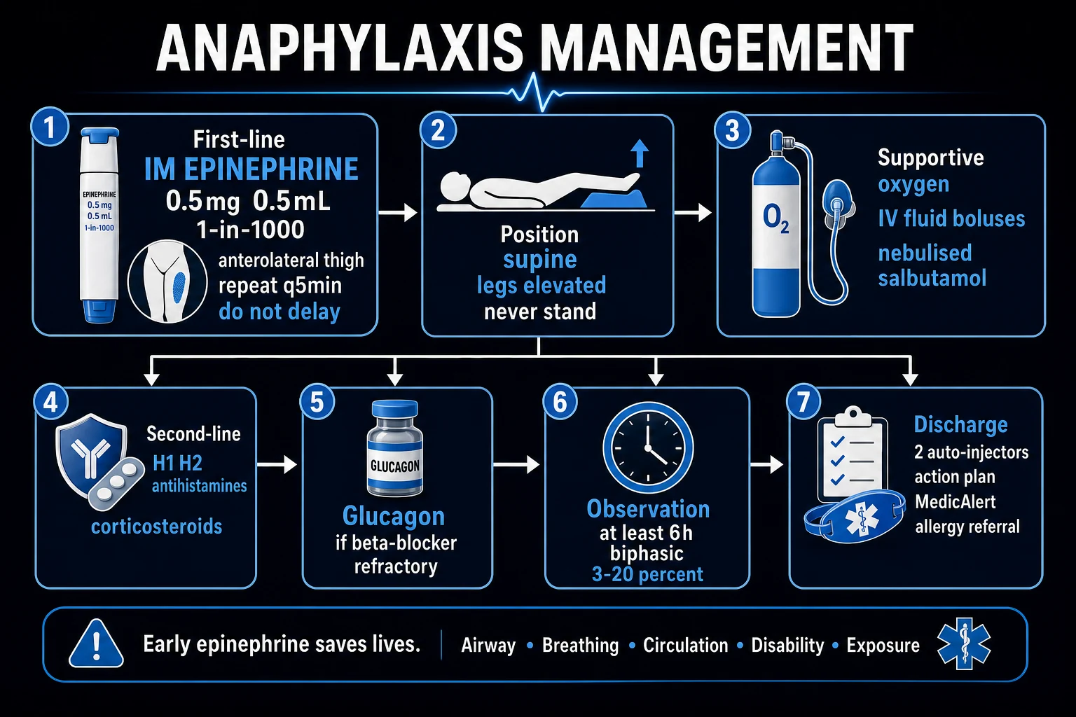
The resuscitation bundle is time-critical and delivered in parallel. The order of priorities is ABCDE; the single most important intervention is IM adrenaline into the anterolateral thigh.[1][3]
- Remove the trigger if possible — stop drug infusion, remove the sting, do not give more of the suspected allergen; in food allergy remove residual food from the mouth.
- Call for help — senior clinician, resuscitation team, anaesthetist.
- Position — lie flat with legs elevated; recovery position if vomiting; left lateral tilt in pregnancy; do not let the symptomatic patient sit or stand.
- Airway — clear, high-suction if angioedema or secretions; nasopharyngeal/oropharyngeal airway if tolerated; early intubation if laryngeal oedema is progressing — call an anaesthetist early; surgical airway (cricothyroidotomy) if intubation is impossible.
- Breathing — high-flow oxygen 15 L/min via a non-rebreather mask; bag-valve-mask ventilation if apnoeic or respirations inadequate; nebulised salbutamol 5 mg if bronchospasm persists after adrenaline.
- Circulation — IM ADRENALINE 0.5 mg (adult) into the anterolateral thigh (vastus lateralis) — this is FIRST and MOST IMPORTANT. Establish large-bore IV access; IV crystalloid bolus 500 to 1000 mL rapidly (adult; 10 mL/kg in children), repeated as guided by response. [1]
Why IM adrenaline first, not IV? Intramuscular adrenaline into the thigh is safer, faster to administer, and pharmacokinetically reliable — peak plasma concentration is reached within about 10 minutes, compared with the much higher risk of ventricular arrhythmia, hypertension, and myocardial ischaemia from a bolus of IV adrenaline. Subcutaneous adrenaline is no longer recommended (unreliable absorption in shock).[3]
Why is adrenaline given BEFORE antihistamines and steroids? Adrenaline acts on alpha-1 (vasoconstriction, increases BP, reduces mucosal oedema), beta-1 (increases heart rate and contractility), and beta-2 (bronchodilation, stabilises mast cells by raising intracellular cAMP and reducing degranulation). Antihistamines and steroids have no role in the acute life-saving moment — they are adjuncts that relieve cutaneous features and may reduce recurrence, but they do not save lives.[1]
Management — Definitive & Stepwise
Adrenaline (epinephrine) — the first-line drug
Adrenaline is a non-selective alpha- and beta-adrenergic agonist. Onset after IM injection is about 5 minutes; duration of action 5 to 10 minutes.[3]
IM adrenaline (1 mg/mL = 1:1000) dosing by age
Repeat at 5-minute intervals if there is no response, up to a maximum of 5 doses. Most patients respond to one or two doses. Failure to respond to two doses prompts escalation to an IV adrenaline infusion (refractory anaphylaxis).[3]
The weight-based equivalent is 0.01 mg/kg IM (max 0.5 mg) for children — useful when age is uncertain or the child is small/large for age. [1]
Adrenaline autoinjectors (discharge)
Adrenaline autoinjector dose bands (EpiPen, Jext, Emerade)
Always prescribe TWO autoinjectors on discharge because a single dose may be insufficient or misfired, and because the recurrence is biphasic. Train the patient and family in the specific device (EpiPen, Jext, Emerade) — they have different administration techniques.[3][6]
IV fluids
The shock of anaphylaxis is distributive with relative hypovolaemia — large volumes of intravascular fluid leak into the tissues. Resuscitate aggressively with crystalloid.[3]
- Adult — 500 to 1000 mL bolus of 0.9 percent sodium chloride or Hartmann's rapidly (over 5 to 10 min), repeated as guided by response; severely shocked patients may need 2 to 4 L in the first hour.
- Child — 10 mL/kg bolus, repeated; use warmed fluid.
- Titrate to BP, capillary refill, mentation, urine output; avoid over-resuscitation in the elderly and in cardiac disease. [1]
Adjuncts (after adrenaline, never before)
- Antihistamine — chlorphenamine 10 mg IV (adult) or oral cetirizine 10 mg; paeds weight-banded (e.g. 2.5 to 5 mg). Relieves cutaneous features (urticaria, pruritus) and rhinitis. Has no effect on airway, breathing, or circulation. H1 + H2 blockade (e.g. add famotidine) is sometimes used but evidence is weak.[1]
- Corticosteroid — hydrocortisone 200 mg IV (adult) or methylprednisolone 1 to 2 mg/kg IV; paeds weight-based. No proven benefit on the acute reaction or on biphasic recurrence in modern trials, but traditionally given, especially for asthma coexisting or severe reactions, and still recommended in UK/EAACI algorithms.[3]
- Bronchodilator — nebulised salbutamol 5 mg (child 2.5 to 5 mg) for refractory bronchospasm after adrenaline; IV salbutamol or IV magnesium sulphate (2 g over 20 min) for severe refractory cases.
- Glucagon — for the beta-blocked patient who is refractory to adrenaline. Glucagon 1 to 5 mg IV over 5 minutes (child 20 to 30 micrograms/kg, max 1 mg), followed by an infusion of 5 to 15 micrograms/min titrated to response. Glucagon bypasses the beta-receptor and directly activates adenylate cyclase — useful when beta-blockade prevents adrenaline from working.[4]
- Atropine — for bradycardia (a late, pre-terminal sign of anaphylactic shock); 0.5 to 1 mg IV (child 20 micrograms/kg).
Refractory anaphylaxis — escalation
Refractory anaphylaxis is defined as failure to respond to two doses of IM adrenaline with ongoing airway, breathing, or circulatory compromise. The Resuscitation Council UK (2021) algorithm escalates as follows:[3]
- Establish definitive airway — early intubation by an experienced operator; surgical airway if impossible.
- IV adrenaline infusion — titrated: adults 0.25 to 0.5 microgram/kg IV bolus cautiously OR an infusion of 0.05 to 0.1 microgram/kg/min (a typical adult starts at 2 to 10 micrograms/min, doubling every 5 min as needed, up to 10 to 25 micrograms/min for profound shock). Prepare by adding 1 mg adrenaline to 100 mL 0.9 percent saline (10 micrograms/mL); a common adult starting rate is 30 to 60 mL/h.
- Glucagon for beta-blocked patients as above.
- Vasopressors — vasopressin or noradrenaline infusion if adrenaline-resistant shock; methylene blue 1 to 2 mg/kg IV is increasingly reported for refractory vasoplegic anaphylaxis (it inhibits NO-cGMP vasodilation) — used investigationally in ICU.
- IV fluid — large-volume crystalloid resuscitation; consider albumin in profound capillary leak.
- Extracorporeal membrane oxygenation (ECMO) — last-resort salvage for refractory cardiac arrest.[2]
Observation after recovery — the biphasic trap
Because a biphasic reaction recurs in up to 15 percent of patients (typically within 10 h, but up to 72 h), every patient must be observed after recovery:[3][5]
[1]The AAAAI 2020 practice parameter suggests 4 to 6 h observation for low-risk patients (longer for severe reactions or those with risk factors for a biphasic reaction).[4]
Discharge bundle
Every patient admitted with anaphylaxis must leave with:[3][6]
- Two adrenaline autoinjectors (correct dose for weight/age) — demonstrated to patient and family.
- A written anaphylaxis action plan (visual, colour-coded for severity).
- Allergen avoidance advice specific to the trigger.
- Antihistamine (cetirizine) and short course of oral corticosteroid (prednisolone) for residual cutaneous and systemic symptoms.
- Urgent allergy specialist referral (within 2 to 4 weeks) for investigation, skin tests, and immunotherapy where indicated.
- MedicAlert identification.
- Education — recognition of early signs, allergen avoidance (including label-reading, restaurant advice), and autoinjector technique with annual refresher.
- Cofactor avoidance (e.g. NSAIDs, alcohol, exercise) where relevant.
- School/workplace notification and a school management plan for children.
- Venom immunotherapy referral for insect-venom anaphylaxis (90 to 98 percent protection). [1]
Specific Subtypes & Scenarios
Drug-induced anaphylaxis
Drugs are the leading cause of fatal anaphylaxis in adults. The commonest culprits: beta-lactam antibiotics (penicillin, cephalosporin), NSAIDs (ibuprofen, diclofenac, aspirin), neuromuscular blockers, radiocontrast media, chemotherapy (platinum, taxanes), monoclonal antibodies, proton-pump inhibitors, chlorhexidine, patent blue dye.[1][2]
Beta-lactam anaphylaxis is IgE-mediated against the beta-lactam ring or its side chains. Cross-reactivity between penicillin and cephalosporin is now under 2 percent for second- and third-generation cephalosporins; aztremonam is safe, and meropenem can usually be given. A documented history mandates skin testing and, if needed, desensitisation or drug provocation in a specialist setting.[1]
NSAID anaphylaxis is often non-allergic (cross-reactive COX-1 inhibition) — symptoms with one NSAID predict symptoms with all non-selective NSAIDs. Selective COX-2 inhibitors (celecoxib) are usually tolerated. A minority have a single-NSAID IgE allergy (tolerate other NSAIDs).[6]
Perioperative anaphylaxis
Incidence 1 in 10 000 to 20 000 anaesthetics; mortality 3 to 9 percent. The leading triggers are, in order: neuromuscular blocking agents (rocuronium, suxamethonium, vecuronium — quaternary ammonium IgE), antibiotics (teicoplanin, beta-lactam, vancomycin), chlorhexidine, latex, dyes (patent blue, isosulfan blue), colloids, opioids.[2]
Latex anaphylaxis has fallen sharply with latex-free policies but remains relevant in spina bifida, multi-surgery, and healthcare-worker patients. Chlorhexidine is the rising culprit — found in skin prep, mouthwash, lubricant gels, and urinary catheters.[2]
Distinguishing features: sudden hypotension, bronchospasm, or cardiac arrest within minutes of induction, often without skin signs (under drapes, the patient is anaesthetised). Management is identical — stop suspected agents, call for help, IM adrenaline, IV fluids, oxygen; exclude other causes of intraoperative collapse (gas embolism, tension pneumothorax, hypovolaemia, high spinal). Send acute and 1 to 2 hour and 24 hour tryptase; refer to a specialist allergy clinic for the EAACI/ESAIC investigation pathway.[2]
Food-induced anaphylaxis
The commonest cause of anaphylaxis in children and a leading cause in adults. Top triggers: peanut, tree nuts (cashew, walnut, hazelnut, almond), shellfish, fish, milk, egg, sesame, wheat, soy, mustard, kiwi. Peanut and tree nut cause the most severe and recurrent reactions. Onset is usually within minutes to 2 hours of ingestion.[6]
Cofactor-augmented food anaphylaxis — exercise, NSAIDs, alcohol, heat, infection, or menstruation at the time of exposure converts a tolerable dose into a life-threatening reaction. Food-dependent exercise-induced anaphylaxis (FDEIA) is the archetype: anaphylaxis occurs only when the trigger food (often wheat, celery, shellfish) is followed by exercise within 4 to 6 hours.[3]
Management is identical. Long-term: strict allergen avoidance, component-resolved diagnostics (e.g. Ara h 2 for peanut) to predict severity, oral immunotherapy (PALFORZIA for peanut) for selected patients, and two autoinjectors.[6]
Venom anaphylaxis
Honeybee, wasp, yellow-jacket, hornet, fire-ant stings cause systemic reactions in 0.3 to 8.9 percent of adults; venom anaphylaxis accounts for a disproportionate share of anaphylaxis deaths (often older men with cardiovascular comorbidity).[1]
Indications for venom immunotherapy (90 to 98 percent protective): any systemic reaction with more than just skin features in adults; any systemic reaction in children; a systemic reaction with a positive diagnostic test. Therapy is given for 3 to 5 years (lifelong in mastocytosis and high-risk patients).[3]
Idiopathic anaphylaxis
No trigger identified after a full workup (about 20 percent of cases). Diagnosis of exclusion; workup must include mastocytosis (baseline tryptase, KIT D816V, bone marrow biopsy if indicated), hidden allergens (spices, uncommon foods), and mast-cell-activating syndromes. Management: autoinjectors, oral prednisolone and antihistamine prophylaxis (in refractory cases — now used less commonly), omalizumab (anti-IgE monoclonal) for refractory cases.[3]
Exercise-induced and physical anaphylaxis
Exercise-induced anaphylaxis — anaphylaxis during or shortly after exercise; food-dependent (wheat, celery, shellfish, alcohol) or non-food-dependent; stop exercise at the first prodromal symptom. Cold-induced anaphylaxis (after cold water immersion) and cholinergic anaphylaxis (with sweating and a rise in core temperature) are rarer physical forms.[6]
Complications & Pitfalls
Biphasic reaction
- Recurrence 1 to 72 h (typically within 10 h) after apparent recovery, without re-exposure; up to 15 percent of cases
- Predictors: severe initial reaction, two or more adrenaline doses, wide pulse pressure, delayed adrenaline, oral drug or unknown trigger, high tryptase
- Prevent by adequate observation (at least 6 h UK; longer if risk factors); discharge with autoinjector and plan
Hypoxic brain injury
- From delayed adrenaline or ventilation; the chief determinant of long-term outcome in survivors
- Preventable by early adrenaline, oxygen, and airway management
Adrenaline-related adverse effects
- Tachycardia, palpitations, tremor, anxiety, headache, hyperglycaemia, hypokalaemia
- Rare: ventricular ectopy, myocardial ischaemia (especially in elderly/cardiac disease), Takotsubo cardiomyopathy
- ST-elevation can occur with normal coronions — adrenaline effect, usually self-limiting; do not withhold life-saving adrenaline for fear of these
Aspiration / pulmonary oedema
- Vomiting and reduced level of consciousness → aspiration; treat in recovery position
- Non-cardiogenic pulmonary oedema from capillary leak and adrenaline — treat with oxygen and PEEP
MI / arrhythmia / stroke (Kounis syndrome)
- Allergic ACS — mast-cell mediator coronary vasospasm or plaque rupture; check troponin and ECG in elderly, perioperative, or chest-pain patients
- Manage both anaphylaxis and the cardiac event
Psychological — anxiety, fear, PTSD
- Common after a life-threatening episode; school refusal in children; food-related avoidance behaviour
- Counselling, allergy support groups, education, and confidence-building with the autoinjector all help
- Treating the rash with antihistamine instead of giving adrenaline for airway/BP. The rash is harmless; airway and BP are not.
- Giving adrenaline subcutaneously — absorption is unreliable in shock; always IM into the thigh.
- Giving adrenaline IV as a first-line bolus — risk of arrhythmia and hypertensive crisis; IV bolus is for cardiac arrest or carefully titrated in refractory shock by experienced staff.
- Failing to repeat adrenaline at 5 minutes — one dose is often insufficient.
- Letting the symptomatic patient sit up to "feel better" — sudden fatal collapse.
- Discharging too early and missing the biphasic reaction.
- Forgetting the beta-blocker history — adrenaline will not work; glucagon is needed.
- Missing the mastocytosis diagnosis — a persistently elevated baseline tryptase is the clue.
- Not prescribing two autoinjectors at discharge.
- Confusing hereditary/acquired angioedema (bradykinin-mediated) with anaphylaxis — adrenaline does NOT work; the patient needs C1-inhibitor concentrate or icatibant. [1]
Prognosis & Disposition
Case-fatality is low (0.03 to 0.3 percent of episodes), but because anaphylaxis is common, the absolute number of deaths is significant. Death is typically from asphyxia (airway obstruction) or cardiovascular collapse. Predictors of death: delayed adrenaline, asthma (especially uncontrolled), cardiovascular disease, mastocytosis, beta-blocker/ACE-inhibitor therapy, intravenous or oral drug triggers, adolescent/young-adult age.[1][6]
Safe discharge criteria (after an uncomplicated reaction responding to one dose of adrenaline, no risk factors):[3]
- Asymptomatic with normal vital signs and peak flow for at least 6 h (UK) or 4 to 6 h (AAAAI);
- Two adrenaline autoinjectors prescribed and technique demonstrated;
- Written action plan, MedicAlert, allergen avoidance advice, and allergy clinic referral provided;
- A reliable carer/companion;
- Cofactors and high-risk features excluded. [1]
Admit or observe longer (overnight) for: two or more adrenaline doses, refractory anaphylaxis, airway involvement, asthma, cardiovascular disease, beta-blocker therapy, mastocytosis, pregnancy, elderly, poor social support, presentation at night, a history of biphasic reactions, a wide initial pulse pressure, or a high tryptase.[3][5]
ICU admission for: refractory anaphylaxis needing an adrenaline infusion, ventilated patients, those with profound shock or cardiac arrest, and those needing vasopressors or ECMO.[2]
Special Populations
Children
Most paediatric anaphylaxis is food-induced (peanut, tree nut, milk, egg, sesame). Onset is rapid. Children cannot always describe prodromal symptoms — drooling, behavioural change (clingy, irritable, quiet), cyanosis, collapse are red flags. Adrenaline dose is weight-based (0.01 mg/kg IM, max 0.5 mg) with age-banded doses (0.15 mg under 6 y; 0.3 mg 6 to 12 y; 0.5 mg over 12 y). The autoinjector band is 150 micrograms under 25 kg / under 6 y, 300 micrograms over 25 to 30 kg / 6 to 12 y, 300 to 500 micrograms over 12 y.[6]
Children with a first anaphylactic reaction must be referred to a paediatric allergy clinic; two autoinjectors are prescribed (one for home, one for school); a school health-care plan is mandatory. Biphasic reactions are well described in children — observe at least 6 hours.[6]
Pregnancy
Anaphylaxis in pregnancy threatens both mother and fetus. The fetal effects of maternal hypotension — fetal bradycardia, distress, miscarriage, preterm labour — are more dangerous than adrenaline. Adrenaline is safe and MUST NOT be withheld; treat the mother first. Position in the left lateral tilt (or left lateral) to relieve aortocaval compression. IV fluids are given generously. After stabilisation, monitor the fetus (cardiotocography) and obstetric review.[3]
Differentials in pregnancy include amniotic fluid embolism, eclampsia, pulmonary embolism, and septic shock. Common triggers in pregnancy: antibiotics (for cesarean prophylaxis), oxytocin, anaesthetic agents, latex.[3]
Elderly
Reduced physiological reserve, ischaemic heart disease, hypertension, heart failure, and beta-blocker/ACE-inhibitor/digoxin therapy all increase the risk of severe and fatal anaphylaxis and of adrenaline-related complications (arrhythmia, ischaemia). Adrenaline is still first-line — do not withhold, but titrate carefully in the elderly with cardiac disease; monitor ECG continuously.[2]
Beta-blocked patients
Beta-blockade attenuates the beta-1 (cardiac) and beta-2 (bronchodilator, mast-cell stabilisation) effects of adrenaline; the patient may have severe refractory hypotension and bradycardia. Glucagon bypasses the beta-receptor and activates adenylate cyclase directly via its own Gs-protein-coupled receptor — 1 to 5 mg IV over 5 minutes (child 20 to 30 micrograms/kg, max 1 mg), then 5 to 15 micrograms/min infusion. Atropine 0.5 to 1 mg IV for bradycardia. Do not stop chronic beta-blocker routinely, but consider withholding in patients with recurrent severe reactions under specialist guidance.[4]
Patients on ACE inhibitors
ACE also degrades bradykinin; ACE-inhibitor therapy augments bradykinin-mediated angioedema and vasodilation, making anaphylaxis more severe and adrenaline-resistant. Stop the ACE inhibitor and consider icatibant (bradykinin B2 antagonist) and C1-inhibitor concentrate in addition to standard therapy.[2]
Mastocytosis (systemic / clonal mast-cell disorder)
Patients with systemic mastocytosis (especially KIT D816V mutation) have a markedly elevated mast-cell burden and a high risk of severe anaphylaxis (often venom-triggered), frequently without skin signs and often refractory. Baseline serum tryptase is persistently elevated (over 11.4 to 20 micrograms/L). Every patient with a baseline tryptase above the threshold after anaphylaxis — especially after a sting — should be referred for bone-marrow biopsy and management by an allergist and haematologist. Life-long venom immunotherapy is recommended for venom-triggered mastocytosis anaphylaxis. Adrenaline autoinjectors always; H1 + H2 antihistamine prophylaxis and mast-cell stabilisers (cromoglicate, omalizumab) in selected patients.[3]
Evidence, Guidelines & Regional Differences
The two foundational diagnostic frameworks are the NIAID/FAAN criteria (Sampson 2006) — still widely taught and reproduced above — and the WAO 2020 criteria, which add that anaphylaxis is also highly probable when an acute skin/mucosal reaction occurs with at least one of respiratory or cardiovascular features (acknowledging the difficulty in identifying a trigger at first presentation).[3][4]
The major current guidelines:[3][4]
- EAACI 2021 update (Muraro et al., Allergy 2022) — the European consensus; emphasises IM adrenaline first-line, IV adrenaline infusion for refractory, observation 6 to 12 h, two autoinjectors on discharge, and the perioperative investigation pathway.
- AAAAI/ACAAI 2020 practice parameter (Shaker et al., JACI 2020) — the US consensus; supports 4 to 6 h observation for low-risk patients (longer for severe or with risk factors) and corticosteroid use is optional (no proven effect on biphasic rate).
- Resuscitation Council UK (2021) algorithm — the standard in the UK; IM adrenaline 0.5 mg adult, repeat at 5 min, IV infusion for refractory, glucagon for beta-blocked, at least 6 h observation. [1]
Regional deltas
- Adrenaline autoinjectors — EpiPen, Jext, Emerade in the UK and Europe; EpiPen and generic equivalents in the US and India. Emerade (0.5 mg adult dose) is unique to parts of Europe; the EpiPen/Jext 0.3 mg is the standard adult dose elsewhere.
- Autoinjector transition weight — UK Resuscitation Council allows the 0.5 mg dose for over 12 y / over 30 kg; many guidelines use 0.3 mg up to 12 y / 25 to 30 kg.
- Observation duration — UK at least 6 h; AAAAI 4 to 6 h for low-risk; many Asian and Indian protocols 6 to 24 h because of late presentation and rural access issues.
- Intranasal adrenaline — under active research for community use (intranasal adrenaline has a faster onset than IM and may be easier to administer), but not yet standard.
- Cost and access — adrenaline autoinjectors are expensive and not universally available in low- and middle-income countries; in India, adrenaline ampoules with a syringe and needle are an affordable alternative that requires teaching. [1]
Controversies
- IV adrenaline bolus in non-arrested patients — risk of arrhythmia and ischaemia; reserved for refractory anaphylaxis under experienced supervision.
- Glucagon — increasingly recommended for beta-blocked patients, but evidence is largely from case series.
- Corticosteroids — modern trials have not demonstrated a reduction in biphasic reactions, yet they remain in most guidelines; the AAAAI 2020 parameter makes their use optional.[4]
- Optimal observation time — the 4-to-6-hour (AAAAI) versus 6-hour (UK) debate, and whether observation is needed at all in low-risk patients.
- Visually-impaired or dexterity-limited patients and children — autoinjector design and education.
Exam Pearls
Anaphylaxis first-line reflexes — REACT
REACT
any ONE of three criteria — treat on suspicion, do not wait for the rash
FIRST and MOST IMPORTANT; repeat at 5 min; up to 5 doses
15 L/min via NRB; early intubation if laryngeal oedema progresses
500-1000 mL bolus adult; senior help early
never let the symptomatic patient sit/stand — empty vena cava causes arrest
- IM ADRENALINE 0.5 mg into the anterolateral thigh — FIRST, before anything else. Repeat after 5 minutes. This single reflex saves lives.
- Position: SUPINE with legs elevated. NEVER let the symptomatic patient sit/stand — sudden fatal collapse from empty vena cava syndrome.
- Anaphylaxis can occur WITHOUT skin signs in up to 20 percent of cases — do not wait for the rash.
- Antihistamines and steroids are ADJUNCTS — they do not save lives, adrenaline does.
- Check acute tryptase (peak 1 to 2 h) AND a baseline (24 h or follow-up) — a persistently raised baseline means systemic mastocytosis.
- Beta-blocked patient not responding to adrenaline → GLUCAGON 1 to 5 mg IV.
- Biphasic reaction recurs in up to 15 percent within 1 to 72 h (typically within 10 h) — observe at least 6 hours; discharge with TWO adrenaline autoinjectors and a plan.
- Refractory anaphylaxis (two IM doses failed) → IV adrenaline infusion; consider vasopressin, methylene blue, ECMO.
- Perioperative anaphylaxis — first suspect is a NEUROMUSCULAR BLOCKER (rocuronium, suxamethonium), not latex.
- ACE-inhibitor angioedema is bradykinin-mediated — adrenaline does NOT work; give icatibant / C1-inhibitor concentrate.
- Vasovagal: bradycardia + pallor, NO rash; anaphylaxis: tachycardia + rash + airway.
- Median time to cardiorespiratory arrest: food 25-35 min, venom 10-15 min, IV drug under 5 min.
- Always prescribe TWO adrenaline autoinjectors on discharge. [1]
Exam application bank (NEET-PG / INICET)
One-line answer
Anaphylaxis is a severe, life-threatening systemic hypersensitivity reaction that is rapid in onset (minutes to hours) and may cause death. It is a CLINICAL diagnosis — treat on suspicion. FIRST-LINE treatment is INTRAMUSCULAR ADRENALINE 0.5 mg (adult) into the anterolateral thigh, repeat after 5 minutes. Position the patient SUPINE with legs elevated (never sit/stand). Adjuncts (antihistamine, corticosteroid, oxygen, IV fluid) come AFTER adrenaline. Observe for 6 to 12 hours because a BIPHASIC reaction recurs in up to 15 percent of cases. Discharge with TWO adrenaline autoinjectors, an action plan, and an allergy referral. [1]
Worked stems (answer without another resource)
Stem 1 — Classic presentation. Map symptoms to mechanism; name the first investigation and first treatment step with dose/route if drug therapy is standard. [1]
Stem 2 — Unstable / complicated. List red flags that force immediate resuscitation, theatre, ICU, antidote, or reperfusion — and what you do in the first 15 minutes. [1]
Stem 3 — Atypical group. Elderly, pregnancy, child, or immunocompromised: how presentation and thresholds change. [1]
Stem 4 — Differential trap. Name the three closest mimics and one discriminator for each. [1]
Stem 5 — Disposition. Who goes home with safety-netting, who is admitted, who needs HDU/ICU/theatre, and what follow-up is mandatory. [1]
Rapid viva checklist
- Definition + classification
- Pathophysiology chain
- Bedside signs / criteria
- Score with exact components (if any)
- Emergency bundle
- Definitive therapy with doses
- Complications of disease and of treatment
- Special populations
- Guideline/trial name if classic
- Three exam traps
Coverage self-check
If you cannot answer any stem above from this page alone, re-read the matching section — the page is intended to be self-sufficient for final-prof and NEET-PG/INICET questions on Anaphylaxis.
References
- [1]Pflipsen MC, Colavincenzo MJ, Espinola JA, et al. Anaphylaxis: Recognition and Management Am Fam Physician, 2020.PMID 32931210
- [2]Ma M, Tomita Y, Yoshida N, Horiuchi T, Takazawa T. Perioperative anaphylaxis: an update on pathophysiology, diagnosis, and management Can J Anaesth, 2025.PMID 40244358
- [3]Muraro A, Worm M, Alviani C, et al. EAACI guidelines: Anaphylaxis (2021 update) Allergy, 2022.PMID 34343358
- [4]Shaker MS, Wallace DV, Golden DBK, et al. Anaphylaxis-a 2020 practice parameter update, systematic review, and Grading of Recommendations, Assessment, Development and Evaluation (GRADE) analysis J Allergy Clin Immunol, 2020.PMID 32001253
- [5]Lee S, Sadosty AT, Campbell RL. Time of Onset and Predictors of Biphasic Anaphylactic Reactions: A Systematic Review and Meta-analysis J Allergy Clin Immunol Pract, 2015.PMID 25680923
- [6]Abrams EM, Trautman CM, O'Keefe A, et al. Anaphylaxis Allergy Asthma Clin Immunol, 2024.PMID 39654057