Emergency & Toxicology · General Medicine
Hypothermia
Also known as Hypothermia · Accidental hypothermia · Environmental hypothermia · Environmental emergency · Rewarming
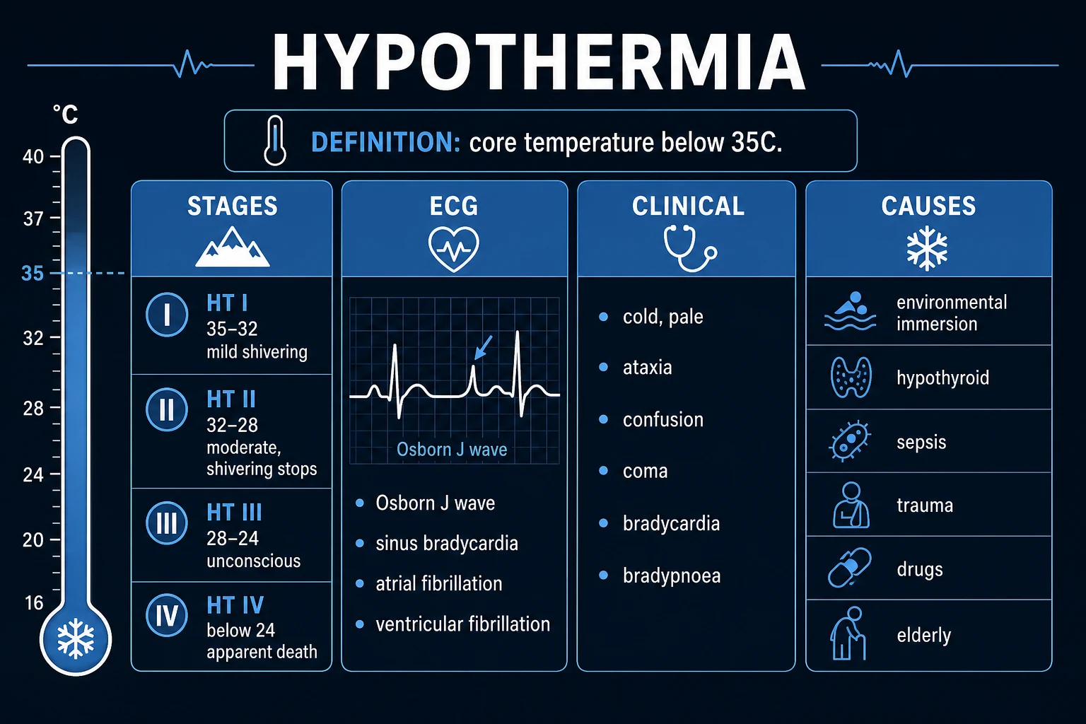
Hypothermia is defined as a core (rectal, bladder or oesophageal) body temperature below 35 degrees C (95 F), caused by excessive heat loss, impaired thermogenesis, or both. Severity by core temperature: mild 35 to 32 C — conscious, shivering; moderate 32 to 28 C — impaired consciousness, shivering ceases; severe 28 to 24 C — unconscious with vital signs possibly present, arrhythmia risk; profound below 24 C — apparent death (coma, fixed dilated pupils, asystole possible). The Swiss (Durrer) HT staging (HT I to HT V) maps clinical state to temperature and drives the rewarming strategy. Presentation: cold pale skin, shivering (which stops below 32 C), bradycardia, bradypnoea, confusion then coma, and the classic Osborn (J) wave on ECG. Management: handle GENTLY (rough handling triggers ventricular fibrillation), remove from cold, and rewarm by stage — passive external (mild) then active external (forced warm air) then active internal (warmed fluids and gases, lavage, and ECMO/CPB for severe/arrest). The cardinal rule of arrest is 'no one is dead until warm and dead' — continue CPR and rewarm to at least 32 C before ceasing. Always search for a secondary cause (myxoedema coma, sepsis, hypoglycaemia, drugs).
On this page & tools
Your progress
Saved locally on this device.
Exam tags
Red flags

Overview & Definition
Hypothermia is a reversible, time-critical environmental emergency defined as a core (central) body temperature below 35 degrees C (95 F), measured at a reliable site (rectal, bladder, or oesophageal). It results when heat loss exceeds heat production — from excessive environmental cold exposure, from impaired thermogenesis, or (most often) from a combination of both.[1][3]
The clinical skill in hypothermia is not the diagnosis (that is core temperature plus context) but four interlocking judgements: [1]
- Stage severity by core temperature — the temperature stage (mild/moderate/severe/profound, or Swiss HT I to IV) dictates the rewarming strategy and the cardiac-arrest algorithm.
- Handle the patient GENTLY — the cold myocardium is electrically irritable; rough moving, central-line insertion, or cold intravenous fluid can precipitate lethal ventricular fibrillation.[1]
- Search for a secondary cause — hypothermia is frequently the presenting feature of myxoedema coma, sepsis, hypoglycaemia, or drug overdose; failing to identify and treat the driver is fatal even if the temperature is corrected.
- Apply the 'warm and dead' rule in arrest — a hypothermic patient in apparent death (fixed dilated pupils, absent reflexes, no palpable pulse) may be fully salvageable; resuscitation continues until the core reaches at least 32 degrees C before death is declared.[1][2]
Classification
Hypothermia is classified along four axes — aetiology (primary vs secondary), severity (by core temperature), clinical stage (the Swiss/Durrer HT system), and the time-course of cooling. The examiner-critical classifications are severity and the Swiss HT stage, because together they determine rewarming.[1][3]
By aetiology: [1]
- Primary (accidental / environmental) hypothermia — a previously healthy thermoregulatory system is overwhelmed by cold exposure: immersion in cold water, avalanche burial, mountain/wilderness exposure, inadequate clothing, or simple indoor cold in the vulnerable. The thermoregulatory response is intact but outmatched.
- Secondary hypothermia — heat production is impaired (so even modest cold triggers hypothermia). Causes: hypothyroidism / myxoedema coma, sepsis, hypoglycaemia, malnutrition / low body mass, central nervous system lesions (stroke, tumour, spinal-cord injury), and drugs (alcohol, opioids, sedatives, phenothiazines, general anaesthetics, beta-blockers). Secondary hypothermia demands you treat the cause as well as rewarm.[5]
By severity (the classical core-temperature bands): [1]
- Mild — 35 to 32 degrees C.
- Moderate — 32 to 28 degrees C.
- Severe — 28 to 24 degrees C.
- Profound — below 24 degrees C. [1]
By Swiss (Durrer) clinical stage — HT I to V (reproduced verbatim):[2]
- HT I — clear consciousness with shivering (core approximately 35 to 32 C).
- HT II — altered consciousness (conscious but confused/drowsy), shivering absent (approximately 32 to 28 C).
- HT III — unconscious, vital signs (pulse and breathing) present (approximately 28 to 24 C).
- HT IV — absent vital signs (no pulse, no respiration); apparent death but not irreversibly dead (approximately 24 to 13 C).
- HT V — irreversible hypothermia; death (core below approximately 13 C with solid freezing of the chest wall, or irreversible biochemical/cellular death). [1]
By time-course (which governs the physiology): [1]
- Acute (minutes, e.g. cold-water immersion) — the body cools rapidly; core falls before significant fluid shifts or electrolyte derangement; the brain and heart are protected by rapid cooling and prognosis is comparatively good. Shivering may never have occurred.
- Subacute / chronic (hours to days, e.g. the elderly indoors, the exhausted mountaineer) — slower cooling allows fluid shifts, electrolyte derangement, exhaustion of metabolic reserves, and dehydration; physiological reserve is depleted and rewarming is more hazardous.[3]
Primary (environmental)
- Previously HEALTHY thermoregulation overwhelmed by cold
- Cold-water immersion, avalanche, wilderness, inadequate clothing
- Thermoregulatory response intact but outmatched (shivers vigorously)
- Rewarm by stage; search for a co-existing secondary cause
- Often good prognosis if no asphyxia/anoxia
Secondary (impaired thermogenesis)
- Heat PRODUCTION impaired — hypothermia at modest cold
- Myxoedema coma, sepsis, hypoglycaemia, malnutrition, CNS lesion, drugs
- Shivering may be absent even when 'only' mildly cold
- Must TREAT THE CAUSE as well as rewarm
- Prognosis dominated by the underlying disease
Epidemiology & Risk Factors
Hypothermia is a global killer that is under-recognised. It accounts for a significant fraction of winter excess mortality in temperate countries and is a leading cause of death in mountain, wilderness, and maritime settings. In the developing world and in north-Indian winters, indoor hypothermia in the elderly and neonatal cold stress are important and preventable.[3]
Risk factors cluster into three groups: [1]
- Excess heat loss — cold/wet/windy environment, inadequate clothing, cold-water immersion (water conducts heat away about 25 times faster than still air), high altitude, alcohol-induced vasodilation.
- Impaired heat production (secondary hypothermia) — extremes of age (neonates with their huge surface-area-to-mass ratio and limited glycogen; the elderly with blunted shivering and reduced muscle mass), malnutrition and low body mass, hypothyroidism / myxoedema, hypopituitarism, adrenal insufficiency, sepsis, hypoglycaemia, exhaustion, and central nervous system depression (stroke, head injury, spinal-cord injury with loss of autonomic vasoconstriction).
- Impaired behavioural response / impaired judgement — alcohol intoxication (the single commonest co-factor: it causes cutaneous vasodilation, impairs shivering, impairs judgement so clothing/shelter are not sought, and precipitates hypoglycaemia), dementia, psychiatric illness, drug overdose (opioids, sedatives, phenothiazines, barbiturates, general anaesthesia), and any cause of collapse or immobility in the cold.[5]
Why alcohol is the classic co-factor (high-yield): ethanol (a) causes cutaneous vasodilation increasing heat loss, (b) impairs shivering thermogenesis, (c) impairs judgement so protective behaviour is not undertaken, (d) causes hypoglycaemia (inhibits hepatic gluconeogenesis), and (e) predisposes to aspiration and trauma. A cold, intoxicated, hypoglycaemic patient is a textbook presentation.[3]
Neonates and infants are at particular risk because of a large surface-area-to-mass ratio, thin subcutaneous fat, limited glycogen stores, and dependence on non-shivering (brown-fat) thermogenesis — the basis of the WHO 'warm chain' for newborn care. The elderly lose thermogenic capacity (less muscle mass, blunted shivering, comorbidity, polypharmacy) and frequently develop indoor hypothermia at ambient temperatures an adult would tolerate.[5]
Pathophysiology
Normal body core temperature is held within a narrow band (approximately 36.5 to 37.5 C) by the pre-optic area of the hypothalamus, which integrates afferent input from skin and core thermoreceptors and mounts three efferent responses when cold is sensed: behavioural (put on clothes, seek shelter — the most powerful, and the first lost with intoxication or CNS depression), shivering thermogenesis (involuntary skeletal-muscle contraction that generates heat), and non-shivering thermogenesis (oxidation of brown adipose tissue, important in neonates), supported by peripheral vasoconstriction (sympathetic-mediated, reducing cutaneous heat loss).[1][5]
Hypothermia results when heat loss exceeds heat production. The four mechanisms of heat loss, in roughly descending order of importance in a clothed adult, are: radiation (the largest contributor in still air — the body radiates infrared to a colder environment), convection (wind/air movement, and the dominant mechanism in cold water), conduction (direct contact with a cold surface — the ground, cold water), and evaporation/respiration (latent heat of vaporisation from wet skin/clothes and from humidifying inspired air). Water is devastating because it conducts heat away roughly 25 times faster than still air.[3]
Mechanistic consequences of falling core temperature (organ by organ): [1]
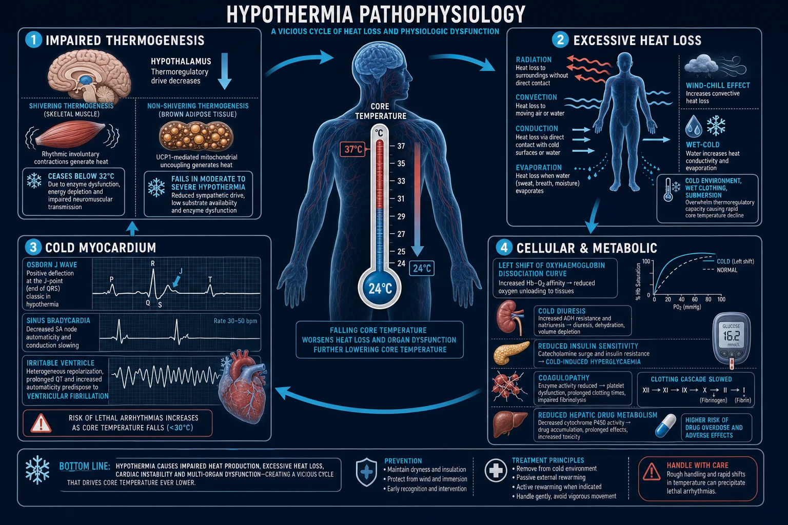
- Central nervous system — cerebral metabolism falls (roughly 6 to 7 per cent per degree C), which is neuroprotective (the basis of 'warm and dead' salvage and of therapeutic hypothermia after cardiac arrest) but produces progressive apathy, confusion, ataxia, drowsiness, coma, areflexia, and fixed dilated pupils. The pupil response is abolished below about 30 C, so dilated non-reactive pupils do NOT confirm death in hypothermia.[1]
- Cardiovascular — initial tachycardia from sympathetic drive and shivering gives way to progressive bradycardia and conduction slowing (prolonged PR, widened QRS, prolonged QT). The Osborn (J) wave — a positive hump at the J point (junction of the QRS and ST segment) — appears below about 32 C, becomes more prominent as temperature falls, and is the single most testable ECG sign of hypothermia. Below 28 to 30 C the myocardium is electrically irritable: atrial fibrillation is common, progressing to ventricular ectopics, ventricular fibrillation, and asystole. The myocardium in VF is refractory to defibrillation when cold — typically a single sequence of (up to) three stacked shocks is tried, then further shocks and drugs are deferred until the patient is warmer.[1][4]
- Respiratory — initial tachypnoea then bradypnoea, with bronchorrhoea and depressed cough reflex (aspiration risk). Hypoventilation produces a respiratory acidosis, and the cold-shifted oxyhaemoglobin curve impairs tissue oxygen delivery.
- Renal and fluid balance — cold diuresis: cold-induced renal medullary response plus suppression of ADH produces a water diuresis and volume depletion, compounded by plasma transudation into cold tissues. The patient is usually volume-depleted, which drives rewarming shock (see Complications).[3]
- Metabolic — reduced insulin sensitivity and impaired insulin release cause hyperglycaemia (do NOT diagnose new diabetes in the hypothermic patient); glycogen depletion in the exhausted; impaired hepatic metabolism of lactate and drugs.
- Haematology — coagulopathy: the enzyme reactions of the clotting cascade are temperature-dependent, so the in-vivo clotting function is impaired even though the in-vitro (37 C-warmed) PT/APTT returns 'normal'. In trauma this is the 'trauma triad of death' (hypothermia + coagulopathy + acidosis) — each worsens the others and is lethal if not broken. Thrombocytopenia (splenic sequestration) and DIC may occur in severe cases.[1][3]
Two phenomenon that decide the rewarming plan (high-yield): [1]
- Core temperature afterdrop — when a cold patient is rewarmed, the periphery warms and vasodilates first, returning cold, acidic, hyperkalaemic blood to the core and dropping the core temperature further (by 1 to 2 C, occasionally more). Afterdrop can precipitate VF. It is minimised by active core (internal) rewarming rather than active external warming alone in the moderate-to-severe patient, and by avoiding excessive movement.
- Rewarming shock (rewarming injury) — peripheral vasodilation on rewarming causes relative hypovolaemia and hypotension (the patient was volume-depleted from cold diuresis); combined with a cold, bradycardic, poorly responsive myocardium and acid washout, this can cause cardiovascular collapse. Anticipate it: give warmed IV crystalloid and titrate to response during rewarming.[3]
Why drugs fail in the cold heart: adrenaline (epinephrine), amiodarone, and lidocaine are less effective and more slowly cleared in the hypothermic myocardium, and repeated dosing accumulates to toxic levels as the patient rewarms. This is the rationale for hypothermia-modified ACLS (withhold adrenaline and antiarrhythmics below 30 C; give them at extended intervals — every 6 to 9 minutes — between 30 and 35 C; resume standard dosing above 35 C).[1][4]
Clinical Presentation
The clinical picture tracks the core temperature in stages — the stage is the temperature is the management.[1][3]
- Mild hypothermia (HT I, 35 to 32 C): the patient is alert and shivering vigorously, with tachycardia, tachypnoea, hypertension (sympathetic surge), and cold-induced diuresis (polyuria). Extremities are pale and cold; there may be ataxia, dysarthria, and impaired fine motor control (the 'umbles': stumbles, mumbles, fumbles, grumbles). The patient is still generating heat.
- Moderate hypothermia (HT II, 32 to 28 C): shivering ceases (a critical threshold — its absence signals loss of the major endogenous heat source). Consciousness clouds (confusion, drowsiness, apathy). Bradycardia and bradypnoea develop. The patient is now losing heat faster than they can generate it and needs active rewarming.
- Severe hypothermia (HT III, 28 to 24 C): the patient is unconscious (comatose), with slow, shallow or absent respiration and a slow, weak or impalpable pulse. Arrhythmias (atrial fibrillation, then ventricular ectopics and VF) appear. Reflexes are depressed or absent, pupils may be fixed and dilated. Skin is cold, pale, and may be oedematous.
- Profound hypothermia (HT IV, below 24 C): apparent death — no palpable pulse, no detectable respiration, fixed dilated pupils, areflexia; the ECG may show VF or asystole. Rigidity and rigor-like muscle stiffness may mimic death. Yet the patient may be fully recoverable with extracorporeal rewarming — hence 'no one is dead until warm and dead.' [1]
The ECG of hypothermia (reproduced — a favourite):[1]
- Sinus bradycardia (earliest).
- Osborn (J) waves — positive deflection at the J point, best seen in the lateral precordial leads; appear below about 32 C and grow with cooling. (Differential of a J wave: early repolarisation — benign, young, male — vs hypothermia vs hypercalcaemia; in the cold patient it is hypothermia until proven otherwise.)
- Prolonged PR, widened QRS, prolonged QT.
- Atrial fibrillation (common, usually benign if rewarmed).
- Ventricular ectopics, ventricular tachycardia, ventricular fibrillation, asystole.
- Muscle tremor artefact (fine baseline oscillation from shivering) — may masquerade as arrhythmia. [1]
Atypical presentation in the elderly: hypothermia is frequently indoor and insidious. The elderly patient may present with confusion or 'dementia', falls, or functional decline rather than shivering. There may be no history of cold exposure. A low threshold to measure core temperature (and to check for sepsis, hypothyroidism, and hypoglycaemia) is essential. Hypothermia in the elderly is a sentinel for serious underlying illness.[5]
When hypothermia is a marker of a secondary cause: suspect a driver when the cold exposure seems trivial for the degree of hypothermia, when shivering is absent at a temperature where it should be present, or when there are pointers in the history/examination. Always check glucose, and consider sepsis, myxoedema coma, and drug overdose. [1]
Differential Diagnosis
A low measured temperature with altered consciousness is not always simple environmental hypothermia. Distinguish:[3][5]
- Faulty reading / device error — many standard thermometers cannot read below 34 C; oral and tympanic readings are unreliable. Repeat with a low-reading rectal, bladder, or oesophageal probe. Confirm the reading before acting on a spurious value.
- Sepsis — the septic patient may be hypothermic rather than febrile (particularly the elderly and immunocompromised); hypothermia in sepsis is a poor prognostic sign. Look for the source, take blood cultures, and give empirical antibiotics within one hour (Surviving Sepsis). Sepsis and hypothermia frequently co-exist and each worsens the other.
- Myxoedema coma — hypothyroidism impairs thermogenesis; the patient is hypothermic, bradycardic, hyponatraemic, and comatose with a preceding history of thyroid disease, lethargy, cold intolerance, constipation. Treat with IV levothyroxine plus hydrocortisone (co-existent adrenal insufficiency) AND rewarm.
- Hypoglycaemia — causes coma and impairs thermogenesis; alcohol suppresses gluconeogenesis. Always check capillary glucose immediately and treat.
- Drug / toxin overdose — opioids (pinpoint pupils, respiratory depression), alcohol, sedatives, phenothiazines, carbon monoxide (check in the unconscious patient found indoors with a heat source). Naloxone trial if opioid suspected.
- Central nervous system event — stroke, subarachnoid haemorrhage, head injury (the patient collapsed then became hypothermic). Consider CT once stabilised.
- Adrenal crisis — hyponatraemia, hyperkalaemia, hypotension, hypoglycaemia; treat with parenteral hydrocortisone. [1]
The discriminating principle: every hypothermic patient gets glucose checked at the bedside, and a directed search for sepsis, myxoedema, and overdose — because missing the driver kills even when the temperature is corrected. [1]
Clinical & Bedside Assessment
The first measured number is the CORE TEMPERATURE, taken at a reliable site.[1]
- Reliable sites: rectal (the practical default — insert 15 cm; lag the reading by a minute), bladder (continuous if a Foley catheter with a thermistor is in place), and oesophageal (most accurate for cardiac/core temperature, used in ICU/theatre).
- Unreliable sites: oral (mouth breathes cold air), tympanic (poorly perfused tympanic membrane, affected by external cold/wax), axillary, and forehead. Use a low-reading thermometer capable of reading to 25 C or below — many ward thermometers bottom out at 34 C and falsely 'normalise' a profoundly hypothermic patient. [1]
Focused examination: level of consciousness (and the presence/absence of shivering — its absence at a temperature where it should be present is a red flag), pulse and rhythm (bradycardia; feel carefully — the pulse may be imperceptible; auscultate for a full minute), respiratory rate (bradypnoea), skin (cold, pale, cyanotic, oedematous), pupils (may be fixed and dilated below 30 C — NOT a death sign), and a search for injury and cause (signs of sepsis, myxoedema, head injury, injection marks). Check capillary glucose immediately.[3]
Handle GENTLY — the cardinal manoeuvre. The cold myocardium is poised on the edge of ventricular fibrillation. Rough moving, unnecessary central-line or pulmonary-artery catheter insertion, cold intravenous fluid, and even vigorous chest compressions have all been documented to precipitate VF. Move the patient on a scoop or vacuum mattress; remove wet clothing by cutting (not pulling); keep manipulation to the minimum consistent with the emergency.[1]
In apparent death (HT IV): assess for any sign of life — listen for heart sounds for a full minute (the rate may be 10 to 20 per minute and easy to miss), feel a major pulse for a minute, and look for any respiratory effort. If there is genuinely no pulse and no respiration, start CPR. The decision to not resuscitate rests on the potassium / asphyxia rule (see Management). [1]
Hypothermia — the numbers that decide management
Investigations
Investigations serve two purposes: to stage the patient and to find the secondary cause and the complications.[3][5]
Mandatory at presentation: [1]
- 12-lead ECG (and continuous monitoring) — Osborn waves, bradycardia, prolonged intervals, atrial fibrillation, ventricular arrhythmias. Continuous monitoring is essential because VF may develop spontaneously.
- Capillary glucose — at the bedside, immediately; hypoglycaemia is common (especially with alcohol) and is rapidly lethal and rapidly treatable.
- Arterial blood gas — to assess ventilation, acid-base (respiratory acidosis from hypoventilation; metabolic acidosis from lactate/shock), and lactate. Interpretation caveat: ABG machines warm the sample to 37 C; the uncorrected (machine) values are the ones to act on clinically (alpha-stat approach) — do not 'temperature-correct' the numbers, which falsely worsens the apparent pH and PaO2.
- Urea and electrolytes — for volume status, AKI, and the potassium level (the key prognostic marker in HT IV — see Management).
- Full blood count, coagulation, group and save — for anaemia, infection, and coagulopathy (remember in-vivo coagulopathy may exist with 'normal' warmed in-vitro PT/APTT).
- Creatine kinase (CK) — for rhabdomyolysis (prolonged immobility, compression, frostbite), which complicates severe cases.
- Blood cultures, lactate, and a septic work-up — if sepsis is suspected.
- Thyroid function (TSH, free T4) and cortisol — if myxoedema coma or adrenal crisis is possible (do not delay empirical hydrocortisone).
- Drug/toxin screen and alcohol level — in the unconscious or uncertain case; a trial of naloxone if opioids are possible.
- Pregnancy test — in any woman of childbearing age (affects imaging and management).
- Chest X-ray — for aspiration, pneumonia, pulmonary oedema (once stabilised).
- CT head — once stabilised, if a primary CNS event or head injury is suspected. [1]
The potassium rule in HT IV (examiner-favourite):[1][2]
In hypothermic cardiac arrest, the serum potassium discriminates asphyxia (poor prognosis, futile resuscitation) from pure hypothermia (good prognosis, full recovery possible with ECLS): [1]
- Potassium below 8 mmol/L — hypothermia is likely the sole problem; full resuscitation and ECLS are indicated.
- Potassium over 12 mmol/L — strongly suggests asphyxia or prolonged anoxia (e.g. an avalanche victim whose airway was buried); the resuscitation is likely futile and may reasonably be withheld or ceased. [1]
The HOPE (Hypothermia Outcome Prediction) score / Swiss staging for HT IV is used to predict survival and guide the decision to institute ECLS — the key inputs are core temperature, serum potassium, age, sex, mode of cooling, and asphyxia.[2]
Why clotting studies lie in hypothermia (the trauma pitfall): the clotting cascade enzymes are temperature-dependent. The in-vivo patient is coagulopathic (bleeding), but the PT and APTT are measured on a sample warmed to 37 C in the laboratory, so they return 'normal' or only mildly abnormal. Do not be reassured by a 'normal' PT/APTT in a cold, bleeding trauma patient — rewarm and treat the coagulopathy clinically (this is the rationale for damage-control resuscitation).[1]
Management — Resuscitation
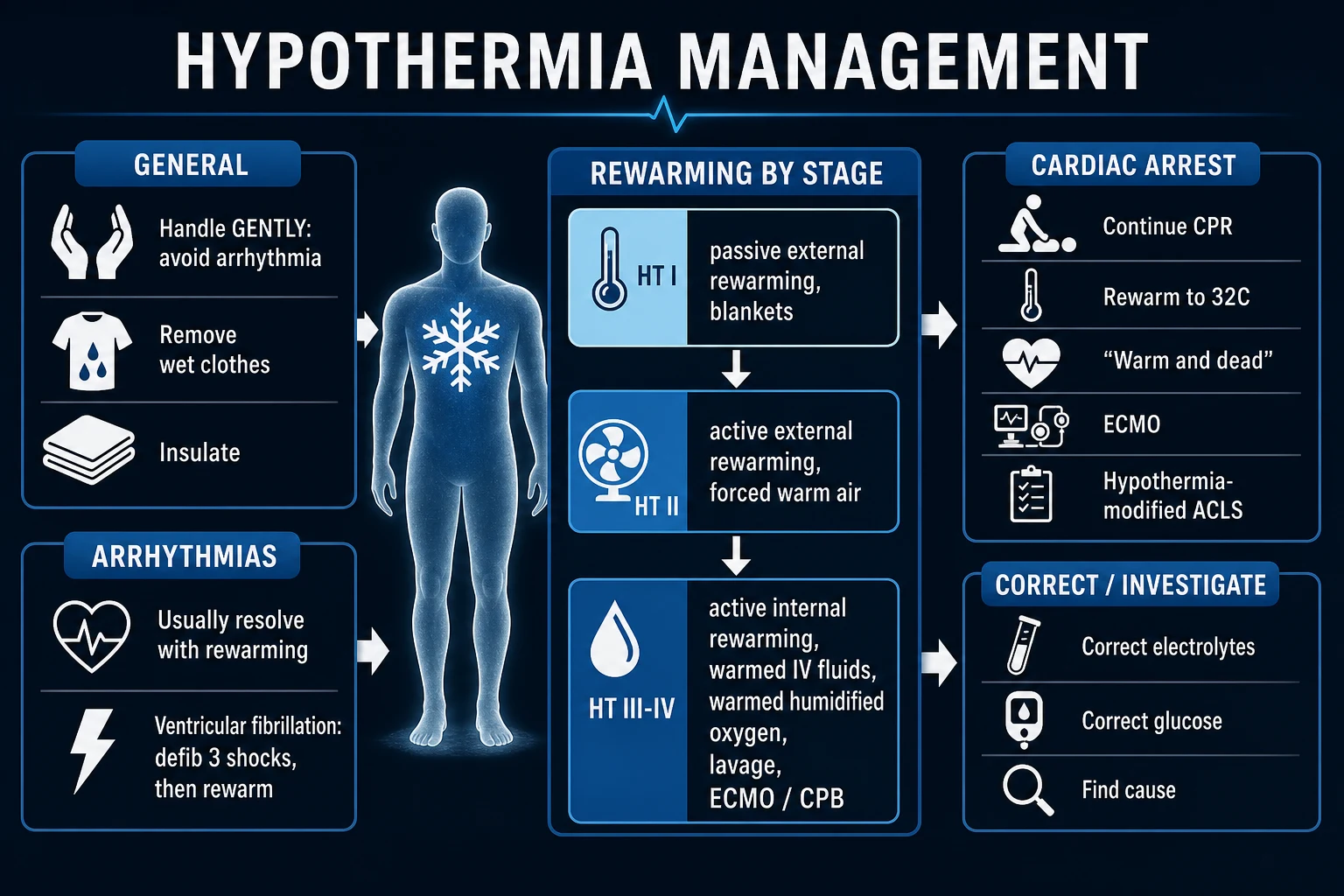
Management is staged by the Swiss HT stage / core temperature, with three universal rules applied to every patient.[1][4]
Universal first actions (all stages): [1]
- Remove from the cold environment; remove wet clothing by CUTTING (not pulling); dry the skin; insulate with dry blankets; protect from wind. Move the patient on a scoop/vacuum mattress.
- Handle GENTLY — avoid rough moving, unnecessary line insertion, and cold fluid (all precipitate VF).
- Monitor: continuous core temperature (rectal/bladder/oesophageal), continuous ECG, capillary glucose (treat hypoglycaemia), SpO2, and blood pressure.
- Airway and breathing: give warmed (42 to 46 C), humidified oxygen; intubate if comatose or unable to protect the airway (do it gently and skilfully — the act of intubation can precipitate VF, but a definitive airway is safer than aspiration). Avoid hyperventilation (it worsens the cold-shifted oxyhaemoglobin curve and may precipitate arrhythmia).
- Circulation: establish IV access; give only WARMED (39 to 42 C) crystalloid — cold or room-temperature fluid worsens hypothermia. Treat hypovolaemia (cold diuresis) and hypoglycaemia.
- Find and treat the secondary cause — glucose, sepsis, myxoedema, drugs. [1]
Cardiac arrest in hypothermia — the 'warm and dead' rule and hypothermia-modified ACLS:[1][4]
- 'No one is dead until warm and dead.' Continue CPR and rewarm the patient to a core temperature of at least 32 degrees C before ceasing resuscitation. A patient in apparent death (fixed dilated pupils, areflexia, no palpable pulse) may recover fully if the brain was protected by rapid cooling and the cause was pure hypothermia.
- Start CPR if there is no pulse and no respiration (after auscultating/feeling for a full minute). In a patient with a perfusing rhythm, do NOT start chest compressions for a slow pulse alone — compressions can precipitate VF in the cold, irritable heart.
- Defibrillation: if in VF/pulseless VT, deliver one sequence of up to three stacked shocks (standard energy). If VF persists, defer further shocks until the core temperature exceeds 30 C (the cold myocardium is refractory to defibrillation).
- Drugs — hypothermia-modified ACLS:
- Below 30 C: withhold adrenaline (epinephrine) and antiarrhythmics (amiodarone, lidocaine) — they are ineffective, accumulate, and become toxic on rewarming. Continue CPR and rewarm.
- 30 to 35 C: give adrenaline and antiarrhythmics at extended intervals (every 6 to 9 minutes) rather than every 3 to 5 minutes.
- Above 35 C: resume standard ACLS drug dosing.
- Rewarm aggressively — the definitive treatment of hypothermic arrest is rewarming, ideally by extracorporeal life support (ECLS — ECMO or cardiopulmonary bypass), which both rewarms (up to 10 C per hour) and provides circulatory support.
- When to stop: cease resuscitation when the core reaches at least 32 C and there is persistent asystole/PEA unresponsive to standard ACLS, OR when the serum potassium is very high (over 12 mmol/L) indicating asphyxia/irreversible cell death, OR when there are other clear signs of irreversibility (e.g. a frozen, solid chest wall).[2]
Management — Definitive & Stepwise
Rewarming is stratified by severity/stage. The principle: match the aggressiveness of rewarming to the depth of hypothermia, and minimise afterdrop and rewarming shock.[1][3]
1. Passive external rewarming (mild, HT I, 35 to 32 C): [1]
- Indication: the alert, shivering patient with mild hypothermia.
- Method: move to a warm room; remove wet clothes; cover with dry insulating blankets (head covered too — significant heat is lost from the scalp).
- Mechanism: the patient rewarms themselves through their own intact shivering thermogenesis. The clinician simply prevents further heat loss.
- Expected rate: 0.5 to 2 degrees C per hour.
- Contraindications (when passive is not enough): a patient who is not shivering (has lost the heat-generating mechanism), who is unconscious or cardiovascularly unstable, or whose temperature is not rising — all need active rewarming. [1]
2. Active external rewarming (moderate, HT II, 32 to 28 C): [1]
- Indication: the conscious but impaired (confused/drowsy), non-shivering patient with moderate hypothermia, and as adjunct in any patient needing more than passive warming.
- Method: forced warm-air blanket (e.g. Bair Hugger) set to 38 to 42 degrees C, warming mattresses/pads, radiant warmers; plus warmed IV fluid.
- Mechanism: external heat is actively transferred to the body surface and conducted inward.
- Expected rate: 1 to 2 degrees C per hour.
- Cautions: active external warming of the limbs (rather than the trunk) can worsen afterdrop and precipitate VF (core rewarming of the trunk is preferred). Avoid burns (monitor skin under heating devices). Not sufficient on its own for the unconscious or arrested patient. [1]
3. Active internal (core) rewarming (severe, HT III to IV, below 28 C): [1]
This is required for the unconscious, cardiovascularly unstable, or arrested patient. Techniques are cumulative — start with the simpler and escalate.[1][4]
- Warmed, humidified oxygen (42 to 46 C) — via mask or endotracheal tube; an easy, safe, modest-impact core warmer (the lungs are a large surface area).
- Warmed intravenous crystalloid (39 to 42 C) — normal saline or balanced crystalloid in a blood/fluid warmer; large volumes may be needed (cold diuresis plus rewarming vasodilation). Never give cold or room-temperature fluid.
- Body-cavity lavage — gastric, bladder, peritoneal, or pleural lavage with warmed (40 to 42 C) crystalloid via catheter (in-and-out or continuous); provides significant core heat transfer. Pleural lavage requires a chest tube (leave one chest tube for lavage in, one out).
- Cardiopulmonary bypass (CPB) or extracorporeal membrane oxygenation (ECMO) — the gold standard for severe hypothermia with cardiac instability or arrest. ECLS rewams at up to 10 degrees C per hour, provides full circulatory support (so it sustains the arrested patient while rewarming), and allows control of acid-base and electrolytes. Indication: HT IV (arrest) or HT III with cardiac instability, especially if not responding to less invasive measures. Transfer the arrest patient to an ECMO-capable centre.
- Haemodialysis / continuous renal replacement therapy — useful for rewarming AND for managing the hyperkalaemia, acidosis, and rhabdomyolysis that complicate severe hypothermia; an option when ECMO is unavailable. [1]
Expected rates (high-yield): passive external 0.5 to 2 C/h; active external 1 to 2 C/h; active internal (fluids/gases/lavage) 1 to 2 C/h; ECLS/CPB up to 10 C/h.[3]
Management of arrhythmias during rewarming:[1]
- Atrial fibrillation is common and usually benign — it typically reverts to sinus rhythm as the patient rewarms; anticoagulate if sustained. Do not cardiovert a haemodynamically stable cold patient in AF.
- Ventricular fibrillation — deliver up to three shocks (standard energy); if it persists, stop shocking and focus on rewarming (defibrillation is ineffective below about 30 C); resume shocks once the patient is warmer. Give amiodarone only once the core is above 30 C (it is ineffective when cold). Magnesium and bretylium have been used but are second-line.
- Bradycardia is physiological in hypothermia — it usually responds to rewarming; do NOT give atropine routinely (it is typically ineffective and the bradycardia is not vagally mediated). Pacing is rarely needed. [1]
Supportive care during rewarming: [1]
- Fluid resuscitation — anticipate rewarming shock (peripheral vasodilation); give warmed crystalloid titrated to blood pressure, lactate, and urine output.
- Treat hypoglycaemia — IV dextrose (e.g. 50 mL of 50 per cent dextrose IV in an adult, or 5 mL/kg of 10 per cent dextrose in a child).
- Watch for rhabdomyolysis — rising CK, dark urine; give IV fluid to target a urine output of approximately 1 to 1.5 mL/kg/h.
- Watch for compartment syndrome — in limbs with frostbite or prolonged compression.
- VTE prophylaxis, pressure-area care, and gentle warming to avoid burns. [1]
Specific Subtypes & Scenarios
- Drowning / near-drowning in cold water — combined asphyxia, hypothermia, and (often) injury. Cold-water submersion can produce protective hypothermia before anoxic brain injury, which is the basis of prolonged successful resuscitation (especially in children). Resuscitate vigorously even after prolonged submersion; correct hypoxia first (the primary driver of death); rewarm; treat ARDS. A submersion victim in cardiac arrest who is also hypothermic is resuscitated and rewarmed in parallel.[1]
- Avalanche burial — distinguish asphyxia (the airway was occluded by snow; poor prognosis; high potassium) from hypothermia (the airway was patent, allowing breathing around the snow; good prognosis with ECLS). Apply the potassium rule: potassium over 8 to 12 mmol/L suggests asphyxia and futility. Avalanche victims in HT IV arrest are the prime candidates for ECLS.[2]
- The elderly indoor hypothermia patient — search hard for sepsis and myxoedema coma; rewarm more slowly (the elderly myocardium tolerates rapid rewarming poorly) with active external plus warmed fluid; treat the underlying driver. Consider social and capacity issues.[5]
- The trauma patient — the 'trauma triad of death' — hypothermia, coagulopathy, and acidosis form a lethal vicious cycle in the bleeding patient (each worsens the others). The intervention is damage-control resuscitation: active warming in resus and theatre (warm fluids, forced-air warmer, warmed theatre, minimise exposure), permissive hypotension until bleeding is controlled, damage-control surgery (stop the bleeding fast, pack, and get out), and ratio-based transfusion. Hypothermia in trauma is never benign — prevent and correct it aggressively.[1]
- Hypothermia with a secondary cause — myxoedema coma — the patient is hypothermic, comatose, bradycardic, hyponatraemic. Treat both: rewarm AND give IV levothyroxine (e.g. a loading dose followed by daily maintenance, guided by endocrinology) plus IV hydrocortisone (co-existing adrenal insufficiency is common and lethal if missed). Rewarm gently (rapid rewarming can precipitate cardiovascular collapse in myxoedema).[3]
- Neonatal / paediatric hypothermia — neonates depend on brown-fat non-shivering thermogenesis and have a huge surface-area-to-mass ratio; prevention is the WHO 'warm chain' (warm delivery room, immediate drying, skin-to-skin, early breastfeeding, delayed bathing, appropriate clothing, warm transport). A hypothermic neonate is rewarmed under a radiant warmer/incubator. Children tolerate deep hypothermia remarkably — prolonged cold-water submersion in a child may be followed by full neurological recovery; resuscitate vigorously ('children are not small adults — they are cold survivors').[1]
Complications & Pitfalls
Complications of the hypothermia itself:[1][3]
- Cardiac: ventricular fibrillation, cardiac arrest, asystole.
- Respiratory: aspiration pneumonia, ARDS, bronchorrhoea.
- Renal: acute kidney injury (cold diuresis plus volume depletion plus rhabdomyolysis).
- Metabolic: hyperglycaemia, lactic acidosis, pancreatitis (a recognised complication of hypothermia and rewarming).
- Haematology: coagulopathy (especially in trauma), DIC, thrombocytopenia.
- Soft tissue: frostbite, pressure injury, rhabdomyolysis with compartment syndrome.
- Infection: sepsis (impaired immunity in the cold). [1]
Complications of REWARMING: [1]
- Core temperature afterdrop — minimised by active core (internal) rewarming and gentle handling.
- Rewarming shock — peripheral vasodilation, hypotension, cardiovascular collapse; anticipate with warmed IV fluid.
- Rewarming acidosis — washout of lactate and acidic metabolites from the periphery.
- Cerebral oedema and acute pulmonary oedema (rare but described, especially with rapid rewarming of chronic hypothermia).
- Compartment syndrome from rhabdomyolysis. [1]
Classic pitfalls (high-yield):[1][5]
- Declaring death too early — not applying the 'warm and dead' rule; failing to rewarm to at least 32 C before ceasing.
- Rough handling precipitating VF.
- Giving cold or room-temperature IV fluid (worsens hypothermia; can trigger VF).
- Relying on oral or tympanic temperature (under-reads; many thermometers bottom out at 34 C).
- Failing to find a secondary cause (sepsis, myxoedema, hypoglycaemia, overdose) — the temperature is corrected but the patient dies of the driver.
- Over-using drugs in the cold heart — repeated adrenaline/amiodarone below 30 C is ineffective and toxic on rewarming.
- Misinterpreting the ABG by 'temperature-correcting' the values (use uncorrected/machine values).
- Misinterpreting 'normal' coagulation studies in a cold, bleeding trauma patient (in-vivo coagulopathy despite normal warmed in-vitro PT/APTT).
- Cardioverting stable AF in a cold patient (usually reverts with rewarming).
- Giving atropine for hypothermic bradycardia (ineffective; the bradycardia is physiological). [1]
Prognosis & Disposition
Determinants of survival: the depth and duration of hypothermia, the presence of asphyxia or anoxia (the dominant determinant in submersion/avalanche), the underlying cause (secondary hypothermia from sepsis/myxoedema carries the prognosis of the driver), age and comorbidity, and the serum potassium in HT IV.[1][2]
- Pure hypothermia without asphyxia carries a good prognosis even at very low core temperatures — full neurological recovery is reported after hours of CPR with ECLS.
- Asphyxiated hypothermia (submersion with anoxia, avalanche with airway occlusion) carries a poor prognosis, reflected in the potassium rule (over 12 mmol/L suggests asphyxia and futility). [1]
The 'warm and dead' rule governs termination: resuscitation is continued until the core reaches at least 32 C; if asystole/PEA persists despite rewarming to 32 C and standard ACLS, death may be declared. A very high potassium (over 12 mmol/L) or a frozen chest wall justify ceasing earlier.[2]
ECLS outcomes: observational data show survival of 47 to 100 per cent in selected HT IV patients (no asphyxia, potassium below 8 to 12) rewarmed with ECLS — striking, and the basis for transferring arrest patients to an ECMO centre.[1]
Disposition by stage:[3]
- HT I (mild): can often be observed and rewarmed in the emergency department, discharged once alert, normothermic, and with a safe plan (or admitted briefly if elderly/comorbid).
- HT II (moderate): admit to a monitored bed; active external rewarming.
- HT III (severe): ICU admission; active internal rewarming; prepare for possible deterioration to arrest.
- HT IV (arrest): ICU with ECLS — transfer to an ECMO-capable centre. [1]
Long-term outlook: survivors of pure hypothermia (no anoxia) typically make a full neurological recovery. Those with anoxic brain injury (prolonged submersion/asystole) carry the prognosis of the anoxic insult. [1]
Special Populations
- Elderly — impaired thermogenesis (less muscle mass, blunted shivering), polypharmacy, comorbidity, and indoor hypothermia at modest cold. Lower threshold to admit and to search for sepsis and myxoedema. Rewarm gently. Address social, capacity, and safeguarding factors.[5]
- Neonates and children — large surface-area-to-mass ratio, dependence on brown fat, limited glycogen. Prevention via the WHO warm chain. Children tolerate deep hypothermia and prolonged submersion remarkably — resuscitate vigorously. Weight-based fluid and drug dosing.[1]
- Pregnancy — maternal hypothermia can precipitate preterm labour and fetal distress; rewarm the mother and monitor the fetus; magnesium sulphate for fetal neuroprotection is not specifically contraindicated by hypothermia but the picture is dominated by maternal stabilisation. Liaise early with obstetrics.
- The intoxicated patient — alcohol plus hypothermia plus hypoglycaemia (alcohol suppresses gluconeogenesis); check glucose; give dextrose and thiamine (Wernicke risk in the malnourished alcoholic); beware aspiration and protect the airway.[3]
- The anticoagulated / trauma patient — hypothermic coagulopathy worsens bleeding (trauma triad of death). Damage-control resuscitation with active warming, permissive hypotension, ratio-based transfusion, and damage-control surgery. Reverse anticoagulation per protocol.[1]
- High-altitude / military (e.g. Ladakh, Siachen) — combined hypothermia, frostbite, hypoxia, and dehydration; long retrieval times; field rewarming and rapid evacuation to an ECLS centre are life-saving.
Evidence, Guidelines & Regional Differences
- Wilderness Medical Society (WMS) 2019 guidelines (Dow et al.) provide the international standard for out-of-hospital evaluation and treatment: stage the patient, rewarm by stage, handle gently, and use ECLS for HT IV with cardiac instability/arrest where feasible. They endorse the potassium/asphyxia rule and the 'warm and dead' principle.[4]
- ICAR MEDCOM (International Commission for Mountain Emergency Medicine) recommendations (Paal et al.) underpin the alpine/avalanche management: rapid extrication, gentle handling, field staging, and transfer to an ECLS centre for HT IV without asphyxia.[2]
- UK (NICE / RCPUK / NHS Cold Weather Plan) focus on prevention in the elderly (insulation, heating, benefits uptake) and on recognising indoor hypothermia; NICE guidance targets vulnerable older people in cold housing.
- India — no central heating in most homes; winter hypothermia contributes to excess mortality in north India (Delhi, UP, Bihar) among the homeless, elderly, and neonates; high-altitude military hypothermia (Siachen/Ladakh) is a specialist problem with field-to-ECLS protocols. The WHO warm chain underpins neonatal prevention.
- ECLS evidence — observational registry data (e.g. from alpine centres in Switzerland and France) show the highest survival for HT IV arrest treated with ECMO/CPB; this is the strongest evidence for the 'transfer to an ECLS centre' recommendation.[1]
- Not supported by evidence (do NOT use routinely): prophylactic antibiotics, prophylactic steroids or barbiturates, and active external rewarming of the limbs (worsens afterdrop).[3]
Exam Pearls
- Definition: core temp below 35 degrees C (95 F). Measure rectally, by bladder, or oesophageally — oral/tympanic are unreliable, and many thermometers bottom out at 34 C.
- Stages = temperature = management. Mild 35 to 32 (conscious, shivering — passive external). Moderate 32 to 28 (altered, shivering stops — active external). Severe 28 to 24 (unconscious, vital signs — active internal). Profound below 24 (apparent death — ECLS).
- Osborn (J) wave = hypothermia — positive deflection at the J point, appears below about 32 C.
- HANDLE GENTLY — rough handling, central lines, or cold fluid precipitate ventricular fibrillation.
- Hypothermia-modified ACLS: withhold adrenaline/antiarrhythmics below 30 C; give at extended intervals (every 6 to 9 min) 30 to 35 C; standard above 35 C. Up to three shocks for VF, then defer until warmer.
- 'No one is dead until warm and dead' — continue CPR and rewarm to at least 32 C before ceasing. ECMO/CPB is the gold standard for HT IV arrest.
- Potassium rule in HT IV: potassium over 12 mmol/L suggests asphyxia/futility; below 8 suggests pure hypothermia and supports full ECLS.
- Always check glucose and search for sepsis, myxoedema coma, and overdose — hypothermia is often the presenting feature of a lethal secondary cause.
- Trauma triad of death: hypothermia + coagulopathy + acidosis — break it with active warming and damage-control resuscitation.
- Rewarming hazards: afterdrop, rewarming shock, rewarming acidosis — anticipate with warmed fluid and active core rewarming. [1]
Hypothermia — the 'COLDD' workup
COLDD
Measure CORE temperature (rectal/bladder/oesophageal), not oral/tympanic; stage severity
ECG for Osborn (J) waves; give warmed humidified OXYGEN; continuous ECG monitoring
LOOK for the secondary cause — glucose, sepsis, myxoedema, overdose — and treat it
DRUGS (adrenaline/antiarrhythmics) DEFERRED below 30 C in arrest; give only warmed IV fluid
DEAD only if WARM — continue CPR, rewarm to at least 32 C; ECMO/CPB gold standard
Exam application bank (NEET-PG / INICET)
One-line answer
Hypothermia is defined as a core (rectal, bladder or oesophageal) body temperature below 35 degrees C (95 F), caused by excessive heat loss, impaired thermogenesis, or both. Severity by core temperature: mild 35 to 32 C — conscious, shivering; moderate 32 to 28 C — impaired consciousness, shivering ceases; severe 28 to 24 C — unconscious with vital signs possibly present, arrhythmia risk; profound below 24 C — apparent death (coma, fixed dilated pupils, asystole possible). The Swiss (Durrer) HT staging (HT I to HT V) maps clinical state to temperature and drives the rewarming strategy. Presentation: cold pale skin, shivering (which stops below 32 C), bradycardia, bradypnoea, confusion then coma, and the classic Osborn (J) wave on ECG. Management: handle GENTLY (rough handling triggers ventricular fibrillation), remove from cold, and rewarm by stage — passive external (mild) then active e
Worked stems (answer without another resource)
Stem 1 — Classic presentation. Map symptoms to mechanism; name the first investigation and first treatment step with dose/route if drug therapy is standard. [1]
Stem 2 — Unstable / complicated. List red flags that force immediate resuscitation, theatre, ICU, antidote, or reperfusion — and what you do in the first 15 minutes. [1]
Stem 3 — Atypical group. Elderly, pregnancy, child, or immunocompromised: how presentation and thresholds change. [1]
Stem 4 — Differential trap. Name the three closest mimics and one discriminator for each. [1]
Stem 5 — Disposition. Who goes home with safety-netting, who is admitted, who needs HDU/ICU/theatre, and what follow-up is mandatory. [1]
Rapid viva checklist
- Definition + classification
- Pathophysiology chain
- Bedside signs / criteria
- Score with exact components (if any)
- Emergency bundle
- Definitive therapy with doses
- Complications of disease and of treatment
- Special populations
- Guideline/trial name if classic
- Three exam traps
Coverage self-check
If you cannot answer any stem above from this page alone, re-read the matching section — the page is intended to be self-sufficient for final-prof and NEET-PG/INICET questions on Hypothermia.
References
- [1]Brown DJ, Brugger H, Boyd J, Paal P. Accidental hypothermia N Engl J Med, 2012.PMID 23150960
- [2]Paal P, Gordon L, Strapazzon G, et al. Accidental hypothermia-an update : The content of this review is endorsed by the International Commission for Mountain Emergency Medicine (ICAR MEDCOM) Scand J Trauma Resusc Emerg Med, 2016.PMID 27633781
- [3]Avellanas Chavala ML, Ayala Gallardo M, Soteras Martinez I, et al. Management of accidental hypothermia: A narrative review Med Intensiva (Engl Ed), 2019.PMID 30683520
- [4]Dow J, Giesbrecht GG, Danzl DF, et al. Wilderness Medical Society Clinical Practice Guidelines for the Out-of-Hospital Evaluation and Treatment of Accidental Hypothermia: 2019 Update Wilderness Environ Med, 2019.PMID 31740369
- [5]Falat C, Tucker MA, Miller KC. Environmental Hypothermia Emerg Med Clin North Am, 2024.PMID 38925770