Emergency & Toxicology · Emergency & Toxicology
Iron Overdose
Also known as Iron poisoning · Iron overdose · Iron toxicity · Ferrous sulphate poisoning · Desferrioxamine · Deferoxamine · Ferrioxamine
Iron overdose is one of the leading causes of accidental poisoning death in children (adult prenatal/iron tablets look like sweets) and an occasional means of deliberate self-harm in adults. Iron is corrosive to the gastrointestinal mucosa (haemorrhagic gastritis, vomiting, diarrhoea) and, once absorbed iron exceeds transferrin binding capacity, becomes a systemic free-radical toxin via the Fenton reaction — it uncouples oxidative phosphorylation, blocks the Krebs cycle and produces lactic (high-anion-gap) acidosis, centrilobular hepatic necrosis, coagulopathy (direct thrombin inhibition), shock and multi-organ failure. Toxic dose (elemental iron): under 20 mg/kg usually asymptomatic; over 20 mg/kg mild; over 40 mg/kg significant; over 60 mg/kg severe, potentially lethal. Four clinical stages: Stage 1 (0 to 6 h) GI corrosive injury + hypovolaemic shock; Stage 2 (6 to 24 h) deceptive latent phase; Stage 3 (12 to 48 h) shock + acidosis + hepatic/renal failure + coma (the killer); Stage 4 (4 to 6 weeks) gastric/pyloric outlet stricture. Diagnosis: serum iron at 4 to 6 h (over 500 microgram/dL significant), abdominal X-ray (radiopaque tablets), anion-gap metabolic acidosis with hyperglycaemia and leucocytosis. Management: ABCDE + aggressive crystalloid; activated charcoal is USELESS — whole bowel irrigation is the decontamination of choice; IV desferrioxamine 15 mg/kg/h (urine turns 'vin rose') for iron over 500, acidosis, or shock; supportive care for organ failure.
On this page & tools
Your progress
Saved locally on this device.
Exam tags
Red flags
Overview & Definition
Iron overdose is a high-yield topic because it brings together four classic examination themes: a stereotyped four-stage clinical course with a deceptive latent phase that catches the unwary, a specific chelating antidote (desferrioxamine/deferoxamine) with a memorable urinary sign ('vin rose' urine), a critical decontamination distinction (iron is a metal and is not adsorbed by activated charcoal — whole bowel irrigation is used instead), and a precise dose-to-toxicity relationship based on the elemental iron content of the tablet rather than the salt weight.[1]
Iron is essential but, uniquely among nutrients, the body has no physiological excretory mechanism for it. All iron balance is regulated at absorption. In overdose, this conserved, easily-absorbed metal overwhelms the iron-binding capacity of transferrin, and the surplus circulates as free (non-transferrin-bound) iron — a powerful redox catalyst. Two injuries follow. First, free iron is directly corrosive to the gastrointestinal mucosa, producing haemorrhagic gastritis, vomiting, diarrhoea and fluid loss. Second, absorbed free iron enters cells and catalyses the Fenton reaction, generating hydroxyl radicals that peroxidise mitochondrial membranes, uncouple oxidative phosphorylation, and inhibit the Krebs cycle. The metabolic consequence is a high-anion-gap lactic acidosis; the tissue consequences are centrilobular hepatic necrosis, coagulopathy (direct inhibition of thrombin), acute tubular necrosis, and shock from a combination of hypovolaemia, venodilatation and myocardial depression.[5]
Iron overdose is a leading cause of accidental poisoning death in children (adult prenatal and iron tablets look like sweets to a toddler) and a recognised method of deliberate self-harm in adults, typically young women with access to iron tablets.[8] The clinical skill the examiner is testing is whether you can stratify severity using the elemental-iron dose, the serum iron level and the acid–base status, avoid the trap of the latent phase, refuse to give charcoal, and start IV desferrioxamine early for significant toxicity.
Classification
Iron poisoning is classified along two axes that the examiner will probe: the elemental-iron dose (which predicts severity) and the clinical stage (which dictates what you do at the bedside). A third axis — the pharmaceutical formulation (immediate-release versus sustained-release/enteric-coated) — modifies the timing of both the symptoms and the serum-iron peak.[1]
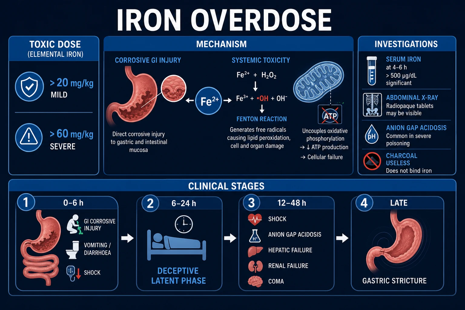
Toxic-dose thresholds (ELEMENTAL iron)
The dose that matters is the elemental iron content, not the weight of the iron salt. This is the single most common calculation error in MCQ stems.[1]
Elemental-iron content of common formulations
The salt weight on the packet is not the iron dose. Always convert to elemental iron.[1]
Ferrous sulphate
- 325 mg tablet contains 65 mg elemental iron (20 percent)
- Ferrous sulphate 300 mg = 60 mg elemental
- The classic paediatric culprit (prenatal tablets)
- As few as 7 tablets in a 10 kg child exceed 40 mg/kg
Ferrous fumarate
- 325 mg contains 106 mg elemental iron (33 percent)
- Higher elemental fraction — fewer tablets needed to overdose
- Common in combined haematinics
- Calculate carefully: 3 x ferrous fumarate 325 mg = 318 mg elemental
Ferrous gluconate
- 325 mg contains 38 mg elemental iron (12 percent)
- Lower elemental fraction — larger tablet numbers needed to overdose
- Used in pregnancy
- Still dangerous in sustained-release form
Polysaccharide-iron complex
- 150 mg capsule = 150 mg elemental iron (100 percent)
- Liquid and elixir preparations exist
- Deceptively 'gentle on the stomach' — still toxic in overdose
- Whole dose is elemental — small volume, big toxicity
The four clinical stages
The four-stage course is the central teaching point of the topic. The danger of stage 2 is that the patient looks well precisely while iron is being sequestered by transferrin and about to spill into tissues.[1]
Stage 1 (0 to 6 h)
- GI CORROSIVE injury — vomiting, often haematemesis; abdominal pain; diarrhoea (may be bloody)
- Dehydration and HYPOVOLAEMIC SHOCK from fluid loss and GI haemorrhage
- Reflects direct caustic effect of free iron on gastric/duodenal mucosa
- May be the only stage in moderate poisoning; severity predicts progression
Stage 2 (6 to 24 h)
- APPARENT IMPROVEMENT — the latent (quiescent) phase
- Patient looks well; GI symptoms settle
- DECEPTIVE — iron is being taken up by transferrin and reticuloendothelial cells
- Free iron is about to saturate binding capacity and spill into tissues — stage 3 is coming
- Never discharge on clinical grounds alone in this window
Stage 3 (12 to 48 h)
- The KILLER — SYSTEMIC free-radical toxicity
- High-anion-gap METABOLIC ACIDOSIS, SHOCK (distributive plus cardiogenic), HEPATIC FAILURE (centrilobular necrosis, AST/ALT over 1000), COAGULOPATHY, RENAL FAILURE (ATN), CNS depression (coma, seizures)
- May progress to ARDS and multi-organ failure
- High mortality without desferrioxamine; the focus of intensive care
Stage 4 (4 to 6 weeks)
- LATE — GASTRIC or PYLORIC OUTLET OBSTRUCTION
- Scarring and stricture of the corrosively injured gastric antrum / pylorus
- Presents with early satiety, post-prandial vomiting, weight loss
- Managed by endoscopic balloon dilation or surgery
Epidemiology & Risk Factors
Iron poisoning is a global problem with a paediatric face. Before the introduction of child-resistant packaging in the late 1990s and 2000s, iron was one of the leading causes of accidental poisoning death in children under six in the United States and Western Europe; mortality has fallen dramatically since, but clusters of severe cases persist wherever adult iron or prenatal tablets are stored unsafely.[2][8] In South Asia, where ferrous sulphate and fumarate are cheap, widely-available prenatal supplements, accidental childhood poisoning remains a public-health concern and intentional self-harm with iron is a recognised pattern in young women.[8]
The risk factors for a severe outcome cluster around three questions: how much elemental iron was taken, what formulation, and how quickly treatment began.[1]
Dose-related
- Elemental iron over 60 mg/kg = severe, potentially lethal
- Over 180 mg/kg = massive, high mortality even with chelation
- A few adult tablets exceed 40 mg/kg in a toddler (10 kg)
- Large intentional ingestions in adults (often over 100 tablets)
Formulation-related
- SUSTAINED-RELEASE / ENTERIC-COATED iron delays absorption
- Delayed serum-iron peak beyond 6 h — needs repeat levels and longer observation
- Prolongs and distorts stage 1 and the latent phase
- Liquid iron: rapid absorption, early peak
Patient-related
- Young age (under 6) — accidental, low weight-based threshold
- Hepatic preconditioning (malnutrition, chronic illness) worsens hepatic outcome
- Pregnancy (iron is physiological; overdose may present late)
- Co-ingestion (especially paracetamol) worsens hepatic prognosis and masks the picture
Treatment-related
- Delay to desferrioxamine is the strongest modifiable predictor of death
- Failure to give whole bowel irrigation when tablets remain
- Premature discharge during the latent phase
- Missing co-ingested paracetamol at 4 h
Modern series report mortality under 5 percent with prompt chelation, but historical mortality in severe untreated ingestions reached up to 45 percent.[2] The lesson the examiner wants: severity is dose-and-time dependent, and the dose is the elemental iron.
Pathophysiology
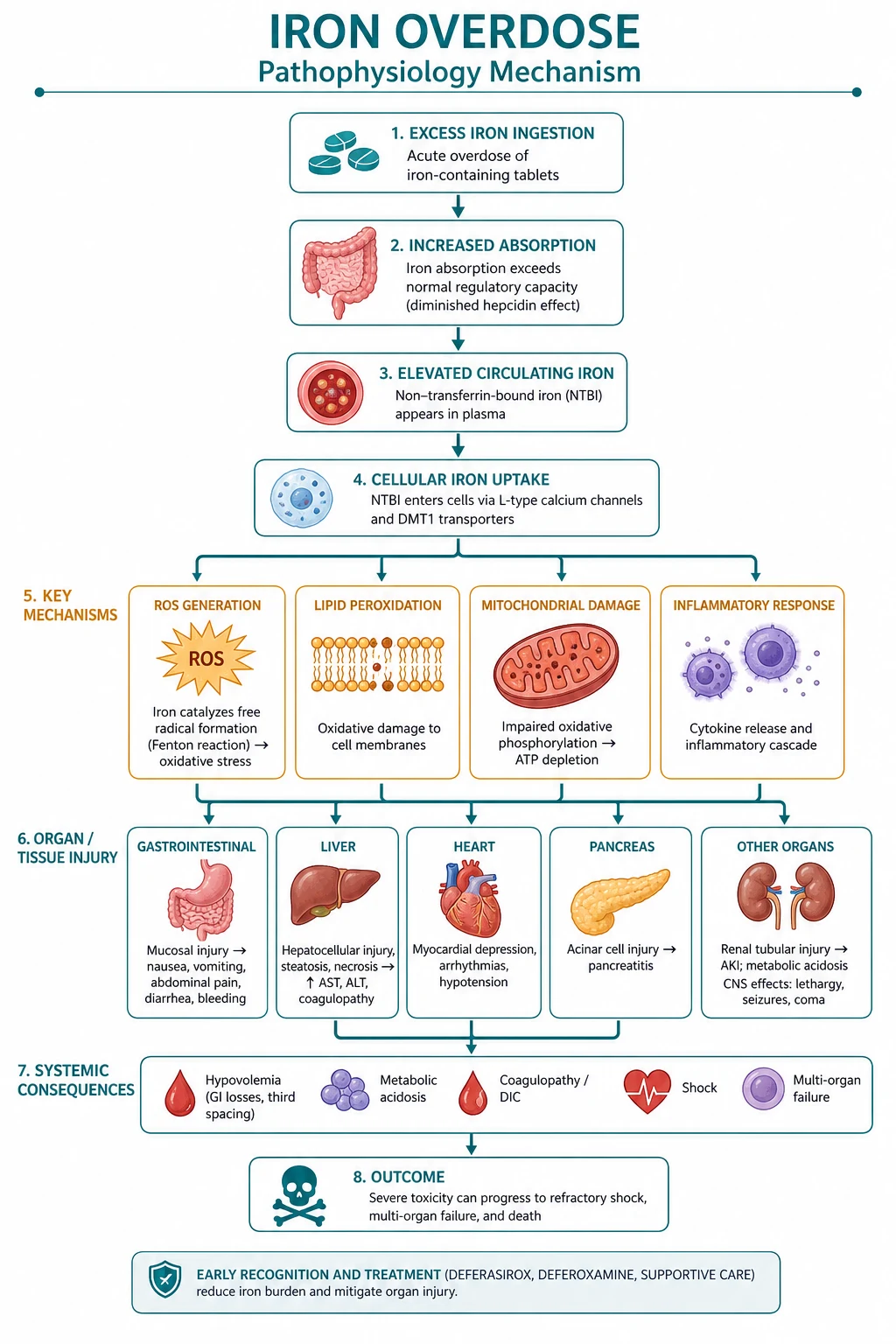
Iron injures the body in two mechanistically distinct waves: a direct corrosive effect on the gastrointestinal mucosa, and a systemic free-radical toxicity mediated by catalytic free (non-transferrin-bound) iron once the binding capacity of transferrin is exceeded. Understanding the second wave is the key to understanding every clinical and biochemical feature of stage 3.[5]
Wave 1 — gastrointestinal corrosive injury
In the stomach and duodenum, free iron in the lumen is directly caustic to the mucosa. The result is haemorrhagic gastritis (often erosive and confluent in the gastric antrum and pylorus), with vomiting that is frequently haematemetic, abdominal pain, and diarrhoea that may be frankly bloody. The fluid sequestration into the injured gut, plus overt GI bleeding, produces the hypovolaemic shock of stage 1.[1] The same corrosive injury is the substrate for the late scarring and gastric-outlet obstruction of stage 4.
Wave 2 — systemic free-radical toxicity (the Fenton reaction)
Once absorbed iron exceeds the iron-binding capacity of transferrin, the surplus circulates as free (non-transferrin-bound) iron. Free iron is taken up by cells (especially hepatocytes, via a transferrin-independent pathway) and becomes a redox catalyst in the Fenton reaction:[5]
The reaction is: Fe2+ + H2O2 -> Fe3+ + hydroxyl radical (OH•) + hydroxide ion (OH-). Iron is regenerated by reduction back to Fe2+, so a small amount of free iron catalyses a large, self-sustaining flux of hydroxyl radicals. [1]
The hydroxyl radical (OH•) is among the most reactive species in biology. It abstracts hydrogen atoms from polyunsaturated fatty acids in mitochondrial and cell membranes, initiating lipid peroxidation that propagates as a chain reaction. The membrane damage has three downstream consequences that explain the entire stage-3 phenotype:[5]
Mitochondrial uncoupling
- Lipid peroxidation of the inner mitochondrial membrane
- Electron leak from the electron transport chain — UNCOUPLES oxidative phosphorylation
- Impairs ATP synthesis; cells switch to anaerobic glycolysis
- Net result: accumulation of LACTATE — the high-anion-gap metabolic acidosis
Krebs-cycle blockade
- Free iron damages iron-sulphur (Fe-S) cluster enzymes, notably ACONITASE
- Krebs cycle stalls — no NADH/FADH2 substrate for the electron transport chain
- Compounds the ATP deficit and the lactate load
- Same biochemical lesion as cyanide/carbon monoxide, but with a different trigger
Na+/K+ ATPase inhibition
- Free iron inhibits the Na+/K+ ATPase of cell membranes
- Cell swelling, dysfunction and eventually necrosis
- Contributes to hepatocyte, renal tubular and myocardial injury
- Note the contrast with digoxin (which also inhibits Na+/K+ ATPase) — a classic comparison question
Why shock, hepatic necrosis and coagulopathy?
The cellular damage does not strike all organs equally.[5]
Shock. Free iron causes venodilatation and reduced systemic vascular resistance (peripheral vasodilatation) and depresses myocardial contractility. The shock of stage 3 is therefore a combination of hypovolaemia (residual GI losses), distributive (vasodilatation) and cardiogenic (myocardial depression) shock — one of the few poisons to produce all three patterns together.[1]
Hepatic necrosis. The liver is the preferential target because hepatocytes take up free iron avidly via a transferrin-independent pathway, and because zone 3 (centrilobular) hepatocytes — the most metabolically active, with the highest cytochrome P450 activity and oxygen demand — are the most vulnerable to oxidative injury. The result is centrilobular (zone 3) necrosis, the same pattern seen in paracetamol hepatotoxicity and ischaemic hepatitis.[5]
Coagulopathy. Free iron directly inhibits thrombin generation and the vitamin-K-dependent factors. The early, sometimes isolated, rise in PT/INR (within the first 12–24 h, before AST/ALT have peaked) is therefore an anticoagulant effect of iron itself, not yet hepatic synthetic failure. The later coagulopathy (after 24–48 h) is true hepatic synthetic failure. This distinction matters: an early INR rise does not by itself prove liver failure, but a rising INR with rising transaminases does.[1]
The basis of the latent phase
Stage 2 looks deceptively quiet because absorbed iron is being temporarily sequestered by transferrin in plasma and by the reticuloendothelial system. Once these binding sites are saturated — typically between 6 and 24 h after ingestion — free iron spills into tissues and stage 3 begins. The clinical lesson, repeated because examiners test it: in the latent window, clinical appearance is a lie; only the serum iron and the metabolic acidosis tell the truth.[1]
Clinical Presentation
The clinical presentation is the four-stage course described above, made real. The examiner will test whether you can recognise each stage, anticipate the next, and avoid being reassured by stage 2.[1]
Stage 1 (0 to 6 h) — gastrointestinal corrosive injury. Within the first one to two hours, the patient develops vomiting (often haematemetic), abdominal pain and diarrhoea (may be bloody). The abdomen may be diffusely tender. Dehydration is rapid in children. With significant doses, hypovolaemic shock is established within hours — tachycardia, delayed capillary refill, hypotension, oliguria, altered consciousness. The severity of stage 1 predicts progression: a patient who is shocked in stage 1 is at high risk of progressing to stage 3. [1]
Stage 2 (6 to 24 h) — the deceptive latent phase. The GI symptoms settle and the patient looks better. This is the dangerous window: clinicians may be falsely reassured and discharge the patient. The truth is that iron is being sequestered by transferrin and is about to saturate binding capacity. Disposition in this window must be based on the serum iron, the anion gap and the formulation, not on clinical appearance.[1]
Stage 3 (12 to 48 h) — systemic toxicity. This is the killer stage and the focus of intensive care. The patient develops, in variable combination:[1]
SHOCK-A-LC
Stage 4 (4 to 6 weeks) — late gastric-outlet obstruction. Weeks after recovery, scarring of the gastric antrum or pylorus (the sites of maximal corrosive injury) produces gastric or pyloric outlet obstruction. The patient presents with early satiety, post-prandial vomiting (sometimes of food eaten a day earlier) and weight loss. A succussion splash may be present. Upper GI endoscopy confirms the stricture; management is endoscopic balloon dilation, repeated as needed, with surgery for refractory cases.[1]
Atypical and modified presentations
Examiners deliberately probe the corners. Three atypical patterns deserve attention.[2]
Sustained-release / enteric-coated
- Delays stage 1 AND the serum-iron peak
- Peak iron may be at 8 to 12 h rather than 4 to 6 h
- Draw levels at 4 to 6 h AND repeat at 8 to 12 h
- Observe for at least 12 to 24 h; do not discharge early
Co-ingestion (paracetamol)
- Self-harm with iron plus paracetamol is common
- Paracetamol hepatotoxicity compounds the iron hepatitis
- Always send a paracetamol level at 4 h and apply the nomogram
- Treat BOTH — NAC plus desferrioxamine
The anuric / chronically-ill child
- May present late, after stage 1 has been attributed to gastroenteritis
- Low physiological reserve; decompensates rapidly
- Treat empirically with resuscitation and chelation while awaiting the iron level
- Beware fluid overload in the anuric patient
Differential Diagnosis
The differential of a patient presenting with vomiting, haematemesis, diarrhoea and a high-anion-gap metabolic acidosis is broad, and iron is only one cause. The discriminator is the history of iron-tablet ingestion, supported by radiopaque tablets on the abdominal X-ray and a serum iron level that is high, with hyperglycaemia and leucocytosis as clues.[2]
Iron overdose (the diagnosis)
- History of iron-tablet ingestion; calculate elemental iron mg/kg
- High-anion-gap metabolic ACIDOSIS, often with raised lactate
- RADIOPAQUE tablets on abdominal X-ray (30 to 50 percent of cases)
- Serum IRON over 500 microgram/dL; hyperglycaemia and leucocytosis
- Normal OSMOLAL gap; no visual symptoms (contrast methanol)
Salicylate overdose
- Tinnitus, hyperpnoea, mixed respiratory alkalosis THEN anion-gap acidosis
- Serum salicylate level high; history of aspirin ingestion
- Can co-exist; check BOTH salicylate and iron
- Treat with alkalinisation and haemodialysis (not desferrioxamine)
Toxic alcohols (methanol / ethylene glycol)
- High-anion-gap acidosis PLUS elevated OSMOLAL GAP (the dual gap)
- Methanol: visual symptoms, optic disc hyperaemia
- Ethylene glycol: calcium oxalate crystals, AKI, hypocalcaemia
- Normal serum iron; treat with fomepizole and haemodialysis
Metformin-associated lactic acidosis
- Diabetic patient on metformin; AKI; profound lactic acidosis
- Normal serum iron; no radiopaque tablets
- History of metformin ingestion; high lactate out of proportion
- Treat with haemodialysis and supportive care
Severe gastroenteritis / septic shock
- Fever, infective source, no iron-tablet history
- Normal serum iron; no radiopaque tablets
- Iron can MIMIC sepsis with leucocytosis and hyperglycaemia
- Cultures and lactate; the iron level settles it
Ischaemic hepatitis ('shock liver')
- Preceded by a profound hypotension episode (arrest, haemorrhage, sepsis)
- AST/ALT over 1000 that normalise within days
- Serum iron normal; iron overdose itself causes shock liver secondarily
- Treat the underlying cause
Paracetamol-induced hepatotoxicity
- Detectable paracetamol level; history; the nomogram
- Centrilobular necrosis (same pattern as iron)
- Co-ingestion common — check paracetamol in EVERY iron overdose
- Treat with N-acetylcysteine
The key manoeuvre at the bedside is to send serum iron, salicylate and paracetamol levels together, and to calculate the anion gap and (if any doubt) the osmolal gap. A normal osmolal gap with radiopaque tablets and a high iron level closes the case for iron.[2]
Clinical & Bedside Assessment
The bedside assessment has three objectives: estimate the elemental-iron dose, grade the current severity, and decide whether to decontaminate and chelate now.[1]
History. Establish the time since ingestion, the number and type of tablets (and calculate the elemental iron), the formulation (immediate-release versus sustained-release), any co-ingestants, the intent (accidental versus self-harm), and — in children — the access to adult iron tablets. Ask specifically about prenatal tablets, ferrous sulphate versus fumarate versus gluconate, and any sustained-release preparation.[4]
Examination. Perform a focused ABCDE: vital signs (HR, BP, RR, temperature, SpO2, GCS), hydration and perfusion (capillary refill, mucus membranes, skin), the abdomen (tenderness, peritonism, distension — beware perforation), and a search for stage-3 features (shock, jaundice, bleeding, oliguria, altered consciousness). A succussion splash weeks later suggests the stage-4 stricture.[1]
Estimate the elemental-iron dose at the bedside. Multiply the number of tablets by the elemental-iron content per tablet (ferrous sulphate 325 mg = 65 mg; ferrous fumarate 325 mg = 106 mg; ferrous gluconate 325 mg = 38 mg) and divide by the weight in kg. Stratify immediately:[4]
Plain abdominal X-ray. A KUB or abdominal X-ray may show radiopaque iron tablets in the stomach or small bowel. They are visible in roughly 30 to 50 percent of significant ingestions. Their presence confirms the diagnosis and guides decontamination (continue whole bowel irrigation until no tablets remain). Their absence does NOT exclude iron poisoning — tablets may have dissolved, been crushed, or be enteric-coated.[1]
Severity markers triggering desferrioxamine. Any one of: shock, metabolic acidosis, severe GI symptoms, altered consciousness, serum iron over 500 microgram/dL, or serum iron over 350 microgram/dL with symptoms.[1]
Investigations
The investigation bundle has three layers: the iron-specific tests, the severity / acid–base tests, and the rule-out co-ingestion tests.[2]
Iron-specific
- SERUM IRON at 4 to 6 h post-ingestion (the peak for immediate-release)
- Repeat at 8 to 12 h for sustained-release / enteric-coated formulations
- Abdominal X-ray for radiopaque tablets
- (Total iron-binding capacity is unreliable in acute overdose — do NOT wait for it)
Severity / acid-base
- VENOUS BLOOD GAS — pH, bicarbonate, lactate, base excess
- Serum ELECTROLYTES + ANION GAP (Na minus Cl plus HCO3; normal 8 to 12)
- GLUCOSE — iron causes hyperglycaemia
- FBC — iron causes leucocytosis
- COAGULATION (PT/INR) and LFTs (AST/ALT) — hepatic injury and coagulopathy
- UREA, creatinine, eGFR — acute kidney injury
Rule-out co-ingestion
- SERUM PARACETAMOL level at 4 h (apply the Rumack-Matthew nomogram)
- SERUM SALICYLATE level
- Beta-hCG in women of childbearing age
- Serum ethanol / toxic-alcohol screen if the anion gap plus osmolal gap pattern fits
Serum-iron thresholds
The serum iron drawn at 4 to 6 h is the central laboratory number. It must be interpreted alongside the acid–base status — a borderline iron level with a high-anion-gap acidosis is still significant toxicity.[1]
Timing caveat
For immediate-release formulations, the peak serum iron is at 4 to 6 h. For sustained-release and enteric-coated formulations, absorption is delayed and the peak may not occur until 8 to 12 h or later. Always draw a level at 4 to 6 h AND repeat it at 8 to 12 h for sustained-release formulations, and treat a rising or high level.[2]
The WBC–glucose–iron triad
Three non-specific clues support the diagnosis and are favourite exam material:[1]
W-G-I triad
The total iron-binding capacity (TIBC) — a trap
Historically a serum iron exceeding the TIBC (ratio over 1) was taken to indicate free iron. In practice, TIBC assays are unreliable in acute overdose (binding proteins are themselves affected, and the assay is slow). Do not wait for, or rely on, the TIBC ratio — treat on the iron level and the acid–base status.[1]
The desferrioxamine challenge test — abandoned
An older practice was to give 50 mg/kg intramuscular desferrioxamine (maximum 1 g) and observe for vin-rose urine ('rose test'). A positive result supposedly indicated free iron. Modern toxicology has abandoned the challenge test because it delays definitive IV chelation and is unreliable. Treat on the serum iron and acidosis.[3]
Management — Resuscitation
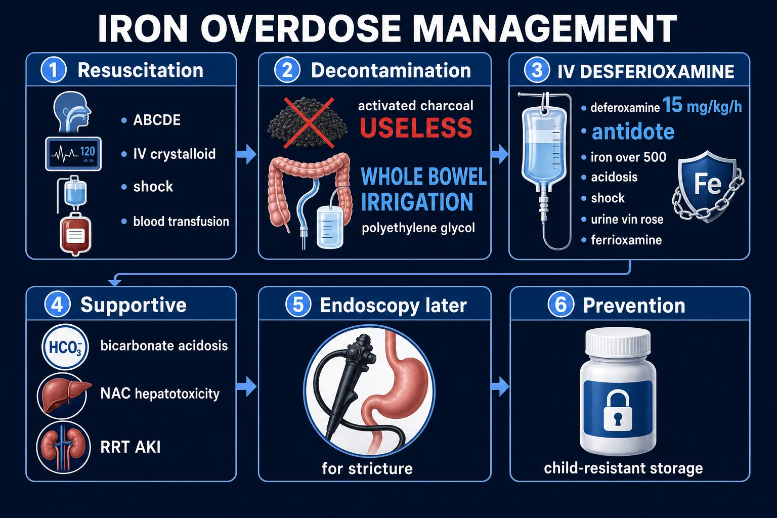
The resuscitation bundle is run concurrently with the investigations, not after them. The aims are to restore perfusion, secure the airway, control GI haemorrhage, prepare for decontamination and chelation, and avoid the useless and harmful interventions.[1]
ABCDE. Secure the airway (intubate early if comatose or shock is severe); give high-flow oxygen if hypoxic or in shock; establish two large-bore IV cannulae with continuous cardiac monitoring and (in severe cases) a urinary catheter to track output and the vin-rose colour.[1]
Fluid resuscitation. Give isotonic crystalloid (0.9% sodium chloride or a balanced crystalloid such as Hartmann's) in 10 to 20 mL/kg boluses, titrated to perfusion (capillary refill, blood pressure, urine output, lactate clearance). Children tolerate and may need larger relative boluses; the elderly and the anuric need caution (smaller boluses, frequent reassessment) to avoid pulmonary oedema. The shock of iron poisoning is multifactorial (hypovolaemic + distributive + cardiogenic), so vasopressors (noradrenaline) may be needed once intravascular volume is restored.[1]
Bloods before treatment. Draw the iron level, VBG, electrolytes, anion gap, glucose, FBC, PT/INR, LFTs, renal function, paracetamol and salicylate levels, and beta-hCG before starting desferrioxamine, because the chelator alters the iron assay. Cross-match if there is haematemesis.[2]
GI haemorrhage. Transfuse for significant bleeding; start a proton-pump inhibitor infusion; involve endoscopy and surgery early for uncontrolled haemorrhage or a large tablet bezoar. Iron-tablet bezoars may need endoscopic or surgical removal.[1]
The metabolic acidosis. The definitive treatment is desferrioxamine, which removes the iron generating the acid. Sodium bicarbonate has a limited adjunctive role for a pH under 7.1 to 7.2 — it improves haemodynamics and increases iron binding to transferrin — but it must not delay chelation. Do not chase the pH with bicarbonate at the expense of starting the chelator.[1]
What NOT to do. The single most important 'do not' in iron overdose: do not give activated charcoal. Iron is a metal, not an organic molecule, and charcoal does not adsorb it. Charcoal is useless here, obscures the view at endoscopy, and is not the decontamination of choice. The correct decontamination is whole bowel irrigation.[6][12]
Management — Definitive & Stepwise
The definitive management ladder runs from decontamination through chelation to organ-failure support. Each step has explicit triggers.[1]
Step 1 — Resuscitation (above)
ABCDE, crystalloid, oxygen, bloods before treatment; treat shock, acidosis and GI haemorrhage. [1]
Step 2 — Decontamination: whole bowel irrigation (NOT charcoal)
Whole bowel irrigation (WBI) is the decontamination of choice for iron. It is endorsed by the AACT/EAPCCT position statements.[11][12]
The AACT/EAPCCT position statement (Tenenbein 1997; updated 2023) endorses whole bowel irrigation with polyethylene-glycol electrolyte solution for iron ingestion, sustained-release/enteric-coated drugs, and body-packers. The UK NPIS (TOXBASE), Australasian poisons centres and the AAPCC consensus guidance all use the same protocol.[6]
Solution
- Polyethylene glycol (PEG) electrolyte solution — isosmotic, non-absorbed
- Same preparation used for bowel preparation before colonoscopy
- Given via a NASOGASTRIC TUBE
- Continue until the rectal effluent is clear AND no residual tablets on repeat KUB
Adult rate
- 1.5 to 2 L per hour via NG tube
- Continue for several hours (typically 4 to 6 h, longer for large bezoars)
- Metoclopramide 10 mg IV may help tolerate the rate
- Anti-emetics as needed
Paediatric rate
- 0.5 L per hour (or 20 to 40 mL/kg/h) via NG tube
- Careful attention to airway and aspiration risk in small children
- Continue until effluent clear
- A nasogastric tube is mandatory — a child will not drink this volume
Contraindications / cautions
- Bowel obstruction, perforation, ileus, haemodynamic instability
- Unprotected airway (aspiration risk)
- Massive GI haemorrhage
- Continue WBI even after starting desferrioxamine if tablets remain on imaging
Step 3 — Antidote: IV desferrioxamine (deferoxamine)
Desferrioxamine (deferoxamine, DFO) is a siderophore that binds free iron in a stable 1:1 complex (ferrioxamine), which is then excreted in the urine, giving it the characteristic vin-rose (pink-red) colour. It is the definitive treatment for significant iron toxicity.[1]
Indications (any one)
- Serum iron over 500 microgram/dL (90 micromol/L)
- High-anion-gap METABOLIC ACIDOSIS (the most reliable bedside trigger)
- SHOCK (any cause in this context)
- Severe GI symptoms (haematemesis, profuse diarrhoea)
- Altered consciousness, seizures, coma
- Serum iron over 350 microgram/dL WITH symptoms
Dose
- 15 mg/kg/h IV — the standard initial infusion rate
- Titrate to clinical response and falling serum iron
- Maximum roughly 6 g per 24 h in an adult
- Avoid rapid IV bolus — causes hypotension
- Typical total duration 6 to 12 h in moderate poisoning; up to 24 h in severe
When to stop
- 12 h AFTER the urine is no longer vin-rose
- AND the serum iron is falling
- AND the metabolic acidosis has resolved
- Do NOT chelate to zero — prolonged infusion causes ARDS and Yersinia sepsis
- Recheck iron level and VBG before stopping
Step 4 — Supportive care for organ failure
Stage-3 patients need intensive-care support for failing organs.[1]
Shock
- Noradrenaline is the first-line vasopressor (distributive + cardiogenic shock)
- Add vasopressin or adrenaline if refractory
- Continue fluid resuscitation guided by perfusion and lactate
- Consider bedside echocardiography for myocardial depression
Acidosis
- Desferrioxamine is definitive; bicarbonate adjunctive for pH under 7.1 to 7.2
- Do not chase the pH with bicarbonate at the expense of chelation
- Renal replacement therapy if AKI contributes
Hepatic failure / coagulopathy
- Fresh-frozen plasma and vitamin K for coagulopathy with bleeding
- N-acetylcysteine as an antioxidant adjunct (limited evidence, commonly used)
- Liver-transplant assessment for refractory acute liver failure (rare)
Renal failure
- Renal replacement therapy (haemofiltration / dialysis) for AKI
- Note: ferrioxamine is renally cleared — in AKI, chelation products accumulate
- Haemofiltration may remove some ferrioxamine but is not a primary iron-removal strategy
ARDS / respiratory
- Mechanical ventilation with lung-protective settings for ARDS
- Beware: BOTH iron toxicity AND prolonged desferrioxamine cause ARDS — distinguish by stopping chelation if iron is already falling
Seizures / coma
- Benzodiazepines (lorazepam 4 mg IV or diazepam 10 mg IV) for seizures
- Intubate and ventilate for airway protection in coma
Escalation triggers to ICU, endoscopy and surgery
ICU
- Any stage-3 feature: shock, acidosis, hepatic failure, ARDS, coma
- Need for vasopressors, mechanical ventilation or RRT
- Massive ingestion (over 60 mg/kg or sustained-release) regardless of initial appearance
Endoscopy
- Suspected tablet bezoar that does not progress on WBI
- Uncontrolled or recurrent GI haemorrhage
- To retrieve large adherent tablet masses
Surgery
- Surgical gastrotomy for an unremovable bezoar causing obstruction
- Suspected bowel perforation or ischaemia
- Stage-4 stricture refractory to endoscopic dilation
Specific Subtypes & Scenarios
Paediatric accidental ingestion (the commonest scenario)
The typical victim is a toddler (1 to 4 years) who has found adult prenatal or iron tablets. Calculate the elemental iron mg/kg at once.[4]
Under 20 mg/kg, asymptomatic
- Observe at home with poison-centre advice
- Return precautions: vomiting, drowsiness
- No investigations needed
Over 20 mg/kg or symptomatic
- ED assessment; serum iron at 4 to 6 h
- Abdominal X-ray if dose over 40 mg/kg
- Whole bowel irrigation if tablets present
Over 40 mg/kg
- Resuscitate as needed; admit
- Serum iron, VBG, glucose, FBC, PT/INR, LFTs
- IV desferrioxamine if iron over 500, acidosis, shock or severe symptoms
Prevention
- Child-resistant packaging is the single most effective measure
- Store iron and prenatal tablets locked and out of reach
- Poison-control education for families
Typically a young woman with access to iron tablets, often ingesting a large number of tablets, frequently sustained-release. Draw levels at 4 to 6 h AND repeat at 8 to 12 h; observe for at least 6 to 12 h (longer for sustained-release); low threshold for WBI and chelation; psychiatric assessment after medical stabilisation; secure the iron supply at discharge.[8]
Sustained-release and enteric-coated formulations
Absorption is delayed. Stage 1 may be prolonged or biphasic, the latent phase is distorted, and the serum-iron peak shifts to 8 to 12 h or beyond. Draw levels at 4 to 6 h AND repeat at 8 to 12 h, prolong WBI, and admit and observe for at least 12 to 24 h. Never discharge a sustained-release ingestion at 6 h.[2]
Pregnancy
Iron overdose in pregnancy typically involves prenatal iron. IV desferrioxamine IS indicated for severe maternal toxicity — the modern consensus is that maternal survival is the priority and deferring chelation for fear of fetal metal chelation costs maternal lives. Desferrioxamine crosses the placenta, but untreated maternal iron toxicity is far more dangerous to the fetus than chelation. Involve obstetrics, monitor the fetus, and remember that maternal outcome determines fetal outcome.[9][10]
Massive ingestion with tablet bezoar
A visible radiopaque mass on KUB that does not progress despite WBI may be a tablet bezoar. Endoscopic removal is first-line; surgical gastrotomy is reserved for unremovable bezoars causing obstruction or perforation. Continue WBI and chelation meanwhile.[1]
Co-ingestion (especially paracetamol)
Always send a paracetamol level at 4 h and apply the Rumack-Matthew nomogram. Co-ingestion worsens the hepatic prognosis (additive centrilobular necrosis) and may mask iron hepatitis. Treat both: N-acetylcysteine for the paracetamol, desferrioxamine for the iron.[2]
Complications & Pitfalls
Acute complications
ACID-ORGAN
Late complications
Gastric or pyloric outlet obstruction from scarring (4 to 6 weeks) is the classic late complication — early satiety, post-prandial vomiting, weight loss, succussion splash; managed by endoscopic balloon dilation or surgery. Less common: bowel stricture, intestinal perforation, chronic liver injury.[1]
Classic pitfalls
Latent-phase reassurance
- Discharging the patient who 'looks well' in stage 2
- Severity is determined by serum iron and acidosis, not appearance
- Always check both before discharge
Activated charcoal
- Giving charcoal — useless, iron is not adsorbed
- Obscures endoscopic view
- Use whole bowel irrigation instead
Single early iron level
- Sustained-release peak is delayed to 8 to 12 h
- Repeat the level; treat a rising or high level
- Do not be reassured by a single low early level
Under-dosing desferrioxamine
- 15 mg/kg/h is the standard; titrate to response
- Do not delay for a 'challenge test'
- Stop only when iron is falling and acidosis resolves
Prolonged desferrioxamine
- Over 24 h or very high dose causes ARDS ('desferrioxamine lung')
- Predisposes to Yersinia enterocolitica sepsis (siderophore effect)
- Stop once iron falling and acidosis resolved
Early PT rise misread
- Early INR rise is the anticoagulant effect of iron, NOT yet liver failure
- A rising INR with rising transaminases IS hepatic failure
- Distinguish the two at the bedside
Missing paracetamol
- Co-ingestion common in self-harm
- Always send a paracetamol level at 4 h
- Treat BOTH if positive
The desferrioxamine–ARDS and Yersinia pitfalls
Prolonged or very-high-dose desferrioxamine is associated with an acute lung injury picture ('desferrioxamine lung') and with an increased risk of Yersinia enterocolitica sepsis, because desferrioxamine is itself a siderophore that Yersinia uses to acquire iron. Suspect Yersinia sepsis in any febrile patient on desferrioxamine. The counter-measure is to stop chelation once the iron level is falling and the acidosis has resolved — do not chelate to zero.[7]
Prognosis & Disposition
The overall mortality of iron poisoning is under 5 percent in modern series with prompt chelation, but historically reached up to 45 percent in severe untreated ingestions. Severe poisoning — serum iron over 1000 microgram/dL, refractory acidosis, multi-organ failure — still carries significant mortality even with chelation.[2]
Predictors of poor outcome: large elemental-iron dose (over 60 mg/kg), sustained-release formulation, delay to desferrioxamine, anion-gap acidosis with pH under 7.1, hepatic failure with coagulopathy, multi-organ failure, and young age.[1]
Disposition criteria
Discharge from ED
- Asymptomatic throughout
- Peak serum iron under 350 microgram/dL
- No metabolic acidosis (normal venous pH and bicarbonate)
- No residual tablets on KUB
- Immediate-release formulation observed at least 6 h
- Psychiatric assessment completed (self-harm)
- Return precautions and poison-centre number given
Admit / HDU
- Symptomatic but stable moderate toxicity
- Treated with desferrioxamine, responding
- Sustained-release ingestion — observe 12 to 24 h
- Co-ingestion being worked up
ICU
- Any stage-3 feature (shock, acidosis, hepatic failure, ARDS, coma)
- Need for vasopressors, ventilation or RRT
- Massive ingestion regardless of initial appearance
Special Populations
Children (1 to 4 years)
- The typical accidental victim — weight-based elemental-iron calculation is essential
- Over 40 mg/kg: aggressive resuscitation, serum iron, consider WBI and chelation
- Child-resistant packaging is the single most effective prevention
- Poison-control referral for any paediatric iron ingestion
Pregnant women
- Iron overdose may involve prenatal iron
- IV desferrioxamine IS indicated for severe maternal toxicity
- Maternal survival is the priority; do not defer chelation
- Obstetric involvement and fetal monitoring; maternal outcome determines fetal outcome
Elderly
- May present atypically (confusion, multiple comorbidities)
- Reduced physiological reserve — lower threshold for ICU
- Caution with fluid resuscitation (heart failure risk)
- Watch for drug interactions and polypharmacy
Chronic iron overload / haemochromatosis
- Context of chronic chelation therapy
- Acute overdose on this background is rare but worsens hepatic prognosis
- Distinguish acute overdose from chronic iron overload — different management
Deliberate self-harm / psychiatric
- Safeguarding; secure the iron supply
- Psychiatric referral AFTER medical stabilisation
- Assess intent and underlying mental-health condition
- Do not discharge without psychiatric assessment
Evidence, Guidelines & Regional Differences
The 2005 AACT/EAPCCT consensus (Manoguerra et al.) on out-of-hospital management of iron ingestion set the triage thresholds still in use: refer to the emergency department for any symptoms, any ingestion over 40 mg/kg elemental iron (or any sustained-release ingestion over 40 mg/kg), or any intentional ingestion; home observation is acceptable only for asymptomatic ingestions under 20 mg/kg.[4]
Whole bowel irrigation is endorsed by the AACT/EAPCCT 1997 position statement (Tenenbein) and its 2023 update, as the decontamination modality of choice for iron, sustained-release/enteric-coated drugs, and body-packers; it is powered by polyethylene-glycol electrolyte solution run until the rectal effluent is clear.[11][12]
The 2026 Clinical Toxicology Recommendations Collaborative reaffirmed that decontamination remains useful in selected poisonings, including iron, and that activated charcoal has no role in metal (iron) poisoning — WBI is the modality.[6]
Controversies
The desferrioxamine challenge test. Historically, 50 mg/kg IM desferrioxamine with observation for vin-rose urine was used to 'test' for free iron. It has been abandoned in favour of early IV chelation based on the serum iron and acidosis, because it delays definitive treatment and is unreliable.[3]
Desferrioxamine dosing. Historical high-dose protocols (15 mg/kg/h uptitrated to 90 mg/kg/h) caused more ARDS and hypotension. Current practice favours 15 mg/kg/h titrated to clinical response with earlier cessation, balancing efficacy against the pulmonary and infectious complications of prolonged chelation.[1]
Total iron-binding capacity (TIBC). A serum iron exceeding the TIBC was once a chelation trigger. TIBC is unreliable in acute overdose and is not used to guide modern therapy.[1]
Regional deltas
Australasian poisons centres use the same WBI and desferrioxamine protocol; poison-information line (13 11 26) for triage.
In South Asia, where ferrous sulphate is a cheap, widely-available prenatal supplement, accidental paediatric poisoning remains a public-health issue. Prevention priorities are child-resistant packaging, poison-control education and safe storage. The management protocol (WBI, IV desferrioxamine) is identical.[8]
Exam Pearls
Mechanism one-liner
- Iron = CORROSIVE to GI mucosa + SYSTEMIC free-radical toxin
- Fenton reaction: Fe2+ + H2O2 -> Fe3+ + hydroxyl radical
- Uncouples oxidative phosphorylation -> lactic acidosis
- Centrilobular hepatic necrosis; inhibits thrombin -> coagulopathy
Toxic dose one-liner
- Under 20 mg/kg asymptomatic; over 20 mild
- Over 40 significant; over 60 severe/lethal
- ELEMENTAL iron (ferrous sulphate 325 mg = 65 mg elemental)
- Ferrous fumarate 325 mg = 106 mg; ferrous gluconate 325 mg = 38 mg
Four-stage course
- (1) 0-6 h GI corrosive + hypovolaemic shock
- (2) 6-24 h DECEPTIVE latent phase
- (3) 12-48 h shock + acidosis + hepatic/renal failure + coma (the killer)
- (4) 4-6 weeks gastric/pyloric stricture
Investigations
- Serum iron at 4-6 h (over 500 = chelate); repeat at 8-12 h for sustained-release
- Anion-gap metabolic ACIDOSIS
- Abdominal X-ray = RADIOPAQUE tablets
- Hyperglycaemia + leucocytosis (the WBC-glucose-iron triad)
Decontamination
- Activated charcoal is USELESS
- Iron is a metal, not adsorbed
- Use WHOLE BOWEL IRRIGATION (polyethylene glycol)
- Adult 1.5-2 L/h; child 0.5 L/h via NG, until effluent clear
Antidote
- IV DESFERRIOXAMINE (deferoxamine) 15 mg/kg/h
- For iron over 500, acidosis, or shock
- Urine turns VIN ROSE (ferrioxamine)
- Stop when iron falling + acidosis resolved (avoid ARDS, Yersinia)
Latent-phase pitfall
- Never discharge on clinical grounds in 6-24 h window
- Use serum iron AND metabolic acidosis
- Sustained-release: delayed peak, longer observation
Pregnancy
- IV desferrioxamine IS indicated for severe maternal toxicity
- Maternal survival is the priority
- Do not defer chelation
Late complication
- Gastric/pyloric outlet obstruction at 4-6 weeks
- Scarring of the corrosively injured antrum/pylorus
- Endoscopic balloon dilation or surgery
Exam application bank (NEET-PG / INICET)
One-line answer
Iron overdose is one of the leading causes of accidental poisoning death in children (adult prenatal/iron tablets look like sweets) and an occasional means of deliberate self-harm in adults. Iron is corrosive to the gastrointestinal mucosa (haemorrhagic gastritis, vomiting, diarrhoea) and, once absorbed iron exceeds transferrin binding capacity, becomes a systemic free-radical toxin via the Fenton reaction — it uncouples oxidative phosphorylation, blocks the Krebs cycle and produces lactic (high-anion-gap) acidosis, centrilobular hepatic necrosis, coagulopathy (direct thrombin inhibition), shock and multi-organ failure. Toxic dose (elemental iron): under 20 mg/kg usually asymptomatic; over 20 mg/kg mild; over 40 mg/kg significant; over 60 mg/kg severe, potentially lethal. Four clinical stages: Stage 1 (0 to 6 h) GI corrosive injury + hypovolaemic shock; Stage 2 (6 to 24 h) deceptive late [1]
Worked stems (answer without another resource)
Stem 1 — Classic presentation. Map symptoms to mechanism; name the first investigation and first treatment step with dose/route if drug therapy is standard. [1]
Stem 2 — Unstable / complicated. List red flags that force immediate resuscitation, theatre, ICU, antidote, or reperfusion — and what you do in the first 15 minutes. [1]
Stem 3 — Atypical group. Elderly, pregnancy, child, or immunocompromised: how presentation and thresholds change. [1]
Stem 4 — Differential trap. Name the three closest mimics and one discriminator for each. [1]
Stem 5 — Disposition. Who goes home with safety-netting, who is admitted, who needs HDU/ICU/theatre, and what follow-up is mandatory. [1]
Rapid viva checklist
- Definition + classification
- Pathophysiology chain
- Bedside signs / criteria
- Score with exact components (if any)
- Emergency bundle
- Definitive therapy with doses
- Complications of disease and of treatment
- Special populations
- Guideline/trial name if classic
- Three exam traps
Coverage self-check
If you cannot answer any stem above from this page alone, re-read the matching section — the page is intended to be self-sufficient for final-prof and NEET-PG/INICET questions on Iron Overdose.
References
- [1]Fine JS. Iron poisoning Curr Probl Pediatr, 2000.PMID 10742921
- [2]Chang TP, Rangan C. Iron poisoning: a literature-based review of epidemiology, diagnosis, and management Pediatr Emerg Care, 2011.PMID 21975503
- [3]Madiwale T, Vereb S, Muzumdar H, Donn S, Rosenfeld W. Iron: not a benign therapeutic drug Curr Opin Pediatr, 2006.PMID 16601499
- [4]Manoguerra AS, Erdman AR, Booze LL, Christianson G, Wax PM, Scharman EJ, Woolf AD, Keyes DC, Olson KR, Chyka PA, Caravati EM, Troutman WG. Iron ingestion: an evidence-based consensus guideline for out-of-hospital management Clin Toxicol (Phila), 2005.PMID 16255338
- [5]Rafati Rahimzadeh M, Rafati Rahimzadeh M, Kazemi S, Moghadamnia AA. Iron; Benefits or threatens (with emphasis on mechanism and treatment of its poisoning) Hum Exp Toxicol, 2023.PMID 37526177
- [6]Hoegberg LCG, Good AM, Tracy JA, Rubino MM, Höjer J, Cottrell E, Buckley NA, Thanacoody HKR, Greene SL, Cincinnati Clinical Toxicology Recommendations Collaborative. Recommendations from the Clinical Toxicology Recommendations Collaborative on the administration of activated charcoal in acute oral overdose Clin Toxicol (Phila), 2026.PMID 41906697
- [7]Mofenson HC, Caraccio TR, Sharieff N. Iron sepsis: Yersinia enterocolitica septicemia possibly caused by an overdose of iron N Engl J Med, 1987.PMID 3561464
- [8]Chandran J, Khanna K, Jayashree M, Gautam V, Sharma A, Singh M. Accidental iron poisoning in children - Experience from a teaching institution J Family Med Prim Care, 2023.PMID 38074262
- [9]Tran T, Waxman B, Akgur FM, Hcl T, Tanyel FC, Turkmen MA, Buyukpamukcu N, Oudesluys-Murphy AM. Intentional iron overdose in pregnancy--management and outcome J Emerg Med, 2000.PMID 10699527
- [10]Tran T, Waxman B, Nguyen A, Leong M, Tsueda K, Ueland K, Vohra BK, Roszler MH. Acute intentional iron overdose in pregnancy Obstet Gynecol, 1998.PMID 9764661
- [11]Tenenbein M, Lheureux P, Position Statement Writing Group for the AACT/EAPCCT. The role of whole bowel irrigation in the treatment of toxic ingestions Br J Clin Pharmacol, 2023.PMID 36639859
- [12]Tenenbein M. Position statement: whole bowel irrigation. American Academy of Clinical Toxicology; European Association of Poisons Centres and Clinical Toxicologists J Toxicol Clin Toxicol, 1997.PMID 9482429