Emergency & Toxicology · General Medicine
Lead Poisoning (Plumbism)
Also known as Lead poisoning · Plumbism · Lead toxicity · Saturnism · Chelation therapy
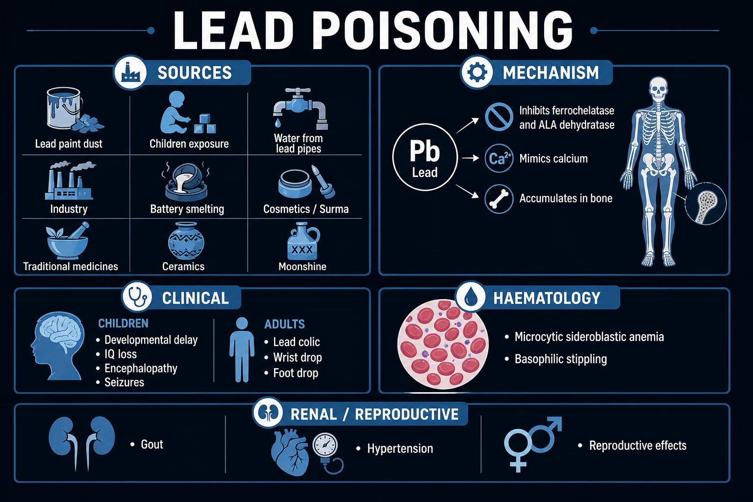
Lead poisoning (plumbism/saturnism) is toxic accumulation of lead (Pb), a heavy metal with NO biological role and NO safe blood level, causing multisystem harm — neurotoxic (especially irreversible IQ loss in children), microcytic sideroblastic anaemia with basophilic stippling, lead colic, motor peripheral neuropathy (wrist drop), nephropathy, saturnine gout, hypertension, and reproductive damage. Sources: lead-based paint (pre-1970s housing, children ingest flakes/dust), contaminated water (lead pipes/solder), industrial exposure (smelting, battery recycling, ammunition, soldering, foundry, demolition), traditional cosmetics (surma/kohl, sindoor), Ayurvedic/herbal medicines, ceramic glazes, leaded petrol (legacy environmental), moonshine, toys/jewellery, retained bullets. Lead mimics calcium (deposits in bone/teeth — metaphyseal 'lead lines'; crosses placenta and blood-brain barrier) and inhibits ALA dehydratase and ferrochelatase → sideroblastic anaemia with basophilic stippling. Diagnosis: whole blood lead level + FBC (basophilic stippling). Treat: remove source + chelation — succimer (DMSA, oral) for moderate; calcium disodium EDTA (IV) for moderate-severe; dimercaprol (BAL, IM) FIRST then EDTA for encephalopathy. Prevention (public health) is paramount — neurodevelopmental damage is irreversible.
On this page & tools
Your progress
Saved locally on this device.
Exam tags
Red flags
Overview & Definition
Lead poisoning (plumbism, saturnism) is a toxic accumulation of the heavy metal lead (Pb) — an element with no biological role and no safe blood level — producing multisystem harm: neurotoxicity (dominant in children), haematological (sideroblastic anaemia), gastrointestinal (lead colic), peripheral nervous system (motor neuropathy — wrist drop), renal (chronic tubulointerstitial nephritis, Fanconi syndrome), cardiovascular (hypertension), and reproductive effects. The names reflect its history: plumbum is the Latin for lead (hence the chemical symbol Pb); Saturn was the alchemical symbol for lead, hence saturnism.[6][1]
Lead is high-yield because of its classic multisystem features (especially the haematological and neurological), its concentration in children (where it causes irreversible neurodevelopmental harm at low levels), its specific chelation therapy, and its public-health dimension (prevention is the only effective strategy for the cognitive damage). The clinical skill is in suspecting lead exposure from the history (housing, occupation, traditional remedies/cosmetics), recognising the syndrome — abdominal pain + microcytic anaemia with basophilic stippling + motor neuropathy or encephalopathy — and confirming with a whole blood lead level. There is no safe blood lead level in children: even levels under 5 micrograms/dL are associated with measurable IQ loss, and in 2021 the CDC lowered the blood lead reference value to 3.5 micrograms/dL.[1][1]
Classification
Lead poisoning is classified in three complementary axes that together determine the clinical picture and the response: [1]
By chronicity: [1]
- Acute high-dose poisoning — large recent ingestion/inhalation; presents with lead colic, encephalopathy, seizures, haemolytic anaemia; blood lead often over 70 micrograms/dL. Seen in pica (lead paint chips), massive occupational exposure, lead dust inhalation.
- Chronic low-dose exposure — the commonest modern pattern; insidious, with subtle neurodevelopmental IQ loss in children, anaemia, hypertension, nephropathy; blood lead 5–44 micrograms/dL. The pattern that matters most for population health. [1]
By age / host: [1]
- Paediatric (under 6) — neurodevelopmental phenotype; higher absorption (50% vs 10%), developing blood-brain barrier, pica behaviour.
- Adult occupational — GI + peripheral nerve phenotype; inhalation of lead dust/fumes; wrist drop, lead colic, nephropathy, hypertension.
- Pregnancy — transplacental transfer; fetal neurotoxicity, miscarriage. [1]
By severity / blood lead level (drives management — see Investigations and Management for full thresholds): [1]
- Reference range / no action (adults): blood lead under 5 micrograms/dL.
- CDC reference value (children): 3.5 micrograms/dL (97.5th percentile of NHANES — triggers public health action, NOT a toxicity threshold).[1]
- Mild (5–44 micrograms/dL): source removal, surveillance, nutritional support; no chelation.
- Moderate (45–69 micrograms/dL, children; 50–70, adults): chelation indicated — oral succimer (DMSA) first-line.
- Severe (over 70 micrograms/dL) or any encephalopathy: IV EDTA ± dimercaprol (BAL); ICU.[2]
Epidemiology & Risk Factors
Lead poisoning is a global environmental disease. Worldwide, lead exposure accounts for hundreds of thousands of attributable deaths and over 20 million disability-adjusted life-years (DALYs) annually, with the burden concentrated in low- and middle-income countries where lead remains in paint, petrol-legacy soil, informal industry (battery recycling, artisanal mining), traditional medicines, and cosmetics.[1]
Populations at highest risk: [1]
- Children under 6 — higher GI absorption (~50% vs ~10% in adults), developing nervous system (irreversible IQ loss), pica behaviour (hand-to-mouth activity, eating paint chips/dust), and a shorter blood-brain barrier maturation window.
- Occupational adults — battery manufacture/recycling, lead smelting/refining, ammunition manufacture/use (firing ranges), soldering/welding, foundry, demolition, radiator repair, painting/paint removal, stained-glass artistry, pottery/ceramics with lead glaze. Inhalation of lead dust and fumes is the principal route; take-home exposure on work clothing/hair exposes the worker's children.
- Low-income families in older housing — pre-1970s housing with deteriorating lead-based paint (banned for residential use in many countries from the 1970s–80s).
- Cultural/regional exposure — surma/kohl (eyeliner applied to children in South Asian and Middle Eastern communities), sindoor (vermillion, may contain lead oxide), Ayurvedic Bhasma medicines, Mexican pottery with lead glaze, Gasoline legacy in countries slow to ban leaded petrol.
- Pregnant women and their fetuses — lead mobilised from maternal bone during pregnancy (calcium turnover); crosses placenta. [1]
Landmark outbreaks and contexts: [1]
- Flint, Michigan water crisis (2014–) — switch of water source caused corrosive water to leach lead from old pipes; population-level elevation of blood lead in children.
- Zamfara, Nigeria (2010) — artisanal gold mining using lead-contaminated ore processed at home; over 400 children died of acute lead encephalopathy; mass oral succimer chelation deployed.[3]
- Leaded petrol phase-out — completed globally only by 2021 (Algeria last); a major public-health triumph that halved population blood lead over 30 years.[1]
Pathophysiology
Lead poisoning is fundamentally a story of how a metal that mimics calcium hijacks the body's storage and signalling machinery and poisons the haem-synthetic and neuronal pathways.[6][1]
Absorption. Lead is absorbed by three routes: (i) gastrointestinal — the principal route in children; absorption is markedly higher in children (~50%) than adults (~10%) and is further enhanced by iron, calcium, and zinc deficiency and by fasting; (ii) respiratory — near-complete absorption of inhaled lead dust and fumes, the dominant route in occupational exposure; (iii) skin — negligible for inorganic lead, but organic lead (tetraethyl lead, the former petrol additive) is well-absorbed through skin, causing predominantly CNS toxicity ("loony gas" / encephalopathy). [1]
Distribution. Once absorbed, lead distributes across three compartments: (i) blood — where over 99% is bound to erythrocytes (chiefly haemoglobin), with a half-life of about 30 days; blood lead therefore reflects recent exposure; (ii) soft tissues (liver, kidney, brain, bone marrow) — also a half-life of weeks; (iii) bone — the major reservoir, holding over 90% of total body lead in adults (about 70% in children) with a half-life of years to decades. Lead in bone is mobilised during pregnancy, lactation, menopause, osteoporosis, and hyperthyroidism (calcium turnover) — so a woman with childhood exposure can re-expose herself and her fetus decades later. [1]
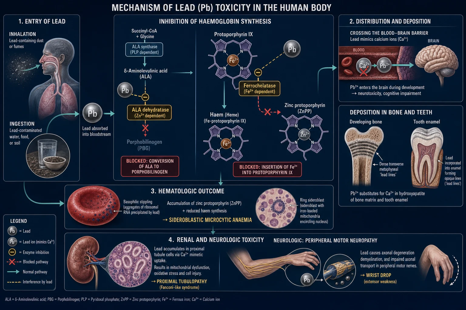
Lead mimics calcium. This is the central toxicological principle. Lead substitutes for calcium in bone hydroxyapatite and in cellular signalling; it is deposited in developing bone matrix and tooth enamel (the radiographic "lead lines"), crosses the blood-brain barrier and the placenta, and is taken up by developing neurons in preference to calcium.[6]
Haematological toxicity — the classic mechanism. Lead poisons the haem biosynthetic pathway at two enzyme steps: [1]
- delta-aminolaevulinic acid dehydratase (ALA dehydratase / porphobilinogen synthase) — inhibition blocks the condensation of two ALA molecules to porphobilinogen → urinary ALA rises.
- ferrochelatase — inhibition blocks the insertion of Fe2+ into protoporphyrin IX to form haem; zinc instead inserts, producing zinc protoporphyrin (ZPP), which accumulates in red cells. [1]
The net result is impaired haemoglobin synthesis → sideroblastic (microcytic, hypochromic) anaemia with ringed sideroblasts in the marrow and, on the blood film, the pathognomonic basophilic stippling — coarse blue-purple granules representing aggregated degenerated ribosomes that the lead-damaged red cell cannot clear. Lead also shortens red-cell survival (mild haemolysis), compounding the anaemia. [1]
Neurotoxicity — the irreversible injury. In the developing brain, lead disrupts synaptogenesis, neurotransmitter release (dopaminergic, glutamatergic, cholinergic), NMDA receptor function, second-messenger signalling (protein kinase C, calmodulin), and myelination, and induces neuronal apoptosis. The consequence is irreversible IQ loss, behavioural change (attention deficit, aggression, impulsivity), and at high levels acute lead encephalopathy (cerebral oedema, seizures, coma). The injury is dose-dependent with no threshold.[1][1]
Peripheral nervous system. Lead produces a predominantly motor axonal neuropathy with segmental demyelination, preferentially affecting long motor nerves — classically the radial nerve (wrist drop) and the common peroneal nerve (foot drop) — with relative sparing of sensation. The mechanism is axonal transport disruption and Schwann-cell damage. [1]
Renal toxicity. Lead accumulates in proximal tubular cells, producing (in children with high acute exposure) Fanconi syndrome (generalised proximal tubular dysfunction — glycosuria with normal blood glucose, aminoaciduria, phosphaturia, renal tubular acidosis) and, with chronic exposure, chronic tubulointerstitial nephritis with progressive renal impairment, hyperuricaemia, and hypertension.[2]
Gastrointestinal (lead colic). Lead causes spasm of intestinal smooth muscle and altered enteric neurotransmission → severe cramping abdominal pain with constipation (lead colic) — a symptom that has masqueraded as an acute abdomen. [1]
Hyperuricaemia and saturnine gout. Lead inhibits renal uric acid excretion → hyperuricaemia and gout ("saturnine gout"), classically in chronically exposed adults. [1]
Hypertension. Chronic lead exposure is an under-recognised cause of hypertension, via renal tubular damage, oxidative stress, and altered nitric oxide / renin-angiotensin signalling.[2]
Reproductive toxicity. Lead reduces fertility in both sexes (sperm abnormalities, menstrual disturbance), causes miscarriage, prematurity, and low birth weight, and is excreted in breast milk. [1]
Clinical Presentation
The clinical face of lead poisoning depends on the age of the patient and the level and chronicity of exposure.[5][1]
Children — CNS dominant. Chronic low-level exposure produces developmental delay, speech delay, learning disability, behavioural change (attention deficit, hyperactivity, aggression, impulsivity), IQ loss, and dropping school performance — often the only clues, easily mistaken for primary behavioural/learning disorders. Higher levels produce lethargy, irritability, anorexia, intermittent vomiting, constipation, and the life-threatening acute lead encephalopathy — ataxia, drowsiness, seizures, coma, with signs of raised intracranial pressure (vomiting, papilloedema). Encephalopathy typically occurs at blood lead over 70 micrograms/dL in children and is a medical emergency. [1]
Adults — GI and peripheral-nerve dominant. The classical picture is lead colic — intermittent, severe, cramping abdominal pain with constipation, often localised and without peritonism or fever, easily confused with an acute surgical abdomen. Motor peripheral neuropathy develops insidiously: wrist drop (radial nerve; weakness of wrist and finger extensors) is the hallmark, less commonly foot drop (common peroneal nerve). Sensation is usually spared. Constitutional symptoms — fatigue, malaise, headache, irritability, memory and concentration impairment, sleep disturbance, reduced libido — are common. Anaemia causes pallor and exertional dyspnoea. [1]
Signs and physical findings across ages: [1]
- Burton's line (gum lead line) — a blue-grey line at the gingival margin (lead sulfide precipitated by bacterial hydrogen sulfide), more prominent with poor dental hygiene; an exam-classic sign but absent in edentulous patients and in many cases.[6]
- Pallor (anaemia), hypertension, signs of chronic kidney disease or gout (tophi) in long-standing exposure.
- Metaphyseal 'lead lines' are radiological, not clinical.
- Neurological: wrist/foot drop, in severe cases encephalopathy (confusion, seizures, ataxia, coma).
Atypical presentations: [1]
- Elderly — hypertension or chronic kidney disease as the sole manifestation; osteoporosis-related lead mobilisation from bone; cognitive decline misattributed to dementia.
- Pregnancy — unexplained miscarriage, pre-eclampsia, or reduced fetal growth; lead is mobilised from maternal bone.
- Lactation — lead passes into breast milk, exposing the infant.
- Retained bullet/lead fragment — chronic lead leaching, especially if the fragment is intra-articular, in a joint, or in contact with synovial fluid/bursa; may present years after injury with abdominal pain, anaemia, or neuropathy.
- Acute massive ingestion (pica) — abdominal pain with radiopaque material on abdominal X-ray. [1]
Sources of lead — 'PLUMBER'
Differential Diagnosis
Lead poisoning mimics several commoner conditions; the differentiator is always the exposure history plus the blood lead level.[2][5]
Differential diagnosis of the three cardinal lead syndromes
Microcytic anaemia + basophilic stippling
- Iron-deficiency anaemia — low ferritin, no stippling, no lead exposure; correct with iron.
- Thalassaemia — microcytosis out of proportion to anaemia, target cells, basophilic stippling rare; Hb electrophoresis confirms; family history.
- Primary sideroblastic anaemia — ringed sideroblasts on marrow; lead level normal.
- Differentiator: blood lead level elevated + exposure history + ZPP elevated.
Cramping abdominal pain + constipation (lead colic)
- Acute abdomen (appendicitis, cholecystitis, perforation) — peritonism, fever, raised inflammatory markers; lead colic has NO peritonism.
- Acute intermittent porphyria (AIP) — ALSO an ALA-pathway defect; abdominal pain + neuropathy + psychiatric features; PLUS photosensitivity, red wine-coloured urine, family history; no lead exposure.
- Opioid / anticholinergic constipation — drug history; no anaemia or stippling.
- Differentiator: lead colic — diffuse tenderness without guarding, microcytic anaemia with stippling, elevated blood lead.
Motor peripheral neuropathy (wrist drop)
- Guillain-Barre syndrome — ascending, predominantly motor with reflex loss, often sensory involvement, CSF albuminocytologic dissociation.
- Motor neuron disease — progressive pure motor weakness without sensory loss, no abdominal pain or anaemia.
- Cervical radiculopathy / radial-nerve entrapment ('Saturday-night palsy') — unilateral, isolated, no systemic features.
- Differentiator: lead — occupational exposure, sensory usually spared, abdominal pain + anaemia point the way.
Encephalopathy in a child
- Meningitis / encephalitis — fever, meningeal signs, CSF pleocytosis; blood lead normal.
- Reye syndrome — antecedent viral illness + aspirin, hepatic dysfunction with microvesicular steatosis.
- Inborn errors of metabolism — recurrent, episodic, specific biochemical markers.
- Differentiator: lead — exposure history, basophilic stippling, blood lead, abdominal X-ray radiopaque flakes.
The single most examined differential is lead poisoning vs acute intermittent porphyria (AIP) — both block the haem/porphyrin pathway and produce the triad of abdominal pain + peripheral neuropathy + neurological/psychiatric symptoms. AIP additionally causes photosensitivity, red/wine-coloured urine on standing, and a positive family history, and shows elevated urinary porphobilinogen (PBG); lead poisoning shows elevated urinary ALA with normal or low PBG, basophilic stippling, a gum lead line, and an exposure history. Both can coexist with the same enzymes affected — but the enzyme blocks differ (AIP = porphobilinogen deaminase; lead = ALA dehydratase + ferrochelatase). [1]
Clinical & Bedside Assessment
The diagnosis of lead poisoning begins at the bedside history, not the laboratory.[1][2]
History — the decisive step. Take a structured exposure history: (i) Housing — age of home (pre-1970s), condition of paint (peeling/flaking), recent renovation, presence of lead pipes or lead-soldered plumbing; (ii) Occupation — battery work, smelting, foundry, demolition, ammunition manufacture/use (firing ranges), soldering, welding, painting/paint removal, radiator repair, stained glass, pottery with lead glaze; (iii) Hobbies — pottery, stained glass, jewellery-making with lead solder, fishing-weight casting, reloading ammunition, restoring old furniture/houses; (iv) Traditional remedies and cosmetics — surma/kohl, sindoor, Ayurvedic Bhasmas, folk remedies, imported herbs; (v) Behaviour — pica in children (eating paint chips, soil, paper); (vi) Water and food — source (private well, lead service line), ceramic-glaze food storage, moonshine consumption; (vii) Migration/travel — recent immigration from high-exposure countries; (viii) Past injury — retained bullet or lead fragment. Take also a family/household history (other children, take-home exposure). [1]
Examination — named signs to actively look for: [1]
- Vital signs — blood pressure (lead-related hypertension), conscious level (encephalopathy).
- Mouth — Burton's line (blue-grey gingival margin), dental hygiene.
- Abdomen — diffuse tenderness without peritonism, constipation (palpable faeces); exclude surgical pathology.
- Neurological — motor strength (wrist drop, foot drop; sensory usually intact), reflexes, coordination (ataxia in encephalopathy), developmental assessment in children ( milestones, behaviour, attention, IQ estimate).
- Skin — pallor (anaemia), tophi (saturnine gout).
- Growth in children — failure to thrive, developmental regression. [1]
Bedside manoeuvres / radiographs: [1]
- Abdominal X-ray — may show radiopaque flecks of ingested lead paint or pica material; guides whole-bowel irrigation.
- Long-bone X-ray (knee/wrist) in children — dense transverse metaphyseal 'lead lines' (lead deposited in the zone of provisional calcification at growing ends); a high-burden sign. [1]
Investigations
The definitive test is the whole blood lead level.[1][2]
First-line investigations: [1]
- Whole blood lead level (venous) — the gold standard. Use venous blood (capillary finger-prick samples are prone to skin contamination and are only for screening; a high capillary result MUST be confirmed with venous blood). Reflects recent exposure (blood half-life ~30 days). Reference thresholds:
- Adults — under 5 micrograms/dL normal; over 5 warrants investigation/source removal.
- Children — CDC blood lead reference value 3.5 micrograms/dL (the 97.5th percentile of NHANES childhood levels); this is a public-health action level, NOT a toxicity threshold.[1]
- Chelation thresholds — children over 45 micrograms/dL or adults over 50–70 → succimer (DMSA); over 70 or any encephalopathy → EDTA ± BAL.[2][3]
- Full blood count (FBC) — microcytic, hypochromic anaemia (sideroblastic; may mimic iron deficiency but iron stores are normal/high).
- Peripheral blood film — basophilic stippling (coarse blue-purple granules — pathognomonic clue), may also show polychromasia from haemolysis.
- Iron studies — to distinguish from iron deficiency; in lead, ferritin and marrow iron are normal or increased (sideroblastic block).
- Blood film / marrow — ringed sideroblasts on Prussian-blue staining of marrow.
Supporting tests (chronic exposure): [1]
- Zinc protoporphyrin (ZPP) / free erythrocyte protoporphyrin (FEP) — elevated; reflects ferrochelatase inhibition and integrates over the red-cell lifespan (~120 days), so a marker of chronic exposure (more useful than blood lead for long-term burden).
- Urinary delta-aminolaevulinic acid (ALA) — elevated (ALA dehydratase inhibition); largely a research/specialised test now.
- Renal function (U&E, creatinine, eGFR), urinalysis — chronic tubulointerstitial nephritis; Fanconi syndrome in children (glycosuria with normal blood glucose, aminoaciduria, phosphaturia).
- Serum uric acid — elevated (saturnine gout).
- Liver function tests — usually normal; may be mildly abnormal.
- Pregnancy test — in women of reproductive age (lead crosses placenta; affects management).
- Hb electrophoresis — if thalassaemia in the differential. [1]
Imaging: [1]
- Long-bone X-ray (children) — dense transverse metaphyseal 'lead lines' at the distal femur, proximal tibia, and distal radius — reflect lead deposited at the growing metaphysis during chronic exposure.
- Abdominal X-ray — radiopaque lead paint flakes or pica material; guides whole-bowel irrigation in acute ingestion.
- K-shell X-ray fluorescence (XRF) of bone — non-invasive measure of cumulative bone lead (research / specialist occupational medicine). [1]
Blood lead thresholds — what to do
Management — Resuscitation
The patient with acute lead encephalopathy is a time-critical medical emergency.[2][5]
ABCDE first. Secure the airway (encephalopathic patients may lose airway reflexes; intubate if GCS impaired), give high-flow oxygen, establish IV access, and monitor continuous ECG, SpO2, BP, neurology. [1]
Seizures and raised intracranial pressure: [1]
- Treat seizures with IV lorazepam (adult 4 mg, child 0.1 mg/kg; repeat once) or diazepam; if recurrent, phenytoin/fosphenytoin or levetiracetam.
- Manage raised ICP — head-of-bed elevation 30 degrees, normocapnia, hypertonic saline or mannitol; avoid over-hydration (worsens cerebral oedema). Neurosurgical/intensive-care input. [1]
Immediate chelation for encephalopathy — the critical reflex: [1]
- Dimercaprol (BAL) FIRST — 75 mg/m2 deep intramuscular injection every 4 hours for the first 5 days; this chelates lead in the brain and soft tissues and prevents the rise in brain lead that would otherwise occur when EDTA mobilises lead from bone.[2][4]
- Calcium disodium EDTA IV — start 4 hours AFTER the first BAL dose (so BAL is already chelating), 1500 mg/m2/day by continuous IV infusion over 5 days. EDTA is given as the calcium disodium salt (CaNa2 EDTA) — NEVER the disodium salt alone, which chelates serum calcium and causes fatal hypocalcaemia.
- Add succimer (DMSA) orally once the patient can tolerate enteral medication.
Decontamination: [1]
- Remove the patient from the source — strip contaminated clothing, wash skin and hair.
- Whole-bowel irrigation with polyethylene glycol electrolyte solution if radiopaque lead material is seen on abdominal X-ray (e.g. paint chips, pica) — until effluent is clear.
- Identify and eliminate the source before discharge (environmental investigation) — otherwise re-exposure is certain. [1]
Management — Definitive & Stepwise
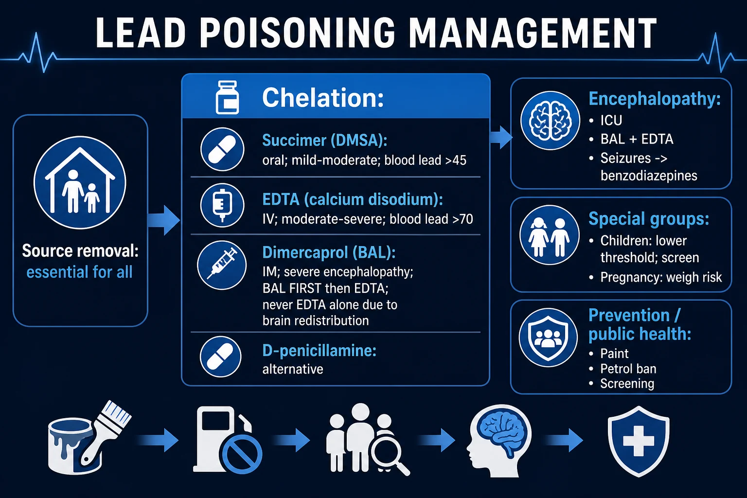
The two pillars of definitive management are (1) source removal and environmental remediation — essential in EVERY case, without which chelation is futile — and (2) chelation therapy for symptomatic poisoning or blood lead above threshold.[1][2][4]
Source removal (all patients): [1]
- Identify the source via environmental investigation (home paint/dust, water, occupation, cosmetics, remedies, retained bullet).
- Remediate — lead-paint abatement by certified contractors (do NOT dry-sand or scrape — generates dust; use wet methods and HEPA vacuuming), replace lead plumbing/service lines, change occupation or institute workplace controls, stop use of cosmetics/remedies, surgically excise an intra-articular retained lead fragment if symptomatic with elevated levels.
- Nutritional support — correct iron, calcium, and zinc deficiency (deficiency increases lead absorption); adequate dietary calcium and iron reduce GI lead uptake in children.
- Public health notification — report to public health authorities for environmental investigation and case-finding (other household members, especially children). [1]
Chelation therapy — agent, dose, route, timing, rationale:[2][3]
The three chelating agents in lead poisoning
Succimer (DMSA) — oral, first-line for moderate
- Water-soluble analogue of dimercaprol; ORAL — well-tolerated, outpatient-possible for stable patients.
- Children: 10 mg/kg (or 350 mg/m2) every 8 hours for 5 days, then every 12 hours for 14 days.
- Adults: 10 mg/kg every 8 hours for 5 days, then every 12 hours for 14 days.
- Use: blood lead over 45 micrograms/dL (children) or over 50–70 (adults) WITHOUT encephalopathy.
- Also chelates mercury and arsenic; can deplete zinc/iron/copper — monitor and supplement.
- Adverse: GI upset, transaminitis, rash, neutropenia, kidney stones (monitor LFTs, FBC).
Calcium disodium EDTA (CaNa2 EDTA) — IV, moderate–severe
- Mobilises lead from bone and soft tissue; excreted in urine (so requires adequate renal function / urine output).
- Adults & children: 1000–1500 mg/m2/day by continuous IV infusion in divided doses over 5 days (max 2 g/day in adults).
- Use: blood lead over 70 micrograms/dL, OR with succimer for moderate–severe, OR with BAL for encephalopathy.
- ALWAYS the calcium disodium salt — disodium EDTA alone chelates serum Ca2+ → fatal hypocalcaemia.
- Adverse: nephrotoxicity (maintain hydration, monitor U&E/Cr), zinc/copper/iron depletion, thrombophlebitis.
Dimercaprol (BAL) — IM, severe / encephalopathy
- Lipid-soluble — crosses blood-brain barrier; chelates lead in brain and soft tissue; given FIRST in encephalopathy.
- Dose: 75 mg/m2 deep IM every 4 hours for 5 days (children and adults).
- Use: encephalopathy or blood lead over 70 — GIVEN BEFORE EDTA, with EDTA added 4 hours later.
- Also chelates arsenic and mercury.
- Adverse: local pain/sterile abscess, fever, hypertension, tachycardia, nausea/vomiting, lacrimation/salivation.
- CONTRAINDICATED in severe G6PD deficiency (haemolysis) and peanut allergy (formulated in peanut oil).
Sequence in encephalopathy — the exam-defining rule:[2]
- Dimercaprol (BAL) FIRST, deep IM, then
- Calcium disodium EDTA IV, starting 4 hours after the first BAL dose (so BAL is already chelating brain lead), then
- Succimer orally once enteral route tolerated, for the remainder of the course and any repeat courses. [1]
The rationale: EDTA alone mobilises lead from bone, transiently raising brain lead and worsening encephalopathy; BAL chelates the brain/tissue lead first, after which EDTA safely clears the blood lead. [1]
Monitoring during and after chelation: [1]
- Blood lead levels — expect a fall during chelation, then a rebound (lead redistributes from bone) after the course ends; repeat courses may be needed until levels and symptoms are controlled.
- Renal function (EDTA nephrotoxicity — maintain hydration, aim for adequate urine output), LFTs (succimer transaminitis), FBC (succimer neutropenia), serum zinc/iron/copper (chelation depletes essential metals).
- Neurology — serial mental state, motor power. [1]
Supportive care: [1]
- Treat hypertension (lead-related; standard antihypertensives).
- Manage constipation (lead colic) with fluids and laxatives.
- Anaemia — correct coexisting iron deficiency (iron supplementation reduces lead absorption), but do NOT give iron alone in suspected lead poisoning without confirming — iron deficiency increases lead absorption; treat the lead first, then the iron.
- Gout — standard urate-lowering if saturnine gout. [1]
Repeat courses and follow-up: [1]
- Repeat blood lead at intervals after the course; chelate again if rebound into the moderate/severe range.
- Repeat blood lead every 1–3 months until two consecutive levels are below the action range, then lengthen the interval.
- Ensure environmental remediation is complete before the child returns home. [1]
Specific Subtypes & Scenarios
-
Paediatric lead poisoning (the most important subtype) — children under 6 absorb ~50% of ingested lead and have a developing brain with no safe level. Screen high-risk children (Medicaid-eligible in the US; those in older housing; recent immigrants; siblings of cases) at ages 9–12 months and again at 24 months, or whenever exposure is suspected. Confirm capillary results with venous blood. Weight-based chelation; developmental surveillance and early-intervention services for any developmental delay. The TLC Trial confirmed succimer lowers blood lead but does not improve long-term IQ — the brain injury is permanent; prevention is the only effective strategy.[4]
-
Occupational lead poisoning (adults) — battery recycling/manufacture, smelting, foundry, demolition, ammunition, soldering, radiator repair, painting/paint removal, firing ranges, pottery with lead glaze. Predominantly inhalation of lead dust and fumes; take-home exposure on work clothes/hair/vehicle is a major source of childhood lead in workers' families. Workplace surveillance — biological monitoring (blood lead, ZPP), engineering controls (local exhaust ventilation, enclosure), PPE (respirators, protective clothing laundered on-site), and medical removal from exposure when blood lead exceeds workplace action/limits (e.g. US OSHA removes workers from exposure at blood lead 50 micrograms/dL average).[2]
-
Lead encephalopathy (life-threatening) — at blood lead typically over 70 micrograms/dL (children) — ataxia, seizures, coma, raised ICP. Emergency chelation: BAL IM FIRST, then CaNa2 EDTA IV 4 hours later, succimer added orally when tolerated; ICU support (airway, seizure control, ICP management). Mortality 25–40% even with treatment; survivors often have permanent neurological and cognitive deficits.[2][5]
-
Pregnancy and lead — lead crosses the placenta; fetal effects include neurotoxicity, miscarriage, prematurity, and low birth weight. Lead is mobilised from maternal bone during pregnancy (calcium turnover), so a previously exposed woman can re-elevate. Reduce maternal exposure, supplement calcium and iron, and chelate only if levels are severe (succimer data in pregnancy are limited — weigh fetal risk vs benefit). Breast-feeding mothers: lead is excreted in breast milk — weigh benefits of breast-feeding vs exposure; if maternal blood lead is high, use formula.[1]
-
Retained lead bullet/fragment — lead leaches from retained projectiles over years, especially if intra-articular (e.g. bullet in a joint) or in contact with a synovial bursa. Chelation only if symptomatic with elevated blood lead; surgical removal of an intra-articular fragment is the definitive treatment. Many retained soft-tissue bullets cause no toxicity — monitor blood lead. [1]
-
Ayurvedic/traditional medicine and cosmetic (surma, kohl, sindoor) — culturally specific exposure, common in South Asian and Middle Eastern families; surma/kohl is applied to infants' and children's eyes, and sindoor to the scalp/hair. Screen children from these communities; counsel families to use only lead-free products; report contaminated products to public health.[1]
-
Acute massive ingestion (pica) — abdominal X-ray may show radiopaque lead material; whole-bowel irrigation with polyethylene glycol until effluent clear, then chelation by level. Common in toddlers with pica for paint chips.[5]
Complications & Pitfalls
Neurological — irreversible IQ loss and neurodevelopmental harm in children (no threshold; persists for life despite chelation), lead encephalopathy (seizures, coma, death; survivors with permanent cognitive and behavioural deficits), chronic cognitive decline in adults. [1]
Renal — chronic tubulointerstitial nephritis with progressive CKD, Fanconi syndrome (children), saturnine gout, hypertension.[2]
Haematological — chronic sideroblastic anaemia (fatigue, pallor); rarely haemolysis at high levels. [1]
Reproductive — miscarriage, prematurity, low birth weight, reduced fertility, sperm abnormalities.[1]
Chelation-related complications: [1]
- EDTA nephrotoxicity — maintain hydration and urine output; reversible on stopping.
- Dimercaprol (BAL) — local pain/sterile abscess, fever, hypertension, tachycardia, haemolysis in G6PD deficiency (contraindicated), peanut allergy (contraindicated — formulated in peanut oil).
- Succimer (DMSA) — GI upset, transaminitis, rash, neutropenia, kidney-stone risk.
- All chelators deplete essential metals (zinc, copper, iron) — monitor and supplement. [1]
Rebound lead levels — lead redistributes from bone after chelation ends; repeat courses often needed. [1]
Classic pitfalls: [1]
- Re-exposure if the source is not remediated — the single commonest management failure; never discharge a child back into an unremediated home.
- Confusing lead colic with an acute surgical abdomen — leading to unnecessary laparotomy; lead colic has no peritonism.
- Confusing lead with iron deficiency or thalassaemia — check the blood lead and ZPP; iron deficiency increases lead absorption, so do NOT give iron alone in suspected lead poisoning without confirming the diagnosis.
- Using EDTA alone in encephalopathy — worsens brain lead; ALWAYS give BAL first.[2]
- Using the disodium EDTA salt instead of the calcium disodium salt — causes fatal hypocalcaemia.
- Missing take-home exposure in workers' children — investigate the whole household.
- Treating the level but not the patient — chelation does not reverse established cognitive damage; counselling and developmental support matter.
Prognosis & Disposition
Children — neurodevelopmental effects (IQ loss, behavioural change, learning disability) are permanent, persisting for life even with chelation that lowers blood lead. Prevention (lead-free housing, water, petrol, paint, consumer products) is therefore the only effective public-health strategy. The magnitude of IQ loss is dose-dependent with no threshold.[1][4]
Adults with chronic exposure — hypertension, CKD, gout, and reproductive effects often persist despite reduced blood lead; long-term cardiovascular and renal surveillance is warranted. [1]
Lead encephalopathy — mortality 25–40% even with chelation; survivors frequently have permanent cognitive, behavioural, and motor deficits.[2]
Disposition: [1]
- Encephalopathy / severe (over 70 micrograms/dL) — ICU, immediate BAL + EDTA chelation, supportive care.
- Moderate (chelation indicated) — inpatient for chelation course, monitoring of blood lead, renal function, and LFTs.
- Mild, asymptomatic — outpatient with source removal, nutritional support, and repeat blood lead at 1–3 month intervals until levels fall; public-health follow-up and environmental investigation.
- Follow-up — confirm source remediation before discharge; developmental and educational support for children; occupational-medicine input for workers. [1]
Special Populations
-
Children (under 6) — the most vulnerable group: higher GI absorption (~50%), developing nervous system with irreversible IQ loss, pica behaviour, and no safe blood level. Screen high-risk children (older housing, low income, recent immigrants, siblings of cases). Use weight-based chelation doses. Provide developmental and early-intervention services for any delay. The CDC reference value is 3.5 micrograms/dL — any level above this triggers public-health action.[1][1]
-
Pregnancy — lead crosses the placenta; fetal neurotoxicity, miscarriage, prematurity, low birth weight. Lead is mobilised from maternal bone (calcium turnover). Reduce maternal exposure, supplement calcium and iron; chelate only if severe (succimer data limited in pregnancy). At-risk women (occupational, cosmetic/remedy exposure, childhood exposure with bone stores) should be screened.[1]
-
Lactation — lead is excreted in breast milk; weigh the benefits of breast-feeding against exposure; if maternal blood lead is high, use formula. [1]
-
Elderly — hypertension or CKD as the sole manifestation; osteoporosis mobilises lead from bone, re-elevating blood lead; cognitive decline may be misattributed to dementia. Screen at-risk elderly. [1]
-
Occupational workers — biological monitoring (blood lead, ZPP); engineering controls and PPE; medical removal from exposure when blood lead exceeds workplace limits; prevent take-home exposure (shower and change before going home; launder work clothes separately / on-site).[2]
-
Coexisting iron/calcium/zinc deficiency — increases lead absorption; correct deficiencies as part of management. [1]
-
G6PD deficiency — dimercaprol (BAL) is contraindicated (risk of haemolysis); use succimer/EDTA. [1]
-
Peanut allergy — dimercaprol (BAL) is contraindicated (formulated in peanut oil). [1]
Evidence, Guidelines & Regional Differences
CDC blood lead reference value (2021). The US CDC lowered the blood lead reference value from 5 to 3.5 micrograms/dL, defined as the 97.5th percentile of NHANES childhood blood lead levels. Crucially, this is a public-health action level, NOT a clinical toxicity threshold — its purpose is to target resources to children with the highest exposure so that sources can be identified and eliminated. There is no safe blood lead level.[1]
TLC Trial (Treatment of Lead-Exposed Children). A landmark randomised trial of succimer in children with blood lead 20–44 micrograms/dL. Succimer lowered blood lead but did NOT improve long-term IQ or behaviour, confirming that chelation does not reverse established cognitive damage and that prevention is paramount.[4]
Zamfara, Nigeria lead epidemic (2010). Artisanal gold mining using lead-contaminated ore processed at home caused the world's largest modern lead poisoning outbreak — over 400 child deaths from acute encephalopathy. Mass oral succimer chelation (Thurtle et al, PLoS Med 2014 — over 3180 courses in children under 5) demonstrated the safety and effectiveness of oral DMSA in resource-limited, severe paediatric lead poisoning, and the necessity of combining chelation with environmental remediation.[3]
Adult lead management — Kosnett et al (2007). The consensus recommendations for medical management of adult lead exposure (Environ Health Perspectives) provide the framework for occupational surveillance, medical removal, and chelation thresholds in adults (chelation generally reserved for symptomatic poisoning or blood lead over 50–70 micrograms/dL).[2]
Leaded petrol phase-out. Completed globally only in 2021 (Algeria was the last country); this public-health intervention halved population blood lead over 30 years and is among the most cost-effective environmental health measures ever implemented.[1]
Regional deltas: [1]
- India — high residual exposure from surma/kohl, sindoor, Ayurvedic Bhasma medicines, and lead-based paint still in widespread use; informal battery recycling; lead in spices/turmeric reported. Screen children from high-risk communities; counsel on cosmetics/remedies.[1]
- United States (CDC) — reference value 3.5 micrograms/dL; mandatory reporting; environmental investigation at elevated levels; Medicaid screening requirements.[1]
- United Kingdom (NICE/UK) — clinical lead exposure is rare but seen in occupational, traditional-medicine/cosmetic, and shooting-range contexts; public-health investigation by the UK Health Security Agency.
- Low- and middle-income countries — informal (often household) battery recycling, artisanal gold mining, lead-glaze pottery, and lead-paint exposure remain major sources; population blood lead often far exceeds high-income country levels.
Controversies: [1]
- Chelation in asymptomatic mild–moderate poisoning — the TLC Trial does not support chelation purely to lower blood lead in asymptomatic children with levels 20–44; the standard is source removal, nutrition, and surveillance, reserving chelation for levels over 45 or symptomatic disease.[4]
- EDTA mobilisation test (provocative chelation) — once used to estimate body burden; not recommended — risk of redistributing lead and limited clinical utility.[4]
- Succimer in pregnancy — limited safety data; reserved for severe poisoning.
Exam Pearls
- Microcytic anaemia with BASOPHILIC STIPPLING = think lead. (Also seen in thalassaemia and sideroblastic anaemias — but in the exam, lead is the answer when there's an exposure history.)[1]
- Lead inhibits ALA dehydratase AND ferrochelatase — NOT ALA synthase (that's the porphyria difference). Result: raised urinary ALA, raised zinc protoporphyrin (ZPP).[6]
- Lead MIMICS CALCIUM — deposits in bone/teeth (metaphyseal 'lead lines'), crosses the BBB and the placenta.[6]
- Sources — mnemonic 'PLUMBER': Paint/dust, Lead pipes/water, Unregulated industry (battery/smelting), Moonshine, Bullets/ammunition, Eyeliner (surma/kohl), Remedies (Ayurvedic).[1]
- WRIST DROP = radial-nerve (motor) palsy from lead; foot drop = common peroneal. Sensation usually spared.[2]
- SATURNINE GOUT = lead-induced gout (lead inhibits renal urate excretion).
- BURTON'S LINE = blue-grey gum line (lead sulfide), more prominent with poor dental hygiene.[6]
- Encephalopathy: BAL (dimercaprol) FIRST, then EDTA 4h later — NEVER EDTA alone (mobilises lead from bone, raises brain lead, worsens encephalopathy).[2]
- Succimer (DMSA) = ORAL chelator (moderate, over 45 in children); EDTA = IV (moderate–severe, over 70); BAL = IM (severe/encephalopathy).[3]
- CaNa2 EDTA, NEVER disodium EDTA — the disodium salt chelates serum calcium and causes fatal hypocalcaemia.
- BAL is CONTRAINDICATED in G6PD deficiency (haemolysis) and peanut allergy (formulated in peanut oil); EDTA is nephrotoxic (maintain hydration).[4]
- No safe lead level in children — IQ loss is permanent; PREVENTION is paramount.[1][1]
- Lead vs acute intermittent porphyria (AIP): BOTH block the haem pathway and cause abdominal pain + neuropathy + neurological symptoms. AIP = photosensitivity, red urine, raised urinary porphobilinogen, family history. Lead = basophilic stippling, gum line, exposure history, raised urinary ALA with normal porphobilinogen.[5]
- Lead lines on X-ray = dense transverse metaphyseal bands in growing bones of children (distal femur, proximal tibia).[1]
- Absorption: children ~50% vs adults ~10% GI; inhalation near-complete; deficiency of iron/calcium/zinc increases absorption.[1]
- Bone holds over 90% of body lead in adults (half-life years–decades); mobilised in pregnancy, lactation, osteoporosis — source of re-exposure decades later.
Exam application bank (NEET-PG / INICET)
One-line answer
Lead poisoning (plumbism/saturnism) is toxic accumulation of lead (Pb), a heavy metal with NO biological role and NO safe blood level, causing multisystem harm — neurotoxic (especially irreversible IQ loss in children), microcytic sideroblastic anaemia with basophilic stippling, lead colic, motor peripheral neuropathy (wrist drop), nephropathy, saturnine gout, hypertension, and reproductive damage. Sources: lead-based paint (pre-1970s housing, children ingest flakes/dust), contaminated water (lead pipes/solder), industrial exposure (smelting, battery recycling, ammunition, soldering, foundry, demolition), traditional cosmetics (surma/kohl, sindoor), Ayurvedic/herbal medicines, ceramic glazes, leaded petrol (legacy environmental), moonshine, toys/jewellery, retained bullets. Lead mimics calcium (deposits in bone/teeth — metaphyseal 'lead lines'; crosses placenta and blood-brain barrier) a
Worked stems (answer without another resource)
Stem 1 — Classic presentation. Map symptoms to mechanism; name the first investigation and first treatment step with dose/route if drug therapy is standard. [1]
Stem 2 — Unstable / complicated. List red flags that force immediate resuscitation, theatre, ICU, antidote, or reperfusion — and what you do in the first 15 minutes. [1]
Stem 3 — Atypical group. Elderly, pregnancy, child, or immunocompromised: how presentation and thresholds change. [1]
Stem 4 — Differential trap. Name the three closest mimics and one discriminator for each. [1]
Stem 5 — Disposition. Who goes home with safety-netting, who is admitted, who needs HDU/ICU/theatre, and what follow-up is mandatory. [1]
Rapid viva checklist
- Definition + classification
- Pathophysiology chain
- Bedside signs / criteria
- Score with exact components (if any)
- Emergency bundle
- Definitive therapy with doses
- Complications of disease and of treatment
- Special populations
- Guideline/trial name if classic
- Three exam traps
Coverage self-check
If you cannot answer any stem above from this page alone, re-read the matching section — the page is intended to be self-sufficient for final-prof and NEET-PG/INICET questions on Lead Poisoning (Plumbism).
References
- [1]Mayans L. Lead Poisoning in Children Am Fam Physician, 2019.PMID 31259498
- [2]Kosnett MJ, Wedeen RP, Rothenberg SJ, et al. Recommendations for medical management of adult lead exposure Environ Health Perspect, 2007.PMID 17431500
- [3]Thurtle N, Greig J, Cooney L, Amitai Y, et al. Description of 3,180 courses of chelation with dimercaptosuccinic acid in children ≤ 5 y with severe lead poisoning in Zamfara, Northern Nigeria: a retrospective analysis of programme data PLoS Med, 2014.PMID 25291378
- [4]Kosnett MJ. Chelation for heavy metals (arsenic, lead, and mercury): protective or perilous? Clin Pharmacol Ther, 2010.PMID 20664538
- [5]Miracle VA. Lead Poisoning in Children and Adults Dimens Crit Care Nurs, 2017.PMID 27902665
- [6]Landrigan PJ, Todd AC. Lead poisoning West J Med, 1994.PMID 7941534