Emergency & Toxicology · General Medicine
Methaemoglobinaemia
Also known as Methaemoglobinaemia · Methemoglobinemia · MetHb · Methylene blue antidote · Chocolate-brown blood · Blue baby syndrome · Cytochrome b5 reductase deficiency · Haemoglobin M disease
Methaemoglobinaemia is the presence of methaemoglobin (MetHb) — haemoglobin in which the haem iron is in the ferric (Fe3+) state (normal haemoglobin is ferrous, Fe2+) — which cannot bind or transport oxygen and, worse, shifts the oxygen-haemoglobin dissociation curve to the LEFT, so that oxygen bound to neighbouring normal haemoglobin subunits is held more tightly and released less readily to tissues. Normal MetHb is under 1 percent of total haemoglobin; levels over 1.5 percent are abnormal. Two mechanisms: ACQUIRED (oxidant drugs/chemicals — over 99 percent of cases) — nitrates/nitrites (contaminated well water in infants, sodium nitrite food preservative, amyl/sodium/butyl nitrite 'poppers'), local anaesthetics (benzocaine, prilocaine, lidocaine), dapsone (hydroxylamine metabolite), aniline dyes, chlorates, phenazopyridine, nitroprusside, nitroglycerin, sulphonamides, primaquine, smoke; and CONGENITAL (rare) — cytochrome b5 reductase deficiency (autosomal recessive) and haemoglobin M disease (autosomal dominant). The cardinal clinical clue is cyanosis REFRACTORY to oxygen with chocolate-brown blood and a saturation gap (SpO2 stuck around 85 percent with a normal PaO2). Diagnose with CO-OXIMETRY (the only test that measures MetHb directly). Treat by stopping the oxidant, high-flow oxygen, and the elegant cofactor antidote METHYLENE BLUE 1-2 mg/kg IV — which acts via the NADPH-methaemoglobin reductase pathway and is therefore CONTRAINDICATED in G6PD deficiency (it fails and causes haemolysis). Alternatives in G6PD deficiency or methylene-blue failure are ascorbic acid, N-acetylcysteine, exchange transfusion and hyperbaric oxygen.
On this page & tools
Your progress
Saved locally on this device.
Exam tags
Red flags
Overview & Definition
Methaemoglobinaemia earns its high-yield status because of the way it tests three habits at once: a clinician's reflex to trust the monitor, their knowledge of a single elegant antidote, and their awareness of the one absolute contraindication that turns the antidote into a poison. The pathology is deceptively simple. Haemoglobin normally carries oxygen with its iron in the ferrous (Fe2+) state; in methaemoglobinaemia the iron is oxidised to the ferric (Fe3+) state, forming methaemoglobin (MetHb). Ferric iron cannot bind oxygen, and by stabilising the relaxed (R) conformation of the neighbouring subunits it shifts the oxygen–haemoglobin dissociation curve to the left — so the oxygen that is bound by the remaining normal haemoglobin is held more tightly and released less readily to the tissues. The patient therefore suffers a double hit: a loss of oxygen-carrying capacity and impaired unloading of the oxygen that remains.[1]
A small amount of MetHb forms constantly by spontaneous auto-oxidation (~0.5–3 percent of circulating haemoglobin each day), and an efficient red-cell reducing system holds the steady-state level under 1 percent of total haemoglobin. Disease is defined as a MetHb level over 1.5 percent. The clinical syndrome to recognise at the bedside is sudden cyanosis after a procedure (benzocaine spray), a drug (dapsone, phenazopyridine), or an exposure (nitrates, aniline) that does NOT improve with oxygen — accompanied by chocolate-brown blood and a saturation gap (the pulse oximeter reads ~85 percent while the arterial PaO2 is normal).[2]
This topic covers both acquired (toxic, ~99 percent of cases) and congenital (hereditary, rare) methaemoglobinaemia, the molecular basis of the methylene-blue antidote and its G6PD contraindication, the bedside and laboratory diagnosis, and the full treatment ladder including the alternatives for when methylene blue cannot be used. [1]
Classification
Methaemoglobinaemia is classified along two axes: mechanism (acquired vs congenital) and severity (MetHb percentage), because the two together determine treatment. [1]
ACQUIRED (toxic) — ~99 percent
- Oxidant drugs/chemicals overwhelm the red-cell reducing system
- Any age; acute-onset after a precipitating exposure; high MetHb; previously well
- Leading drug causes: DAPSONE (hydroxylamine metabolite; chronic use in leprosy/dermatology/HIV prophylaxis), BENZOCAINE (topical spray before endoscopy/intubation — abrupt onset), PRILOCAINE (EMLA, dental)
- Leading chemical causes: NITRATES/NITRITES (contaminated well water in infants; sodium nitrite food preservative; amyl/sodium/butyl nitrite 'poppers'), ANILINE dyes, CHLORATES, NAPHTHALENE, NITROBENZENE
- Other drugs: PHENAZOPYRIDINE (Pyridium), NITROPRUSSIDE (releases nitrite), NITROGLYCERIN, SULPHONAMIDES, PRIMAQUINE, METOCLOPRAMIDE
- Reversible with the antidote methylene blue (except in G6PD deficiency)
CONGENITAL (hereditary) — under 1 percent
- Genetic enzyme deficiency or structural globin abnormality
- LIFELONG cyanosis from infancy; family history; MetHb only 10-30 percent (chronic compensation); otherwise well
- CYTOCHROME b5 REDUCTASE (NADH-methaemoglobin reductase) DEFICIENCY — autosomal RECESSIVE; type I (erythroid — cyanosis only, benign) and type II (ubiquitous — severe CNS, microcephaly, fatal in infancy)
- HAEMOGLOBIN M DISEASE — autosomal DOMINANT; point mutations (HbM Boston/Iwate/Saskatoon/Hyde Park/Milwaukee) stabilise iron as Fe3+
- CYTOCHROME b5 DEFICIENCY — autosomal recessive, very rare, cyanosis + intellectual disability
- Confirm with enzyme assay / haemoglobin electrophoresis / globin gene sequencing
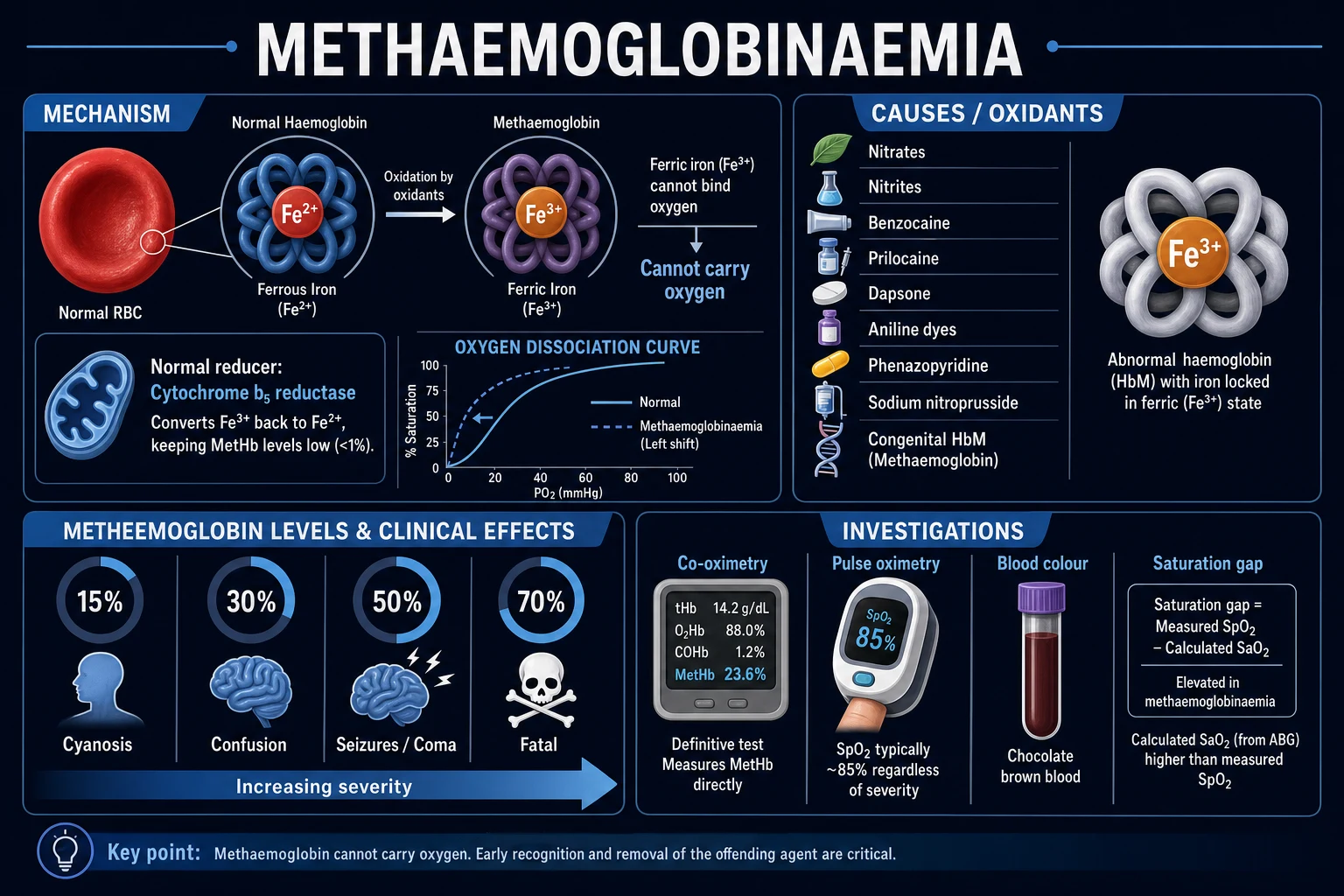
A second axis — severity by MetHb percentage — determines who to treat: [1]
Mild (under 10 percent)
- Often asymptomatic or mild headache
- Visible cyanosis usually appears around 10 percent (equivalent to ~1.5 g/dL MetHb absolute)
- Manage by withdrawing the oxidant + oxygen; no antidote usually needed
Moderate (10–30 percent)
- Cyanosis (slate-grey/dusky, NOT improving with oxygen), exertional dyspnoea, headache, fatigue
- Methylene blue if symptomatic; observe with serial MetHb if asymptomatic
Severe (30–50 percent) / Very severe (50–70 percent)
- Dyspnoea at rest, confusion, syncope, chest pain, ischaemic ECG changes; seizures, coma, metabolic (lactic) acidosis, arrhythmia at higher levels
- Methylene blue 1–2 mg/kg IV + ICU; prepare exchange transfusion if refractory
Epidemiology & Risk Factors
Acquired methaemoglobinaemia is uncommon but under-recognised — the monitor looks reassuring (SpO2 ~85 percent with a normal PaO2), so the diagnosis is missed until someone thinks to send a co-oximetry sample. The largest modern series (Ash-Bernal, 138 cases over 2 academic centres in 28 months, Medicine 2004) identified dapsone and topical/local anaesthetics as the leading drug causes, with a case-fatality around 1–3 percent in treated cases that rises sharply once MetHb exceeds 50–60 percent.[4]
The principal causes are grouped below; the oxidant is the unifying theme. [1]
OXIDANTS
Sources and risk groups: [1]
- Nitrates/nitrites — sodium nitrite (meat curing/food preservative), potassium/sodium nitrate in contaminated well water (infantile 'blue baby syndrome' — gut flora reduces nitrate to the far more potent nitrite), recreational 'poppers' (alkyl nitrites inhaled for sexual enhancement, often co-used with phosphodiesterase inhibitors that compound hypotension), silver nitrate (burn dressings), nitrate fertiliser.[2]
- Local anaesthetics — benzocaine (topical spray/gel; the classic iatrogenic cause after endoscopy, bronchoscopy, transoesophageal echo, intubation; over-the-counter teething gels — withdrawn by the FDA in 2018), prilocaine (in EMLA cream, dental; in high doses), lidocaine (rarer), articaine.[3]
- Antimicrobials — dapsone (leprosy, dermatitis herpetiformis, Pneumocystis prophylaxis — its N-hydroxylamine metabolite is a potent oxidant; the leading drug cause in chronic users), sulphonamides, primaquine (also causes G6PD haemolysis), trimethoprim, chloroquine.[4]
- Other drugs — phenazopyridine (Pyridium — urinary analgesic; the orange/red urine is a clue), nitroprusside (releases cyanide AND nitrite), nitroglycerin, metoclopramide, rasburicase (contraindicated in G6PD), flutamide, zopiclone.
- Industrial/chemical — aniline dyes and inks (absorbed through skin and lungs), chlorates (match industry, weedkiller), nitrobenzene, toluidine, naphthalene (mothballs — also haemolysis in G6PD), nitroethane (artificial-nail glue remover).[6]
- Smoke/fire inhalation — combustion products generate oxidants; combined carbon monoxide, cyanide AND methaemoglobinaemia.
Risk factors that raise susceptibility: [1]
- Infants under 4–6 months — fetal haemoglobin oxidises more readily; gut flora reduces nitrate to nitrite; cytochrome b5 reductase activity is only ~50–60 percent of adult until ~4 months (the molecular basis of well-water 'blue baby syndrome').
- G6PD deficiency — impaired NADPH-dependent reduction and a heightened risk of oxidative haemolysis; methylene blue is contraindicated.
- Anaemia or cardiopulmonary disease — reduced physiological reserve; lower tolerance for a given MetHb percentage.
- Chronic exposure — dapsone therapy, occupational aniline workers.
- Genetic enzyme deficiency — undiagnosed cytochrome b5 reductase deficiency or haemoglobin M (precipitated or worsened by oxidants).[5]
Pathophysiology
The entire syndrome flows from one number — the steady-state balance between haemoglobin auto-oxidation and the red-cell reducing systems that keep it in check. [1]
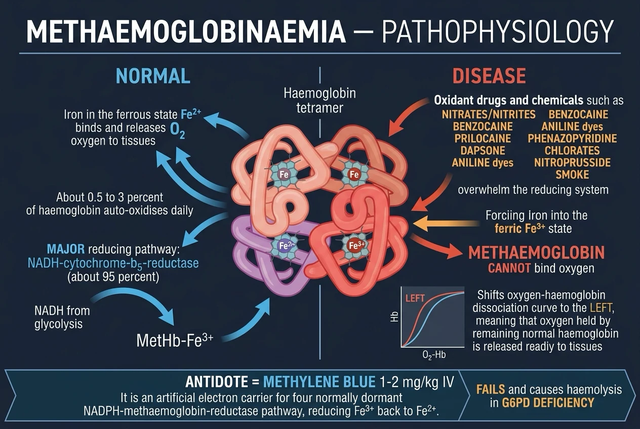
The normal redox balance
Spontaneous auto-oxidation of the haem iron converts a small fraction of circulating oxyhaemoglobin to MetHb each day, releasing superoxide in the process. Under normal conditions an efficient reducing system keeps steady-state MetHb under 1 percent of total haemoglobin. There are two enzymatic routes and two minor non-enzymatic routes.[1]
- MAJOR pathway (~95 percent of daily reduction): NADH–cytochrome b5 reductase (also called NADH-methaemoglobin reductase or diaphorase I). NADH generated from glycolysis reduces cytochrome b5, which in turn reduces MetHb-Fe3+ back to functional Hb-Fe2+. This is the pathway that is genetically deficient in cytochrome b5 reductase deficiency (congenital methaemoglobinaemia).
- MINOR pathway (~5 percent of daily reduction): NADPH–methaemoglobin reductase (diaphorase II). It uses NADPH from the hexose-monophosphate (pentose-phosphate) shunt but is normally dormant because it lacks an endogenous electron carrier. Methylene blue supplies that missing carrier — it is reduced to leukomethylene blue by NADPH-methaemoglobin reductase, and leukomethylene blue non-enzymatically reduces MetHb-Fe3+ back to Hb-Fe2+. This is the molecular basis of methylene-blue therapy and the reason it is completely dependent on G6PD (no G6PD → no NADPH → no reduction, and methylene blue itself, an oxidant dye, then causes haemolysis).
- Non-enzymatic direct reduction by ascorbic acid (vitamin C) and reduced glutathione — small in magnitude, slow, but the rationale for ascorbic acid as an alternative antidote in G6PD deficiency and in congenital disease.[2]
Why ferric iron is doubly harmful
Ferric (Fe3+) iron cannot bind oxygen — so each MetHb subunit is a lost oxygen-carrying site. Worse, by stabilising the R (relaxed) state of the neighbouring ferrous subunits within the tetramer, MetHb shifts the oxygen–haemoglobin dissociation curve to the left. The practical consequence: the oxygen that is bound to the remaining normal haemoglobin is held more tightly and released less readily to the tissues. The patient suffers both a loss of carrying capacity (functional anaemia) and impaired unloading — the molecular basis of tissue hypoxia, lactic acidosis and end-organ (brain, heart) injury at high MetHb levels.[1]
The cyanosis threshold
MetHb is dark (chocolate-brown), and clinically visible cyanosis appears when MetHb reaches ~1.5 g/dL absolute (analogous to the 5 g/dL deoxyhaemoglobin threshold for ordinary cyanosis). This absolute threshold explains two clinical points examiners test: anaemic patients cyanose at a HIGHER MetHb percentage (they may be profoundly hypoxic with NO cyanosis, because total Hb is too low to reach 1.5 g/dL MetHb), and polycythaemic patients cyanose at a LOWER percentage. Always interpret MetHb percentage against total haemoglobin.[3]
The pulse-oximetry paradox
A standard pulse oximeter uses two wavelengths (660 nm red, 940 nm infrared) calibrated only for oxy- and deoxyhaemoglobin. MetHb absorbs strongly at both wavelengths, so as MetHb rises the SpO2 reading is driven toward ~85 percent and becomes insensitive to changes in true arterial oxygenation — giving the classic saturation gap: the patient looks hypoxic on the monitor but the PaO2 is normal. Giving more oxygen does not move the number.[3]
The arterial-blood-gas paradox
A standard (non-co-oximetric) blood-gas machine does not measure SaO2 — it calculates it from PaO2, pH and temperature using the oxyhaemoglobin dissociation equation, on the assumption that all haemoglobin is normal. It therefore reports a normal or near-normal calculated SaO2 despite the true SaO2 being low. Only CO-oximetry (multi-wavelength, 4+ wavelengths that directly measure oxy-, deoxy-, carboxy- and methaemoglobin) reveals the true picture.[2]
Why methylene blue can fail or harm
G6PD deficiency
- Absolute contraindication — methylene blue FAILS (no NADPH to power the NADPH-reductase shunt)
- And it WORSENS the patient: methylene blue is itself an oxidant dye and causes oxidative HAEMOLYSIS
- Use ascorbic acid + N-acetylcysteine + exchange transfusion instead
Haemoglobin M / cytochrome b5 reductase deficiency
- The NADPH pathway is intact, but the structural enzyme defect persists, so the response is poor
- Methylene blue generally unhelpful; treat with ascorbic acid / supportive
Sulphaemoglobinaemia (irreducible)
- A sulphur atom binds haem; methylene blue CANNOT reduce it
- Lasts the red-cell lifespan (~120 days); withdraw the drug and wait
Overdose of methylene blue itself
- Doses over 7 mg/kg in 24 h are themselves oxidant — they CAUSE haemolysis and paradoxically RAISE MetHb
- The antidote becomes the toxin
Clinical Presentation
The presentation is dominated by cyanosis that does not respond to oxygen, with severity tracking the MetHb percentage. Recognising the cardinal triad and the atypical contexts is what the examiner probes. [1]
The signature clinical triad
- Cyanosis REFRACTORY TO 100 percent oxygen — the cardinal clue. The cyanosis has a characteristic slate-grey/dusky ('muddy') quality rather than the bluer cyanosis of deoxyhaemoglobin; it affects lips, nail beds, earlobes and oral mucosa and does not improve with supplemental oxygen.[2]
- Chocolate-brown blood — draw blood into a tube; MetHb blood is brown and does not turn bright red on exposure to air/oxygen (shake the tube in air — normal blood turns red, MetHb blood stays brown). The bedside 'filter-paper' drop test is a classic manoeuvre.[1]
- The saturation gap — the pulse oximeter reads ~85 percent and does not rise with oxygen, while the calculated SaO2 on a standard ABG is normal and the PaO2 is normal.
Severity by MetHb level — reproduced verbatim
Mild (1.5–10 percent)
- Often asymptomatic or mild headache
Moderate (10–30 percent)
- Cyanosis, exertional dyspnoea, headache, fatigue
Severe (30–50 percent)
- Dyspnoea at rest, syncope, confusion, tachycardia, chest pain, ischaemic ECG changes
Very severe (50–70 percent)
- Seizures, coma, metabolic (lactic) acidosis, arrhythmia, cardiogenic shock
Over 70 percent
- Usually LETHAL — coma, shock, severe acidosis, death
Note that chronic (congenital) MetHb of 15–30 percent is tolerated remarkably well compared with an acute level of the same magnitude — chronic compensation upregulates red-cell mass and non-enzymatic reduction, and tissue adaptation occurs. A patient with congenital methaemoglobinaemia may walk into clinic with a MetHb of 25 percent looking only mildly dusky.[5]
Atypical and special-context presentations
Infantile well-water nitrate ('blue baby')
- Rural infant fed formula made with nitrate-contaminated well water (over 50 mg/L)
- Gut flora reduces nitrate to the more potent nitrite; fetal Hb oxidises readily; low cytochrome b5 reductase activity
- Well-appearing slate-grey ('blue') baby; lethargy, poor feeding, irritability
Post-procedure benzocaine
- Abrupt cyanosis MINUTES after topical anaesthesia (endoscopy, bronchoscopy, transoesophageal echo, intubation)
- Previously well patient; high index of suspicion from the temporal link
Dapsone therapy
- Insidious onset dyspnoea and cyanosis over weeks (leprosy, dermatitis herpetiformis, HIV/transplant prophylaxis)
- May co-present with oxidative haemolysis (Heinz bodies, falling Hb, jaundice) — especially in G6PD
'Poppers' (alkyl nitrites)
- Sudden cyanosis, often with hypotension (co-inhaled phosphodiesterase-5 inhibitors)
- Recreational sexual-enhancement use; abrupt onset
Anaemic patient
- May have SEVERE tissue hypoxia with NO visible cyanosis (total Hb too low to reach the 1.5 g/dL MetHb threshold)
- Use absolute MetHb (g/dL), not percentage, to gauge severity; transfuse to raise functional Hb
Smoke/fire inhalation
- Combined carbon monoxide + cyanide + methaemoglobinaemia from combustion
- Sequence treatment by the dominant toxin (avoid sodium thiosulphate/nitrite, which raise MetHb)
Congenital (lifelong)
- Lifelong slate-grey cyanosis from infancy; otherwise well; family history
- MetHb 10-30 percent chronically; type II cytochrome b5 reductase deficiency — microcephaly, developmental delay
Differential Diagnosis
The unifying clinical problem is cyanosis that does not respond to oxygen. The differential splits into the dyshaemoglobins (MetHb, CO-Hb, sulph-Hb) and the non-dyshaemoglobin causes (severe cardiopulmonary shunt, pseudocyanosis). [1]
Methaemoglobinaemia
- Slate-grey/chocolate cyanosis; blood CHOCOLATE-BROWN (does not turn red on oxygen)
- SpO2 plateaus at ~85 percent; normal PaO2; normal calculated SaO2 (gap)
- Triggered by oxidant drug/chemical (benzocaine, dapsone, nitrates, aniline)
- MetHb measured directly on CO-OXIMETRY; methylene blue reduces it (unless G6PD)
Carboxyhaemoglobinaemia (CO poisoning)
- CHERRY-RED skin/lips/blood (not brown); no true cyanosis
- Normal PaO2 (CO does not affect dissolved O2); pulse oximetry FALSELY HIGH (CO-Hb misread as oxy-Hb, SpO2 near 100 percent)
- Headache, neuropsychiatric symptoms; history of fire/fumes/exhaust
- Treat with HIGH-FLOW 100% O2 / HYPERBARIC O2; NOT methylene blue
Sulphaemoglobinaemia
- GREENISH blood; cyanosis at a lower threshold (~0.5 g/dL sulph-Hb)
- Same oxidant drugs as MetHb (sulphonamides, phenazopyridine, metoclopramide) — can coexist
- IRREDUCIBLE — lasts the RBC lifespan (~120 days); methylene blue does NOT work
- Measured by CO-oximetry (some analysers cannot distinguish sulph-Hb from MetHb)
Severe cardiopulmonary disease (true hypoxaemia)
- PaO2 LOW (not normal); SpO2 tracks PaO2 and IMPROVES with oxygen
- Pneumonia, pulmonary oedema, ARDS, right-to-left shunt, severe COPD
- No dyshaemoglobin; cyanosis improves with oxygen
Pseudocyanosis (pigment deposition)
- Slate-grey skin WITHOUT blood/gas abnormality (argyria from chronic silver; gold; chlorpromazine, amiodarone, chloroquine)
- Normal blood colour, normal SpO2/PaO2; history of chronic exposure
- Not a true hypoxia — distinguished by normal gases
Two distinctions deserve emphasis. MetHb versus CO-Hb at the pulse oximeter: MetHb drives SpO2 toward ~85 percent; CO-Hb is misread as oxyhaemoglobin so the pulse oximeter reads falsely high (near 100 percent) — a useful discriminator. MetHb versus sulph-Hb: both are measured by co-oximetry and produced by the same oxidants, but sulph-Hb is irreducible (no response to methylene blue) and lasts the red-cell lifespan.[2][6]
Distinguishing acquired from congenital at the bedside: congenital presents with lifelong cyanosis from infancy, a family history, a MetHb of only 10–30 percent (chronic compensation) and no precipitating exposure; acquired is acute-onset in a previously normal patient with a clear drug/chemical trigger. Confirm congenital with a cytochrome b5 reductase enzyme assay and/or haemoglobin electrophoresis / globin gene sequencing for HbM.[5]
Clinical & Bedside Assessment
The focused assessment is built around the cardinal triad and the search for a precipitant. [1]
History. Establish the precipitating exposure — prescription drugs (dapsone, sulphonamides, primaquine, phenazopyridine, nitroprusside, nitroglycerin, local anaesthetics especially benzocaine/prilocaine), occupational/recreational (aniline dyes, nitrites/'poppers', nitrobenzene, chlorates, naphthalene), recent procedures (endoscopy, bronchoscopy, dental work, intubation, TEE with benzocaine spray), water source (well water in an infant), smoke inhalation. Establish the timing of onset relative to exposure, the symptoms (cyanosis, dyspnoea, headache, chest pain, altered consciousness, dark/abnormal urine), comorbidity (anaemia, cardiac, respiratory, G6PD status, pregnancy) and — for suspected congenital disease — a family history of chronic cyanosis.[3]
Examination. Vital signs (tachypnoea, tachycardia, hypotension in severe disease; Kussmaul breathing if metabolic acidosis). GCS. Cardiovascular — tachycardia, signs of myocardial ischaemia, arrhythmia. Respiratory — central and peripheral slate-grey cyanosis that does not improve with oxygen. Neurologic — confusion, seizures, coma at high levels. Skin — signs of haemolysis (jaundice, splenomegaly) in dapsone/G6PD; signs of chronic dapsone use.[4]
Bedside tests. [1]
- Blood-colour test — draw blood into a heparinised tube and compare its colour with a control (a normal staff member's blood, or a reference chart). MetHb blood is chocolate-brown and does not turn bright red on shaking in air; a drop on filter paper that stays brown is a classic bedside manoeuvre.[1]
- Oxygen challenge — apply 100 percent oxygen; the cyanosis of MetHb does not improve (unlike true hypoxaemic cyanosis, which does).
- Recognise the saturation gap — pulse oximeter ~85 percent with a normal PaO2 and a normal calculated SaO2.
Apply ABCDE in the unstable patient — airway (intubate if GCS under 8), high-flow oxygen, IV access, continuous ECG and pulse oximetry, bedside glucose (exclude hypoglycaemia), assess for shock.[3]
Investigations
Investigations serve three purposes: confirm the dyshaemoglobin, quantify MetHb, and exclude mimics and assess severity. [1]
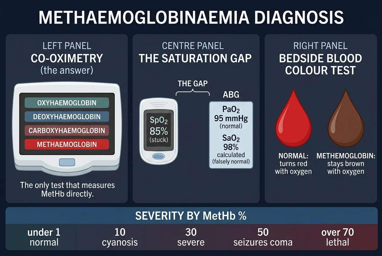
First-line panel
- CO-OXIMETRY (gold standard) on arterial or venous blood — directly measures the four haemoglobin species and reports MetHb as a percentage of total Hb. Over 1.5 percent is abnormal. A venous sample is acceptable and avoids arterial puncture.[2]
- Venous/arterial blood gas — confirms the metabolic picture (severe MetHb causes a metabolic/lactic acidosis with low bicarbonate); PaO2 is normal (the lung works).
- Full blood count, reticulocytes, blood film — anaemia; Heinz bodies and bite cells in oxidative haemolysis (dapsone, G6PD); polychromasia.
- LDH, haptoglobin, bilirubin — haemolysis screen.
- G6PD assay — ideally before methylene blue (do not delay treatment in a severely symptomatic patient). Caveat: G6PD levels are falsely elevated during acute haemolysis (the most deficient cells have lysed) — recheck 2–3 months later.
- U&E, lactate, glucose, troponin, ECG — severity and end-organ injury; lactate reflects tissue hypoxia.
- Drug/oxidant screen — paracetamol, salicylate, ethanol in overdose; consider specific levels.
The saturation gap — reproduced verbatim
Standard pulse oximetry
- Two wavelengths (660 nm, 940 nm) calibrated for oxy-/deoxy-Hb only
- MetHb absorbs strongly at BOTH wavelengths -> SpO2 driven toward ~85 percent
- SpO2 INSENSITIVE to changes in true arterial O2 (plateau)
- SpO2 does NOT rise with oxygen
Standard (non-co-oximetric) ABG
- Calculates SaO2 from PaO2/pH/temperature assuming all Hb is normal
- Reports a NORMAL or near-normal SaO2 despite the true SaO2 being low
- PaO2 NORMAL (lung function intact; PaO2 reflects dissolved O2)
CO-OXIMETRY (the answer)
- Multi-wavelength (4+) measures oxy-, deoxy-, carboxy-, methaemoglobin directly
- Reports the true (low) SaO2 and the MetHb percentage (high)
- The ONLY test that measures MetHb directly
Limitations of CO-oximetry to know
Some older co-oximeters cannot distinguish sulphhaemoglobin from methaemoglobin (sulph-Hb can be misread as MetHb); very high MetHb can saturate the assay and report falsely low; frozen/mishandled samples artefactually raise MetHb. A venous sample is acceptable.[3]
When to investigate for congenital causes
Lifelong cyanosis, family history, a MetHb of 10–30 percent in an otherwise well patient with no precipitant — send a cytochrome b5 reductase enzyme assay (low in deficiency) and haemoglobin electrophoresis / globin gene sequencing (identifies HbM variants).[5]
Management — Resuscitation
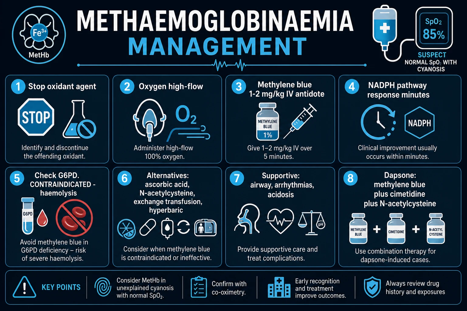
Resuscitation and the first specific measures are simultaneous: stop the oxidant and give oxygen while you prepare the antidote. [1]
- ABCDE. Secure the airway (intubate and ventilate if GCS under 8, severe respiratory distress, or rapid deterioration); give HIGH-FLOW 100 percent oxygen via a non-rebreather mask (maximises the oxygen carried by remaining functional haemoglobin and increases dissolved O2); establish IV access; continuous cardiac monitoring (arrhythmia risk at high MetHb); isotonic crystalloid for hypotension; bedside glucose.[3]
- STOP and REMOVE the oxidant — discontinue dapsone, withhold benzocaine, remove the patient from the chemical source, change the infant's water source. Ongoing exposure defeats therapy.
- Confirm with CO-OXIMETRY and check G6PD where practical — but do NOT delay methylene blue in a severely symptomatic patient.[2]
Management — Definitive & Stepwise
The definitive ladder integrates resuscitation, antidotal reduction, alternatives for when the antidote cannot be used, and source control. The key decision is whether methylene blue is appropriate — that hinges on G6PD status and co-medication. [1]
Step 1 — Methylene blue (first-line antidote)
METHYLENE BLUE — first line
- Dose: 1-2 mg/kg IV (= 0.1-0.2 mL/kg of the 1 percent solution, 10 mg/mL) given SLOWLY OVER 5 MINUTES
- Response within 30-60 minutes (patient visibly turns pink as MetHb falls)
- REPEAT 1-2 mg/kg after 30-60 minutes if symptoms persist or MetHb remains high
- Maximum 7 mg/kg in 24 hours (larger doses cause haemolysis and paradoxically RAISE MetHb)
- Mechanism: artificial electron CARRIER (cosubstrate) for the NADPH-dependent methaemoglobin reductase pathway; reduced to leukomethylene blue, which reduces MetHb-Fe3+ to Hb-Fe2+
- Indicated for SYMPTOMATIC disease OR MetHb over 30 percent (some guidelines over 25 percent if symptomatic)
CONTRAINDICATIONS
- G6PD DEFICIENCY (absolute) — methylene blue fails (no NADPH) AND causes oxidative haemolysis
- Haemoglobin M disease / severe cytochrome b5 reductase deficiency — response poor
- Recent SEROTONERGIC drugs (SSRIs, SNRIs, MAOIs, tramadol, linezolid, methadone) — methylene blue is a weak MAO inhibitor -> SEROTONIN SYNDROME risk (FDA warning)
- Large doses (over 7 mg/kg/24 h) — oxidant; cause haemolysis and raise MetHb
- Use cautiously in pregnancy and renal failure
Step 2 — Alternatives when methylene blue is contraindicated or ineffective
ASCORBIC ACID (vitamin C)
- 300-1000 mg/day orally in divided doses (or IV for rapid effect)
- Slow non-enzymatic direct reduction of MetHb over 24-48 hours
- Preferred when methylene blue is contraindicated (G6PD) or in congenital disease
- Long-term oral ascorbic acid 200-500 mg/day for congenital cytochrome b5 reductase deficiency
N-ACETYLCYSTEINE (NAC)
- Provides cysteine for glutathione regeneration -> supports non-enzymatic reduction
- Adjunct for DAPSONE-induced MetHb (may also conjugate the hydroxylamine metabolite)
- Dosing analogous to paracetamol-overdose NAC (150 mg/kg IV load over 1 h, then 50 mg/kg over 4 h, then 100 mg/kg over 16 h)
EXCHANGE TRANSFUSION
- Severe MetHb unresponsive to methylene blue (over 50-60 percent with ongoing symptoms)
- G6PD-deficient patient with severe disease (methylene blue contraindicated)
- Massive ongoing oxidant load; co-existent severe haemolysis/anaemia
- Physically removes MetHb and the oxidant and supplies fresh functional Hb
HYPERBARIC OXYGEN
- Salvage therapy for critically ill severe MetHb unresponsive to other measures
- Provides enough DISSOLVED oxygen (independent of Hb carriage) to sustain life temporarily
Step 3 — Source-specific measures
- Dapsone-induced MetHb — dapsone's N-hydroxylamine metabolite is the oxidant, and the drug's long half-life (20–40 h) means MetHb RECURS after each methylene-blue dose. Give repeated doses or a continuous infusion, ADD CIMETIDINE (inhibits the CYP450 generation of the hydroxylamine — typically cimetidine 300 mg orally every 6 h, or 300 mg IV every 6 h), consider N-acetylcysteine, and monitor MetHb every 4–6 hours for 24–48 hours.[4]
- Infantile well-water nitrate — change to an alternative water source (bottled/municipal); do NOT boil the water (boiling concentrates nitrate); methylene blue 1 mg/kg IV cautiously if symptomatic (check G6PD); ascorbic acid for mild disease; supportive care; notify public health; test the well water.[5]
- Smoke inhalation — combined CO + cyanide + MetHb; treat CO with high-flow/hyperbaric oxygen, cyanide with hydroxocobalamin (avoid sodium thiosulphate/nitrite which themselves generate MetHb), and MetHb with methylene blue — sequence by the dominant toxin.[6]
Step 4 — Supportive care and disposition
- Supportive/ICU care — severe MetHb (over 50–60 percent), coma, seizures, severe acidosis, arrhythmia or haemodynamic instability → ICU. Treat seizures with benzodiazepines, arrhythmia per ACLS, shock with fluids and vasopressors; transfuse for anaemia/haemolysis.
- Observation — asymptomatic with MetHb under 20–30 percent and the oxidant stopped → observe with serial MetHb every 4–6 h; discharge when MetHb is falling and symptoms absent.
- Congenital forms — outpatient management with lifelong oral ascorbic acid 200–500 mg/day (or riboflavin 20–30 mg/day); reassure that chronic cyanosis is well tolerated; genetic counselling. [1]
Specific Subtypes & Scenarios
Drug-induced (commonest acquired)
- BENZOCAINE (abrupt onset post-procedure), DAPSONE (insidious, recurrent — add cimetidine), sulphonamides, primaquine (also G6PD haemolysis), phenazopyridine (orange urine clue), nitroprusside (cyanide + nitrite), nitroglycerin
- Withdraw the drug + oxygen + methylene blue
Nitrates / nitrites
- Contaminated WELL WATER (infants — 'blue baby syndrome'); sodium nitrite food preservative; amyl/sodium/butyl/isobutyl nitrite 'poppers' (sudden onset ± hypotension with PDE-5 inhibitors); nitrobenzene; silver nitrate burns
- Remove the source, oxygen, methylene blue; for infants change water source and notify public health
Industrial / chemical
- ANILINE dyes/inks (skin + lung absorption), chlorates, naphthalene (mothballs — also haemolysis in G6PD), toluidine, nitroethane (nail-glue remover)
- Decontamination (remove clothing, wash skin), oxygen, methylene blue, supportive; occupational health reporting
Smoke inhalation
- Combined CO + cyanide + MetHb from combustion
- Treat CO with high-flow/hyperbaric O2; cyanide with HYDROXOCOBALAMIN (avoid sodium thiosulphate/nitrite — they raise MetHb); MetHb with methylene blue
Congenital — cytochrome b5 reductase deficiency
- Autosomal RECESSIVE; TYPE I (erythroid — cyanosis only, benign); TYPE II (ubiquitous — severe CNS, microcephaly, fatal in infancy)
- Lifelong oral ascorbic acid 200-500 mg/day; genetic counselling
Congenital — haemoglobin M disease
- Autosomal DOMINANT; HbM Boston/Iwate (alpha), HbM Saskatoon/Hyde Park/Milwaukee (beta)
- Point mutations stabilise iron as Fe3+; chronic cyanosis from birth, otherwise well
- Methylene blue INEFFECTIVE; no specific therapy; reassure (cosmetic concern)
Dapsone-therapy MetHb
- Recurrent MetHb in leprosy, dermatitis herpetiformis, Pneumocystis prophylaxis
- Long half-life causes recurrence; CO-PRESCRIBE CIMETIDINE; lowest effective dose; monitor MetHb; consider alternative therapy (atovaquone, inhaled pentamidine)
Pregnancy
- Foetus highly vulnerable (fetal Hb oxidises readily; low foetal enzyme activity)
- Methylene blue crosses placenta — historical associations with foetal haemolysis, hyperbilirubinaemia, jejunal atresia (debated)
- Ascorbic acid first-line for mild disease; methylene blue reserved for severe life-threatening maternal disease; obstetric + foetal-medicine input
G6PD-deficient patient
- Methylene blue CONTRAINDICATED (fails AND causes haemolysis)
- Ascorbic acid + NAC + exchange transfusion for severe disease; transfuse for haemolysis; avoid the trigger for life
Refractory MetHb despite methylene blue
- Re-examine for ONGOING EXPOSURE (dapsone — give cimetidine), check G6PD and HbM, consider continuous methylene-blue infusion
- Exchange transfusion, hyperbaric oxygen; reconsider sulph-Hb (irreducible)
Complications & Pitfalls
Disease complications — tissue hypoxia: myocardial ischaemia and infarction, arrhythmia, seizures, coma, metabolic (lactic) acidosis, shock, multiorgan failure, death (MetHb over 70 percent usually lethal). Haemolysis (especially dapsone and G6PD — Heinz bodies, bite cells, jaundice, dark urine, renal injury). In congenital disease, type II cytochrome b5 reductase deficiency causes intellectual disability, microcephaly and early death.[5]
Treatment-related complications — methylene blue itself: haemolysis (over 7 mg/kg or G6PD deficiency); paradoxical rise in MetHb at high dose; blue/green discolouration of skin and urine (harmless but alarming); serotonin syndrome in patients on SSRIs/SNRIs/MAOIs (FDA warning); phlebitis at infusion site; nausea, vomiting, dizziness; false pulse-oximetry readings during infusion.[3]
Prognosis & Disposition
Overall prognosis. Mild-to-moderate acquired methaemoglobinaemia responds rapidly to withdrawal of the oxidant + oxygen ± a single dose of methylene blue — response within minutes to an hour, the patient visibly turning pink. Mortality is low in treated cases (under 1–3 percent in the Ash-Bernal series) but rises sharply with severe MetHb (over 50–60 percent), delayed treatment, comorbidity (anaemia, cardiac, respiratory) and G6PD-related haemolysis.[4]
Predictors of poor outcome. High MetHb (over 50–60 percent); delayed presentation; severe metabolic (lactic) acidosis; seizures/coma; myocardial ischaemia or arrhythmia; co-existent haemolysis (especially G6PD); pre-existing anaemia or cardiopulmonary disease; ongoing oxidant exposure (long-half-life drugs like dapsone); co-poisoning (CO, cyanide). [1]
Disposition. [1]
- Symptomatic or MetHb over 30 percent → Emergency Department, methylene blue.
- Severe MetHb, coma, haemodynamic instability, need for exchange transfusion → ICU.
- Asymptomatic with MetHb under 20–30 percent and the oxidant stopped → observe with serial MetHb every 4–6 h; discharge when MetHb is falling and symptoms absent.
- Congenital forms → outpatient with ascorbic acid and reassurance (no admission). [1]
Response-to-therapy. Methylene blue reduces MetHb within 30–60 minutes — a visible 'turning pink' with rising SpO2 (toward true saturation). Failure to respond suggests: (1) G6PD deficiency; (2) ongoing oxidant exposure (especially dapsone); (3) haemoglobin M disease or cytochrome b5 reductase deficiency; (4) sulphhaemoglobinaemia (irreducible); (5) too-low initial dose — re-dose and reassess.[2]
Follow-up. Identify and remove the precipitant (change medication, occupational controls, water-source testing); G6PD testing after the acute episode (recheck at 2–3 months if haemolysis falsely elevated the level); genetic counselling for congenital forms; advise the patient to avoid the trigger for life; document the reaction in the medication record. [1]
Prevention. Regulation of well-water nitrate (WHO limit 50 mg/L); FDA withdrawal of OTC benzocaine teething products (2018); avoid benzocaine in infants; occupational exposure limits for aniline/chlorates; G6PD screening before oxidant drugs (dapsone, primaquine, rasburicase, sulphonamides) in high-risk populations; patient education about triggers. [1]
Special Populations
Paediatric (especially under 6 months)
- Highly susceptible — fetal Hb oxidises more readily; gut flora reduces nitrate to nitrite; cytochrome b5 reductase only ~50-60% of adult until ~4 months
- Well-water 'blue baby syndrome'; avoid benzocaine in infants (FDA 2018 withdrawal)
- Methylene blue 1 mg/kg IV cautiously if symptomatic (check G6PD); ascorbic acid for mild; change water source; weight-based dosing throughout
Pregnancy
- Foetus highly vulnerable (fetal Hb, low foetal enzyme activity); methylene blue crosses placenta
- Ascorbic acid preferred for mild disease; methylene blue reserved for severe life-threatening maternal disease
- Obstetric and foetal-medicine input; monitor foetus
Elderly
- Reduced cardiopulmonary reserve — even modest MetHb causes ischaemia and arrhythmia
- Polypharmacy increases oxidant exposure (nitroprusside, nitroglycerin, sulphonamides, phenazopyridine)
- Lower threshold to treat; check renal function (methylene blue excretion); beware serotonin syndrome with concurrent SSRI
G6PD-deficient
- Methylene blue CONTRAINDICATED (fails AND causes haemolysis)
- Ascorbic acid + NAC + exchange transfusion; transfuse for haemolysis; avoid the trigger for life
- Recheck G6PD 2-3 months after the acute episode (falsely elevated during active haemolysis)
Anaemic
- Cyanosis appears at a HIGHER MetHb percentage (need 1.5 g/dL absolute); may be profoundly hypoxic WITHOUT cyanosis
- Use absolute MetHb (g/dL), not percentage; transfuse to raise functional Hb
- Lower threshold for exchange transfusion
Immunocompromised (HIV/transplant on dapsone)
- Dapsone is a leading cause in this group
- Co-prescribe cimetidine; lowest effective dose; consider alternative prophylaxis (atovaquone, inhaled pentamidine); check G6PD before starting
Resource-limited / rural (well-water nitrate)
- Test the well water (WHO limit 50 mg/L nitrate)
- Use an alternative water source for formula (NOT boiled — boiling concentrates nitrate)
- Methylene blue widely available; ascorbic acid adjunct; notify public health for community outbreaks
On serotonergic psychotropics
- Methylene blue (weak MAO inhibitor) + SSRIs/SNRIs/MAOIs/tramadol/linezolid -> SEROTONIN SYNDROME
- Hold psychotropics where possible; if urgent, use ascorbic acid and exchange transfusion, or methylene blue with intensive monitoring
Evidence, Guidelines & Regional Differences
Landmark evidence. The canonical review is Wright, Lewander and Woolf (Ann Emerg Med 1999) — 'Methemoglobinemia: etiology, pharmacology, and clinical management' — covering mechanism, MetHb thresholds and methylene-blue dosing and still the most-cited reference.[1] The largest modern case series, Ash-Bernal, Wise and Wright (Medicine 2004, 138 cases over 2 academic centres in 28 months), established dapsone and topical anaesthetics as the leading acquired causes and the clinical epidemiology.[4] Skold, Cosco and Klein (South Med J 2011) and Cortazzo and Lichtman (J Cardiothorac Vasc Anesth 2014) provide comprehensive general and perioperative/anaesthetic reviews with the pathophysiology and treatment ladder.[2][3] Iolascon, Bianchi, Andolfo and Russo (Am J Hematol 2021) 'Recommendations for diagnosis and treatment of methemoglobinemia' is the contemporary international consensus covering both congenital and acquired forms.[5] The Borron and Bebarta (Emerg Med Clin North Am 2015) 'Asphyxiants' review places MetHb among the toxin-induced histotoxic hypoxias (alongside CO, cyanide, hydrogen sulphide).[6]
Guidelines. International consensus (AACT/EAPCCT, UK NPIS/TOXBASE, US FDA) endorses methylene blue 1–2 mg/kg IV as first-line for symptomatic acquired methaemoglobinaemia (MetHb over 30 percent or symptomatic); check G6PD where possible; alternatives ascorbic acid, exchange transfusion, hyperbaric oxygen for refractory or G6PD-deficient disease.[5]
Regional deltas. [1]
- UK (NPIS/TOXBASE) — emphasises benzocaine as a leading iatrogenic cause; conservative threshold in mild cases; check G6PD before methylene blue where practical.
- US (FDA) — boxed warning + 2018 withdrawal of OTC benzocaine teething products after severe infantile methaemoglobinaemia; FDA warning on methylene blue and serotonergic drugs and on G6PD deficiency.
- India / South-Asia — well-water nitrate 'blue baby syndrome' remains a public-health problem in rural agricultural regions (WHO limit 50 mg/L); dapsone (leprosy) and occupational aniline-dye exposure are leading; G6PD testing is often unavailable so empiric caution is required.
- WHO — drinking-water nitrate limit 50 mg/L to prevent infantile methaemoglobinaemia. [1]
Controversies. (1) The exact threshold for treating asymptomatic disease (under 30 percent with conservative monitoring vs treat over 20–25 percent) — individualise to comorbidity. (2) Whether to test G6PD before methylene blue in an emergency (do not delay severe-treatment; test where feasible). (3) The role and dose of N-acetylcysteine and riboflavin. (4) Whether exchange transfusion or hyperbaric oxygen adds value in severe refractory disease. (5) The strength of the association between maternal methylene blue and foetal jejunal atresia (debated).[5]
Exam Pearls
METHYLENE BLUE
Must-know facts
- MetHb = Hb iron in Fe3+ state; normal under 1 percent; over 1.5 percent abnormal
- Cardinal clue: CYANOSIS REFRACTORY TO OXYGEN (benzocaine, dapsone, nitrates)
- Diagnose with CO-OXIMETRY (the ONLY direct measure of MetHb)
- Antidote: METHYLENE BLUE 1-2 mg/kg IV over 5 min; repeat in 30-60 min; max 7 mg/kg/24 h
- Methylene blue mechanism: artificial electron carrier for the NADPH-methaemoglobin reductase pathway
- CONTRAINDICATED in G6PD deficiency (fails + haemolysis); weak MAO inhibitor -> serotonin syndrome risk
Discriminating clues
- MetHb -> slate-grey cyanosis, chocolate-brown blood; SpO2 ~85 percent
- CO-Hb -> cherry-red blood, SpO2 FALSELY HIGH (near 100 percent) -> NOT methylene blue, use O2/HBO
- Sulph-Hb -> greenish blood, IRREDUCIBLE (methylene blue useless), lasts RBC lifespan
- Acquired vs congenital: lifelong cyanosis + family history + MetHb 10-30 percent = congenital
- Dapsone -> recurrent MetHb (long half-life); add cimetidine; monitor 24-48 h
Must-avoid pitfalls
- TRUSTING the pulse oximeter or the calculated SaO2 (both misleading in MetHb)
- GIVING methylene blue to G6PD-deficient patient (haemolysis + failure)
- EXCESSIVE methylene blue (over 7 mg/kg/24 h) -> causes MetHb and haemolysis
- MISSING the diagnosis in an anaemic patient (no cyanosis but severe hypoxia)
- BOILING nitrate-contaminated water (concentrates nitrate)
- CONFUSING sulph-Hb (irreducible) with MetHb
Exam application bank (NEET-PG / INICET)
One-line answer
Methaemoglobinaemia is the presence of methaemoglobin (MetHb) — haemoglobin in which the haem iron is in the ferric (Fe3+) state (normal haemoglobin is ferrous, Fe2+) — which cannot bind or transport oxygen and, worse, shifts the oxygen-haemoglobin dissociation curve to the LEFT, so that oxygen bound to neighbouring normal haemoglobin subunits is held more tightly and released less readily to tissues. Normal MetHb is under 1 percent of total haemoglobin; levels over 1.5 percent are abnormal. Two mechanisms: ACQUIRED (oxidant drugs/chemicals — over 99 percent of cases) — nitrates/nitrites (contaminated well water in infants, sodium nitrite food preservative, amyl/sodium/butyl nitrite 'poppers'), local anaesthetics (benzocaine, prilocaine, lidocaine), dapsone (hydroxylamine metabolite), aniline dyes, chlorates, phenazopyridine, nitroprusside, nitroglycerin, sulphonamides, primaquine, smoke
Worked stems (answer without another resource)
Stem 1 — Classic presentation. Map symptoms to mechanism; name the first investigation and first treatment step with dose/route if drug therapy is standard. [1]
Stem 2 — Unstable / complicated. List red flags that force immediate resuscitation, theatre, ICU, antidote, or reperfusion — and what you do in the first 15 minutes. [1]
Stem 3 — Atypical group. Elderly, pregnancy, child, or immunocompromised: how presentation and thresholds change. [1]
Stem 4 — Differential trap. Name the three closest mimics and one discriminator for each. [1]
Stem 5 — Disposition. Who goes home with safety-netting, who is admitted, who needs HDU/ICU/theatre, and what follow-up is mandatory. [1]
Rapid viva checklist
- Definition + classification
- Pathophysiology chain
- Bedside signs / criteria
- Score with exact components (if any)
- Emergency bundle
- Definitive therapy with doses
- Complications of disease and of treatment
- Special populations
- Guideline/trial name if classic
- Three exam traps
Coverage self-check
If you cannot answer any stem above from this page alone, re-read the matching section — the page is intended to be self-sufficient for final-prof and NEET-PG/INICET questions on Methaemoglobinaemia.
References
- [1]Wright RO, Lewander WJ, Woolf AD. Methemoglobinemia: etiology, pharmacology, and clinical management Ann Emerg Med, 1999.PMID 10533013
- [2]Skold A, Cosco DL, Klein R. Methemoglobinemia: pathogenesis, diagnosis, and management South Med J, 2011.PMID 22024786
- [3]Cortazzo JA, Lichtman AD. Methemoglobinemia: a review and recommendations for management J Cardiothorac Vasc Anesth, 2014.PMID 23953868
- [4]Ash-Bernal R, Wise R, Wright SM. Acquired methemoglobinemia: a retrospective series of 138 cases at 2 teaching hospitals Medicine (Baltimore), 2004.PMID 15342970
- [5]Iolascon A, Bianchi P, Andolfo I, Russo R. Recommendations for diagnosis and treatment of methemoglobinemia Am J Hematol, 2021.PMID 34467556
- [6]Borron SW, Bebarta VS. Asphyxiants Emerg Med Clin North Am, 2015.PMID 25455664