Emergency & Toxicology · General Medicine
Rhabdomyolysis
Also known as Rhabdomyolysis · Myoglobinuria · Crush syndrome · Exertional rhabdomyolysis · Myonecrosis
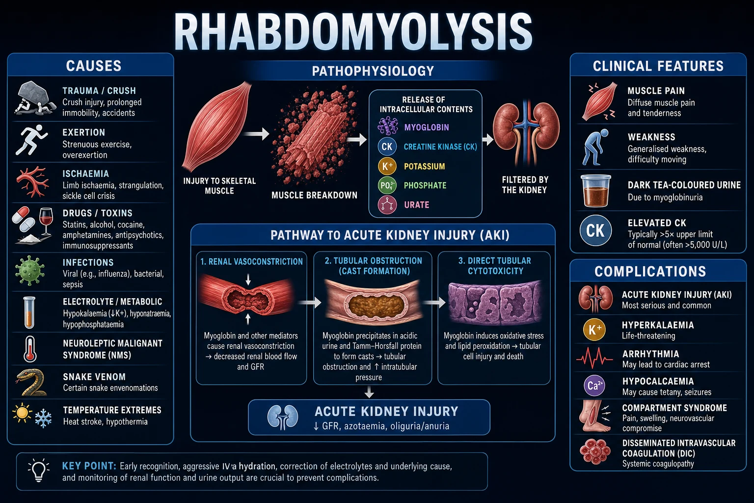
Rhabdomyolysis is the breakdown of skeletal muscle with release of intracellular contents (myoglobin, creatine kinase, potassium, phosphate, urate, lactate dehydrogenase) into the circulation, causing acute kidney injury, electrolyte disturbance, compartment syndrome and disseminated intravascular coagulation. Causes span trauma/crush (earthquakes, prolonged immobilisation), exertion (strenuous exercise, seizures, delirium), muscle ischaemia (arterial occlusion, compartment syndrome), drugs and toxins (statins, fibrates, alcohol, cocaine, amphetamines, MDMA, succinylcholine, neuroleptic malignant syndrome, serotonin syndrome, snake venom), infection (influenza, coxsackie, malaria, legionella, sepsis), electrolyte disorders (hypokalaemia, hypophosphataemia), temperature extremes (heat stroke, hypothermia) and inherited metabolic myopathies (McArdle, carnitine palmitoyltransferase II deficiency). Presents with the classic triad of muscle pain, weakness and dark tea-coloured urine (often incomplete). Diagnosis rests on a raised creatine kinase (over 5 times the upper limit of normal, frequently over 1000 U/L; over 5000 U/L marks high acute-kidney-injury risk), urine dipstick positive for blood but with no red cells on microscopy (myoglobin), hyperkalaemia, and a creatinine that rises disproportionate to urea. The cornerstone of treatment is aggressive IV crystalloid to maintain urine output 1 to 3 mL/kg/h (around 300 mL/h, often 6 to 12 L in the first 24 h), started before extrication in crush injury, with treatment of hyperkalaemia, treatment of the cause, fasciotomy for compartment syndrome, and renal replacement therapy for established AKI. Sodium bicarbonate and mannitol are controversial adjuncts, not first-line; early hypocalcaemia is not treated.
On this page & tools
Your progress
Saved locally on this device.
Exam tags
Red flags
Overview & Definition
Rhabdomyolysis is a syndrome of skeletal-muscle injury in which disruption of the sarcolemma (the muscle-cell membrane), or rapid intracellular ATP depletion, allows the leakage of intracellular muscle constituents — most importantly myoglobin, creatine kinase, potassium, phosphate and urate — into the extracellular fluid and bloodstream.[1] The leaked myoglobin is small enough to be freely filtered at the glomerulus, and it is toxic to the renal tubule; the leaked potassium and phosphate disturb the heart and the calcium-phosphate balance. The clinical consequences — acute kidney injury (AKI), hyperkalaemic arrhythmia, compartment syndrome, hypocalcaemia and disseminated intravascular coagulation (DIC) — are what make rhabdomyolysis a time-critical emergency.[5]
The syndrome is high-yield because it connects many emergencies that look different but share one final pathway. The earthquake crush victim, the untrained athlete after squats, the status-epilepticus or delirium patient, the statin user, the cocaine or MDMA overdose, the heat-stroke casualty, the Russell's viper bite, and the unconscious patient found on the floor all converge on the same problem — muscle dying and poisoning the kidney and the heart. The diagnosis rests on one cheap, universally available biomarker (creatine kinase, CK), and the single most effective treatment — aggressive IV fluids — is cheap, simple and time-critical. [1]
Rhabdomyolysis is a final-common-pathway emergency: recognise the syndrome early, start fluids before myoglobin precipitates, and treat the cause.[1]
The classic clinical picture — muscle pain and weakness with dark urine and a massive CK — is unmistakable, but mild or occult cases present as unexplained AKI with no muscle symptoms at all; a urine dipstick positive for blood with no red cells on microscopy is the bedside clue that should never be missed.[1]
Classification
Rhabdomyolysis has one pathological endpoint — muscle-cell death and content leak — but a very broad aetiology. The clinically useful classification is by cause, because treatment of the cause is as important as fluid resuscitation.[1][5]
By cause (the classification examiners expect): [1]
- Trauma / crush / ischaemia — earthquakes and building collapse (crush syndrome), prolonged immobilisation (unconscious on the floor, prolonged coma, drug overdose), compartment syndrome, arterial occlusion, tourniquet, burns, electrical injury and lightning.
- Exertion — strenuous exercise in the untrained (squats, Spin class, military training, marathons), status epilepticus, delirium/agitation, severe asthma, tetany; worse when hot, dehydrated or at altitude.
- Drugs and toxins — statins are the single commonest drug cause (especially high-dose and with interacting drugs — fibrates, macrolides, cyclosporin, azole antifungals, daptomycin), fibrates, alcohol (direct toxicity plus withdrawal and electrolyte disturbance), cocaine, amphetamines and MDMA (ecstasy), neuroleptic malignant syndrome, serotonin syndrome, succinylcholine (especially in burns, denervation or prolonged immobility — hyperkalaemic response).
- Infection — influenza A and B, coxsackievirus, EBV, HIV, legionella, Streptococcus pyogenes (necrotising myositis), Clostridium perfringens (gas gangrene/myonecrosis), malaria (Plasmodium falciparum — blackwater fever), and any severe sepsis.
- Electrolyte and endocrine — hypokalaemia (predisposes to muscle ischaemia and is the commonest "silent" precipitant), hypophosphataemia, hyponatraemia, hypothyroidism (myxoedema myopathy), diabetic ketoacidosis and hyperosmolar hyperglycaemic state (hypokalaemia/hypophosphataemia plus osmotic diuresis).
- Temperature extremes — heat stroke, hypothermia, malignant hyperthermia (anaesthetic-triggered).
- Inherited / metabolic myopathies — McArdle disease (myophosphorylase deficiency), carnitine palmitoyltransferase II (CPT II) deficiency, mitochondrial myopathies, muscular dystrophies. Suspect when rhabdomyolysis is recurrent, exertion-triggered, or in a child or young adult with a family history.
- Inflammatory — polymyositis, dermatomyositis.
- Others — snake bite (Russell's viper, sea snake), hornet/wasp sting, electrocution, prolonged tourniquet, sickle-cell trait (exertional sickling).[1][2]
Epidemiology & Risk Factors
Rhabdomyolysis accounts for roughly 7 to 15 per cent of all cases of acute kidney injury in adults; the true incidence is underestimated because mild cases are never diagnosed. Among patients who present with established rhabdomyolysis, AKI develops in 15 to 33 per cent, and the risk rises steeply once the peak CK exceeds 5000 U/L.[5][6] The commonest single cause in adults presenting to an emergency department is prolonged immobilisation (the patient "found on the floor"), followed by drugs/toxins and exertion; in children, viral myositis (especially influenza B) and exertion dominate.[1]
Risk factors that increase susceptibility: [1]
- Prior statin or fibrate use, high-dose, or interacting drugs (fibrates, macrolides, cyclosporin, azole antifungals) and hypothyroidism.
- Dehydration, heat, exertion at altitude — amplifies exertional injury.
- Electrolyte disturbance, especially hypokalaemia and hypophosphataemia.
- Underlying inherited or inflammatory myopathy — lowers the threshold.
- Sickle-cell trait — predisposes to exertional sickling with rhabdomyolysis and sudden death in athletes and military recruits.
- Chronic kidney disease — amplifies the renal consequence of any given muscle injury.
- Male sex and young age — exertional cases cluster in young men; the untrained "weekend warrior" is the classic stem.[2][5]
Crush syndrome (mass-casualty context): after earthquakes and building collapse, crush injury to a limb produces rhabdomyolysis on reperfusion — the toxin surge released when the crushing weight is lifted can trigger sudden lethal hyperkalaemia and AKI. Mortality of crush-syndrome-associated AKI without dialysis is very high; the Renal Disaster Relief Task Force protocol (start saline before extrication) was developed precisely to prevent this.[3]
Pathophysiology
Whatever the trigger, the final common pathway is the same: sarcolemma disruption (mechanical, thermal, electrical, ischaemic or toxin-mediated) OR intracellular ATP depletion (anaerobic ischaemia, glycolytic enzyme defects) leads to a rise in free intracellular calcium, which activates neutral proteases (calpains) and phospholipases that digest the contractile and membrane proteins, completing the destruction of the myocyte.[1]
Stepwise mechanism: [1]
- Insult damages the sarcolemma directly (crush, exertion, toxin, heat) and/or depletes ATP (ischaemia, glycolytic defect).
- Sodium/potassium and calcium pumps fail (ATP-dependent) — sodium and water enter the cell (oedema), calcium floods in.
- Intracellular calcium accumulation activates calpains (proteases) and phospholipase A2 — these digest the contractile apparatus and the membrane itself.
- Cell death (myonecrosis) releases the intracellular contents — myoglobin, CK, potassium, phosphate, urate and lactate dehydrogenase — into the circulation.
- Volume sequestration into injured muscle produces hypovolaemia, which compounds renal hypoperfusion.
- Myoglobin reaches the kidney and causes pigment nephropathy (the three mechanisms below).
- Potassium causes hyperkalaemic arrhythmia; phosphate precipitates calcium; activated clotting factors and tissue factor drive DIC.[1][5]
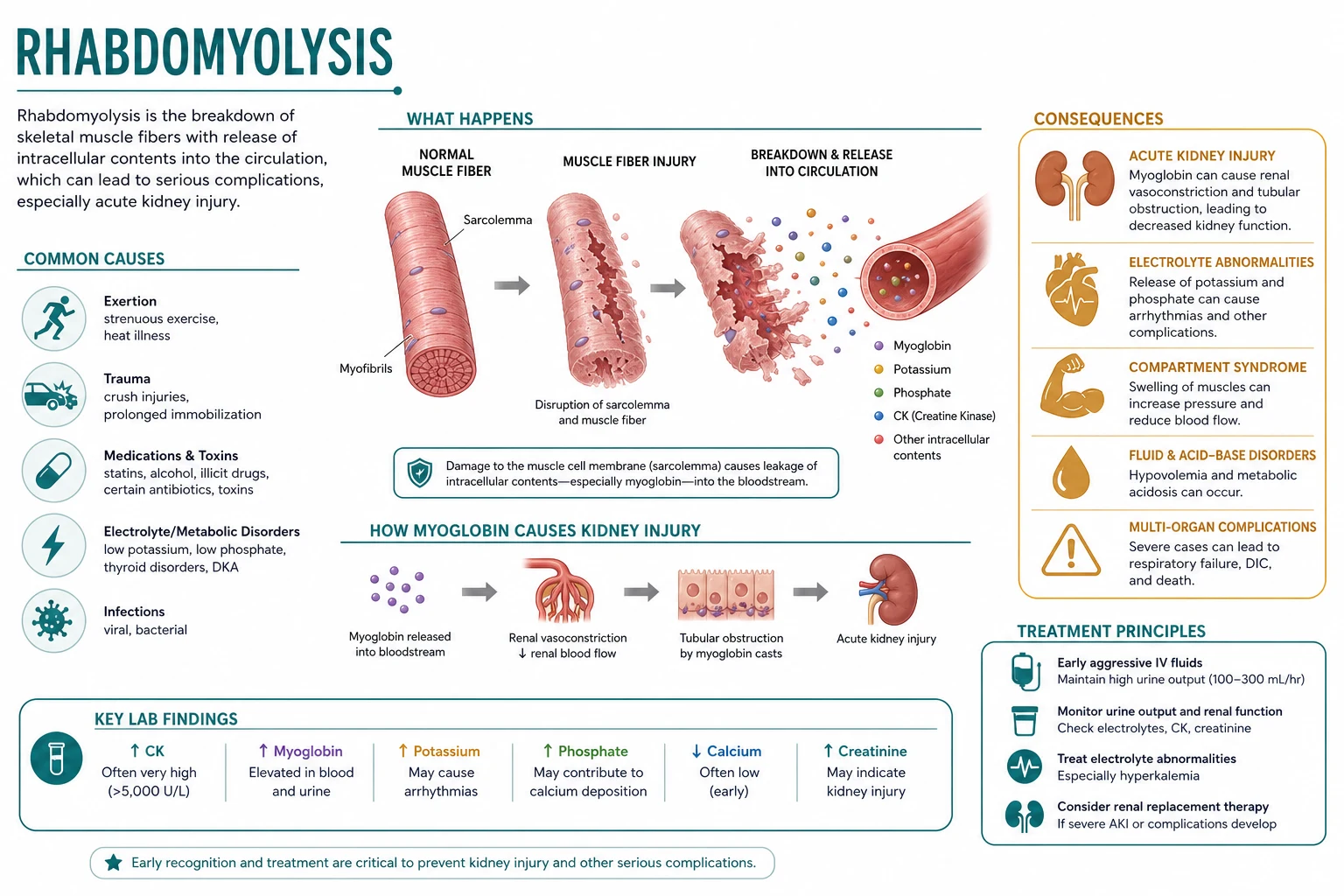
Why myoglobin is the kidney-killer — three mechanisms of pigment nephropathy:[1][5]
- Renal vasoconstriction — myoglobin is a nitric-oxide scavenger; the resulting vasoconstriction plus hypovolaemia reduces renal blood flow and glomerular filtration.
- Tubular cast obstruction — at acidic urine pH, myoglobin (specifically the dissociated ferrihemate) precipitates with Tamm-Horsfall protein to form pigmented granular casts that obstruct the distal tubule.
- Direct free-radical cytotoxicity — free iron from myoglobin generates reactive oxygen species that injure the tubular epithelium, producing acute tubular necrosis. [1]
The electrolyte derangements, and why they happen: [1]
- Hyperkalaemia — intracellular potassium released from dead muscle; compounded by AKI and acidosis. The commonest cause of EARLY death (arrhythmia).
- Hypocalcaemia (early) — calcium precipitates as calcium-phosphate in necrotic muscle (hyperphosphataemia drives this). Do NOT treat early hypocalcaemia unless symptomatic, because it rebounds.
- Rebound hypercalcaemia (late, during recovery) — as the deposited calcium is mobilised.
- Hyperphosphataemia and hyperuricaemia — released from muscle; worsen tubular injury.
- High anion-gap metabolic acidosis — from released organic acids plus AKI. [1]
The compartment-syndrome vicious cycle: muscle oedema within a tight fascial compartment raises the interstitial pressure, occludes venous then capillary flow, worsens ischaemia, and extends the muscle injury — which is why a swollen, tight, painful limb in rhabdomyolysis demands urgent pressure measurement and fasciotomy.[1]
Clinical Presentation
The classic triad — muscle pain, muscle weakness and dark (tea- or cola-coloured) urine — is present in only a minority; many patients, especially the unconscious or sedated, have NO muscle symptoms, and the first clue is dark urine, oliguria, a rising creatinine or hyperkalaemia.[1]
Typical features: [1]
- Muscle — aching, tenderness, swelling and stiffness, most often in the calves, thighs, lower back and shoulders; weakness (proximal, sometimes ascending). Exertional cases localise to the worked muscle group.
- Urine — dark, tea- or cola-coloured (myoglobin). Dipstick positive for "blood" but microscopy shows no (or very few) red cells — the cardinal bedside clue.
- General — fever, nausea and vomiting, malaise, dehydration (from fluid sequestration into muscle).
- Compartment features — a swollen, tense, tender compartment with pain on passive stretch, paraesthesia — see below.
- Hyperkalaemia features — palpitations, chest pain, ECG changes (peaked T waves, PR prolongation, QRS widening, sine-wave ventricular tachycardia), sudden collapse.[1][5]
Atypical / occult presentations — the ones that are missed: [1]
- Unconscious / immobilised patient "found on the floor" — no history; first clue is dark urine, raised CK, hyperkalaemia or AKI on the admission bloods.
- Septic or ICU patient — rhabdomyolysis masked by the primary illness; CK sent for unexplained AKI or rising potassium.
- Elderly / diabetic — may have silent muscle injury (neuropathy, immobility); present with falls and confusion.
- Child with influenza and a limp — benign acute childhood myositis (calf pain, refusal to walk) can progress to genuine rhabdomyolysis.[1]
Differential Diagnosis
Differential of dark / pigmented urine:[1]
Pigmented urine — distinguishing rhabdomyolysis
Rhabdomyolysis (myoglobinuria)
- Urine dipstick blood-positive (orthotolidine reacts with myoglobin haem) but MICROSCOPY shows NO (or very few) red cells
- Raised CK over 5x ULN, often over 1000 U/L; myoglobin raised in blood and urine
- Clinical context of muscle injury, exertion, drugs, immobility
- Centrifuged urine: pigmented supernatant with pigmented granular casts; red sediment only if coexistent haematuria
Intravascular haemolysis (haemoglobinuria)
- Same dipstick-positive / no-red-cell pattern (haemoglobin also reacts)
- Haemolysis on blood film, raised unconjugated bilirubin, low haptoglobin, LDH high
- CK normal or only mildly raised; plasma is pink (free haemoglobin) rather than normal
- Causes: mismatched transfusion, malaria (blackwater fever), G6PD, mechanical (valve), autoimmune haemolysis
Haematuria (true)
- Dipstick blood-positive AND red cells on microscopy
- Causes: urological (stones, cancer, BPH), glomerular (IgA nephropathy, nephritic syndrome), infection, trauma
Other pigments (no dipstick reaction)
- Porphyria (acute intermittent — urine darkens on standing), beetroot, rifampicin, phenytoin, myoglobin vs porphobilinogen
- Dipstick is NEGATIVE for blood; microscopy negative
Differential of a raised CK: [1]
- Cardiac — CK-MB fraction or, better, troponin (rhabdomyolysis can co-raise CK-MB from skeletal-muscle MM isoenzyme cross-reactivity; troponin is cardiac-specific).
- Recent exercise, IM injection, seizure, prolonged immobility, hypothyroidism — all elevate CK modestly without true rhabdomyolysis.
- Inflammatory myopathy (polymyositis/dermatomyositis), muscular dystrophy, MELAS / mitochondrial — chronic, often with weakness but a smaller CK rise.
- Macro-CK (macro-enzyme complex, a benign lab artefact) — persistent unexplained CK rise in an asymptomatic adult.[1]
Distinguishing the hyper-metabolic syndromes that cause rhabdomyolysis: [1]
Malignant hyperthermia vs NMS vs serotonin syndrome
Malignant hyperthermia
- Trigger: SUCCINYLCHOLINE or VOLATILE anaesthetic (halothane, sevoflurane)
- Onset: intra-operative, rapid
- Features: masseter spasm, rapid rise in CO2 (hypercapnia), hyperthermia, rigidity, rhabdomyolysis
- Specific treatment: IV DANTROLENE 2.5 mg/kg, stop trigger, cooling
Neuroleptic malignant syndrome (NMS)
- Trigger: neuroleptics (haloperidol, typical antipsychotics; rare with atypicals), or withdrawal of dopamine (Parkinson's)
- Onset: days to weeks, slow (lead-pipe rigidity develops first)
- Features: 'lead-pipe' rigidity, hyperthermia, altered mental state, autonomic instability, raised CK, rhabdomyolysis
- Specific treatment: stop neuroleptic, cooling, IV DANTROLENE, bromocriptine; supportive
Serotonin syndrome
- Trigger: SSRIs/SNRIs/MAOIs/tramadol/linezolid, especially in combination or overdose
- Onset: hours, rapid
- Features: clonus (especially inducible/ocular), hyperreflexia, mydriasis, agitation, diarrhoea, hyperthermia, rigidity (lower-limb dominant)
- Specific treatment: stop serotonergic drug, benzodiazepines, cooling; CYPROHEPTADINE in moderate-severe cases
Clinical & Bedside Assessment
ABCDE first, then a focused search for the cause and the two life-threats (hyperkalaemia and compartment syndrome).[1]
Vital signs and monitoring drive severity — heart rate, blood pressure, respiratory rate, oxygen saturation, temperature, GCS/confusion, and hourly urine output (catheterise). Cardiac monitoring is mandatory from the moment of arrival because hyperkalaemia can kill within minutes; look for peaked T waves, PR prolongation, QRS widening and sine-wave ventricular tachycardia on the ECG. [1]
The 5 Ps of acute compartment syndrome (reproduced verbatim): [1]
- Pain — severe, out of proportion to the injury, and worse on passive stretch of the compartment muscles (the earliest and most sensitive sign).
- Paraesthesia — early; numbness in the distribution of nerves traversing the compartment.
- Pallor, Poikilothermia, Pulselessness — LATE and ominous signs; a pulse is still present until very late, so "the pulse is fine" must NOT reassure.
- Confirm with compartment pressure measurement: a delta pressure (diastolic BP minus compartment pressure) under 30 mmHg, or an absolute pressure over 30 mmHg, mandates urgent fasciotomy.[1]
Bedside assessment of cause: a careful history and examination for trauma, exertion, drugs (prescribed and recreational), infection, seizure, endocrine/electrolyte, hypothyroidism, snake/insect bite, hypothermia/heat exposure, and a family history of recurrent exertional rhabdomyolysis (inherited myopathy). [1]
Bedside assessment of severity: the peak CK correlates loosely with the risk of AKI and is used to triage the intensity of fluid therapy; any CK over 1000 U/L, any hyperkalaemia, any oliguria, or any signs of compartment syndrome mandates aggressive IV fluid therapy and admission.[1][5]
Investigations
- Creatine kinase (CK) — the diagnostic marker (normal under 200 U/L). Rises within 12 hours, peaks at 1 to 3 days, and declines over 3 to 5 days (longer with ongoing injury). A value over 5 times the upper limit of normal (often stated as over 1000 U/L) with the right clinical context is diagnostic; over 5000 U/L marks high AKI risk, and levels above 15,000 to 20,000 U/L carry a very high risk of AKI.
- Urea and electrolytes — potassium elevated (the lethal early derangement); creatinine elevated disproportionate to urea (a creatinine-to-urea ratio that looks "too high" for the dehydration is a classic clue — myoglobin is an extra substrate).
- Calcium, phosphate, magnesium — calcium low initially (precipitation), phosphate high; rebound hypercalcaemia later in recovery.
- AST and ALT — both elevated (released from muscle, not just liver); a "transaminitis" in rhabdomyolysis does not necessarily mean liver injury.
- Urate, LDH — elevated (released from muscle).
- Venous blood gas / lactate — high anion-gap metabolic acidosis, raised lactate.
- Coagulation (PT, aPTT, fibrinogen, D-dimer) — to detect DIC.
- Troponin — to separate cardiac from skeletal-muscle injury.
- FBC — leucocytosis, thrombocytopenia (DIC).
- TSH, cortisol — if an endocrine precipitant is suspected.
- Blood cultures, viral serology (influenza PCR) — when infection is the trigger. [1]
Urine: [1]
- Dipstick — positive for blood (orthotolidine reacts with the haem of myoglobin).
- Microscopy — no (or very few) red cells; pigmented granular casts are characteristic. This dipstick-positive / microscopy-negative pattern is the bedside signature of pigment nephropathy.
- Urine myoglobin — qualitative, slow, NOT routinely needed (serum myoglobin peaks at 6 to 8 hours and clears by 24 hours, so it is often normal by the time the diagnosis is suspected). CK is cheaper, universally available and tracks severity — that is why CK, not myoglobin, is the diagnostic and monitoring marker.[1]
Rhabdomyolysis — the numbers that decide management
Imaging and procedures: [1]
- ECG — mandatory, looking for hyperkalaemic changes (peaked T waves, PR prolongation, QRS widening, sine-wave, VT/VF).
- Compartment pressure measurement — when compartment syndrome is suspected (delta pressure under 30 mmHg is diagnostic).
- Ultrasound / CT — to identify an abscess, necrotising infection, vascular occlusion, or underlying cause; no imaging is needed to diagnose rhabdomyolysis itself (CK is the test).
- Renal ultrasound — to exclude obstruction as a contributor to AKI. [1]
Renal replacement therapy — the A-E-I-O-U indications
Management — Resuscitation
ABCDE. Secure IV access (two large-bore cannulae), attach a cardiac monitor (for hyperkalaemia), and send bloods including CK, potassium, creatinine, calcium, phosphate, coagulation and troponin. Catheterise for hourly urine output.[1]
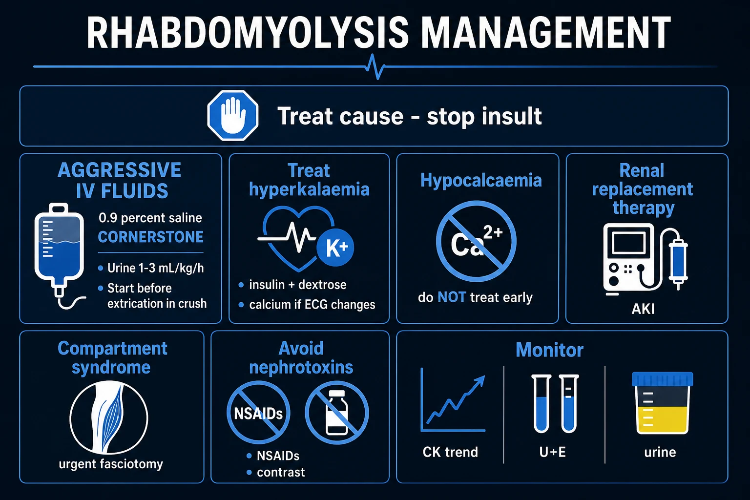
The cornerstone — aggressive IV crystalloid — and why: the goal is to flush myoglobin through the kidney before it precipitates in the tubules, restore intravascular volume (muscle oedema sequesters large volumes), and prevent the renal vasoconstriction that drives AKI. Start fluids EARLY and at high rate.[1][5]
- Fluid: 0.9 per cent saline or balanced crystalloid (Lactated Ringer's / Plasma-Lyte); alternating saline with a balanced solution avoids hyperchloraemic acidosis.
- Rate: 1 to 1.5 L/h initially (up to 1 L/h even before extrication in crush), titrated to a target urine output of 1 to 3 mL/kg/h — roughly 200 to 300 mL/h in an adult (about 300 mL/h) — until the CK is clearly falling. Patients commonly need 6 to 12 L in the first 24 hours; monitor hourly fluid balance to avoid pulmonary/cerebral oedema.
- When to ease back: when the CK is falling, urine output is established, electrolytes are stable — typically at 24 to 72 hours.[1][3]
Treat hyperkalaemia immediately if there are ECG changes (peaked T waves, QRS widening): IV calcium gluconate 10 per cent 10 mL over 2 to 5 minutes (membrane stabilisation), then insulin-dextrose (10 units soluble insulin in 25 to 50 g IV dextrose), nebulised salbutamol 10 to 20 mg, and sodium bicarbonate (especially in this acidotic setting). Shifting potassium into cells buys time; definitive removal needs RRT once AKI is established.[1]
Stop all nephrotoxins: NSAIDs, ACE inhibitors, angiotensin-receptor blockers, iodinated contrast, aminoglycosides, metformin, and the culprit drug (e.g. the statin).[1]
Resuscitation sequence — the order matters:[1][5]
First-hour rhabdomyolysis resuscitation bundle
ABCDE: oxygen if hypoxic, two large-bore IV cannulae, continuous cardiac monitor (hyperkalaemia watch), urethral catheter for hourly urine output
Send bloods: CK, U&E, creatinine, calcium, phosphate, magnesium, coagulation, troponin, VBG/lactate, FBC, group-and-save
ECG now: if peaked T waves or QRS widening, give calcium gluconate 10 per cent 10 mL IV over 2 to 5 min (membrane stabiliser) BEFORE fluids
Start aggressive IV crystalloid: 0.9 per cent saline or balanced solution at 1 to 1.5 L/h; target urine 1 to 3 mL/kg/h (about 300 mL/h); anticipate 6 to 12 L per 24 h
Add hyperkalaemia therapy if ECG changes or potassium over 6.5: insulin 10 units in 25 to 50 g dextrose IV, nebulised salbutamol 10 to 20 mg, sodium bicarbonate if acidotic
Treat the cause: stop statin/toxin, control seizures, cool heat stroke, antivenom for snake bite, dantrolene for MH/NMS
Stop all nephrotoxins: NSAIDs, ACEi/ARB, contrast, aminoglycosides, metformin, the culprit drug
Reassess: hourly urine output and fluid balance, CK and potassium every 6 to 12 h; check for compartment syndrome (delta pressure under 30 mmHg -> fasciotomy); escalate to ICU / RRT for the A-E-I-O-U indications
Management — Definitive & Stepwise
Step 1 — Treat the cause (this is non-negotiable): [1]
- Stop the offending drug (statin, fibrate, cocaine, neuroleptic).
- Treat infection (influenza antivirals, sepsis bundle, antimalarials for falciparum).
- Correct electrolyte and endocrine derangement (potassium, phosphate, thyroid replacement).
- Control seizures, cool the heat-stroke patient, warm the hypothermic patient.
- Give dantrolene for malignant hyperthermia or NMS; cyproheptadine for serotonin syndrome.
- Give snake antivenom for elapid/viper envenomation per regional protocol.[1][1]
Step 2 — Aggressive fluid therapy as above (the cornerstone).[3]
Step 3 — Adjuncts (controversial, not first-line): [1]
Sodium bicarbonate and mannitol — mechanism, role and the controversy
Sodium bicarbonate (urinary alkalinisation)
- Mechanism: raise urine pH above 6.5 to keep myoglobin soluble and reduce tubular cast formation; also treats acidosis and shifts potassium intracellularly
- Typical regimen: 1.4 per cent (isotonic) sodium bicarbonate, titrated to urine pH over 6.5 and systemic pH normalisation
- Role: reasonable when there is a severe metabolic acidosis (pH under 7.1) — the alkalinisation is a bonus of correcting acidosis, not the primary goal
- Controversy: the Brown et al. J Trauma 2004 retrospective review found NO clear mortality benefit over saline alone; risk of hypernatraemia, hypocalcaemia and fluid overload
- Conclusion: NOT routinely recommended; reserve for the acidotic patient
Mannitol
- Mechanism: osmotic diuretic (force urine flow) AND free-radical scavenger; may reduce compartment pressure
- Dose (when used): 0.5 to 1 g/kg IV (up to 200 g per 24 h) once urine output is established
- Two ABSOLUTE contraindications: ANURIA (no urine output) and HYPOVOLAEMIA — mannitol can precipitate AKI in a volume-depleted patient
- Controversy: no survival benefit over saline; trials are observational and small; modern EAST 2022 practice is fluid-first, mannitol rarely
- Conclusion: NOT routinely recommended; check plasma osmolar gap if used; stop if anuric
Step 4 — Manage complications: [1]
- Compartment syndrome — measure compartment pressure; delta pressure under 30 mmHg (or absolute over 30 mmHg) mandates urgent fasciotomy. Restore volume and correct coagulopathy BEFORE fasciotomy to avoid catastrophic bleeding and reperfusion; anticipate a potassium/myoglobin surge at the moment of release.[1]
- DIC — supportive; blood-product support as guided by coagulation and bleeding.
- AKI — fluids (the prevention), avoid nephrotoxins, renal replacement therapy for the A-E-I-O-U indications (intermittent haemodialysis or continuous RRT in the haemodynamically unstable).[1]
Step 5 — Disposition and follow-up: admit any patient with a CK over 1000 U/L, hyperkalaemia, AKI, compartment concerns, or an unstable cause. Discharge when CK is falling, electrolytes and renal function are stable, the cause is addressed, and the patient is mobile. Do NOT re-challenge the culprit statin; review the medication list; consider inherited myopathy workup (forearm exercise test, metabolic/genetic studies) for recurrent or exertional cases in the young.[2]
Specific Subtypes & Scenarios
- Crush syndrome (earthquake / mass casualty) — the Renal Disaster Relief Task Force / Sever protocol: start isotonic saline before extrication (1 L/h during and after release), watch for sudden hyperkalaemia on reperfusion, anticipate a large fluid sequestration into injured muscle, and plan for mass-casualty dialysis resources.[3]
- Exertional rhabdomyolysis (the "weekend warrior") — unaccustomed intense exercise (squats, Spin, military training, marathons), worse in heat, dehydration, sickle-cell trait and at altitude. Most recover with fluids; the rare fatal cases are from hyperkalaemia or compartment syndrome. Gradual training progression, hydration and heat acclimatisation prevent recurrence.[2]
- Statin-associated rhabdomyolysis — the single commonest drug cause; risk rises with high dose, age, hypothyroidism, low body mass, and interacting drugs (fibrates, macrolides, cyclosporin, azole antifungals, daptomycin). Present with muscle pain and a rising CK on a statin. Stop the statin, give fluids, do NOT re-challenge. Immune-mediated necrotising myopathy (anti-HMGCR antibody) is a rare, persistent variant needing immunosuppression.[1]
- Heat stroke — core temperature over 40 degrees C with CNS dysfunction; multi-organ failure includes rhabdomyolysis. Rapid cooling (evaporative, ice-water immersion, intravascular) plus standard fluid management.
- Snake bite (Russell's viper, sea snake, krait) — direct myotoxins cause rhabdomyolysis; species-specific antivenom plus supportive care, AKI management and ventilatory support as needed.[1]
- Status epilepticus / delirium — sustained muscle activity injures muscle; control seizures/agitation and give fluids.
- Malignant hyperthermia / NMS / serotonin syndrome — stop trigger, give dantrolene (MH/NMS) or cyproheptadine/benzodiazepines (serotonin), cool, support.[1]
- Inherited myopathy (McArdle, CPT II) — recurrent exertional rhabdomyolysis in a young person; workup with forearm exercise test, metabolic and genetic studies; counsel on avoiding fasting and prolonged intense exertion.[2]
Complications & Pitfalls
Early complications: acute kidney injury (the principal cause of overall death), hyperkalaemic arrhythmia (the commonest cause of early death), hypocalcaemia (and tetany if over-treated), compartment syndrome, hepatic dysfunction (raised AST/ALT from muscle, sometimes genuine shock liver), DIC, metabolic acidosis, and fluid overload (pulmonary and cerebral oedema from aggressive resuscitation).[1]
Late / reperfusion complications: rebound hypercalcaemia (as deposited calcium mobilises during recovery), calcium-phosphate deposition, progression of AKI to chronic kidney disease, infection of necrotic muscle or fasciotomy wounds, and critical-illness myopathy / neuropathy in the ventilated patient.[5]
Classic pitfalls (examiner favourites): [1]
- Failing to start fluids early — the single most preventable cause of AKI.
- Treating early hypocalcaemia with IV calcium — it precipitates more calcium-phosphate and sets up rebound hypercalcaemia; treat only if symptomatic or for membrane stabilisation in hyperkalaemia.
- Using mannitol in an oliguric/anuric or hypovolaemic patient — it causes AKI and pulmonary oedema.
- Missing compartment syndrome behind the more visible AKI — the swollen, tight, painful limb needs pressure measurement, not just fluids.
- Re-challenging the culprit statin — never re-challenge; switch class or use a non-statin strategy after review.
- Believing a "normal-looking limb" excludes rhabdomyolysis — the unconscious patient may have no external signs; send a CK on every unexplained AKI.[1][4]
Prognosis & Disposition
Overall mortality of rhabdomyolysis is around 5 to 10 per cent, rising sharply with AKI, dialysis need, sepsis, multi-organ failure and extremes of age; mortality of crush-syndrome AKI without dialysis is very high but falls to around 15 to 20 per cent with dialysis support.[3]
Predictors of poor outcome: the cause (sepsis, trauma), a high peak CK (especially over 15,000 to 20,000 U/L), the presence and severity of AKI and need for dialysis, hyperkalaemia, compartment syndrome, DIC, and comorbidity.[5]
Recovery of renal function is the rule in survivors (CK falls over 3 to 5 days; renal function recovers over 2 to 3 weeks), though a minority progress to chronic kidney disease.[1]
Disposition: admit any patient with CK over 1000 U/L, hyperkalaemia, AKI, compartment concerns, or an unstable cause to a monitored bed; ICU for severe hyperkalaemia, acidosis, compartment syndrome needing fasciotomy, multi-organ failure, or established AKI needing RRT. Discharge when CK falling, electrolytes and renal function stable, mobile, and cause addressed, with follow-up of renal function and a medication review, and inherited-myopathy workup for recurrent exertional cases in the young.[2]
Special Populations
- Paediatric — commonest causes are viral myositis (influenza B — the child with calf pain and a limp) and exertion; give weight-based fluids (target urine 1 to 3 mL/kg/h with a maintenance component). Recurrent or exertional rhabdomyolysis in a child mandates a workup for inherited metabolic myopathy and a consideration of non-accidental injury.[2]
- Pregnancy — physiological volume expansion is partly protective, but hyperemesis, magnesium sulfate (for pre-eclampsia), and trauma all raise risk; manage with the same fluid-first approach, adjusting for uterine displacement and foetal monitoring in the third trimester.
- Elderly — less muscle mass raises the CK-to-muscle-mass signal; dehydration, polypharmacy (statins, diuretics) and falls dominate; lower threshold to admit.
- Chronic kidney disease — any muscle injury tips into dialysis-requiring AKI earlier; lower threshold for fluids and for nephrology input.
- Athletes / military recruits — exertional cases cluster in heat, dehydration, sickle-cell trait and at altitude; gradual training progression, hydration, heat acclimatisation and sickle-cell screening prevent recurrence.[2]
Evidence, Guidelines & Regional Differences
KDIGO 2012 / updated AKI guidance provides the universal framework: prevent AKI with volume resuscitation, avoid nephrotoxins, monitor urine output and creatinine, and initiate RRT for the A-E-I-O-U indications; no specific rhabdomyolysis drug beyond fluids.[1]
EAST 2022 practice management guideline (Sawhney et al.) — a modern, evidence-graded trauma-society guideline: early aggressive isotonic crystalloid is conditionally recommended to prevent AKI; routine bicarbonate and mannitol are NOT recommended over saline alone; fasciotomy for established compartment syndrome.[6]
Crush-syndrome (mass casualty) — the Renal Disaster Relief Task Force (Sever, Vanholder) protocol: saline before extrication, anticipate reperfusion hyperkalaemia, plan mass-casualty dialysis.[3]
The bicarbonate-and-mannitol controversy — the often-cited historical practice of forced alkaline-mannitol diuresis is not supported by randomised evidence; a systematic review and the Brown et al. J Trauma 2004 retrospective series found no clear survival benefit of bicarbonate and mannitol over saline alone.[4][5] Modern practice is fluid-first, with bicarbonate reserved for severe acidosis and mannitol rarely used (and never in anuria or hypovolaemia).
In South Asia, common precipitants include snakebite (Russell's viper, krait, saw-scaled viper), heat stroke (pre-monsoon), earthquake crush, counterfeit or contaminated alcohol, H1N1 influenza outbreaks, and traditional / herbal remedies. ICMR/NCDC guidance applies for snakebite antivenom (polyvalent ASV), empirical sepsis and influenza management, and intra-venous fluids; in mass-casualty settings, Renal Disaster Relief Task Force protocols and dialysis surge planning apply. In the United States, statin-associated and opioid-overdose-related immobility dominate; in Europe and Australasia, exertional and alcohol-related cases are common.
Prevention
- Avoid triggers in susceptible people — stop/reduce the statin dose, avoid interacting drugs, treat hypothyroidism, correct electrolyte disturbance.
- Gradual exercise progression for athletes and military recruits — no sudden spikes in volume or intensity; heat acclimatisation, hydration, and rest breaks in hot weather.[2]
- Hydration before, during and after exertion, especially in heat and at altitude.
- Statin monitoring — baseline CK, warn patients about muscle symptoms, check CK if symptomatic; avoid the statin-fibrate/macrolide combination.
- Crush-scenario planning — pre-position saline and dialysis capacity for earthquakes and mass-casualty events (Renal Disaster Relief Task Force).[3]
- Sickle-cell screening in athletes and recruits of relevant ancestry; genetic counselling for inherited myopathies.
Exam Pearls
MUSCLES
earthquake crush, prolonged immobilisation, compartment syndrome, arterial occlusion, burns, electrical injury
squats/Spin in the untrained, status epilepticus, delirium — worse when hot or dehydrated
statins (the number-one drug cause; esp. with fibrates, macrolides, cyclosporin), fibrates, alcohol, cocaine, amphetamines, MDMA, succinylcholine
influenza, coxsackie, EBV, HIV, legionella, malaria (falciparum), Strep pyogenes, Clostridium, sepsis
hypokalaemia, hypophosphataemia, hyponatraemia; heat stroke and hypothermia; hypothyroid
neuroleptic malignant, serotonin, malignant hyperthermia — dantrolene / cyproheptadine
Russell's viper, sea snake, hornet; McArdle, CPT II, mitochondrial myopathies
- Urine dipstick blood-positive with NO red cells on microscopy equals rhabdomyolysis (or haemoglobinuria). The single most examinable bedside clue.[1]
- CK over 5x ULN is diagnostic; over 5000 U/L marks high AKI risk; over 15,000 to 20,000 U/L is very high risk.[5]
- The creatinine rises DISPROPORTIONATE to urea (myoglobin is a substrate) — a classic biochemical stem clue.
- Hyperkalaemia is the commonest cause of EARLY death; AKI is the commonest cause of OVERALL death.
- Calcium gluconate first for hyperkalaemic ECG changes (membrane stabilisation), then insulin-dextrose, salbutamol, bicarbonate.
- Do NOT treat early hypocalcaemia (it rebounds); treat only if symptomatic or for membrane stabilisation.
- Start IV fluids BEFORE extrication in crush (Renal Disaster Relief Task Force / Sever protocol).[3]
- Bicarbonate and mannitol are NOT first-line — no proven survival benefit over saline; reserve bicarbonate for severe acidosis, never give mannitol in anuria/hypovolaemia.[4][6]
- Delta pressure under 30 mmHg = compartment syndrome = urgent fasciotomy.
- A-E-I-O-U = indications for emergency dialysis (Acidosis, Electrolytes, Ingestion, Overload, Uraemia/Oliguria).[1]
- Dantrolene for malignant hyperthermia and NMS; cyproheptadine for serotonin syndrome; antivenom for snake bite.
Exam application bank (NEET-PG / INICET)
One-line answer
Rhabdomyolysis is the breakdown of skeletal muscle with release of intracellular contents (myoglobin, creatine kinase, potassium, phosphate, urate, lactate dehydrogenase) into the circulation, causing acute kidney injury, electrolyte disturbance, compartment syndrome and disseminated intravascular coagulation. Causes span trauma/crush (earthquakes, prolonged immobilisation), exertion (strenuous exercise, seizures, delirium), muscle ischaemia (arterial occlusion, compartment syndrome), drugs and toxins (statins, fibrates, alcohol, cocaine, amphetamines, MDMA, succinylcholine, neuroleptic malignant syndrome, serotonin syndrome, snake venom), infection (influenza, coxsackie, malaria, legionella, sepsis), electrolyte disorders (hypokalaemia, hypophosphataemia), temperature extremes (heat stroke, hypothermia) and inherited metabolic myopathies (McArdle, carnitine palmitoyltransferase II defic
Worked stems (answer without another resource)
Stem 1 — Classic presentation. Map symptoms to mechanism; name the first investigation and first treatment step with dose/route if drug therapy is standard. [1]
Stem 2 — Unstable / complicated. List red flags that force immediate resuscitation, theatre, ICU, antidote, or reperfusion — and what you do in the first 15 minutes. [1]
Stem 3 — Atypical group. Elderly, pregnancy, child, or immunocompromised: how presentation and thresholds change. [1]
Stem 4 — Differential trap. Name the three closest mimics and one discriminator for each. [1]
Stem 5 — Disposition. Who goes home with safety-netting, who is admitted, who needs HDU/ICU/theatre, and what follow-up is mandatory. [1]
Rapid viva checklist
- Definition + classification
- Pathophysiology chain
- Bedside signs / criteria
- Score with exact components (if any)
- Emergency bundle
- Definitive therapy with doses
- Complications of disease and of treatment
- Special populations
- Guideline/trial name if classic
- Three exam traps
Coverage self-check
If you cannot answer any stem above from this page alone, re-read the matching section — the page is intended to be self-sufficient for final-prof and NEET-PG/INICET questions on Rhabdomyolysis.
References
- [1]Bosch X, Poch E, Grau JM. Rhabdomyolysis and acute kidney injury N Engl J Med, 2009.PMID 19571284
- [2]Manspeaker S, Henderson K, Riddle D. Treatment of exertional rhabdomyolysis in athletes: a systematic review JBI Database System Rev Implement Rep, 2016.PMID 27532656
- [3]Sever MS, Vanholder R, Lameire N. Management of crush-related injuries after disasters N Engl J Med, 2006.PMID 16525142
- [4]Brown CV, Rhee P, Chan L, Evans K, Demetriades D, Velmahos GC. Preventing renal failure in patients with rhabdomyolysis: do bicarbonate and mannitol make a difference? J Trauma, 2004.PMID 15211124
- [5]Chavez LO, Leon M, Einav S, Varon J. Beyond muscle destruction: a systematic review of rhabdomyolysis for clinical practice Crit Care, 2016.PMID 27301374
- [6]Sawhney JS, Kasotakis G, Goldenberg A, Abramson S, Dodgion C, Patel N. Management of rhabdomyolysis: A practice management guideline from the Eastern Association for the Surgery of Trauma Am J Surg, 2022.PMID 34836603