Emergency & Toxicology · General Medicine
Toxic Shock Syndrome
Also known as Toxic shock syndrome · TSS · Staphylococcal TSS · Streptococcal toxic shock · STSS · Superantigen · TSST-1 · Tampon disease
Toxic shock syndrome (TSS) is an acute, life-threatening, toxin-mediated multisystem illness caused by bacterial superantigen exotoxins that bypass normal MHC-restricted antigen presentation, activating up to 20 to 30 percent of all T-cells simultaneously (versus ~0.01 percent normally) and producing a massive cytokine storm (IL-1, IL-2, TNF-alpha, IFN-gamma) that drives capillary leak, systemic vasodilation, hypotension and multi-organ failure. Two forms: staphylococcal TSS (Staphylococcus aureus producing TSST-1, staphylococcal enterotoxins B and C — classically associated with tampons/menstruation but now more often non-menstrual — surgical wounds, postpartum, burns, skin infection, nasal packing — and typically NOT bacteraemic; mortality 3 to 5 percent) versus streptococcal toxic shock syndrome (STSS) (Group A beta-haemolytic strep, Streptococcus pyogenes, producing streptococcal pyrogenic exotoxins SpeA, SpeC — associated with invasive soft-tissue infection / necrotising fasciitis, myonecrosis, bacteraemia in ~60 percent, mortality 30 to 60 percent). Clinical: acute high fever (over 38.9 deg C), hypotension, diffuse macular 'sunburn-like' rash that desquamates 1 to 2 weeks later (especially palms and soles), mucous membrane hyperaemia and multi-organ failure. Diagnosis is CLINICAL (CDC criteria) — blood culture isolation is NOT required for staphylococcal TSS. Treat: aggressive fluid resuscitation + immediate SOURCE CONTROL (remove tampon / foreign body, surgical debridement of necrotising fasciitis) + empirical IV antibiotics including an ANTI-TOXIN agent (clindamycin 600 to 900 mg IV q8h) + beta-lactam + IVIG (1 to 2 g/kg) in severe disease + vasopressors + ICU.
On this page & tools
Your progress
Saved locally on this device.
Exam tags
Red flags
Overview & Definition
Toxic shock syndrome is one of the highest-yield emergencies in medicine because three concepts collide in one disease: a single elegant mechanism (the superantigen), a dramatic clinical phenotype (fever, sunburn rash, shock), and a specific mechanism-directed therapy (source control + an anti-toxin antibiotic). The mechanism is worth understanding in full, because it explains every clinical feature — from the cytokine-storm shock to the late desquamation of palms and soles — and it dictates every element of therapy.[2]
Toxic shock syndrome was first described by Todd in 1978 in seven children with high fever, a sunburn-like rash, hypotension and multi-organ involvement from which Staphylococcus aureus phage-group-I exotoxins were isolated. The disease exploded into public consciousness in 1979 to 1980 when an epidemic of menstrual TSS struck young American women using ultra-high-absorbency tampons (notably the Rely brand); incidence peaked at about 13 cases per 100,000 menstruating women and fell sharply after the brand was withdrawn and tampon absorbency was regulated. Today non-menstrual staphylococcal TSS is commoner than menstrual, and streptococcal TSS (STSS) from invasive Group A streptococcal infection is an increasingly recognised, far deadlier, form.[4][2]
The defining clinical tetrad is: fever (over 38.9 deg C), diffuse blanching macular 'sunburn-like' rash, hypotension (systolic BP at or under 90 mmHg in adults), and multi-organ involvement (3 or more systems). The diagnosis is CLINICAL, made by the CDC criteria and reproduced verbatim below. Isolation of the organism is NOT required for staphylococcal TSS (blood cultures are usually negative); for streptococcal TSS, Group A strep is isolated from a sterile site in the majority. Desquamation of the palms and soles 1 to 2 weeks after onset is pathognomonic and allows a retrospective diagnosis — a favourite exam 'late presentation' vignette.[1][3]
The single most important early action in staphylococcal TSS is removing the source — the tampon, contraceptive sponge, nasal packing, or foreign body — which alone can abort the syndrome. In streptococcal TSS the equivalent act is urgent surgical debridement of necrotising fasciitis, which is life-saving and must NOT be delayed to 'stabilise' the patient (the patient stabilises only after source control). Both require an anti-toxin antibiotic (clindamycin) in addition to a beta-lactam, and IVIG in severe disease.[1][6]
Exam application bank (NEET-PG / INICET)
One-line answer
Toxic shock syndrome (TSS) is an acute, life-threatening, toxin-mediated multisystem illness caused by bacterial superantigen exotoxins that bypass normal MHC-restricted antigen presentation, activating up to 20 to 30 percent of all T-cells simultaneously (versus ~0.01 percent normally) and producing a massive cytokine storm (IL-1, IL-2, TNF-alpha, IFN-gamma) that drives capillary leak, systemic vasodilation, hypotension and multi-organ failure. Two forms: staphylococcal TSS (Staphylococcus aureus producing TSST-1, staphylococcal enterotoxins B and C — classically associated with tampons/menstruation but now more often non-menstrual — surgical wounds, postpartum, burns, skin infection, nasal packing — and typically NOT bacteraemic; mortality 3 to 5 percent) versus streptococcal toxic shock syndrome (STSS) (Group A beta-haemolytic strep, Streptococcus pyogenes, producing streptococcal
Worked stems (answer without another resource)
Stem 1 — Classic presentation. Map symptoms to mechanism; name the first investigation and first treatment step with dose/route if drug therapy is standard. [1]
Stem 2 — Unstable / complicated. List red flags that force immediate resuscitation, theatre, ICU, antidote, or reperfusion — and what you do in the first 15 minutes. [1]
Stem 3 — Atypical group. Elderly, pregnancy, child, or immunocompromised: how presentation and thresholds change. [1]
Stem 4 — Differential trap. Name the three closest mimics and one discriminator for each. [1]
Stem 5 — Disposition. Who goes home with safety-netting, who is admitted, who needs HDU/ICU/theatre, and what follow-up is mandatory. [1]
Rapid viva checklist
- Definition + classification
- Pathophysiology chain
- Bedside signs / criteria
- Score with exact components (if any)
- Emergency bundle
- Definitive therapy with doses
- Complications of disease and of treatment
- Special populations
- Guideline/trial name if classic
- Three exam traps
Coverage self-check
If you cannot answer any stem above from this page alone, re-read the matching section — the page is intended to be self-sufficient for final-prof and NEET-PG/INICET questions on Toxic Shock Syndrome.
Classification
TSS is classified along three axes: the causative organism (which determines toxin, source, bacteraemia and mortality), the staphylococcal source (menstrual vs non-menstrual), and the clinical presentation (which CDC criteria are met). [1]
Staphylococcal TSS — menstrual
- Causative organism: Staphylococcus aureus (TSST-1 in >90 percent)
- Source: tampon (high-absorbency, prolonged), contraceptive sponge, diaphragm
- Bacteraemia: UNCOMMON (blood cultures usually negative) — organism isolation NOT required for diagnosis
- Population: menstruating young women (historically the classic vignette; now a minority of all TSS)
- Mortality: 3 to 5 percent with modern care
- Key action: REMOVE tampon/sponge immediately
Staphylococcal TSS — non-menstrual
- Causative organism: S. aureus (TSST-1, staphylococcal enterotoxins B and C)
- Source: surgical wound (often CLOSED and 'benign-looking'), postpartum wound, burn, skin/soft-tissue infection (cellulitis, abscess), nasal packing (epistaxis), barrier contraceptive, foreign body
- Bacteraemia: UNCOMMON
- Population: any age; NOW COMMONER than menstrual TSS
- Mortality: 3 to 5 percent
- Key trap: the wound looks clean — diagnosis is systemic
Streptococcal TSS (STSS)
- Causative organism: Group A beta-haemolytic strep (Streptococcus pyogenes; streptococcal pyrogenic exotoxins SpeA, SpeC). Rarely Group B, C, G strep
- Source: invasive soft-tissue infection — necrotising fasciitis (in ~50 percent), myonecrosis, cellulitis, pneumonia, postpartum sepsis, joint/bone infection; occasionally non-focal flu-like illness
- Bacteraemia: COMMON (in ~60 percent) — Group A strep isolated from a sterile site
- Population: any age; rising after the 2022 to 2023 global surge in invasive GAS disease post-COVID-19
- Mortality: 30 to 60 percent (markedly higher than staphylococcal TSS)
- Key action: URGENT surgical debridement of necrotising fasciitis — life-saving
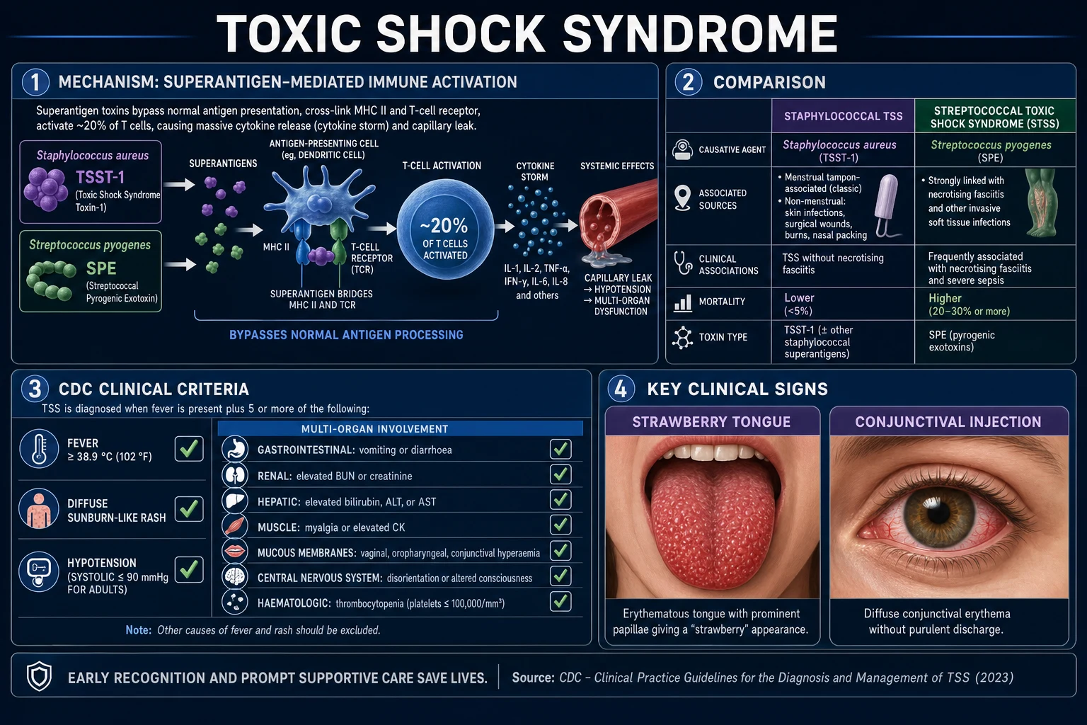
A second classification axis is the CDC case definition status — confirmed versus probable staphylococcal TSS. A confirmed case requires ALL five clinical criteria (fever over 38.9 deg C, diffuse macular rash, desquamation 1 to 2 weeks later, hypotension, and 3 or more organ systems) AND exclusion of other aetiologies. A probable case satisfies all criteria but for desquamation (the patient presents before desquamation has occurred, or dies before it does), or is missing one criterion. Isolation of S. aureus is NOT part of the case definition — a fact examiners test repeatedly. The CDC case definition for streptococcal TSS is structurally different (see Investigations) and DOES require isolation of Group A strep from a normally sterile site in the 'definite' category.[4][2]
Epidemiology & Risk Factors
The historic 1979 to 1980 US epidemic of menstrual TSS, traced to ultra-high-absorbency tampons (notably the Rely brand, made of carboxymethylcellulose and polyester foam), produced a peak incidence of about 13 cases per 100,000 menstruating women. After Rely was withdrawn and tampon absorbency was regulated, incidence fell roughly tenfold to about 1 per 100,000; the Hajjeh CDC surveillance update 1979 to 1996 documents this decline. Today non-menstrual staphylococcal TSS accounts for the majority of cases, and overall US incidence is roughly 3 to 5 per 100,000.[4]
Streptococcal TSS is rarer (about 3 to 4 per 100,000 in developed countries) but far deadlier (30 to 60 percent mortality). A striking global surge in invasive Group A streptococcal disease — and STSS — was observed in 2022 to 2023 in the UK, Europe, the US and elsewhere in the wake of the COVID-19 pandemic, possibly reflecting reduced population GAS exposure during pandemic lockdowns. Children bore a disproportionate burden of this surge.[5]
Risk factors for staphylococcal TSS: [1]
- Tampon use — especially high-absorbency, prolonged or overnight use, and a single tampon left in place. The mechanism: high-absorbency tampons bind magnesium (derepressing TSST-1 production), create an oxygenated, protein-rich, neutral-to-alkaline environment ideal for TSST-1 production, and the vaginal mucosa absorbs the preformed toxin.[3]
- Barrier contraceptives — contraceptive sponge, diaphragm, cervical cap.
- Surgical wounds — especially wounds that are CLOSED and look clean (the 'benign wound' trap).
- Postpartum — vaginal or caesarean delivery; the uterus or wound is the focus.
- Burns — colonised burn wounds.
- Skin and soft-tissue infection — cellulitis, abscess, surgical-site infection.
- Nasal packing for epistaxis.
- Foreign bodies — retained packing, surgical mesh.
- Recent influenza or varicella infection — disrupts mucocutaneous barriers.
Risk factors for streptococcal TSS (STSS): [1]
- Invasive Group A streptococcal infection — necrotising fasciitis, myositis, myonecrosis, cellulitis, pneumonia, postpartum sepsis, joint/bone infection.
- Minor trauma or bruise — the classic 'minor injury, severe illness' history (a small abrasion precedes a catastrophic GAS necrotising infection).
- Varicella (chickenpox) in children — a major risk factor for GAS superinfection and STSS.
- NSAID use — controversial but commonly asked; may mask early symptoms and has been associated with necrotising fasciitis.
- Immunocompromise, diabetes mellitus, alcoholism, IV drug use, advanced age, chronic skin conditions.
- Post-COVID-19 surge — a population-level risk factor for invasive GAS disease (2022 to 2023).[5]
Pathophysiology
The entire clinical syndrome flows from a single molecular event — the superantigen bypass of normal antigen presentation. Understanding this cascade explains every clinical feature and every element of therapy. [1]
Normal antigen presentation (the control)
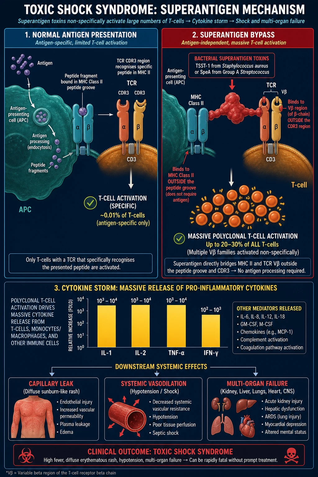
In normal adaptive immunity: (1) an antigen-presenting cell (APC — macrophage, dendritic cell, B-cell) takes up an antigen; (2) it processes the antigen into peptide fragments in endosomes; (3) it loads the peptides into the groove of MHC class II; (4) a T-cell whose TCR CDR3 region (the antigen-specific complementarity-determining region) specifically recognises that peptide-MHC complex is activated. Because the TCR is highly specific, at most about 0.01 percent (1 in 10,000) of all T-cells respond to any one antigen.[2]
The superantigen bypass (the disease)
Bacterial superantigens — TSST-1 and the staphylococcal enterotoxins B and C from S. aureus, and the streptococcal pyrogenic exotoxins A and C (SpeA, SpeC) from Group A strep — short-circuit this elegant specificity in two ways at once:[2]
- They bind DIRECTLY to the OUTER side surface of MHC class II — NOT in the peptide-binding groove, but on the side, outside the cleft — without requiring any specific peptide.
- They bind simultaneously to the OUTER surface of the T-cell receptor beta-chain variable region (V-beta) — NOT the antigen-specific CDR3 region, but a conserved framework region shared by a large family of T-cells. [1]
The superantigen thus forms a bridge between the APC and the T-cell that does NOT require antigen processing or specificity. Because the V-beta regions it binds are shared by many T-cells (different V-beta families are present in different proportions), a single superantigen activates 20 to 30 percent of ALL T-cells simultaneously — a 1,000- to 10,000-fold increase over the normal ~0.01 percent.[3]
The cytokine storm and the clinical phenotype
Massive polyclonal T-cell activation produces a cytokine storm: activated T cells and macrophages release interleukin-1 (IL-1), interleukin-2 (IL-2), tumour necrosis factor-alpha (TNF-alpha) and interferon-gamma (IFN-gamma), with additional IL-6 and macrophage inflammatory proteins. These cytokines are the direct mediators of:[2]
- Capillary leak — diffuse endothelial dysfunction, third-space loss of albumin and fluid -> the diffuse 'sunburn-like' rash, hypotension, oedema, pulmonary oedema, renal and hepatic dysfunction.
- Systemic vasodilation — profound hypotension refractory to fluids.
- Myocardial depression — direct cytokine-mediated cardiomyocyte injury.
- Multi-organ failure — kidneys (AKI), liver (hepatitis), lungs (ARDS), blood (thrombocytopenia, DIC), muscles (rhabdomyolysis), CNS (encephalopathy without focal signs). [1]
The clinical syndrome is mediated ENTIRELY by host cytokines — a fact that explains why staphylococcal TSS is typically NOT bacteraemic: the toxin is produced locally (in the vagina or a wound) and absorbed, and the bacteria themselves need not be present in the bloodstream. This is also why blood cultures are negative in staphylococcal TSS and why organism isolation is NOT required for the CDC diagnosis.[3]
The desquamation and the rash
The diffuse macular erythema ('sunburn-like' or scarlatiniform, blanching, involving the whole body including palms and soles) results from cytokine-mediated dermal capillary leak and T-cell infiltration. The late desquamation of the stratum corneum 1 to 2 weeks later (especially palms and soles, fingertips) reflects the kinetics of epidermal turnover after the inflammatory insult — the damaged superficial epidermis is shed as a new layer regenerates. This is why desquamation is a LATE (and confirmatory) feature, not a presenting one.[2]
Why menstrual TSS followed high-absorbency tampons
TSST-1 production by S. aureus is enhanced by a high-protein, neutral-to-alkaline, oxygenated environment. High-absorbency tampons create exactly these conditions: they absorb menstrual blood (a protein-rich medium), elevate vaginal pH toward neutral/alkaline, and introduce oxygen. Critically, high-absorbency tampons bind magnesium — and magnesium represses TSST-1 production, so its removal derepresses TSST-1 synthesis. The vaginal mucosa then absorbs the preformed toxin. This single biochemical fact explains both the 1980 epidemic and the dramatic fall after tampon regulation.[3]
TSST-1 antibody immunity and recurrence
About 90 percent of adults have protective anti-TSST-1 antibodies — the result of asymptomatic colonisation with TSST-1-producing S. aureus during childhood and adolescence. The ~10 percent who lack antibodies are susceptible to TSS. This explains why TSS recurs in susceptible individuals (about a 30 percent recurrence rate for menstrual TSS without counselling), and it is the rationale for IVIG in severe disease — pooled human immunoglobulin contains high titres of anti-TSST-1 (and anti-Spe) antibodies that neutralise the circulating superantigen.[3]
Clinical Presentation
TSS is an acute, rapidly progressive illness (over hours) dominated by a striking prodrome, the cardinal clinical tetrad, and evolving multi-organ failure. The tempo and the discriminating features between staphylococcal and streptococcal disease are the examiner's favourite test. [1]
Shared early features (both forms)
- High fever — over 38.9 deg C (a CDC criterion).
- Myalgia, headache, arthralgia, pharyngitis, conjunctivitis — a non-specific prodrome.
- Abdominal pain, vomiting and diarrhoea at onset (GI involvement is a CDC criterion) — easily mistaken for gastroenteritis or 'flu', a key diagnostic trap.
- Hypotension — systolic BP at or under 90 mmHg (adults), or an orthostatic drop, with signs of shock (cool peripheries, mottling, tachycardia).
- Altered mental state — disorientation, confusion, agitation, obtundation (CNS involvement is a CDC criterion; no focal neurology).
- Diffuse blanching macular 'sunburn-like' rash — non-pruritic, scarlatiniform, involves the whole body including palms and soles, may be subtle or missed in dark skin. [1]
Staphylococcal TSS — the classic menstrual vignette
A menstruating young woman using tampons presents within 1 to 2 days of menses onset with acute high fever, myalgia, headache, sore throat, vomiting and diarrhoea, confusion and hypotension, and a diffuse sunburn-like rash. The picture progresses over hours to multi-organ failure. Non-menstrual staphylococcal TSS presents identically but the source is a surgical wound (often 'closed' and benign-looking), postpartum wound, burn, skin infection, nasal packing or barrier contraceptive.[1]
The mucous membrane signs are particularly useful: conjunctival hyperaemia, 'strawberry tongue' (white coating with red bumps that peels to leave a bright red tongue with prominent papillae), oropharyngeal hyperaemia, and vaginal hyperaemia — these constitute a 'mucosal triad' that should prompt TSS in any febrile, toxic patient.[3]
Streptococcal TSS (STSS) — the necrotising fasciitis picture
STSS typically presents in one of two ways: [1]
- With a soft-tissue focus (~80 percent) — necrotising fasciitis, myositis, myonecrosis, cellulitis or pneumonia. The classic history is minor trauma or bruise followed by rapidly progressive, excruciatingly painful soft-tissue swelling with pain out of proportion to the visible findings, tense oedema, skin that becomes dusky/blue/gangrenous with haemorrhagic bullae and cutaneous anaesthesia (cutaneous nerves destroyed). Shock, renal failure, ARDS and coagulopathy develop over 24 to 48 hours.
- Without a focus (~20 percent) — a non-localised flu-like illness (fever, myalgia, malaise) that rapidly progresses to shock and multi-organ failure; the source may be an occult deep infection. [1]
Bacteraemia is present in ~60 percent (compared with uncommon bacteraemia in staphylococcal TSS).[6]
Atypical and special-context presentations
- Postpartum TSS — within days of vaginal or caesarean delivery; the uterus or wound may look uninfected; presents as fever, hypotension, rash and diarrhoea; perform uterine exploration, remove any retained products/packing.[1]
- Post-surgical TSS — typically presents within 48 hours of surgery (often much faster than a conventional wound infection); the wound looks clean and closed; a 'benign wound + systemic shock' picture is the trap; remove sutures and explore.[1]
- Burn-associated TSS — colonised burn wounds; high index of suspicion in any burn patient with sudden deterioration and rash.
- Paediatric TSS — often a focus of skin/soft-tissue infection, or varicella (chickenpox) lesions colonised by GAS; must be distinguished from Kawasaki disease (see Differential Diagnosis); weight-based fluid and drug doses.[5]
- Nasal-packing TSS — within 12 to 24 hours of nasal packing for epistaxis; remove the packing immediately.
- Elderly/immunocompromised — blunted fever (may be afebrile or hypothermic), atypical presentation (confusion, collapse), higher mortality (especially STSS), higher risk of MRSA — broaden cover with vancomycin or linezolid.
Differential Diagnosis
The useful framing is the differential of fever + rash + hypotension + multi-organ illness. Several conditions overlap with TSS and examiners test the distinction relentlessly. [1]
FEBRILE RASH SHOCK
The discriminating features examiners expect: [1]
Staphylococcal TSS
- Toxin: TSST-1 / staph enterotoxins (superantigen)
- Bacteraemia: UNCOMMON — blood cultures negative
- Source: colonised focus (tampon, 'benign' wound) — NOT deep invasive infection
- Rash: diffuse blanching macular 'sunburn' + late desquamation of palms/soles
- Mortality: 3 to 5 percent
- Key action: REMOVE tampon / foreign body + clindamycin (anti-toxin)
Streptococcal TSS (STSS)
- Toxin: SpeA / SpeC (superantigen)
- Bacteraemia: COMMON (~60 percent) — Group A strep from sterile site
- Source: invasive soft-tissue infection (necrotising fasciitis, myonecrosis)
- Rash: generalised erythema (may desquamate); soft-tissue focus dominates
- Mortality: 30 to 60 percent
- Key action: URGENT surgical debridement + penicillin + clindamycin + IVIG
Kawasaki disease
- Population: children under 5 years
- Course: SUBACUTE (days to weeks), fever over 5 days
- Shock: ABSENT (no hypotension)
- Platelets: thromboCYTOSIS after day 7 (TSS has thromboCYTOPENIA early)
- Hallmark: coronary artery aneurysm risk
- Treatment: IVIG 2 g/kg + aspirin (anti-inflammatory dose)
Staphylococcal scalded skin syndrome (SSSS)
- Population: neonates and young children
- Toxin: epidermolytic toxin A/B (NOT a superantigen)
- Skin: superficial flaccid bullae, positive NIKOLSKY sign, perioral fissuring
- Mucosae: SPARED (key difference from SJS/TEN and TSS)
- Shock / multi-organ: ABSENT
- Treatment: anti-staphylococcal antibiotic (flucloxacillin); no IVIG
Meningococcaemia
- Organism: Neisseria meningitidis (Gram-negative diplococcus)
- Rash: PETECHIAL / PURPURIC, NON-blanching (TSS is blanching)
- Focus: meningitis / meningococcal septicaemia
- Complication: purpura fulminans, DIC, Waterhouse-Friderichsen (adrenal haemorrhage)
- Treatment: ceftriaxone (meningitis dose) + supportive
DRESS / SJS-TEN
- Onset: delayed (2 to 8 weeks after drug — DRESS); 1 to 3 weeks (SJS/TEN)
- DRESS: eosinophilia, atypical lymphocytes, facial oedema; SJS/TEN: target lesions, mucosal erosions, blistering, positive NIKOLSKY
- Shock / cytokine storm: ABSENT (the key discriminator from TSS)
- Treatment: withdraw offending drug; supportive; IVIG/ciclosporin debated
The most easily missed TSS is post-surgical TSS with a 'benign-looking' wound and postpartum TSS — in both, the wound or uterus often appears uninfected, and the diagnosis rests on the systemic picture. A high index of suspicion is essential: any patient with fever + diffuse rash + hypotension + multi-organ involvement has TSS until proven otherwise.[1]
Clinical & Bedside Assessment
The bedside assessment of suspected TSS is structured, time-critical and source-seeking. [1]
ABCDE priorities
- Airway — protect if altered mental state; prepare for intubation if obtunded.
- Breathing — high-flow oxygen; monitor for ARDS and pulmonary oedema from capillary leak; prepare for mechanical ventilation.
- Circulation — two large-bore IV cannulae; aggressive crystalloid resuscitation; have vasopressors drawn up; arterial line early.
- Disability — GCS, pupils, look for CNS disorientation WITHOUT focal signs (a CDC criterion); exclude hypoglycaemia.
- Exposure — FULL skin examination including palms, soles, all mucous membranes, genitalia, and every wound and burn (even 'closed' and clean-looking ones). [1]
The focused examination for a source
This is the highest-yield part of the assessment. Inspect every potential focus: [1]
- The vagina — remove any tampon, look for a retained foreign body (tampon, contraceptive sponge, diaphragm); send for culture.
- Every surgical wound and burn — even if 'closed' and clean-looking (the trap); remove sutures and explore if suspicious.
- The postpartum uterus — perform bimanual and speculum examination; look for retained products of conception, packing, abscess.
- The nose — if packed for epistaxis, remove the packing.
- The skin — cellulitis, abscess, chickenpox (varicella) lesions, surgical-site infection.
- The soft tissues — actively elicit signs of necrotising fasciitis (pain out of proportion, tense oedema, cutaneous anaesthesia, haemorrhagic bullae, skin necrosis, crepitus from gas-forming organisms). [1]
Cardinal clinical signs to elicit
- Diffuse blanching macular erythema including palms and soles (the 'sunburn' rash).
- Mucous membrane hyperaemia — conjunctivae, oropharynx, 'strawberry tongue', vagina.
- Hypotension — systolic at or under 90 mmHg, or orthostatic drop.
- Signs of necrotising fasciitis (if STSS suspected) — pain out of proportion, tense oedema, skin discoloration, cutaneous anaesthesia, haemorrhagic bullae, crepitus. [1]
The LRINEC score for necrotising fasciitis
Because necrotising fasciitis drives STSS and mandates urgent surgery, the LRINEC score is a useful bedside adjunct to distinguish necrotising fasciitis from simple cellulitis. The components (sum 0 to 13): [1]
| Variable | Score 0 | Score 1 | Score 2 | Score 4 |
|---|---|---|---|---|
| C-reactive protein (mg/L) | under 150 | over 150 | — | — |
| White cell count (x10^9/L) | 15 to 25 | — | over 25 | — |
| Haemoglobin (g/dL) | 11 to 13.5 | — | under 11 | — |
| Sodium (mmol/L) | over 135 | — | under 135 | — |
| Creatinine (umol/L) | under 141 | — | over 141 | — |
| Glucose (mmol/L) | under 10 | over 10 | — | — |
(CRP over 150 = 4 points; WBC 15-25 = 1, over 25 = 2; Hb 11-13.5 = 1, under 11 = 2; Na under 135 = 2; creatinine over 141 = 2; glucose over 10 = 1.) [1]
A LRINEC score of 6 or more indicates a high risk of necrotising fasciitis (positive predictive value ~92 percent in the derivation cohort). Critically, LRINEC is INSENSITIVE — a high clinical suspicion overrides a 'low' LRINEC, and any patient with the clinical features of necrotising fasciitis (pain out of proportion, rapidly spreading, systemic toxicity) must undergo surgical exploration regardless of the score.[6]
Examination for complications during resuscitation
- Mental status — cerebral oedema, hypoperfusion, encephalopathy.
- Urine output — Foley catheter; target at least 0.5 mL/kg/h; renal failure is a CDC criterion.
- Perfusion — lactate (severity and clearance), capillary refill, mottling.
- Signs of DIC — ooze from venepuncture sites, petechiae, purpura, bleeding.
- Cardiac — arrhythmia, myocardial depression, signs of heart failure. [1]
Investigations
TSS is a CLINICAL diagnosis using CDC criteria. There is no single confirmatory laboratory test at presentation; the role of investigations is to support the diagnosis, quantify organ failure, find the source and exclude mimics. Do NOT delay empiric therapy (fluids, source control, antibiotics, IVIG) while waiting for results.[1]
The CDC criteria for staphylococcal TSS (reproduce verbatim)
[1]The CDC criteria for streptococcal TSS (reproduce verbatim)
[1]Note the structural differences: staphylococcal TSS requires 3 or more organ systems, organism isolation is not required, and the rash is a primary criterion; streptococcal TSS requires only 2 or more organ systems, organism isolation is required, and soft-tissue necrosis can substitute for organ failure. Bacteraemia is expected in STSS (in ~60 percent) but uncommon in staphylococcal TSS — a frequently tested discriminator.[2][6]
First-line blood tests
- Full blood count — leukocytosis with a left shift; THROMBOCYTOPENIA (platelets under 100,000/microL) is a CDC criterion.
- Urea and electrolytes — renal failure (BUN/creatinine over twice ULN is a CDC criterion).
- Liver function tests — hepatitis (bilirubin/ALT/AST over twice ULN is a CDC criterion).
- Creatine kinase (CK) — rhabdomyolysis (CK over twice ULN is a CDC criterion).
- CRP and procalcitonin — markers of inflammation/sepsis.
- Coagulation — PT, aPTT, fibrinogen, D-dimer (for DIC).
- Lactate — severity and resuscitation marker (rising lactate = worsening perfusion).
- Venous blood gas — metabolic acidosis.
- Glucose, calcium, albumin — correction targets (hypocalcaemia common in STSS; albumin low from capillary leak). [1]
Microbiological specimens
- Blood cultures — BEFORE antibiotics, repeated; bacteraemia expected in STSS (~60 percent), uncommon in staphylococcal TSS.
- Wound / tissue / swab cultures from any focus — surgical wound, burn, skin, postpartum uterus.
- Vaginal swab and the removed tampon — culture for S. aureus and TSST-1 testing.
- Throat swab — for GAS.
- Urine — Group A strep antigen; microscopy for sterile pyuria (a CDC criterion). [1]
TSST-1 antibody testing is NOT useful acutely (results take weeks; it confirms a diagnosis retrospectively) — but ~90 percent of adults have protective titres, and absence of antibody identifies susceptibility to recurrent disease.[3]
Imaging
- Chest X-ray — ARDS, pulmonary oedema (from capillary leak), pneumonia (a source).
- Ultrasound or CT of soft tissue — for an occult focus (necrotising fasciitis, postpartum uterine infection, abscess, retained foreign body); CT may show gas within soft tissue in clostridial myonecrosis, fascial thickening with fat stranding in necrotising fasciitis.
- Echocardiography — myocardial depression, regional wall motion abnormalities. [1]
Bedside tests for distinguishing mimics
- Blood film — atypical lymphocytes and eosinophilia for DRESS.
- Meningococcal PCR and culture — if meningococcaemia plausible.
- RMSF serology, leptospirosis serology, viral serology (measles, EBV, HIV) — to exclude mimics as required by the CDC criteria. [1]
Management — Resuscitation
TSS is a time-critical emergency. The resuscitation bundle must be delivered concurrently, not sequentially. [1]
The time-critical bundle
- ABCDE — airway protection if obtunded; high-flow oxygen; two large-bore IV cannulae; full monitoring.
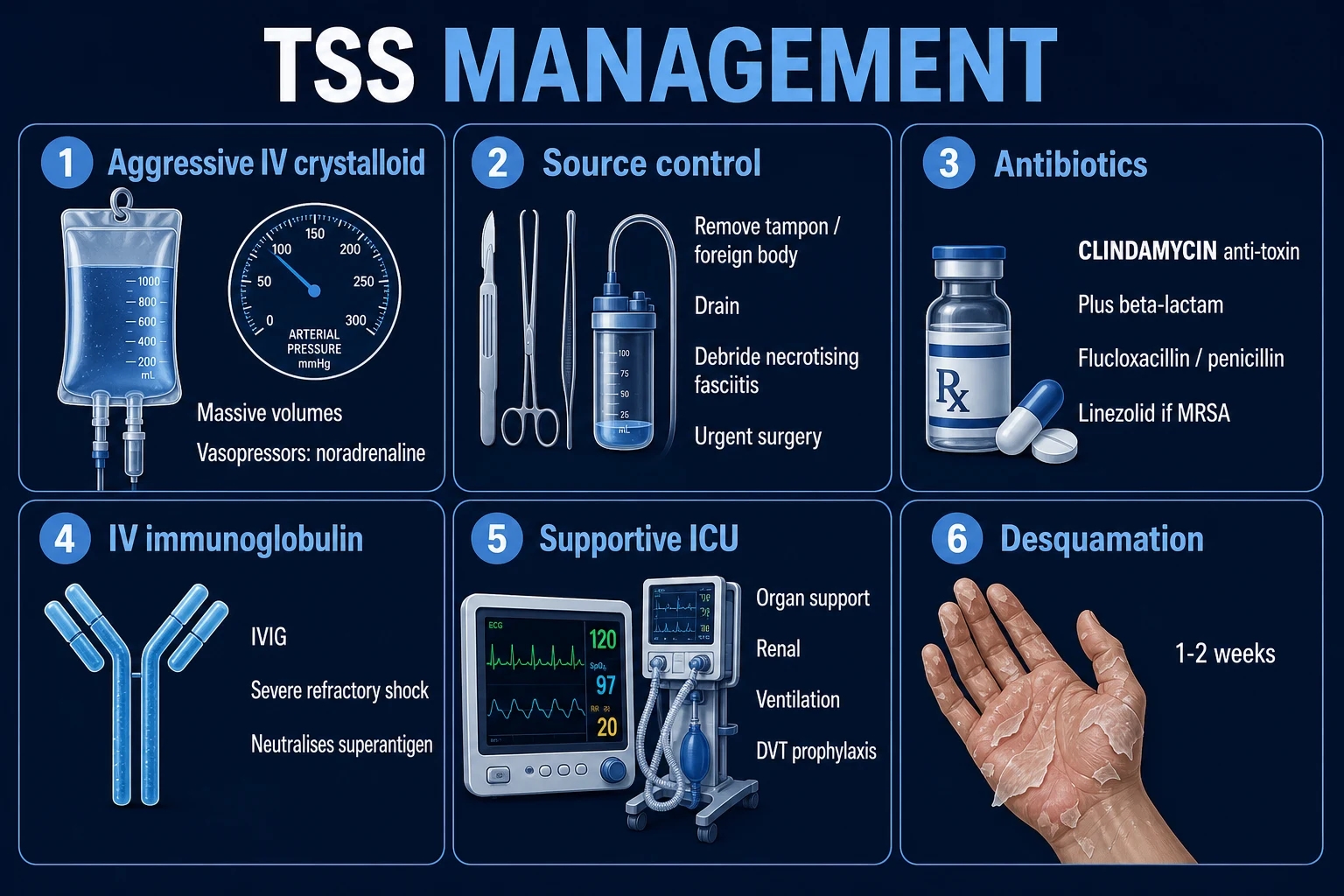
- Aggressive IV fluid resuscitation — crystalloid (balanced or normal saline) 10 to 20 mL/kg boluses repeated up to 30 mL/kg in the first hour, titrated to MAP at least 65 mmHg, urine output at least 0.5 mL/kg/h, and lactate clearance. Severe capillary leak means very large volumes (often 5 to 10 L) may be required; reassess after each bolus for pulmonary oedema.
- Immediate SOURCE CONTROL — REMOVE the tampon or contraceptive sponge/diaphragm AT ONCE (this alone can abort staphylococcal TSS); inspect and debride any wound (necrotising fasciitis needs urgent surgical exploration and debridement); drain abscesses; remove nasal packing/foreign bodies; perform uterine exploration in postpartum TSS. In STSS, do NOT wait for stabilisation before surgery — the patient stabilises only after source control.[6]
- Empirical IV antibiotics within 1 hour — a regimen covering both staph (including MRSA) and strep AND suppressing toxin. The cornerstone is clindamycin (the anti-toxin). See below for full regimens.
- Vasopressors for refractory shock — noradrenaline (norepinephrine) is first-line, titrated to MAP at least 65 mmHg (peripheral or central); add vasopressin if refractory; consider adrenaline if myocardial depression predominates. Do not delay vasopressors while giving more fluids if shock is refractory.
- IVIG in severe / refractory disease — 1 to 2 g/kg IV single dose (or 0.5 g/kg/day for 4 days). Neutralises circulating superantigen and provides anti-TSST-1 antibodies; indicated for refractory shock, deepening multi-organ failure, or STSS.
- Supportive measures — mechanical ventilation for ARDS / respiratory failure, renal replacement therapy for renal failure, glucose control, stress-ulcer and VTE prophylaxis, temperature control, correction of electrolytes / calcium / albumin.
Management — Definitive & Stepwise
The definitive management builds on resuscitation with anti-toxin therapy, source control, IVIG and organ support. The escalation triggers are explicit. [1]
R-A-S-C-I-V
The antibiotic regimen
The empirical regimen must cover both staphylococcal (including MRSA) and streptococcal disease AND suppress toxin production. The cornerstone is clindamycin, which — uniquely among the antibiotics — suppresses TSST-1 and Spe production by inhibiting 50S ribosomal protein synthesis. Penicillin alone may INCREASE toxin release by lysis of bacteria, which is why a beta-lactam must ALWAYS be combined with clindamycin.[3][6]
Empirical (organism unknown)
- Clindamycin 600 to 900 mg IV q8h (anti-toxin — suppresses TSST-1, SpeA, M-protein)
- PLUS flucloxacillin / nafcillin 2 g IV q6h (MSSA) OR vancomycin 15 to 20 mg/kg IV q8 to 12h (trough 15 to 20 mg/L) / linezolid 600 mg IV q12h (if MRSA suspected or known)
- PLUS benzylpenicillin 2.4 g IV q4 to 6h (Group A strep)
- Consider adding a third-generation cephalosporin (ceftriaxone 2 g IV q12 to 24h) for broad Gram-negative cover if source uncertain
Confirmed staphylococcal TSS (MSSA)
- Flucloxacillin / nafcillin 2 g IV q6h (the anti-staphylococcal beta-lactam)
- PLUS clindamycin 600 to 900 mg IV q8h (the anti-toxin)
- Duration typically 10 to 14 days
Confirmed staphylococcal TSS (MRSA)
- Vancomycin 15 to 20 mg/kg IV q8 to 12h (trough 15 to 20 mg/L)
- OR linezolid 600 mg IV q12h (suppresses toxin AND covers MRSA — preferred in severe disease)
- PLUS clindamycin 600 to 900 mg IV q8h (anti-toxin; note MRSA may be clindamycin-resistant — test for inducible resistance / D-test)
Confirmed streptococcal TSS (STSS)
- Benzylpenicillin 2.4 g IV q4 to 6h (Group A strep — note the 'Eagle effect': penicillin less effective in high-inoculum stationary-phase infection)
- PLUS clindamycin 900 mg IV q8h (suppresses SpeA and M-protein; unaffected by Eagle effect)
- IVIG 1 to 2 g/kg IV single dose (evidence strongest for STSS)
- URGENT surgical debridement of necrotising fasciitis (within hours) — life-saving
- Duration typically 10 to 14 days; longer if deep tissue infection / osteomyelitis
Why clindamycin is the cornerstone
Clindamycin inhibits the 50S ribosomal subunit and therefore suppresses bacterial protein (exotoxin) synthesis — reducing TSST-1 and Spe production by about 90 percent, even when organisms are viable. It also suppresses M-protein synthesis in Group A strep (an antiphagocytic virulence factor) and is unaffected by the 'Eagle effect' that limits penicillin efficacy in high-inoculum, stationary-phase GAS infection (where penicillin-binding proteins are down-regulated). Penicillin (and oxacillin/nafcillin), by contrast, may transiently INCREASE toxin release by lysing bacteria — so a beta-lactam must ALWAYS be combined with clindamycin. This single pharmacological fact is one of the most frequently tested in TSS exams.[3][6]
Why linezolid is the MRSA alternative
Linezolid is a 50S protein-synthesis inhibitor (oxazolidinone) that suppresses toxin production AND covers MRSA — making it a single-agent alternative for severe MRSA-associated TSS. Dose 600 mg IV q12h. Cautions: serotonin syndrome (linezolid is a weak MAO-A inhibitor — avoid with serotonergic drugs and SSRIs), thrombocytopenia and anaemia with prolonged use (over 2 weeks), and peripheral and optic neuropathy with very prolonged courses.[1]
IVIG — dose and rationale
Intravenous immunoglobulin provides a bolus of anti-TSST-1 and anti-Spe antibodies from pooled human donors, neutralising circulating superantigen and modulating T-cell and macrophage activation. Dose 1 to 2 g/kg IV as a single infusion over 12 to 24 hours (alternatively 0.5 g/kg/day for 4 days). Evidence is strongest for STSS (one small RCT — Darenberg 2003 — showed a trend to mortality benefit; observational data support use in staphylococcal TSS refractory to conventional therapy). Indications: refractory shock, deepening multi-organ failure, or any STSS. Do not delay conventional therapy (source control, antibiotics, vasopressors) while waiting for IVIG.[2][6]
Surgical principles for STSS with necrotising fasciitis
Surgery is life-saving in STSS with necrotising fasciitis and must NOT be delayed to 'stabilise' the patient — the patient stabilises only after source control. The principles:[6]
- Urgent (within hours) surgical exploration — incise over the area of maximal tenderness; explore down to and along fascial planes.
- Debride all necrotic tissue to bleeding viable tissue — the hallmark of necrotic fascia is that it does not bleed and separates easily ('dishwater' fluid, grey necrotic fascia).
- Re-look in theatre at 24 to 48 hours — necrosis often extends beyond the initial debridement; plan return-to-theatre.
- Culture-guided therapy — send tissue for culture and adjust antibiotics.
- Amputation may be required for limb gangrene — a life-saving measure, not a failure. [1]
Management of complications
- ARDS — lung-protective mechanical ventilation (tidal volume 6 mL/kg ideal body weight, plateau pressure under 30 cmH2O); prone positioning; neuromuscular blockade in severe cases.
- DIC — transfuse platelets and fresh-frozen plasma if bleeding; do NOT treat asymptomatic laboratory DIC.
- Renal failure — renal replacement therapy (continuous for the haemodynamically unstable patient).
- Rhabdomyolysis — IV fluids to target urine output 1 to 2 mL/kg/h; renal replacement therapy if established AKI.
- Myocardial depression — inotropes (dobutamine or adrenaline); consider milrinone if on vasopressors.
- Cerebral oedema — head elevation 30 degrees, normocapnia, osmotic therapy (mannitol or hypertonic saline) if signs of herniation. [1]
Prevention and patient-safety counselling
Counsel recovered patients on tampon hygiene to prevent recurrence: lowest-absorbency tampon that meets needs, change every 4 to 8 hours, avoid overnight use, no tampons at all after an episode of menstrual TSS. Avoid barrier contraceptives (sponge, diaphragm) after staphylococcal TSS. The recurrence risk is about 30 percent for menstrual TSS without counselling — patient education is the single most effective prevention. Consider oral clindamycin eradication therapy for the ~10 percent of adults who lack anti-TSST-1 antibodies and have recurrent disease.[3]
Specific Subtypes & Scenarios
Menstrual TSS
The classic vignette: a menstruating young woman using tampons presents within 1 to 2 days of menses onset with acute high fever, myalgia, vomiting, diarrhoea, confusion, hypotension and a sunburn-like rash. TSST-1-producing S. aureus colonises the vagina and is absorbed across the vaginal mucosa. Management: remove the tampon immediately (this alone can abort the syndrome), aggressive fluid resuscitation, antibiotics with clindamycin + flucloxacillin, IVIG if severe, ICU. Counsel on recurrence prevention.[3]
Non-menstrual staphylococcal TSS
Now commoner than menstrual TSS. Sources: surgical wounds (often CLOSED and benign-looking — the trap), postpartum wounds, burns, skin/soft-tissue infection, nasal packing, barrier contraceptives, foreign bodies. Management is identical: find and remove the source. The wound may need to be opened and explored even when it looks clean — the diagnosis is the systemic picture, NOT the wound appearance.[1]
Streptococcal TSS (STSS)
Group A strep (S. pyogenes) deep soft-tissue infection (necrotising fasciitis in ~50 percent, myositis, cellulitis) or a non-focal flu-like illness. Bacteraemia in ~60 percent; mortality 30 to 60 percent. Management: urgent surgical debridement of necrotising fasciitis within hours, benzylpenicillin + clindamycin, IVIG 1 to 2 g/kg, organ support. Surgery is life-saving and must not be delayed.[6]
Paediatric TSS
Suspect TSS in any febrile child with rash and shock. Focus on skin infection (often varicella/chickenpox lesions colonised by GAS) or post-surgical infection. The Kawasaki overlap is the key trap — Kawasaki is in children under 5, is subacute (fever over 5 days), has no shock and thrombocytosis after day 7 (TSS has thrombocytopenia early), and carries a risk of coronary artery aneurysm (give IVIG 2 g/kg + aspirin for Kawasaki). Use weight-based fluid (10 to 20 mL/kg boluses) and weight-based antibiotic doses (clindamycin 10 mg/kg q8h, flucloxacillin 50 mg/kg q6h, vancomycin 15 mg/kg q6h). Involve PICU.[5]
Postpartum / post-surgical TSS
The uterine or wound focus often looks uninfected — the diagnosis is systemic and the source may only be identified on direct inspection or exploration. Remove sutures / explore the uterus / remove any retained products or packing. High index of suspicion in any postpartum or post-surgical patient with fever + rash + shock + multi-organ involvement, even with a 'clean' wound.[1]
Recurrent TSS
Patients lacking anti-TSST-1 antibody (~10 percent of adults) are susceptible to recurrence (~30 percent for menstrual TSS without counselling). Counsel on avoiding tampons/barriers; some advocate oral clindamycin eradication therapy to clear S. aureus carriage.[3]
Complications & Pitfalls
Early complications
- Refractory shock — mortality from vasoplegia, myocardial depression, capillary leak.
- ARDS — from cytokine-mediated alveolar-capillary leak.
- Acute kidney injury requiring renal replacement therapy.
- Disseminated intravascular coagulation (DIC) — bleeding and microvascular thrombosis.
- Rhabdomyolysis — contributes to AKI.
- Myocarditis / cardiomyopathy with myocardial depression.
- Hepatic failure.
- Cerebral oedema and encephalopathy.
- Gangrene and amputation of limbs (STSS).
- Multi-organ failure and death. [1]
Late complications
- Full-thickness desquamation of palms and soles at 1 to 2 weeks.
- Reversible alopecia and nail shedding (Beau lines, onychomadesis) at 2 to 3 months.
- Chronic kidney disease.
- Neuropsychiatric sequelae — cognitive impairment, depression, anxiety.
- Prolonged fatigue and weakness.
- Amputation and surgical disfigurement (STSS). [1]
Diagnostic pitfalls
- Mistaking the prodromal GI or respiratory symptoms for gastroenteritis or 'flu' — the rash is missed because it is subtle in dark skin or attributed to drug allergy.
- Attributing post-surgical TSS to the wound looking 'clean' — the wound often looks benign; the diagnosis is systemic.
- Treating for sepsis WITHOUT an anti-TOXIN agent (clindamycin or linezolid) — penicillin/oxacillin alone may not suffice and may transiently increase toxin release.
- Failing to REMOVE the source (tampon, foreign body, packing) — the single most important early action in staphylococcal TSS.
- Mistaking STSS necrotising fasciitis for simple cellulitis — use the LRINEC score and the clinical features (pain out of proportion) — a high suspicion overrides a 'low' score. [1]
Therapeutic pitfalls
- Delaying vasopressors while giving excessive fluids — produces pulmonary oedema and ARDS.
- Delaying surgical debridement in STSS to 'stabilise' the patient — the patient stabilises only after source control.
- Omitting clindamycin (the anti-toxin) — the most common pharmacological error.
- Under-dosing IVIG (use 1 to 2 g/kg, not 0.4 g/kg).
- Missing MRSA cover — add vancomycin or linezolid if MRSA is plausible (post-surgical, healthcare-associated, known colonisation). [1]
Prognosis & Disposition
Case-fatality
- Staphylococcal TSS: 3 to 5 percent with modern ICU care.
- Streptococcal TSS (STSS): 30 to 60 percent — markedly higher. Predictors of fatal STSS: advanced age, comorbidity (diabetes, immunocompromise), deep tissue necrosis, delay to surgery, bacteraemia, renal failure, ARDS, hypocalcaemia.[2][6]
Disposition
All suspected TSS require ICU (level 3) admission. STSS and any patient needing vasopressors, ventilation or renal replacement therapy require a tertiary centre with surgical and intensive-care capability. Mobilise the sepsis team and infectious diseases early; involve surgery immediately if necrotising fasciitis is suspected.[1]
Recovery timeline
- Acute phase — hours to days; the period of greatest mortality risk.
- Desquamation — 1 to 2 weeks (palms and soles).
- Full clinical recovery — weeks to months.
- Nail and hair changes (Beau lines, onychomadesis, alopecia) — 2 to 3 months.
- Some patients have persistent fatigue, weakness and neuropsychiatric symptoms for months. [1]
Recurrence risk and follow-up
About 30 percent recurrence for menstrual TSS without counselling. Counsel on tampon/barrier avoidance; consider serology to identify anti-TSST-1-negative patients (susceptible); consider eradication therapy. Public-health notification of confirmed TSS and iGAS disease is mandatory in many jurisdictions.[4]
Special Populations
[1]Paediatrics
Suspect TSS in any febrile child with rash and shock. Weight-based fluid (10 to 20 mL/kg boluses) and antibiotic doses: clindamycin 10 mg/kg IV q8h, flucloxacillin 50 mg/kg IV q6h, vancomycin 15 mg/kg IV q6h (MRSA), benzylpenicillin 50 mg/kg IV q6h (GAS). Distinguish from Kawasaki disease (Kawasaki: under 5 years, fever over 5 days, NO shock, thrombocytosis after day 7, coronary aneurysm — give IVIG 2 g/kg + aspirin). Varicella-associated STSS is common; treat aggressively and involve PICU.[5]
Pregnancy and postpartum
Postpartum TSS (vaginal or caesarean wound) presents within days of delivery; the uterus or wound may look uninfected — perform uterine exploration, remove any retained products or packing. Pregnancy itself does not change management; deliver if chorioamnionitis is the source. Antibiotic choices (clindamycin, flucloxacillin, benzylpenicillin, vancomycin) are safe in pregnancy and breastfeeding.[1]
Elderly and immunocompromised
Blunted fever (may be afebrile or hypothermic); atypical presentation (confusion, collapse, falls); higher mortality (especially STSS); higher risk of MRSA — broaden cover with vancomycin or linezolid. Comorbidities (diabetes, renal failure, immunosuppression) worsen outcome and complicate antibiotic dosing.[2]
Anticoagulated, dialysis-dependent and comorbid
Manage DIC if present (transfuse platelets and plasma if bleeding; do NOT treat asymptomatic laboratory DIC). Adjust antibiotic doses for renal impairment (clindamycin needs no adjustment; vancomycin is level- or trough-guided; linezolid needs no adjustment but monitor for cytopenias with prolonged use). Vasopressors are safe in dialysis patients — do not withhold. Ensure VTE prophylaxis (the patient is critically ill and at high VTE risk). [1]
Evidence, Guidelines & Regional Differences
Landmark historical observations
- Todd (1978) first described TSS in seven children.
- The 1979 to 1980 US tampon epidemic (Rely brand) — Davis, Shands and colleagues identified the tampon link; the epidemic peaked at ~13/100,000 menstruating women.
- The fall in incidence after tampon absorbency regulation is documented in the Hajjeh CDC surveillance update 1979 to 1996.[4]
- The CDC case definition for staphylococcal TSS was established in 1980 to 1981 and refined subsequently; the consensus definition for streptococcal TSS was published in 1993.[2]
IDSA / Stevens guidance on invasive GAS
The IDSA / Stevens guidance on invasive Group A streptococcal infection recommends clindamycin PLUS penicillin for invasive GAS (clindamycin suppresses M-protein and Spe and is unaffected by the Eagle effect that limits penicillin in high-inoculum stationary-phase infection), IVIG for STSS, and urgent surgical debridement for necrotising fasciitis.[6]
The IVIG controversy
Evidence for IVIG is observational with one small RCT (Darenberg 2003) showing a trend to mortality benefit in STSS. Guidelines (especially for STSS) recommend IVIG (1 to 2 g/kg), but the quality of evidence is moderate and dosing varies. The biological rationale is strong (IVIG contains anti-TSST-1 and anti-Spe antibodies that neutralise circulating superantigen).[2]
The 2022 to 2023 post-COVID-19 iGAS surge
A striking global surge in invasive Group A streptococcal disease (and STSS) was observed in 2022 to 2023 in the UK, Europe, the US and elsewhere in the wake of the COVID-19 pandemic, with children disproportionately affected. Public-health alerts were issued (UKHSA, WHO, CDC). The proposed mechanism — reduced population GAS exposure during pandemic lockdowns leading to a susceptibility 'gap' — remains under investigation.[5]
Regional deltas
- India / South Asia — tampon use is uncommon, so menstrual TSS is rare; postpartum, surgical, burn-related and STSS from skin sepsis are commoner. IVIG availability and cost are variable (often limiting use in practice). CDC / IDSA / NICE / WHO guidance broadly converge on source control + clindamycin + beta-lactam + MRSA cover + IVIG.[2]
- UK — TSS and iGAS are notifiable (UKHSA); NICE and UKHSA guidance; IVIG supported for severe STSS.
- USA — CDC case definition is the international standard; IDSA guidance on invasive GAS.
- WHO / ICMR — guidance on menstrual hygiene and tampon use; recognition of the iGAS surge post-COVID-19.
Exam Pearls
- TSS = FEVER + RASH + SHOCK + MULTI-ORGAN — and the superantigen mechanism (TSST-1/Spe bypass MHC-TCR, 20 to 30 percent T-cell activation, cytokine storm). The single most tested concept.[2]
- Staphylococcal TSS is NOT bacteraemic — blood cultures are NEGATIVE — and S. aureus isolation is NOT required for the CDC diagnosis; STSS IS bacteraemic in ~60 percent. The most tested discriminator.[3]
- Desquamation of palms and soles 1 to 2 weeks after onset is PATHOGNOMONIC and confirms a retrospective TSS diagnosis — a favourite exam 'late-presentation' vignette.[2]
- The 'closed wound' trap — post-surgical TSS frequently presents with a wound that looks clean and closed; the diagnosis is the systemic picture, NOT the wound appearance.[1]
- The drug-exam trap — penicillin/oxacillin ALONE may INCREASE toxin release by lysis — ALWAYS ADD CLINDAMYCIN (the anti-toxin); linezolid is the alternative for MRSA (also suppresses toxin).[3][6]
- The recurrence and antibody pearl — ~90 percent of adults have protective anti-TSST-1 antibodies; the ~10 percent who lack them are susceptible to recurrent TSS — IVIG supplies these antibodies.[3]
- The menstrual-hygiene pearl — lowest-absorbency tampon, change every 4 to 8 hours, avoid overnight use, no tampons after an episode of menstrual TSS.[4]
- The mortality discriminator — staphylococcal TSS 3 to 5 percent vs streptococcal TSS 30 to 60 percent — an exam favourite.[2][6]
- The LRINEC pearl — a score of 6 or more indicates a high risk of necrotising fasciitis, but LRINEC is insensitive — a high clinical suspicion overrides a 'low' score.[6]
- The tampon-absorbency mechanism — high-absorbency tampons bind magnesium (derepressing TSST-1 production) and create an oxygenated, protein-rich, neutral-to-alkaline environment ideal for TSST-1.[3]
- The superantigen binding site — TSST-1 binds the outer side surface of MHC class II (NOT the peptide groove) and the V-beta region of the TCR (NOT the antigen-specific CDR3).[2]
References
- [1]Gottlieb M, Long B, Koyfman A. The Evaluation and Management of Toxic Shock Syndrome in the Emergency Department: A Review of the Literature J Emerg Med, 2018.PMID 29366615
- [2]Lappin E, Ferguson AJ. Gram-positive toxic shock syndromes Lancet Infect Dis, 2009.PMID 19393958
- [3]Silversides JA, Lappin E, Ferguson AJ. Staphylococcal toxic shock syndrome: mechanisms and management Curr Infect Dis Rep, 2010.PMID 21308522
- [4]Hajjeh RA, Reingold A, Weil A, Shutt K, Schuchat A, Perkins BA. Toxic shock syndrome in the United States: surveillance update, 1979 1996 Emerg Infect Dis, 1999.PMID 10603216
- [5]Gaensbauer JT, Birkholz M, Smit MA, Garcia R, Todd JK. Epidemiology and Clinical Relevance of Toxic Shock Syndrome in US Children Pediatr Infect Dis J, 2018.PMID 29601458
- [6]Stevens DL, Bryant AE, Goldstein EJ. Necrotizing Soft Tissue Infections Infect Dis Clin North Am, 2021.PMID 33303335