Endocrinology · General Medicine
Acromegaly
Also known as Acromegaly · Growth hormone excess · Somatotroph adenoma · Pituitary gigantism
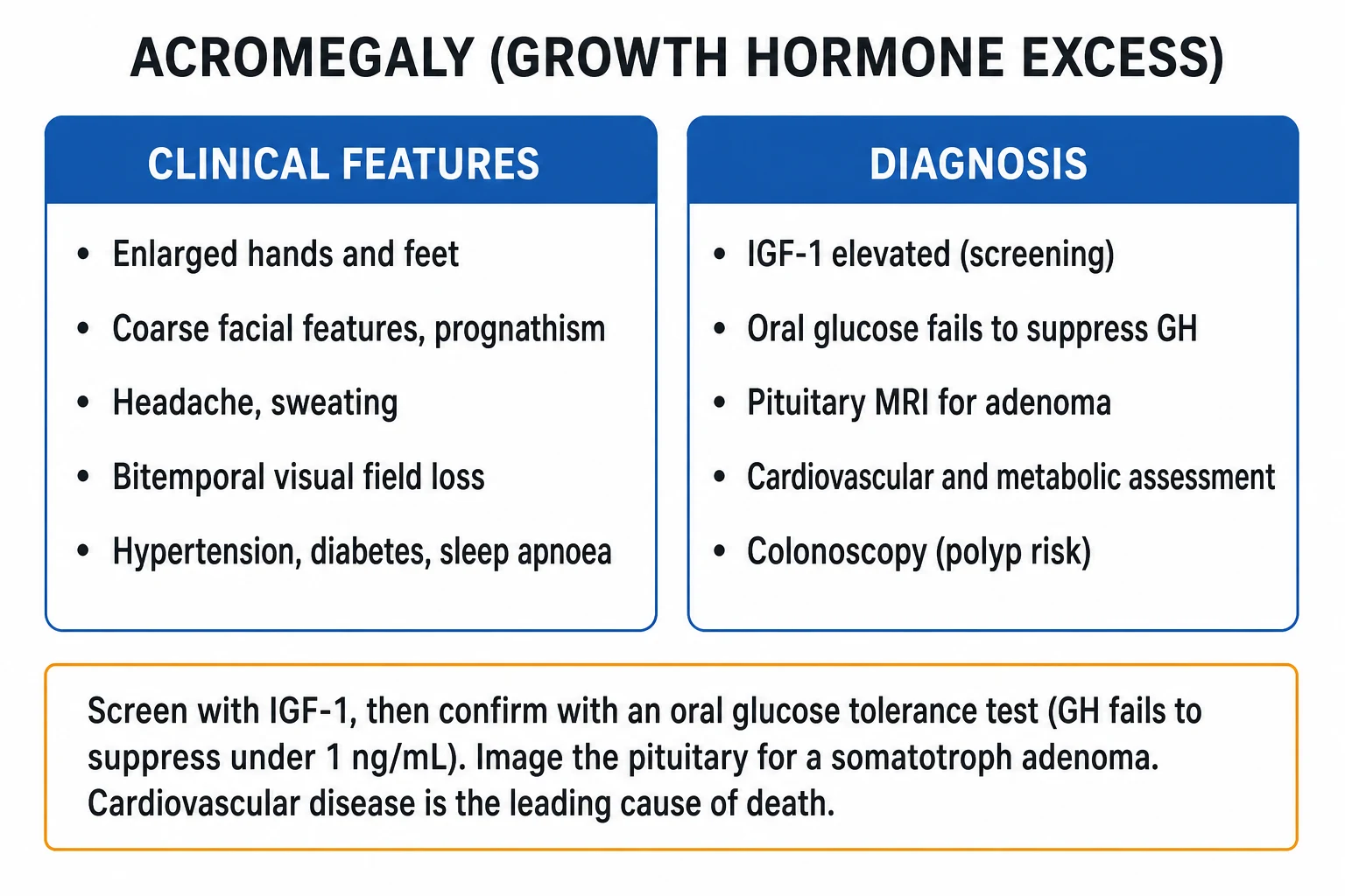
Acromegaly is chronic growth hormone (GH) excess, nearly always from a pituitary somatotroph adenoma, driving hepatic IGF-1 overproduction and progressive somatic overgrowth. Features include enlarging hands and feet, coarse facial features (prognathism, frontal bossing), dental malocclusion, macroglossia, headache, hyperhidrosis, carpal tunnel and bitemporal visual field loss (optic chiasm compression), with hypertension, diabetes, obstructive sleep apnoea and acromegalic cardiomyopathy. Screening is by elevated age/sex-matched IGF-1, confirmed by a 75 g oral glucose tolerance test in which GH fails to suppress under 1 ng/mL, and a pituitary MRI localises the adenoma. Transsphenoidal surgery is first-line and curative when complete; somatostatin receptor ligands (octreotide LAR, lanreotide, pasireotide), the GH receptor antagonist pegvisomant, the dopamine agonist cabergoline and stereotactic radiotherapy treat residual disease. Cardiovascular disease is the leading cause of death.
On this page & tools
Your progress
Saved locally on this device.
Exam tags
Red flags
Overview & Definition
Acromegaly (from the Greek akron = extremity, megas = large) is a chronic, sustained excess of growth hormone (GH) in an adult, driving overproduction of insulin-like growth factor 1 (IGF-1) and producing the insidious, progressive somatic overgrowth that gives the disease its name. In well over 99 percent of cases the source is a monoclonal pituitary somatotroph adenoma; rare ectopic sources of GHRH or GH must be considered when no pituitary tumour is found.[1][2]
The same hormone excess operating before epiphyseal fusion causes pituitary gigantism (excess linear growth, tall stature); after fusion it causes acromegaly (acral and soft-tissue enlargement). The two share identical tumour biology, workup and treatment, and the distinction is purely the developmental state of the growth plate at onset.[1]
The clinical challenge in acromegaly is not the recognition of advanced disease - the phenotype is unmistakable - but the 8 to 10 year diagnostic delay that is typical because the changes creep forward so slowly that patients, their families and their doctors attribute them to "just getting older." The skill is to recognise the phenotype early, screen with a single IGF-1 measurement, confirm with glucose suppression, and localise the tumour - then to control the disease biochemically and manage its comorbidities for life, because untreated acromegaly shortens survival, principally through cardiovascular and respiratory disease.[1][2]
Classification
Acromegaly is classified along four axes that each carry management meaning. [1]
By source. The vast majority - more than 99 percent - are pituitary somatotroph adenomas. The rare remainder are ectopic GHRH from neuroendocrine tumours (pancreatic, bronchial carcinoid, small-cell lung, pheochromocytoma) that produce somatotroph hyperplasia, or, very rarely, ectopic GH from a pancreatic or lung tumour.[1]
By adenoma size. A microadenoma is under 10 mm and a macroadenoma is 10 mm or more. Around 70 percent of acromegaly patients present with a macroadenoma, which is more likely to be invasive, to compress the optic chiasm (bitemporal hemianopia), to invade the cavernous sinus (cranial nerve palsies) and to leave residual disease after surgery. Microadenomas are far more likely to be cured by a single operation.[1][2]
By histological granularity (an increasingly important predictor). Densely granulated (DG) somatotroph adenomas express abundant somatostatin receptor subtype 2 (SSTR2) and respond well to somatostatin receptor ligands (octreotide, lanreotide). Sparsely granulated (SG) adenomas contain fibrous bodies, express less SSTR2, respond more poorly to SRLs (and may do better on the GH receptor antagonist pegvisomant), and tend to behave more aggressively.[7]
By familial context. Most cases are sporadic, but familial syndromes must be considered when presentation is early or there is a family history: MEN1 (MEN1), Carney complex (PRKAR1A), McCune-Albright syndrome (mosaic GNAS), familial isolated somatotroph adenomas (germline AIP mutation - often presenting as gigantism in a young man), and the very rare X-linked acrogigantism (X-LAG) (Xq26.3 duplication, GPR101).[1]
Acromegaly
after epiphyseal fusion
- Acral enlargement - hands, feet, jaw
- Coarse features, prognathism, macroglossia
- Insidious onset, 4th-5th decade
- IGF-1 raised + OGTT fails to suppress GH under 1 ng/mL
Pituitary gigantism
before epiphyseal fusion
- Excess linear growth (tall stature)
- Same somatotroph adenoma biology
- Onset in childhood or adolescence
- Same pathway: surgery first-line
- Screen for AIP mutation
Ectopic GHRH
rare, non-pituitary
- Bronchial carcinoid, pancreatic NET, SCLC
- Pituitary hyperplasia, not adenoma
- High plasma GHRH level
- Treat the tumour + somatostatin analogue
Epidemiology & Risk Factors
Acromegaly is rare. The incidence is around 3 to 4 per million population per year, and the prevalence is 40 to 60 per million in classic series, although active-screening programmes that measure IGF-1 in suspicious cohorts report figures as high as 130 per million - suggesting a substantial burden of undiagnosed disease.[1][2]
The disease affects men and women equally. The mean age at diagnosis is 40 to 45 years (the 4th to 5th decade), but the true onset is much earlier: a typical patient has had symptoms for 8 to 10 years before the diagnosis is made, often coming to attention through a complication (cardiac, metabolic, arthritic) or an incidental observation by a dentist (widening tooth spaces), podiatrist (increasing shoe size) or jeweller (ring resizing) rather than through endocrine suspicion.[1]
Acromegaly - key numbers
Risk factors are largely genetic. Beyond the familial syndromes above (MEN1, Carney complex, McCune-Albright, AIP mutations), most sporadic somatotroph adenomas carry no single environmental precipitant. The clinical risk - more than a cause - is delay: the longer the disease smoulders, the more cardiac, articular and metabolic damage accumulates, much of it only partially reversible.[1]
The case for IGF-1 screening. Because the phenotype is so insidious and the diagnostic delay measured in years, there is a strong argument for a low-threshold IGF-1 in any patient with two or more acromegaly-associated features (new diabetes with acral change, refractory sleep apnoea, treatment-resistant hypertension, bilateral carpal tunnel, enlarging hands or feet, or an incidentally found pituitary lesion). A single IGF-1 is cheap, stable and highly discriminating; the active-screening studies that used it in such cohorts doubled the apparent prevalence - implying that earlier IGF-1 testing is the single highest-yield intervention to shorten the diagnostic delay and prevent the irreversible cardiac and articular damage of late disease.[1][2]
Pathophysiology
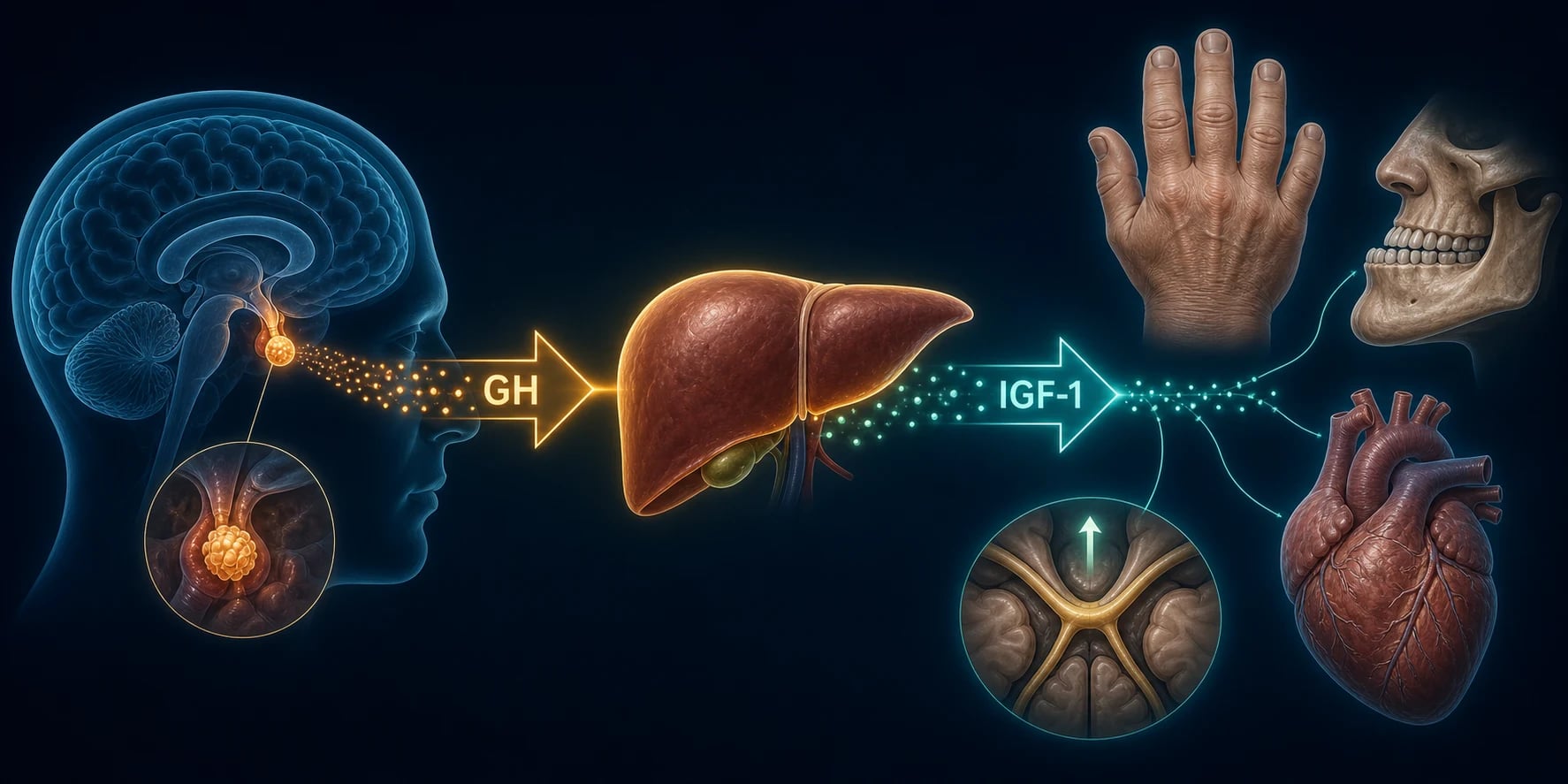
Acromegaly results from autonomous GH hypersecretion, nearly always from a monoclonal pituitary somatotroph adenoma whose cells proliferate from a single transformed progenitor and secrete GH without heeding the normal negative feedback from IGF-1 and somatostatin.[6]
Normal physiology - what the tumour overrides. In health, the hypothalamus balances stimulatory GHRH against inhibitory somatostatin to set the pulsatile rhythm of GH release from the anterior pituitary somatotrophs. GH peaks at night during slow-wave sleep and acts mainly through hepatic IGF-1; both GH and IGF-1 feed back negatively on the somatotroph (short loop) and on the hypothalamus (long loop) to close the loop. Puberty, stress, exercise, fasting, oestrogens and ghrelin all raise GH, while hyperglycaemia, somatostatin and IGF-1 suppress it. The acromegalic adenoma ignores all of these - it secretes GH autonomously, and the glucose load that would normally switch GH off fails to do so, which is precisely the principle exploited in the oral glucose tolerance test.[1][6]
GH to IGF-1: the effector axis. Circulating GH binds the hepatic GH receptor, activating the JAK2-STAT5 signalling cascade and driving transcription of IGF-1. Hepatic (and local tissue) IGF-1 mediates the great majority of somatic growth: it drives bone, cartilage and soft-tissue overgrowth, visceromegaly, cardiac hypertrophy and skin thickening. Because IGF-1 has a long plasma half-life and reflects integrated GH secretion over days, it is the ideal biochemical marker of disease burden - this is why serum IGF-1 is the screening test.[1]
The direct (diabetogenic) actions of GH. GH is not only an anabolic hormone: it has powerful anti-insulin effects. It stimulates lipolysis (raising free fatty acids), promotes hepatic gluconeogenesis, and reduces insulin receptor signalling in muscle and fat. The net result is insulin resistance, impaired glucose tolerance and, in many patients, overt type 2 diabetes. This is why hyperglycaemia is one of the commonest presenting metabolic disturbances.[1]
The molecular driver: the cAMP / PKA pathway and the gsp oncogene. Somatotrophs are physiologically driven by hypothalamic GHRH, which acts through a G-protein-coupled receptor to raise intracellular cyclic AMP (cAMP), activate protein kinase A (PKA), phosphorylate CREB, and stimulate both GH transcription/secretion and somatotroph proliferation. In roughly 30 to 40 percent of sporadic somatotroph adenomas a somatic activating point mutation in the GNAS gene (encoding the Gs-alpha stimulatory subunit) locks the G-protein in its GTP-bound, "on" state - the so-called "gsp oncogene" - producing constitutive, receptor-independent cAMP signalling. The cAMP pathway is the dominant molecular driver of somatotroph adenoma pathogenesis; the same pathway also induces DNA damage and a senescent phenotype tightly linked to GH overproduction. This molecular biology is not arcane - it explains why somatostatin receptor ligands (which suppress cAMP through SSTR2) work, and why densely granulated, SSTR2-rich tumours respond best.[6]
Mass effect. A macroadenoma compresses the optic chiasm (classically a bitemporal hemianopia, beginning superiorly), the surrounding normal pituitary (hypopituitarism - gonadotrophs first, then thyrotrophs, then corticotrophs), and the cavernous sinus (cranial nerves III, IV, V1, V2 and VI).[1]
Molecular drivers of sporadic somatotroph adenomas
Ectopic disease. When a neuroendocrine tumour secretes GHRH, the normal pituitary somatotrophs undergo hyperplasia (not a discrete adenoma) and hypersecrete GH; plasma GHRH is high, and the primary tumour is usually in the lung (carcinoid), pancreas, or occasionally a pheochromocytoma. True ectopic GH secretion from a non-pituitary tumour is very rare. The clue to an ectopic source is acromegaly with a small or normal-looking pituitary on MRI - the tumour is elsewhere.[1]
Clinical Presentation
The clinical face of acromegaly is a syndrome of slow tissue overgrowth layered with metabolic, cardiovascular and mechanical complications that have usually been present for years by the time the diagnosis is made. Examiners reward a system-by-system description.[1][2]
Acral and facial changes (the signature). The hands and feet enlarge (rings no longer fit; shoe size goes up), the skin thickens and becomes oily, the frontal bossing and mandibular prognathism remodel the skull, the interdental spaces widen (diastema, dental malocclusion), and the tongue enlarges (macroglossia). A patient may report needing new shoes every year or two, or a dentist may note the tooth separation. [1]
Neurological. Headache (tumour stretch of the dura, or a true raised-pressure headache), bitemporal hemianopia (the chiasmal pressure sign that demands urgent imaging), and bilateral carpal tunnel syndrome from soft-tissue swelling around the median nerve. A proximal myopathy contributes to fatigue. [1]
Cardiovascular (the lethal system). Hypertension (in roughly a third to half of patients) is the commonest cardiovascular finding, driven by sodium retention and increased cardiac output. The heart develops a specific, biventricular hypertrophic acromegalic cardiomyopathy that evolves through recognisable stages: an early hyperdynamic phase (high-output, preserved systolic function), an intermediate phase of biventricular hypertrophy with diastolic dysfunction (impaired relaxation, exertional dyspnoea), and a late phase of systolic heart failure with dilatation, arrhythmia (atrial fibrillation, ventricular ectopy) and valvular regurgitation. The cardiomyopathy is partly reversible if biochemical control is achieved early, but advanced disease leaves fixed structural damage. Cardiovascular disease is the leading cause of death in acromegaly.[5]
Respiratory. Obstructive sleep apnoea affects over half of patients (macroglossia, thickened pharyngeal soft tissue, large neck), the upper airway is hypertrophied, and a goitre may coexist. Together these make anaesthesia hazardous.[5]
Metabolic. Hyperhidrosis and heat intolerance (GH-driven basal metabolic rate increase), insulin resistance with impaired glucose tolerance or type 2 diabetes, dyslipidaemia, and hypercalciuria with nephrolithiasis. [1]
Musculoskeletal. Arthropathy (knee, hip, spine - from cartilage and bone overgrowth; often irreversible even after biochemical cure), spinal stenosis, osteoarthritis, and carpal tunnel syndrome. [1]
Other. Visceromegaly (cardiomegaly, hepatosplenomegaly), goitre, colonic polyps and an increased risk of colorectal cancer, skin tags (acrochordons), fatigue, menstrual disturbance or erectile dysfunction (from mass effect or co-secreted prolactin), and occasionally galactorrhoea if the tumour co-secretes prolactin.[5]
Atypical presentations. An elderly patient may present with heart failure or new diabetes alone, the acral changes dismissed as ageing. A young man with tall stature and a family history suggests AIP-mutation gigantism. A patient with acromegaly features and a normal pituitary on MRI demands a search for ectopic GHRH.[1]
Differential Diagnosis
Several conditions mimic the coarse phenotype of acromegaly without GH/IGF-1 excess. The discriminating test is always the IGF-1 and OGTT: in true acromegaly IGF-1 is high and GH fails to suppress; in every mimic it is normal with appropriate suppression.[1]
Pseudoacromegaly (severe insulin resistance, typically in poorly controlled type 2 diabetes or lipodystrophy) produces acral overgrowth, skin thickening and multiple skin tags but with normal IGF-1 and normal GH suppression. The clue is the metabolic background rather than the phenotype. [1]
Hypothyroidism / myxoedema gives coarse features, macroglossia, periorbital puffiness, bradycardia and slow-relaxing reflexes; IGF-1 is normal and thyroid function is deranged. [1]
Pachydermoperiostosis (Touraine-Solente-Gole syndrome) is a rare hereditary osteoarthropathy of adolescence with digital clubbing, periosteal new bone formation, coarse facial skin and seborrhoea; IGF-1 is normal. [1]
Cushing syndrome overlaps through central obesity, hypertension, hyperglycaemia and proximal myopathy, but lacks the acral enlargement; 24-hour urinary cortisol, an overnight dexamethasone suppression test and a midnight cortisol distinguish it. [1]
Familial prognathism / isolated macrocephaly and a co-secreting prolactinoma (galactorrhoea with acral change) round out the list.[2]
Acromegaly
GH/IGF-1 excess
- IGF-1 high for age/sex
- OGTT GH fails to suppress under 1 ng/mL
- Pituitary adenoma on MRI
- Bitemporal hemianopia if macroadenoma
Pseudoacromegaly
severe insulin resistance
- Acral overgrowth + skin tags
- Normal IGF-1
- Normal GH suppression on OGTT
- Type 2 diabetes / lipodystrophy background
Hypothyroidism
myxoedema
- Coarse features, macroglossia
- Periorbital puffiness
- Slow-relaxing reflexes
- Normal IGF-1; high TSH
Pachydermoperiostosis
hereditary
- Digital clubbing + periostitis
- Coarse skin, seborrhoea
- Adolescent onset, familial
- Normal IGF-1
Clinical & Bedside Assessment
The bedside assessment is where the diagnosis is first suspected, and it is built on three moves: (1) compare with old photographs, (2) examine the phenotype and the visual fields, and (3) hunt for the comorbidities that will determine perioperative risk and long-term outcome.[1]
Old photographs. Because the change is insidious, the patient and family are often poor historians. Pulling out a driving-licence or wedding photo from 5 to 10 years earlier frequently reveals the slow coarsening that the patient has not noticed. [1]
Phenotype. Examine the hands (size, skin thickening, ring tightness, thenar wasting of carpal tunnel, joint swelling), feet and shoe size, face (frontal bossing, prognathism, dental spacing, macroglossia), skin (oily, skin tags, acanthosis nigricans). [1]
Visual fields by confrontation, then formal Humphrey perimetry for any macroadenoma or any visual symptom - the classic deficit is a bitemporal hemianopia, often beginning in the superior temporal quadrants. Examine the cranial nerves (III, IV, V1, V2, VI) for cavernous sinus involvement, the cardiovascular system (blood pressure, heart sounds, signs of failure), the neck for goitre, and the abdomen for organomegaly. [1]
Comorbidity screen at the bedside and in clinic: glucose/HbA1c, lipids, echocardiogram, sleep study (polysomnography), colonoscopy.[2]
Investigations
The investigation of acromegaly is a three-step biochemical algorithm (screen, confirm, localise) plus a comorbidity assessment. Examiners expect the exact thresholds.[3]
Step 1 - Screening: serum IGF-1
A single serum IGF-1, age- and sex-matched, is the single best screening test, because IGF-1 reflects integrated GH secretion over the preceding days and is far more stable than pulsatile GH. A normal age/sex IGF-1 effectively excludes acromegaly. An elevated IGF-1 (in the right clinical context) mandates the confirmatory test.[3]
Step 2 - Confirmation: the 75 g oral glucose tolerance test (OGTT) [1]
After an overnight fast, give 75 g oral glucose and measure GH at 0, 30, 60, 90 and 120 minutes. In health, glucose suppresses GH to a nadir under 1 ng/mL. Acromegaly is confirmed when GH fails to suppress under 1 ng/mL. With modern high-sensitivity chemiluminescence assays the consensus cut-off is stricter - GH under 0.4 ng/mL - because some residual GH secretion in a "normal" range can still indicate disease with older thresholds.[3][4]
[1]Step 3 - Localisation and tumour assessment: pituitary MRI
A pituitary MRI with gadolinium localises the adenoma, measures its size (micro- vs macroadenoma), defines its relationship to the optic chiasm and cavernous sinus, and assesses invasion. If the MRI shows no adenoma (or a normal-sized pituitary), the next move is to suspect ectopic GHRH: measure a plasma GHRH level (markedly elevated in ectopic GHRH syndrome) and image the chest, abdomen and pelvis with CT (and octreotide scintigraphy / Gallium-68 DOTATATE where available) for a bronchial carcinoid, pancreatic neuroendocrine tumour or other source.[1][3]
Step 4 - Anterior pituitary axis testing
Assess the other pituitary axes for hypopituitarism: 9 am cortisol (or short synacthen test), free T4 and TSH, testosterone (men) or oestradiol (women) with FSH/LH, and prolactin (a macroadenoma may stalk-effect raise prolactin modestly, while a true co-secreting mammosomatotroph adenoma raises it markedly).[2]
Step 5 - Visual fields and comorbidity assessment
Humphrey perimetry for any macroadenoma or visual symptom. The comorbidity bundle - which informs both operative risk and lifelong surveillance - comprises HbA1c / fasting glucose, lipid profile, echocardiogram (LV mass, systolic and diastolic function), polysomnography, and colonoscopy at diagnosis (because of the colorectal polyp/cancer risk).[3][5]
Biochemical control (remission) criteria
Once treated, biochemical control (remission) is defined as a normal age- and sex-matched IGF-1 PLUS a random GH under 1 ng/mL (or an OGTT GH under 0.4 ng/mL with sensitive assays). These criteria, refined at successive Acromegaly Consensus conferences, are the treatment targets that return mortality toward that of the general population.[4]
Interpreting discordant biochemistry. A high IGF-1 with a normal OGTT GH nadir can occur in pregnancy or oestrogen therapy (oestrogen lowers hepatic IGF-1 generation, paradoxically raising measured GH, or in adolescence); a normal IGF-1 with a non-suppressible GH may reflect assay interference or GH-receptor-antagonist therapy (pegvisomant lowers IGF-1 without lowering GH). A random GH alone is never diagnostic - GH is pulsatile, with peaks after meals, exercise and stress - which is why IGF-1 (integrated) screens and the OGTT (dynamic suppression) confirms. Always interpret IGF-1 against age- and sex-matched reference ranges, because IGF-1 declines with age; a value that looks "normal" in an elderly patient may in fact be elevated for their age.[3][4]
Management - Resuscitation
Acromegaly is overwhelmingly a chronic disease; the diagnostic and operative pathway is elective, not a resuscitation. The one true emergency is pituitary apoplexy - haemorrhagic infarction of the adenoma.[3]
Pituitary apoplexy presents with sudden severe headache, vomiting, rapidly progressive visual loss, ophthalmoplegia (cranial nerve III palsy) and, in severe cases, altered consciousness or meningism. The immediate management is: [1]
- Intravenous hydrocortisone 100 to 200 mg stat, then 50 to 100 mg every 6 to 8 hours - because acute cortisol deficiency (corticotroph destruction) is the immediate threat to life.
- Intravenous fluids and haemodynamic support.
- Urgent pituitary MRI to confirm apoplexy.
- Urgent transsphenoidal surgical decompression if there is deteriorating vision or depressed consciousness; conservative management with close monitoring is acceptable in stable patients without visual deficit.
- Endocrine and ophthalmology review.[3]
A patient who presents in decompensated acromegalic cardiomyopathy (acute heart failure) needs standard heart-failure therapy (oxygen, diuretics, afterload reduction), but the disease-modifying step is biochemical control of GH/IGF-1; acute severe hyperglycaemia or DKA is managed with IV fluids, insulin and electrolyte correction in the usual way.[5]
Management - Definitive & Stepwise
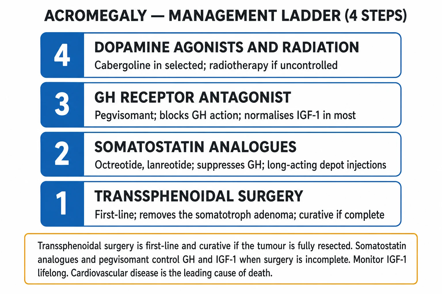
The treatment of acromegaly is a multimodal, lifelong programme built around four modalities - surgery, somatostatin receptor ligands (SRLs), the GH receptor antagonist pegvisomant, and radiotherapy - with cabergoline as an adjunct and comorbidity management running in parallel. The aim is biochemical control (normal IGF-1 + GH under 1 ng/mL), tumour mass control (relieve chiasmal pressure, prevent regrowth) and protection of remaining pituitary function.[1][3]
1. Transsphenoidal surgery - first-line
Endoscopic transsphenoidal surgery by an experienced pituitary neurosurgeon is first-line for nearly all patients with a discrete adenoma, and is curative when the tumour is completely resected. Surgical remission rates are around 75 to 95 percent for microadenomas but only 40 to 60 percent for macroadenomas (lower still for invasive/Knosp grade 3-4 tumours). Surgery promptly relieves chiasmal compression and mass effect, and immediately removes the source of GH. Complications - in expert hands - include hypopituitarism, diabetes insipidus, cerebrospinal fluid leak, meningitis and cranial nerve injury. Biochemical assessment at 12 weeks postoperatively (IGF-1 and a random or OGTT GH) defines remission versus residual disease.[1][2][3]
The surgical approach is endoscopic endonasal transsphenoidal: through the nostril and sphenoid sinus to the sella, using image guidance and intra-operative neuro-navigation to dissect the adenoma off the optic chiasm, cavernous sinus and normal pituitary. Predictors of surgical remission are a small tumour, no cavernous sinus invasion (Knosp grade 0-2), a clearly demarcated adenoma on MRI, lower pre-operative IGF-1/GH, and densely granulated histology; the most important negative predictor is cavernous sinus invasion, where a complete resection is usually impossible and residual disease is the rule. After surgery, postoperative day 1-2 GH can give an early read on remission, but the definitive reassessment is at 12 weeks with an IGF-1 and an OGTT or random GH, together with a full pituitary axis check (cortisol, free T4, sex steroids, sodium/diabetes insipidus). Cortisol replacement is given empirically in the immediate postoperative period until the axis is confirmed.[1][2]
Preoperative medical therapy - a debated role. A short course of a somatostatin receptor ligand before surgery can shrink the tumour and improve biochemical control in selected patients with large macroadenomas, and may ease anaesthetic risk by softening the upper-airway tissues, but it is not routine because it has not been shown to improve surgical cure rates consistently. It is most often used when surgery must be delayed, or for severe pharyngeal/laryngeal overgrowth complicating intubation.[7]
2. Somatostatin receptor ligands (SRLs) - the medical backbone
SRLs bind the somatostatin receptor subtype 2 (SSTR2) (and, for pasireotide, SSTR5) on the somatotroph, suppress GH secretion and shrink the tumour in many patients. They are first-line medical therapy for residual disease after surgery, in patients unfit for or refusing surgery, and - increasingly - as preoperative or primary therapy for large invasive macroadenomas without chiasmal compression.[7]
- Octreotide LAR (long-acting release): 20 to 30 mg intramuscularly every 4 weeks (titrate within the 10 to 40 mg range by IGF-1/GH response). Densely granulated, SSTR2-rich tumours respond best.
- Lanreotide autogel / depot: 60 to 120 mg deep subcutaneously every 4 weeks (some patients on long-term control can extend to every 6-8 weeks). Equivalent to octreotide LAR.
- Pasireotide LAR: 40 to 60 mg intramuscularly every 4 weeks - a multireceptor ligand (SSTR5 > 2 > 3) that achieves higher biochemical control rates than octreotide/lanreotide but carries a significant risk of hyperglycaemia/diabetes (monitor blood glucose).[7]
Common SRL adverse effects include gallstones/sludge, steatorrhoea, abdominal cramps, bradycardia and injection-site reactions.[1]
3. Pegvisomant - the GH receptor antagonist
Pegvisomant is a genetically modified GH analogue that blocks the GH receptor, preventing IGF-1 generation. It normalises IGF-1 in the great majority of patients and is the most biochemically effective monotherapy, but it does not shrink the tumour (because it acts peripherally) - so a tumour with mass effect must be co-treated with an SRL or observed with MRI. It is given as a subcutaneous injection, starting at 10 mg daily and titrating up to 30 mg daily by IGF-1 response. Monitor liver enzymes (transaminase elevation, rare hepatotoxicity).[1][7]
4. Cabergoline - the dopamine agonist adjunct
Cabergoline is a dopamine D2 agonist with modest GH-lowering effect, most useful when the tumour co-secretes prolactin or as an add-on to an SRL in mild residual disease. Doses are higher than for prolactinoma - 1 to 3.5 mg per week (typically 1-2 mg weekly, titrated). It is oral, cheap and well tolerated; the main caveat at high cumulative doses is cardiac valve fibrosis (monitor echocardiogram).[1]
Octreotide LAR / Lanreotide
first-gen SRL - SSTR2
- Octreotide 20-30 mg IM q4wk (10-40 mg range)
- Lanreotide 60-120 mg deep SC q4wk
- Suppress GH + may shrink tumour
- Best for densely granulated tumours
- Adverse: gallstones, bradycardia, GI
Pasireotide LAR
2nd-gen multiligand SRL
- 40-60 mg IM q4wk
- Higher biochemical control rates
- Binds SSTR5 > 2 > 3
- Significant hyperglycaemia / diabetes
- For resistant tumours
Pegvisomant
GH receptor antagonist
- 10-30 mg/day SC (titrate to IGF-1)
- Blocks GH receptor; normalises IGF-1
- Does NOT shrink tumour - co-treat with SRL
- Most effective monotherapy
- Monitor LFTs
Cabergoline
dopamine D2 agonist
- 1-3.5 mg/week (higher than prolactinoma)
- Oral, cheap, well tolerated
- Best if co-secreted prolactin
- Modest effect; useful adjunct
- Cardiac valve fibrosis at high cumulative dose
5. Radiotherapy / stereotactic radiosurgery - third-line
Radiotherapy is reserved for refractory or residual disease after surgery and medical therapy, or for patients who cannot adhere to long-term injections. Stereotactic radiosurgery (gamma knife) delivers a focused single fraction; fractionated stereotactic radiotherapy is used for larger tumours close to the optic nerves. Its drawbacks are a slow onset of biochemical effect (12 to 60 months), the risk of hypopituitarism (the commonest late complication, mandating lifelong axis surveillance), and small long-term risks of cerebrovascular disease and secondary tumours. Conventional fractionated radiotherapy is now seldom first choice.[1][3]
6. Lifelong monitoring and comorbidity management
Whatever the modality, the patient needs lifelong surveillance - the disease and its comorbidities outlast any single intervention. The biochemical schedule is IGF-1 and GH every 3 to 6 months (more often when titrating therapy). Imaging requires an annual pituitary MRI for any residual tumour (and lifelong, less frequently, after radiotherapy until regrowth is excluded). Visual fields are repeated for any macroadenoma or residual suprasellar extension. The comorbidity bundle runs in parallel: blood pressure control (ACE inhibitor / ARB / calcium-channel blocker as needed), glucose and lipids (metformin first-line for the diabetes; statin per CV risk), sleep apnoea (CPAP, weight management), and colonoscopy surveillance at diagnosis and then every 3 to 5 years for the colorectal cancer risk.[4][5]
Lifelong acromegaly surveillance timeline
Escalation triggers. If IGF-1 rises despite first-generation SRL monotherapy, the next moves are (in turn) up-titration of the SRL within its dose range, addition of pegvisomant (combination therapy), switch to pasireotide, or addition of cabergoline; if the tumour is growing despite medical therapy or there is refractory mass effect, stereotactic radiotherapy is considered, and reoperation in selected patients with a surgically accessible residual. A rising IGF-1 with a stable or shrinking tumour on an SRL points to biochemical escape rather than regrowth and is usually managed by adding or switching medical therapy, not by radiation.[7]
Exam application bank (NEET-PG / INICET)
One-line answer
Acromegaly is chronic growth hormone (GH) excess, nearly always from a pituitary somatotroph adenoma, driving hepatic IGF-1 overproduction and progressive somatic overgrowth. Features include enlarging hands and feet, coarse facial features (prognathism, frontal bossing), dental malocclusion, macroglossia, headache, hyperhidrosis, carpal tunnel and bitemporal visual field loss (optic chiasm compression), with hypertension, diabetes, obstructive sleep apnoea and acromegalic cardiomyopathy. Screening is by elevated age/sex-matched IGF-1, confirmed by a 75 g oral glucose tolerance test in which GH fails to suppress under 1 ng/mL, and a pituitary MRI localises the adenoma. Transsphenoidal surgery is first-line and curative when complete; somatostatin receptor ligands (octreotide LAR, lanreotide, pasireotide), the GH receptor antagonist pegvisomant, the dopamine agonist cabergoline and stereo [1]
Worked stems (answer without another resource)
Stem 1 — Classic presentation. Map symptoms to mechanism; name the first investigation and first treatment step with dose/route if drug therapy is standard. [1]
Stem 2 — Unstable / complicated. List red flags that force immediate resuscitation, theatre, ICU, antidote, or reperfusion — and what you do in the first 15 minutes. [1]
Stem 3 — Atypical group. Elderly, pregnancy, child, or immunocompromised: how presentation and thresholds change. [1]
Stem 4 — Differential trap. Name the three closest mimics and one discriminator for each. [1]
Stem 5 — Disposition. Who goes home with safety-netting, who is admitted, who needs HDU/ICU/theatre, and what follow-up is mandatory. [1]
Rapid viva checklist
- Definition + classification
- Pathophysiology chain
- Bedside signs / criteria
- Score with exact components (if any)
- Emergency bundle
- Definitive therapy with doses
- Complications of disease and of treatment
- Special populations
- Guideline/trial name if classic
- Three exam traps
Coverage self-check
If you cannot answer any stem above from this page alone, re-read the matching section — the page is intended to be self-sufficient for final-prof and NEET-PG/INICET questions on Acromegaly.
Specific Subtypes & Scenarios
Pituitary gigantism (pre-epiphyseal fusion). The same somatotroph adenoma in a child or adolescent drives excess linear growth (tall stature) in addition to soft-tissue overgrowth; puberty may be delayed. The workup and surgery-first pathway are identical. AIP-mutation testing is advised, and a GnRH analogue may be used to slow epiphyseal fusion and protect final height while definitive treatment is undertaken.[1]
Ectopic GHRH syndrome. When a bronchial carcinoid, pancreatic neuroendocrine tumour, small-cell lung cancer or pheochromocytoma secretes GHRH, the pituitary shows somatotroph hyperplasia rather than a discrete adenoma, and the plasma GHRH level is markedly elevated. Management is resection of the primary tumour plus a somatostatin receptor ligand to control GH; the pituitary itself is not operated on unless a discrete adenoma coexists.[1]
Ectopic GH secretion (very rare, from a pancreatic or lung tumour) is managed by resecting the source. [1]
Familial acromegaly. In MEN1, somatotroph adenoma may be the presenting tumour; screen relatives. McCune-Albright syndrome (mosaic GNAS, polyostotic fibrous dysplasia, cafe-au-lait spots, precocious puberty) carries constitutive GH excess. Carney complex (PRKAR1A) and familial isolated somatotroph adenomas (germline AIP) should be suspected in any young patient (especially male) with gigantism or a family history of pituitary tumour.[1]
Aggressive / invasive macroadenoma. A large Knosp grade 3-4 tumour invading the cavernous sinus is unlikely to be cured surgically; the typical strategy is surgical debulking to relieve mass effect, then primary medical therapy (SRL +/- pegvisomant), with radiotherapy for refractory growth.[7]
Complications & Pitfalls
The complications of acromegaly are the reason it shortens life and the centre of long-term management. They arise from three converging mechanisms - tissue overgrowth (heart, upper airway, joints, colon), GH-driven metabolic derangement (insulin resistance, lipolysis), and tumour mass effect on surrounding structures - and they persist or progress even after biochemical cure if they were established late. Every complication below is part of the baseline comorbidity screen at diagnosis and the lifelong surveillance programme.[5]
Comorbidity burden at diagnosis (approximate)
Cardiovascular (leading cause of death). Acromegalic cardiomyopathy - biventricular hypertrophy with diastolic then systolic dysfunction, arrhythmia (atrial fibrillation, ventricular ectopy), hypertension and valvular regurgitation. Cardiovascular control tracks biochemical control, but established cardiomyopathy may be only partially reversible. [1]
Respiratory. Obstructive sleep apnoea (OSA) affects over half of patients and is one of the commonest and most under-recognised complications: it arises from macroglossia, thickened pharyngeal and laryngeal soft tissue, a hypertrophied epiglottis and a large neck circumference, and it materially raises both cardiovascular mortality and anaesthetic risk. A formal polysomnogram is part of the baseline workup, and CPAP is first-line treatment. The upper airway compromise also makes intubation difficult - the acromegalic airway is a classic exam stem for a difficult airway.[5]
Musculoskeletal. The acromegalic arthropathy is one of the most disabling and least reversible complications: it begins as cartilage overgrowth and joint-space widening (knees, hips, spine) and progresses to premature osteoarthritis, deformity and chronic pain. Unlike the soft-tissue changes, the joint damage does not reverse with biochemical cure - the articular cartilage has already remodeled - so early diagnosis and treatment are the only way to limit it. Vertebral fractures (from altered bone quality, not classical osteoporosis), spinal stenosis, carpal tunnel syndrome (soft-tissue swelling around the median nerve) and a proximal myopathy complete the picture.[5]
Neoplastic. Acromegaly carries an increased risk of colonic polyps and colorectal cancer - the rationale for a colonoscopy at diagnosis and surveillance every 3 to 5 years. The risk scales with IGF-1 and is one of the drivers of the cancer-related excess mortality. Thyroid nodules and differentiated thyroid cancer, and possibly breast and prostate nodules, are also over-represented, though the magnitude of these associations is debated and surveillance is pragmatic rather than evidence-certain.[5]
Tumour mass effect. A macroadenoma threatens four structures: the optic chiasm (progressive bitemporal hemianopia, which may be permanent if long-standing), the normal pituitary (hypopituitarism, in the order gonadotrophs, thyrotrophs, corticotrophs), the cavernous sinus (cranial nerves III, IV, V1, V2, VI), and - rarely - the hypothalamus. Pituitary apoplexy (haemorrhagic infarction) is the acute mass-effect emergency, presenting with sudden headache, vomiting, visual loss and ophthalmoplegia, and managed with emergency steroids and decompression.[1]
Treatment-related pitfalls. Hypopituitarism after surgery or radiotherapy; hyperglycaemia with pasireotide; hepatotoxicity and injection-site lipodystrophy with pegvisomant (monitor LFTs); gallstones with SRLs; cardiac valve fibrosis with high cumulative cabergoline doses. A common diagnostic pitfall is missing the disease for years; a common management pitfall is declaring "control" on GH alone without confirming the age/sex IGF-1 is normal.[1]
Prognosis & Disposition
Untreated or poorly controlled acromegaly carries a 2- to 3-fold excess mortality, driven by cardiovascular and respiratory disease (and to a lesser extent cancer), shortening median survival by roughly 10 years.[1]
With biochemical control - a normal age/sex IGF-1 plus a random GH under 1 ng/mL - mortality approaches that of the general population, though established cardiomyopathy, arthropathy and sleep apnoea may persist. Earlier diagnosis and aggressive multimodal treatment (surgery, medical therapy, radiotherapy as needed) improve both survival and quality of life.[1][4]
Disposition is lifelong. The patient is managed in a multidisciplinary pituitary tumour centre of excellence (PTCOE) with endocrinology, neurosurgery, radiation oncology, ophthalmology, cardiology, sleep medicine, gastroenterology and anaesthetics, with IGF-1/GH every 3-6 months, annual MRI for residual disease, repeat visual fields for macroadenomas, and structured comorbidity surveillance (echo, sleep study, HbA1c/lipids, colonoscopy).[4][5]
Special Populations
Anaesthetic risk. The acromegalic airway is the classic difficult intubation and a frequent exam stem. The problems stack: macroglossia, thickened pharyngeal and laryngeal soft tissue, a hypertrophied epiglottis, a large mandible, a short neck and a large thyroid make laryngoscopy and bag-mask ventilation difficult; obstructive sleep apnoea raises the risk of rapid desaturation and postoperative respiratory depression; and cardiomyopathy limits cardiovascular reserve under anaesthesia. The safe approach is anticipate, seniorise and prepare: senior anaesthetic and ENT support on standby, a documented plan for awake fibre-optic intubation or videolaryngoscopy, a difficult-airway trolley at the bedside, and optimisation of cardiac status and CPAP preoperatively. Postoperatively, watch for airway oedema and sleep-disordered breathing before extubating.[5]
Pregnancy. Fertility is often impaired through hypogonadotrophic hypogonadism (mass effect) or hyperprolactinaemia; restoration of ovulation may follow surgery or dopamine agonist. Somatostatin analogues are usually stopped in pregnancy (they cross the placenta), though data are limited; pegvisomant has been continued in selected cases where control cannot be maintained otherwise. Monitor visual fields monthly (the tumour may enlarge under oestrogen), because IGF-1 rises physiologically in pregnancy and cannot be used alone for monitoring - rely on GH and clinical signs. If vision is threatened, transsphenoidal surgery in the second trimester is the option.[3]
Gigantism (children and adolescents). Same workup and surgery-first approach; AIP-mutation screening is advised; a GnRH analogue can delay epiphyseal fusion to protect final height while definitive treatment is delivered.[1]
Elderly. May present with heart failure or new diabetes alone, with higher surgical risk; primary medical therapy (SRL +/- pegvisomant) is often preferred when the tumour has no chiasmal compression.[2]
Familial / MEN1. Screen family members; a somatotroph adenoma may be the index tumour, and other endocrine tumours (parathyroid, pancreatic NET) need surveillance.[1]
Evidence, Guidelines & Regional Differences
The Endocrine Society / European Society of Endocrinology 2014 clinical practice guideline (Katznelson et al.) remains the practice-defining document: it established surgery as first-line, defined the biochemical diagnostic and remission criteria, and laid out the medical-therapy algorithm (including pregnancy management).[3] Successive Acromegaly Consensus conferences (2019-2024, Giustina et al.) have refined the multidisciplinary management and complications frameworks, and the most recent (2026) update consolidates the comorbidity agenda.[4][5] The Colao 2019 Nature Reviews Primer is the comprehensive single reference; the Castinetti & Ioachimescu 2026 treatment-landscape review captures the expanding pharmacopoeia (oral octreotide, paltusotine).[1][7]
Some European centres (notably in Germany) use primary medical therapy for large invasive macroadenomas without chiasmal compression, reserving surgery for tumours compressing the optic chiasm. Pasireotide, oral octreotide and pegvisomant are not uniformly approved or affordable in all health systems, so octreotide LAR / lanreotide remain the global workhorses; in resource-variable settings, cabergoline (cheap, oral) has a relatively larger role. In India, the ICMR does not publish an acromegaly-specific guideline; the Endocrine Society algorithm is followed, with cost often steering therapy toward surgery and first-generation SRLs.[1][4]
Where the evidence is weak. The exact GH cut-off for remission (under 1 vs under 0.4 ng/mL) is assay- and consensus-dependent; the role of routine preoperative medical therapy, the place of oral octreotide and paltusotine in the long-term algorithm, and the timing and type of radiotherapy (stereotactic radiosurgery vs fractionated) remain debated.[4][7]
Exam Pearls
Acromegaly is one of the most patterned topics in endocrinology - the phenotype, the biochemical trio, the surgical-first algorithm and the cardiovascular-mortality punchline recur across NEET-PG, INICET, USMLE and PLAB.[1]
The definitional one-liner: chronic GH excess from a pituitary somatotroph adenoma driving hepatic IGF-1 overproduction. The screening test is a single age/sex-matched IGF-1; the confirmatory test is a 75 g OGTT in which GH fails to suppress under 1 ng/mL; the localising test is a pituitary MRI.[3]
The management ladder is fixed: transsphenoidal surgery first-line (curative for microadenoma), then somatostatin receptor ligands (octreotide LAR, lanreotide) to suppress GH, then pegvisomant to block the GH receptor, with cabergoline as an adjunct and radiotherapy third-line.[1][7]
Key contrasts the exam rewards: pegvisomant blocks the GH receptor (normalises IGF-1; does not shrink tumour) versus somatostatin analogues suppress GH secretion (and may shrink the tumour). Gigantism is the same process before epiphyseal fusion. McCune-Albright and AIP mutations cause the familial/gigantism forms; a GNAS (gsp oncogene) mutation drives the cAMP/PKA pathway in about a third of sporadic tumours. If the pituitary looks normal on MRI, think ectopic GHRH (pancreatic/bronchial NET) and check a plasma GHRH. Pseudoacromegaly is severe insulin resistance with acral overgrowth but normal IGF-1.[6]
The single most quotable fact: cardiovascular disease is the leading cause of death, and biochemical control (normal IGF-1 + GH under 1 ng/mL) returns mortality toward the general population.[5]
Quick self-test - cover and answer
Q1. A 42-year-old man has enlarging hands, new diabetes and a bitemporal hemianopia. What is the single best screening test? - Serum IGF-1 (age/sex-matched). Q2. IGF-1 is elevated; what confirms the diagnosis? - A 75 g OGTT in which GH fails to suppress under 1 ng/mL. Q3. MRI shows a macroadenoma. First-line treatment? - Endoscopic transsphenoidal surgery. Q4. Three months after surgery IGF-1 is still high. Which two drugs normalise IGF-1 by different mechanisms? - Somatostatin receptor ligands (octreotide/lanreotide) suppress GH; pegvisomant blocks the GH receptor. Q5. Pituitary looks normal on MRI but acromegaly is biochemically confirmed - what rare cause and what test? - Ectopic GHRH (bronchial/pancreatic NET) - check plasma GHRH and CT chest/abdomen/pelvis.
Acromegaly features - GROWTH
GROWTH
excess linear growth before epiphyseal closure
acral enlargement; rings and shoes no longer fit
cardiomegaly, hepatosplenomegaly; obstructive sleep apnoea
mandibular prognathism, macroglossia, frontal bossing
carpal tunnel syndrome; insulin resistance and diabetes
macroadenoma: chiasmal compression, bitemporal hemianopia
References
- [1]Colao A, Grasso LFS, Giustina A, Melmed S, Chanson P, Pereira AM, Pivonello R. Acromegaly Nat Rev Dis Primers, 2019.PMID 30899019
- [2]Ershadinia N, Tritos NA. Diagnosis and Treatment of Acromegaly: An Update Mayo Clin Proc, 2022.PMID 35120696
- [3]Katznelson L, Laws ER Jr, Melmed S, Molitch ME, Mohmed ME, Arafah BM, et al. Acromegaly: an endocrine society clinical practice guideline J Clin Endocrinol Metab, 2014.PMID 25356808
- [4]Giustina A, Barkhoudarian G, Beckers A, Ben-Shlomo A, Biermasz N, Biller B, et al. Multidisciplinary management of acromegaly: A consensus Rev Endocr Metab Disord, 2020.PMID 32914330
- [5]Giustina A, di Filippo L, Fleseriu M, Pivonello R, Petersenn S, Wass J, et al. Consensus on acromegaly complications: an update Pituitary, 2026.PMID 42050227
- [6]Ben-Shlomo A, Melmed S, et al. Pathogenesis of nonfamilial somatotroph adenomas J Clin Endocrinol Metab, 2026.PMID 41824769
- [7]Castinetti F, Ioachimescu AG. Current treatment landscape of acromegaly J Clin Endocrinol Metab, 2026.PMID 41965092