Endocrinology · Endocrinology
Adrenal Insufficiency
Also known as Addison disease (primary adrenal insufficiency) · Adrenocortical failure · Hypocortisolism · Adrenal crisis (acute decompensation)
Adrenal insufficiency is the clinical syndrome arising from inadequate production of glucocorticoid (cortisol), with or without mineralocorticoid (aldosterone) and adrenal androgen deficiency. Primary adrenal insufficiency (Addison disease) results from destruction of the adrenal cortex — autoimmune in the West, tuberculosis worldwide — producing low cortisol AND low aldosterone with HIGH ACTH-driven hyperpigmentation, hyponatraemia and hyperkalaemia. Secondary adrenal insufficiency results from pituitary ACTH deficiency, and tertiary from hypothalamic CRH deficiency (most often chronic exogenous glucocorticoid therapy); both spare the mineralocorticoid axis (RAAS intact) so there is no hyperpigmentation and no hyperkalaemia. Adrenal crisis — hypotension, abdominal pain, vomiting, hypoglycaemia and hyponatraemia ± hyperkalaemia — is a fatal endocrine emergency treated with parenteral hydrocortisone, intravenous saline and treatment of the precipitant.
On this page & tools
Your progress
Saved locally on this device.
Exam tags
Red flags

Overview & Definition
Adrenal insufficiency is the clinical and biochemical syndrome caused by inadequate secretion of glucocorticoid (cortisol), with or without deficiency of mineralocorticoid (aldosterone) and adrenal androgens (DHEA/DHEAS).[2][3] Cortisol is essential for life: it maintains vascular tone and blood pressure (permissive effect on catecholamines and angiotensin II), supports hepatic gluconeogenesis, suppresses inflammation and immunity, and modulates central nervous system arousal. The condition spans a spectrum from subclinical (compensated, only evident on testing) through overt chronic insufficiency (classical Addison disease) to the decompensated extreme of adrenal crisis — a medical emergency with mortality of 5 to 15 percent even with treatment.[8]
Three anatomical levels of the hypothalamic–pituitary–adrenal (HPA) axis can fail:[1][4]
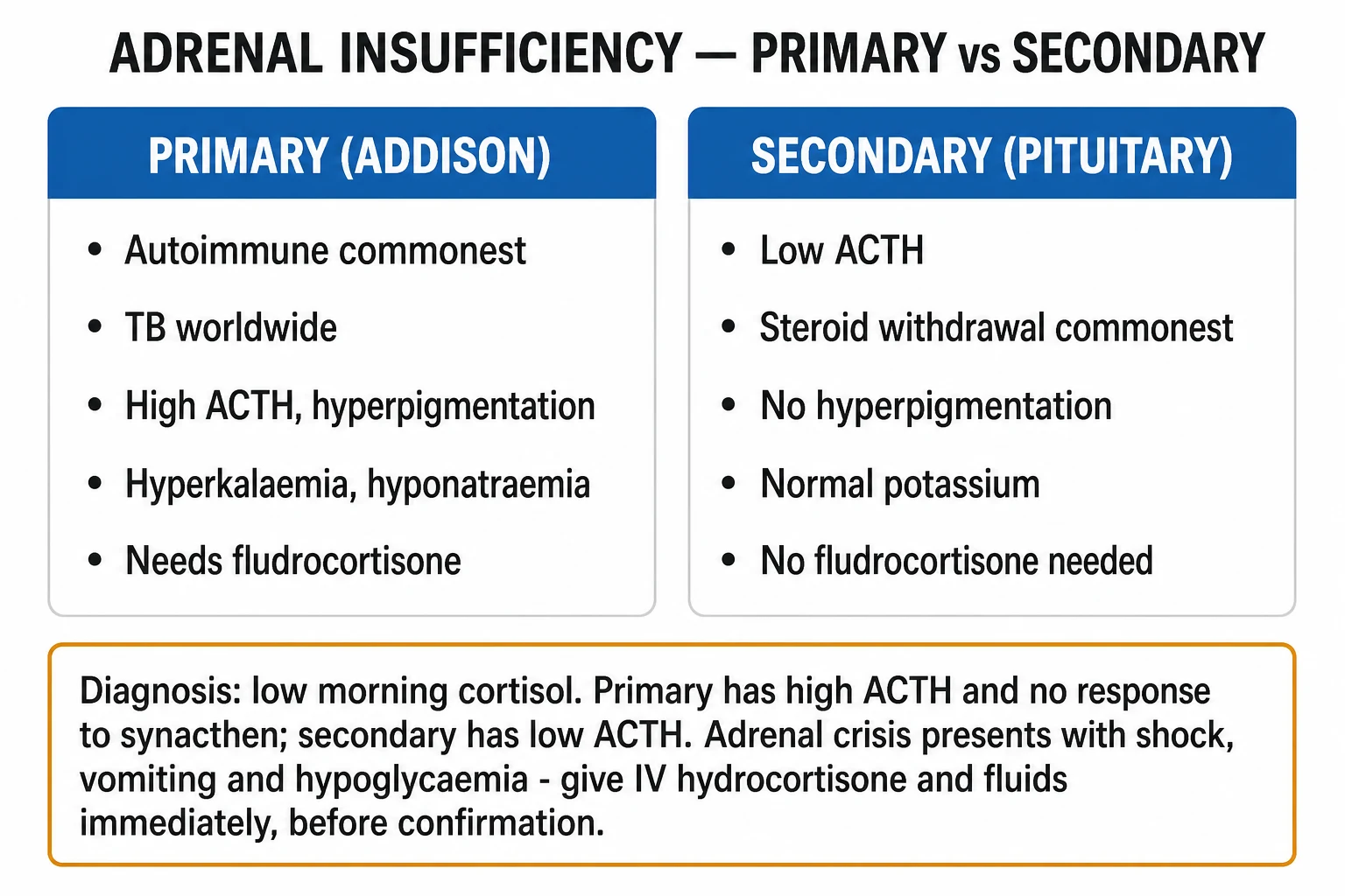
- Primary adrenal insufficiency (Addison disease) — disease of the adrenal cortex itself. Cortisol, aldosterone and adrenal androgens are all deficient; ACTH is high (loss of negative feedback), driving the hallmark hyperpigmentation. The biochemical signature is hyponatraemia WITH hyperkalaemia (aldosterone loss).
- Secondary adrenal insufficiency — disease of the anterior pituitary (ACTH deficiency). Cortisol and adrenal androgens fall; aldosterone is preserved (RAAS still drives the zona glomerulosa). ACTH is low or inappropriately normal; there is no hyperpigmentation and no hyperkalaemia.
- Tertiary adrenal insufficiency — disease of the hypothalamus (CRH deficiency) or, by far the commonest, chronic exogenous glucocorticoid therapy suppressing CRH. Biochemically identical to secondary AI (low ACTH, preserved aldosterone). [1]
The clinical task is two-fold: (a) recognise the chronic syndrome and the acute crisis (the latter is the lethal one), and (b) localise the level — because primary AI needs mineralocorticoid replacement that secondary AI does not, and secondary AI prompts a search for pituitary disease and other pituitary hormone deficiencies.[1]
Two physiology essentials underpin the whole topic. The axis: hypothalamic corticotropin-releasing hormone (CRH) reaches the anterior pituitary via the hypophyseal portal system and drives corticotrophs to cleave pro-opiomelanocortin (POMC) into ACTH (and the by-products alpha-MSH, beta-lipotrophin and beta-endorphin). ACTH binds the melanocortin-2 receptor (MC2R), a Gs-protein-coupled receptor on adrenal cortical cells, raising cAMP and driving StAR-mediated cholesterol delivery to CYP11A1 and the cascade that produces cortisol. The feedback loop: circulating cortisol exerts negative feedback on both the pituitary ACTH and the hypothalamic CRH. This feedback is steep and fast — so chronic glucocorticoid therapy suppresses CRH and ACTH within days to weeks, causing adrenal cortical atrophy; abrupt withdrawal leaves the patient cortisol-deficient for months. The diurnal rhythm: cortisol peaks in the early morning (around 6 to 8 am) and troughs around midnight. This is why a morning cortisol is the screening test — a low morning value is the most sensitive biochemical clue to AI.[2][4]
Aldosterone is regulated differently — this is the single most important physiology fact for the topic. The zona glomerulosa is driven principally by angiotensin II (from the renin-angiotensin system) and serum potassium, with ACTH providing only a permissive tonic support. Therefore pituitary failure does not abolish aldosterone secretion — angiotensin II keeps the glomerulosa working. This is why secondary AI spares the mineralocorticoid axis, producing hyponatraemia without hyperkalaemia. Primary AI destroys all three zones — so aldosterone falls and hyperkalaemia appears alongside hyponatraemia.[3]
Classification
Adrenal insufficiency is classified along two axes — by the level of HPA-axis failure (which determines whether mineralocorticoid replacement is needed and where to look for the cause) and by temporal course (chronic compensated vs acute crisis).[1][2]
By level of HPA-axis failure (the clinically decisive axis)
PRIMARY (Addison)
- Lesion = ADRENAL CORTEX (all three zones destroyed)
- Cortisol LOW, aldosterone LOW, androgens LOW; ACTH HIGH; renin HIGH
- BIOCHEMICAL signature: hyponatraemia WITH HYPERKALAEMIA + hypoglycaemia + metabolic acidosis
- HALLMARK: HYPERPIGMENTATION (palmar creases, buccal mucosa, gums, scars, extensor surfaces, recent sun-exposed areas) — driven by high ACTH/MSH
- Loss of axillary and pubic hair in women (loss of adrenal androgens); salt craving
- Causes: AUTOIMMUNE (commonest in West, anti-21-hydroxylase Ab); TUBERCULOSIS (commonest worldwide); bilateral adrenal haemorrhage (Waterhouse-Friderichsen — meningococcaemia, anticoagulation); congenital adrenal hyperplasia (21-hydroxylase); drugs (etomidate, ketoconazole, mitotane); infiltrative (amyloid, haemochromatosis, sarcoid, metastases — lung, breast, melanoma); infections (HIV, fungal)
- TREATMENT: hydrocortisone + FLUDROCORITISONE + salt; increased dosing on stress
SECONDARY (pituitary)
- Lesion = ANTERIOR PITUITARY (ACTH deficiency)
- Cortisol LOW, ACTH LOW/inappropriately normal; ALDOSTERONE PRESERVED (RAAS intact)
- BIOCHEMICAL: hyponatraemia (cortisol deficiency drives ADH — dilutional) WITHOUT hyperkalaemia; hypoglycaemia
- NO hyperpigmentation (ACTH is low); skin is typically PALE
- Other pituitary hormones often deficient (hypothyroidism, hypogonadism, GH, prolactin) — panhypopituitarism; headache/visual field defect if mass
- Causes: PITUITARY ADENOMA; pituitary surgery or radiotherapy; APPOLEXY; Sheehan syndrome (postpartum necrosis); infiltrative (haemochromatosis, sarcoid, histiocytosis); trauma (TBI); genetic (PROP1); metastases
- TREATMENT: hydrocortisone ONLY (no fludrocortisone — mineralocorticoid axis intact); investigate and treat other pituitary deficits
TERTIARY (hypothalamic)
- Lesion = HYPOTHALAMUS (CRH deficiency) — by far the COMMONEST form overall due to chronic exogenous glucocorticoid therapy
- Biochemistry IDENTICAL to secondary (low ACTH, preserved aldosterone, no hyperpigmentation, no hyperkalaemia)
- Distinguishing feature: history of chronic steroid use for asthma/COPD, rheumatologic disease, transplant, malignancy; SUPPRESSION lasts MONTHS after withdrawal as the suppressed axis recovers
- Other causes: hypothalamic tumour, cranial trauma/radiation, abrupt cessation of long-term opioid or megestrol
- CRH stimulation test (rarely done) may distinguish secondary from tertiary (tertiary responds to CRH)
- TREATMENT: hydrocortisone ONLY; never stop chronic steroids abruptly — taper to allow HPA axis recovery
By temporal course
| Form | Onset | Defining features |
|---|---|---|
| Chronic compensated | Months | Insidious fatigue, weight loss, hyperpigmentation; cortisol low but baseline compensated |
| Acute adrenal crisis | Hours to days | Shock, abdominal pain, vomiting, hypoglycaemia, hyponatraemia ± hyperkalaemia; often the first presentation in a previously undiagnosed patient |
| Critical illness-related corticosteroid insufficiency (CIRCI) | During ICU admission | Transient, reversible HPA dysfunction in sepsis/shock; not classical AI but a related concept (see Special Populations)[7] |
Epidemiology & Risk Factors
The epidemiology of adrenal insufficiency depends entirely on the level and the country.[2][3]
- Tertiary (chronic steroid therapy) is the commonest AI overall. Anyone receiving more than the equivalent of prednisolone 5 to 7.5 mg daily for more than 3 weeks is at risk of HPA-axis suppression; inhaled and nasal steroids (rarely), intra-articular and topical (very rarely) can also suppress.
- Primary (Addison disease) prevalence is roughly 100 to 150 per million in Western populations, with an annual incidence of 4 to 6 per million.[3] The lifetime prevalence is increasing as autoimmune disease rises.
- Autoimmune Addison disease accounts for 70 to 90 percent of primary AI in developed countries; tuberculosis is the leading cause worldwide and remains dominant in TB-endemic regions (India, sub-Saharan Africa, parts of South-East Asia), where TB causes up to half of primary AI.
- Secondary AI is much commoner than primary AI (estimated prevalence 150 to 280 per million), driven largely by steroid use and pituitary disease.
- The female-to-male ratio in autoimmune Addison disease is about 2 to 3 : 1, peaking in the third and fourth decades.
Adrenal insufficiency — key numbers
Host and environmental risk factors (an examiner staple):[1][3]
- Autoimmune clustering — personal or family history of autoimmune thyroid disease, type 1 diabetes, vitiligo, premature ovarian failure, pernicious anaemia, coeliac disease, autoimmune gastritis (autoimmune polyglandular syndromes — see below).
- Chronic glucocorticoid therapy (the dominant cause of tertiary AI), especially at doses over 5 to 7.5 mg prednisolone equivalent for over 3 weeks; risk worsens with evening dosing (suppresses the early-morning ACTH surge).
- Tuberculosis (especially extrapulmonary), HIV/AIDS (opportunistic adrenal infection — CMV, mycobacteria, cryptococcus, histoplasma), and fungal infection (histoplasmosis, paracoccidioidomycosis, cryptococcosis).
- Anticoagulation (heparin, warfarin, direct oral anticoagulants) — risk of bilateral adrenal vein thrombosis and haemorrhage; post-surgical patients in anticoagulant therapeutic range.
- Meningococcaemia (Waterhouse-Friderichsen syndrome) — bilateral adrenal haemorrhage in children and young adults.
- Critically ill patients — sepsis, especially with vasopressor-refractory shock, may develop transient critical illness-related corticosteroid insufficiency.[7]
- Drugs — etomidate (anaesthetic — inhibits 11β-hydroxylase, a single dose can precipitate AI in the critically ill), ketoconazole (inhibits multiple steroidogenic CYP enzymes), metyrapone (11β-hydroxylase inhibitor), mitotane (adrenolytic, for adrenocortical carcinoma), mifepristone (glucocorticoid receptor antagonist), fluconazole (less potent), and rifampicin and phenytoin (accelerate cortisol metabolism — precipitate crisis in established Addison disease).
- Infiltrative disease of adrenals or pituitary — haemochromatosis, amyloidosis, sarcoidosis, metastases (lung, breast, melanoma, kidney), lymphoma.
- Pregnancy and postpartum — Sheehan syndrome (postpartum pituitary necrosis from obstetric haemorrhage).
Pathophysiology
The shared cellular mechanism: why cortisol deficiency causes every symptom
At the molecular level, cortisol acts via the glucocorticoid receptor (GR), a cytoplasmic receptor that, on ligand binding, translocates to the nucleus and acts as a transcription factor (transactivation of metabolic and anti-inflammatory genes, transrepression of pro-inflammatory cytokines such as NF-κB and AP-1).[2] When cortisol falls:
- Loss of vascular catecholamine permissiveness → reduced arteriolar tone, decreased cardiac output, postural hypotension and shock (this is why crisis is vasodilatory shock refractory to catecholamines until cortisol is replaced).
- Loss of hepatic gluconeogenesis → fasting hypoglycaemia (especially in children).
- Loss of ADH suppression (cortisol normally suppresses vasopressin release) → free-water retention → dilutional hyponatraemia (in both primary and secondary AI).
- Loss of aldosterone (primary only) → renal sodium wasting, potassium and hydrogen retention → hyponatraemia with hyperkalaemia and metabolic acidosis, volume depletion and secondary hyper-reninaemia.
- Loss of negative feedback at the pituitary → ACTH rises (primary AI only) → MSH (from POMC cleavage) drives melanocytes → hyperpigmentation.
- Loss of adrenal androgens (primary only) → in women, loss of axillary and pubic hair and reduced libido (ovarian hormones preserve secondary sexual characteristics in men). [1]
Mechanism of autoimmune Addison disease (the prototype)
Autoimmune Addison disease is an organ-specific T-cell-mediated destruction of the adrenal cortex. The immunological lesion:[2][3]
- Loss of self-tolerance to 21-hydroxylase (CYP21A2) — the autoantigen in autoimmune Addison disease; anti-21-hydroxylase antibodies are the diagnostic serological marker (sensitivity over 90 percent in recent-onset autoimmune AI).
- CD4+ and CD8+ T-cell infiltrate of the zona fasciculata and reticularis, with progressive destruction of all three cortical zones (the glomerulosa is also eventually destroyed in primary AI, though sometimes later).
- The glands become small, atrophic with lymphocytic infiltration on histology.
- Strong association with HLA-DR3/DR4-DQ2/DQ8 and CTLA-4 and PTPN22 polymorphisms, and with other organ-specific autoimmune diseases (autoimmune polyglandular syndromes). [1]
Autoimmune polyglandular syndrome type 1 (APS-1, Whitaker syndrome) — autosomal recessive, due to mutations in the AIRE (autoimmune regulator) gene on chromosome 21; the classic triad of chronic mucocutaneous candidiasis (often first, in childhood), hypoparathyroidism and Addison disease; also primary hypogonadism, enamel hypoplasia, alopecia, malabsorption. Diagnose clinically (two of the triad) or by AIRE sequencing.[2]
Autoimmune polyglandular syndrome type 2 (APS-2, Schmidt syndrome) — polygenic, HLA-associated, more common in adults and women; Addison disease + autoimmune thyroid disease (Hashimoto or Graves) ± type 1 diabetes (the three components in various combinations; pernicious anaemia, vitiligo, premature ovarian failure also cluster). APS-2 is far commoner than APS-1.[2]
Mechanism of tuberculous Addison disease
Haematogenous spread of Mycobacterium tuberculosis to the adrenal cortex produces caseating granulomata that progressively destroy both glands; because more than 90 percent of the adrenal cortex must be destroyed before symptoms appear, tuberculous Addison disease is a late, often years-after manifestation of TB, and adrenal calcification on CT is characteristic.[4] The glands are typically enlarged with necrotic centres early and become atrophic and calcified late.
Mechanism of congenital adrenal hyperplasia (CAH)
In classic CAH, an autosomal recessive enzyme defect in cortisol biosynthesis lowers cortisol, which releases feedback on ACTH; ACTH rises and drives adrenal hyperplasia and overproduction of steroid precursors proximal to the block. 21-hydroxylase (CYP21A2) deficiency accounts for over 90 percent of cases; it blocks the conversion of progesterone to 11-deoxycorticosterone (mineralocorticoid pathway) and 17-hydroxyprogesterone to 11-deoxycortisol (glucocorticoid pathway), causing cortisol deficiency, aldosterone deficiency and excess androgen (from shunted 17-OH-progesterone to testosterone). The classic salt-wasting form presents in the neonatal period with vomiting, dehydration, hyponatraemia, hyperkalaemia and hypoglycaemia (a true adrenal crisis) plus virilisation of female infants (ambiguous genitalia) and is screened for on the newborn heel-prick test by elevated 17-hydroxyprogesterone.[2]
11β-hydroxylase deficiency (less common) causes hypertension (excess 11-deoxycorticosterone is a mineralocorticoid) and virilisation; 17α-hydroxylase deficiency causes hypertension and sexual infantilism (low androgen and oestrogen). [1]
Mechanism of adrenal crisis
Adrenal crisis is acute decompensation of AI in the setting of a stressor that demands more cortisol than the failing axis can supply. Cortisol requirements normally rise several-fold during illness, surgery or trauma (via CRH/ACTH); in AI this rise is impossible, so the patient develops:[8]
- Vasodilatory shock refractory to fluids and catecholamines (loss of cortisol's permissive effect on vascular tone and angiotensin II).
- Volume depletion from vomiting (which also prevents absorption of oral hydrocortisone — a vicious cycle) and, in primary AI, mineralocorticoid failure.
- Hypoglycaemia from failed gluconeogenesis.
- Hyponatraemia (worsened by ADH excess) ± hyperkalaemia (primary AI).
- Abdominal pain so severe it can mimic an acute surgical abdomen — historically patients were occasionally laparotomised before the diagnosis was recognised. [1]
Precipitants (examiner favourite) — gastroenteritis or any infection (commonest), surgery, trauma, myocardial infarction, omission or vomiting of replacement therapy, hyperthyroidism, pregnancy/labour, anaesthesia/surgery without stress-dose steroids, abrupt withdrawal of chronic glucocorticoid therapy, and intercurrent illness in an undiagnosed patient.[8][9]
Clinical Presentation
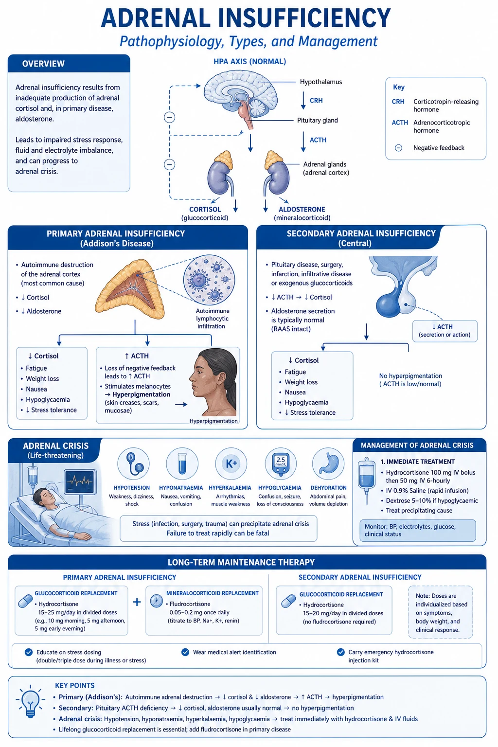
Adrenal insufficiency produces a syndrome of chronic cortisol deficiency with (primary) or without (secondary) mineralocorticoid deficiency. Symptoms and signs are best organised system by system.[2][3]
General (almost universal in chronic AI):
- Fatigue and profound weakness, anorexia and weight loss (typically 5 to 10 kg over months), lethargy, malaise.
- Salt craving (a key historical clue, especially in primary AI — patients add salt to everything).
- Nausea, vomiting, abdominal pain, diarrhoea — the GI symptoms can be so prominent as to mimic a surgical abdomen or gastroenteritis. [1]
Cardiovascular:
- Postural dizziness and syncope, postural hypotension (a near-universal finding in primary AI from volume depletion).
- In crisis, shock — typically vasodilatory and refractory to catecholamines until hydrocortisone is given (the bedside clue that should prompt the diagnosis). [1]
Skin and pigmentation (the distinguishing feature of PRIMARY AI):
- Hyperpigmentation — diffuse dusky, bronzelike darkening of the skin, palmar creases, buccal mucosa, gums, lips, recent surgical scars, extensor surfaces, pressure points, areolae, and sun-exposed areas. Pathognomonic for primary AI (driven by ACTH/MSH); absent in secondary AI. May precede other symptoms by months.
- Vitiligo — in autoimmune Addison disease (a clue to the autoimmune aetiology); coexists with hyperpigmentation, producing striking contrast.
- Loss of axillary and pubic hair in women (loss of adrenal androgens; men are protected by testicular androgens) — a feature of primary AI only. [1]
Musculoskeletal:
- Myalgia, arthralgia, proximal muscle weakness (sometimes severe enough to mimic myopathy). [1]
Neuropsychiatric:
- Depression, apathy, irritability, decreased libido, cognitive slowing; in severe cases, psychosis. Often misattributed to depression.
- Salt craving, hyponatraemic encephalopathy (confusion, seizures) when sodium falls rapidly. [1]
Reproductive:
- Amenorrhoea / oligomenorrhoea, reduced libido, infertility in women; loss of adrenal androgens contributes. Secondary AI may have additional features from other pituitary hormone loss (hypothyroidism, hypogonadism). [1]
Bedside signs that should trigger testing
Hyperpigmentation with weight loss and postural hypotension, hyponatraemia with or without hyperkalaemia, unexplained postural syncope, or refractory vasodilatory shock — check a morning cortisol and ACTH. [1]
Adrenal crisis — the acute presentation
The decompensated extreme. Classic tetrad:[8]
- Hypotension and shock (vasodilatory, often refractory to fluids and vasopressors).
- Abdominal pain, nausea, vomiting, diarrhoea — may mimic an acute surgical abdomen (do not operate first).
- Hypoglycaemia (especially in children and in secondary AI from fasting) — can cause seizures and coma.
- Hyponatraemia with hyperkalaemia (primary) or hyponatraemia alone (secondary), with metabolic acidosis, hypercalcaemia occasionally, eosinophilia.
- Fever may be present (infection is the common precipitant) or hypothermia. [1]
Almost always precipitated in a known or undiagnosed patient by infection (gastroenteritis, pneumonia, UTI), surgery, trauma, vomiting with inability to absorb oral steroids, missed doses, pregnancy/labour, or withdrawal of chronic glucocorticoids.[8][9]
Atypical presentations (examiner favourites)
- Acute abdomen presentation — severe abdominal pain, vomiting, guarding, fever; laparotomy before recognition is a classic avoidable error. Always check cortisol in an undiagnosed patient with an acute abdomen and refractory shock.
- Isolated hyponatraemia — especially in older patients misdiagnosed as SIADH; check cortisol and ACTH before committed to SIADH treatment.
- Refractory septic shock in ICU — consider critical illness-related corticosteroid insufficiency (CIRCI); a 250 mcg SST with a blunted response and vasopressor dependence suggests the diagnosis.[7]
- Addisonian crisis as first presentation in undiagnosed CAH (neonate), autoimmune Addison disease, or after unilateral adrenalectomy in a patient with an unrecognised contralateral atrophic gland.
- Recurrent hypoglycaemia in a type 1 diabetic (clue to APS-2 — coexisting Addison disease reduces insulin requirement and causes unexplained hypoglycaemia).
- Secondary AI masquerading as chronic fatigue or depression — pale skin (no pigment), hyponatraemia without hyperkalaemia, weight loss, other pituitary deficits.
Differential Diagnosis
The differential depends on the presenting syndrome. [1]
Differential of the cause of PRIMARY adrenal insufficiency
| Cause | CT adrenal | Antibodies / other | Distinguishing features |
|---|---|---|---|
| Autoimmune Addison | Small/atrophic, normal | Anti-21-hydroxylase Ab positive; vitiligo; APS-1 or APS-2 clustering | Commonest in the West; female predominance; other autoimmune disease |
| Tuberculosis | Enlarged, caseating ± calcification | TB testing (Quantiferon, AFB); CXR | Commonest worldwide; extrapulmonary TB; calcification late |
| CAH (21-OHD) | Enlarged, lipid-depleted | 17-OH-progesterone markedly raised; neonatal screen | Neonatal salt-wasting crisis; ambiguous genitalia in females |
| Bilateral adrenal haemorrhage | Enlarged, hyperdense (acute); atrophic (chronic) | Coagulation; blood cultures | Meningococcaemia (Waterhouse-Friderichsen), anticoagulation, post-surgery, sepsis |
| Infiltrative (amyloid, sarcoid, haemochromatosis) | Enlarged, infiltrative | Iron studies, ACE, biopsy | Systemic features of the underlying disease |
| Metastases (lung, breast, melanoma, kidney, lymphoma) | Bilateral masses, primary evident | Biopsy | Usually with widespread disease; AI uncommon unless >90 percent destroyed |
| Infections (HIV, CMV, histoplasma, cryptococcus) | Variable | HIV test, fungal serology | Immunocompromise; opportunistic infection |
| Drugs (etomidate, ketoconazole, mitotane, metyrapone) | Normal or atrophic | Drug history | Recent exposure; resolves on withdrawal |
Differential of the syndrome (when cortisol not yet back)
- Chronic fatigue syndrome / depression — fatigue, weight loss, low mood; but normal cortisol, no hyperpigmentation, no electrolyte disturbance.
- Malignancy (occult) — weight loss, anorexia, fatigue; but no hyperpigmentation, imaging/evidence of primary.
- Gastroenteritis / acute abdomen — vomiting, abdominal pain, diarrhoea; but no hyperpigmentation, no chronic weight loss, normal cortisol (crisis can mimic, however — always check cortisol in shock with abdominal pain).
- Septic shock / CIRCI — vasodilatory shock in ICU; baseline cortisol often normal — a dynamic SST defines CIRCI.[7]
- SIADH — hyponatraemia with low sodium and low osmolality, inappropriately concentrated urine; distinguish by checking cortisol and ACTH before labelling SIADH.
- Hyperkalaemia of renal failure — but urea/creatinine proportionate; aldosterone and renin point to AI.
Differential of hyperpigmentation
Distinguish ACTH-driven (Addison) hyperpigmentation (diffuse, palmar creases, mucosal, recent scars, with systemic features and hyponatraemia) from sun exposure, racial/ethnic, haemochromatosis (bronze diabetes with diabetes and cirrhosis, iron studies raised), porphyria cutanea tarda, Peutz-Jeghers syndrome (perioral macules, GI polyps), Cushing disease (ACTH-secreting pituitary adenoma — but with central obesity, striae), Nelson syndrome (after bilateral adrenalectomy for Cushing — high ACTH from a growing pituitary tumour), chronic renal failure, drug-induced (amiodarone, minocycline, chlorpromazine, heavy metals).[3]
Clinical & Bedside Assessment
A focused examination aims to (a) recognise the chronic syndrome, (b) distinguish primary from secondary AI at the bedside, (c) detect adrenal crisis, and (d) seek clues to the cause and to other autoimmune or pituitary disease.[1]
Vital signs and general: postural blood pressure (a drop of more than 20 mmHg systolic on standing is highly suggestive of volume depletion in primary AI); weight loss; fever (precipitant); shock in crisis. [1]
Named bedside signs of primary AI (examiner favourites):
- Hyperpigmentation — examine the palmar creases, knuckles, buccal mucosa, gums, lips, recent surgical scars, extensor surfaces, areolae, and sun-exposed areas. The pigmentation is generalised and includes mucous membranes (mucosal pigmentation distinguishes it from racial pigmentation, which spares mucosa). Pathognomonic for primary AI.
- Vitiligo — patches of depigmentation in autoimmune Addison disease.
- Loss of axillary and pubic hair in women (loss of adrenal androgens).
- Postural hypotension from volume depletion.
- Salt craving (history).
- Cachexia, abdominal tenderness (in crisis). [1]
Distinguishing secondary/tertiary AI at the bedside:
- Pale skin (no ACTH → no MSH → no pigmentation), fine wrinkling around the eyes and mouth, loss of axillary and pubic hair (in both sexes if panhypopituitarism), signs of other pituitary hormone deficiency: pale, cool, dry skin (hypothyroidism), loss of secondary sexual characteristics (hypogonadism), bitemporal hemianopia (pituitary mass on optic chiasm), headache, ophthalmoplegia (pituitary apoplexy), failure of postpartum lactation and amenorrhoea (Sheehan syndrome). [1]
Bedside clues to the cause:
- Tuberculosis — examine for lymphadenopathy, pleural effusion, spinal gibbus (Pott disease), chronic cough.
- APS-1 — chronic mucocutaneous candidiasis, hypocalcaemia signs (Chvostek, Trousseau).
- APS-2 — vitiligo, goitre, type 1 diabetes features, premature ovarian failure.
- Meningococcaemia / Waterhouse-Friderichsen — purpuric rash in a febrile, shocked child or young adult.
- Pituitary apoplexy — sudden severe headache, ophthalmoplegia, visual field defect, meningism. [1]
Investigations
Investigation has three goals: (1) confirm AI biochemically, (2) localise the level (primary vs secondary), and (3) find the cause.[1][2]
Step 1 — Confirm cortisol deficiency
- Morning (8 to 9 am) serum cortisol — the simplest screen, taking advantage of the diurnal peak. Interpret:
- Above 500 nmol/L (18 mcg/dL) — AI effectively excluded (assuming normal cortisol-binding globulin and no recent exogenous steroid in the past 24 hours).
- Below 138 nmol/L (5 mcg/dL) at 8 am — highly suggestive of AI.
- Between 138 and 500 nmol/L (5 to 18 mcg/dL) — equivocal, proceed to a stimulation test.
- Do NOT rely on a single random cortisol outside the early morning window — diurnal variation makes interpretation misleading.
- Exclude exogenous glucocorticoid in the previous 24 to 48 hours (prednisolone and prednisone cross-react in cortisol immunoassays; hydrocortisone IS cortisol and cross-reacts fully; dexamethasone does NOT cross-react — useful when assessing the axis in a steroid-treated patient). [1]
Step 2 — Confirm with a stimulation test (gold standard)
The 250 mcg short Synacthen (cosyntropin) test (SST) — the standard diagnostic test for primary AI.[1]
- Procedure: baseline cortisol at 0 minutes; give 250 mcg tetracosactide (Synacthen = cosyntropin = ACTH 1-24) intravenously or intramuscularly; cortisol at 30 and 60 minutes.
- Normal response: peak cortisol rises above 500 nmol/L (18 mcg/dL) AND a rise of at least 200 nmol/L from baseline.
- Adrenal insufficiency: failure of cortisol to rise above 500 nmol/L. The SST is highly sensitive for primary AI (where the cortex is destroyed and cannot respond to ACTH).
- Caveat in secondary/tertiary AI: recent-onset secondary AI may give a false-normal SST because the atrophic cortex can still respond to a supraphysiological bolus of ACTH — use the low-dose (1 mcg) SST or the insulin tolerance test for suspected recent-onset secondary AI. [1]
Insulin tolerance test (ITT) — the gold standard for secondary/tertiary AI but more invasive and risky (contraindicated in epilepsy, ischaemic heart disease, pregnancy).[1][4]
- Procedure: intravenous insulin 0.1 to 0.15 U/kg body weight to achieve symptomatic hypoglycaemia with blood glucose below 2.2 mmol/L; cortisol at 0, 30, 45, 60, 90, 120 minutes.
- Normal response: cortisol rises above 500 nmol/L.
- Tests the entire HPA axis — needs an intact hypothalamus, pituitary AND adrenal. A subnormal response confirms AI but does not localise; the CRH stimulation test (cortisol response to exogenous CRH) distinguishes secondary (no pituitary ACTH response) from tertiary (delayed/preserved ACTH response).
Metyrapone test — alternative when ITT contraindicated; blocks 11β-hydroxylase, lowering cortisol and (normally) driving ACTH and 11-deoxycortisol rise; subnormal 11-deoxycortisol response indicates AI. [1]
Step 3 — Localise the level
- Plasma ACTH drawn at 8 to 9 am with the baseline cortisol. Inappropriately HIGH ACTH with low cortisol = primary AI (ACTH typically over 300 ng/L, often over 1000). Low or inappropriately normal ACTH with low cortisol = secondary/tertiary AI.[1][4]
- Plasma renin and aldosterone — in primary AI: renin HIGH, aldosterone LOW (mineralocorticoid failure drives renin). In secondary AI: renin and aldosterone normal (RAAS intact). This confirms the mineralocorticoid axis is preserved in secondary AI.
- CRH stimulation test — distinguishes secondary (blunted ACTH) from tertiary (delayed, exaggerated ACTH rise) AI; rarely performed in routine practice.
Reproducing the short Synacthen test interpretation (examiner essentials)
| Result | Interpretation |
|---|---|
| Baseline 8 am cortisol above 500 nmol/L (18 mcg/dL) | AI excluded (assuming no recent steroid) |
| Baseline 8 am cortisol below 138 nmol/L (5 mcg/dL) | AI highly likely — proceed to ACTH and confirm |
| SST peak cortisol above 500 nmol/L (18 mcg/dL) | Normal — AI excluded |
| SST peak cortisol below 500 nmol/L | AI confirmed — check ACTH to localise |
| Plasma ACTH HIGH with low SST cortisol | PRIMARY adrenal insufficiency |
| Plasma ACTH LOW/normal with low SST cortisol | SECONDARY or TERTIARY |
Step 4 — Find the cause
- Anti-21-hydroxylase antibodies — positive in autoimmune Addison disease (sensitivity over 90 percent); if positive, the cause is established.[1]
- 17-hydroxyprogesterone — to exclude classic CAH (21-OHD) in children and young adults with primary AI; the newborn heel-prick test screens for this.
- Tuberculosis testing — Quantiferon-TB Gold, chest X-ray, sputum/AFB, adrenal CT for caseating granulomata or calcification.
- CT or MRI of the adrenal glands — small/atrophic in autoimmune Addison; enlarged with calcification in TB; enlarged with haemorrhage (hyperdense acutely); bilateral masses in metastasis/infiltration/lymphoma; cystic in rare adrenal cysts.
- MRI of the pituitary (secondary AI) — adenoma, empty sella, apoplexy, infiltrative.
- Other autoimmune screening (if autoimmune Addison): TSH, free T4, anti-TPO, vitamin B12, coeliac serology, fasting glucose, FSH/LH/oestradiol, calcium/PTH — to detect APS-1, APS-2 or single autoimmune associations.[2]
- HIV test — if HIV-related adrenal infection suspected.
- Iron studies — haemochromatosis.
- DHEAS — low in primary AI; rarely measured routinely but supports the diagnosis when borderline.
Associated laboratory abnormalities (examiner favourites)
- Hyponatraemia (in over 80 percent of primary AI; from aldosterone loss plus ADH excess).
- Hyperkalaemia (in 50 to 65 percent of primary AI; absent in secondary AI — a key discriminator).
- Hypoglycaemia (especially in children and in fasting).
- Mild normocytic anaemia, eosinophilia, lymphocytosis (loss of cortisol's permissive effect on marrow and lymphoid tissue).
- Mild metabolic acidosis (from mineralocorticoid failure in primary AI).
- Hypercalcaemia occasionally (rare, mechanism uncertain — increased calcium resorption and vitamin D sensitivity).
- Raised urea from volume depletion.
- Mild transaminitis. [1]
Management — Resuscitation
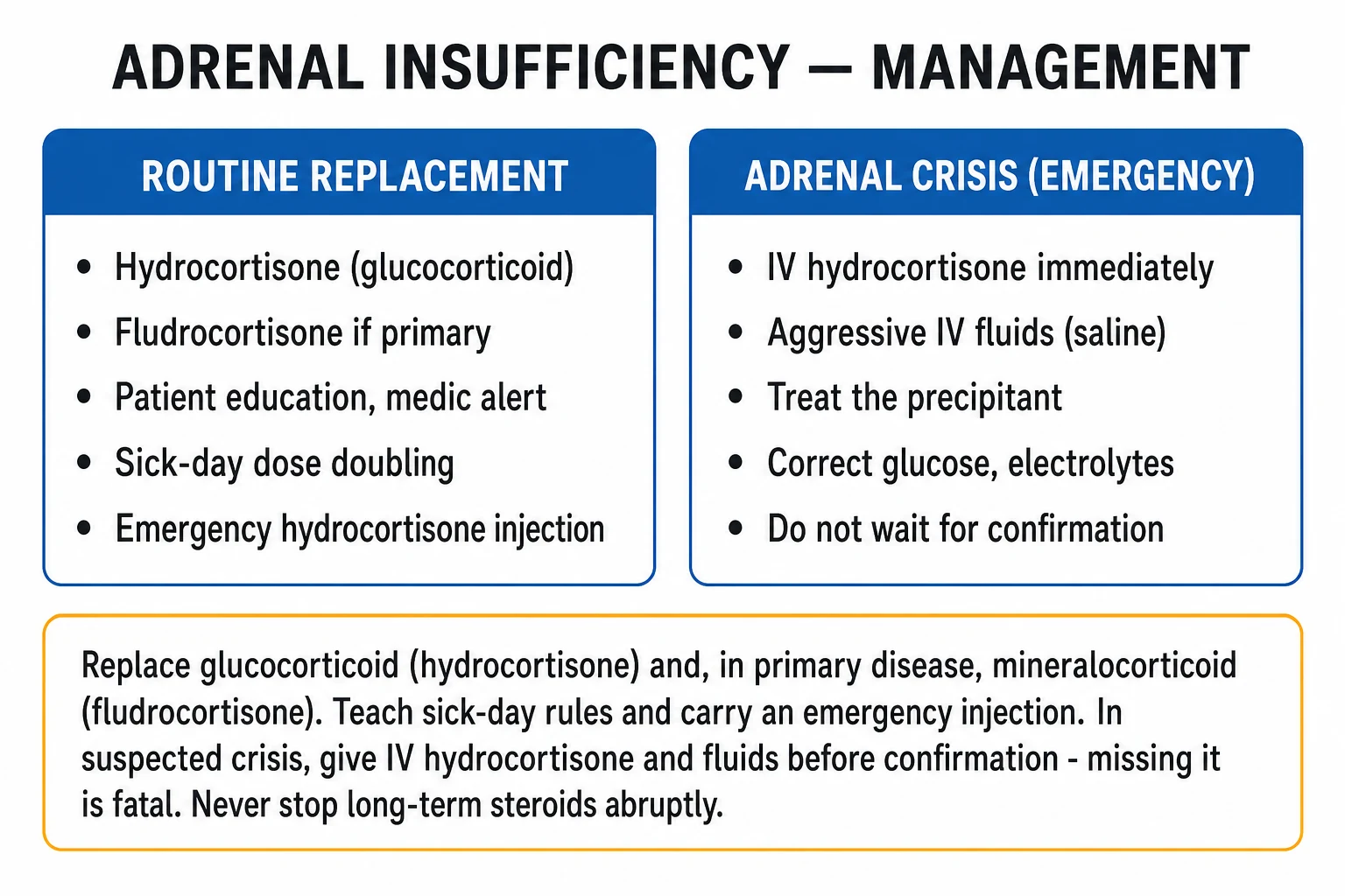
Adrenal crisis is a fatal endocrine emergency and treatment must NOT await cortisol/ACTH confirmation. The resuscitation bundle — apply simultaneously.[1][8]
- ABCDE; high-flow oxygen if hypoxic; cardiac monitoring; two large-bore intravenous cannulae; bloods including cortisol and ACTH BEFORE the first hydrocortisone (the cortisol and ACTH drawn in crisis are diagnostic — but do not delay treatment to obtain them).
- Parenteral hydrocortisone — 100 mg intravenous stat, then 100 mg over 24 hours (either as a continuous infusion of 100 mg in 24 hours, or as 50 mg intravenous or intramuscular every 6 hours, or 200 mg per 24 hours in severe shock). At this dose, hydrocortisone provides sufficient mineralocorticoid activity — fludrocortisone is NOT required acutely.
- Intravenous fluid resuscitation — 0.9 percent sodium chloride 1 L stat over 30 to 60 minutes, then 2 to 3 L over the first 24 hours, guided by clinical and haemodynamic response; add 5 to 10 percent dextrose if hypoglycaemic (children particularly).
- Treat the precipitant — sepsis is the commonest precipitant: take cultures and start broad-spectrum antibiotics empirically (e.g. ceftriaxone 2 g intravenous once daily, modified on culture); treat other identified stressors (surgery, trauma, MI).
- Identify and correct hypoglycaemia, electrolyte disturbance and acidosis — fluid resuscitation and hydrocortisone will correct sodium, potassium and glucose; do not aggressively correct sodium (risk of osmotic demyelination) — let it rise by no more than 8 to 10 mmol/L in 24 hours.
- Intensive care monitoring — shocked or comatose patients; vasopressors (noradrenaline) for refractory shock will work only after hydrocortisone is given (cortisol restores vascular catecholamine sensitivity).
- Taper — once stable (usually within 24 to 72 hours), halve the hydrocortisone dose daily until the oral maintenance dose is reached (15 to 25 mg/day in divided doses), then transition to long-term maintenance including fludrocortisone (primary AI).[1]
Adrenal crisis bundle — A-B-C-DEF
DEFEND
then 100 mg over 24 h (infusion or 50 mg q6h); fludrocortisone NOT needed acutely
then 2 to 3 L in 24 h, guided by response; add dextrose if hypoglycaemic
sepsis (commonest) — cultures and broad-spectrum antibiotics empirically
do NOT correct Na by more than 8 to 10 mmol/L in 24 h (osmotic demyelination)
draw cortisol and ACTH BEFORE the first hydrocortisone, but treat empirically
taper to oral maintenance 15 to 25 mg/day + fludrocortisone (primary AI)
Management — Definitive & Stepwise
For chronic adrenal insufficiency, definitive management has three pillars: (1) hormone replacement (glucocorticoid ± mineralocorticoid ± DHEA), (2) patient education and sick-day rules, and (3) prevention and treatment of crisis.[1][9]
Pillar 1 — Hormone replacement
Glucocorticoid replacement (all forms of AI):[1][2]
- Hydrocortisone is first-line — closest analogue to physiological cortisol.
- Total daily dose 15 to 25 mg in divided doses, mimicking the diurnal rhythm. A common regimen: 10 mg on waking, 5 mg at noon, 5 mg at 4 to 5 pm (some patients tolerate twice-daily dosing). The first dose is largest and on waking (mimics the physiological morning peak); no dose after 6 pm unless evening shift worker, to avoid insomnia.
- Alternative: prednisolone 5 mg once daily in the morning — longer half-life, fewer doses, but less physiological and harder to titrate; useful in patients with adherence or nocturnal hypoglycaemia problems.
- Avoid dexamethasone for routine replacement (long-acting, suppresses the axis, mineralocorticoid activity negligible).
- Monitor clinically — wellbeing, energy, weight, blood pressure, electrolytes; do NOT over-treat (Cushingoid features, osteoporosis, diabetes indicate excess dose). 24-hour urinary free cortisol and day-curve cortisol can be used to titrate in difficult cases.
Mineralocorticoid replacement (primary AI ONLY):[1]
- Fludrocortisone (9α-fludrocortisone) — synthetic mineralocorticoid; 50 to 200 mcg orally once daily (typically 100 mcg daily).
- Monitor with blood pressure (including postural), serum sodium and potassium, and plasma renin — aim for normal electrolytes and renin in the upper normal range. Increase dose if postural hypotension, hyponatraemia or salt craving; decrease if hypertension, hypokalaemia or oedema.
- Not required in secondary/tertiary AI — the RAAS is intact.
- In severe crisis at high hydrocortisone doses (above 50 to 100 mg/day), hydrocortisone saturates the mineralocorticoid receptor, so fludrocortisone is held acutely and resumed when hydrocortisone is tapered to maintenance.
Adrenal androgen replacement (selected patients):
- DHEA (dehydroepiandrosterone) 25 to 50 mg orally each morning — may improve wellbeing, libido and mood in women with primary AI; not routinely needed, monitor serum DHEAS; evidence is mixed.[2]
Pillar 2 — Patient education and sick-day rules (the single most important determinant of long-term outcome)
Patient education reduces adrenal crisis rates by around half in prospective studies.[9]
Sick-day rules (examiner favourite):
- Minor illness (fever over 37.5°C, cold, UTI, minor surgery, dental extraction): double the oral hydrocortisone for the duration of illness (typically 3 days) — usually 20 to 40 mg daily total.
- Severe illness, vomiting, diarrhoea, trauma, severe infection, surgery: triple the dose or give parenteral hydrocortisone 100 mg intramuscularly or intravenously.
- Vomiting or inability to absorb oral hydrocortisone is an emergency: use the emergency hydrocortisone injection kit (100 mg IM) and seek medical review; dehydration precipitates crisis within hours. [1]
Surgical stress-dose steroids (standard schedules):
- Minor procedure (e.g. cataract, hernia, dental extraction, colonoscopy): hydrocortisone 50 mg intravenous or intramuscularly at induction, resume usual oral dose post-op.
- Moderate procedure (e.g. cholecystectomy, joint replacement): hydrocortisone 75 to 100 mg at induction, then 50 mg every 8 hours for 24 hours, taper to maintenance over 1 to 2 days.
- Major surgery (e.g. cardiac, thoracic, major abdominal): hydrocortisone 100 mg at induction, then 200 mg per 24 hours (continuous infusion or 50 mg every 6 hours) for 24 to 48 hours, taper to maintenance over 2 to 3 days.
- Labour and delivery: hydrocortisone 100 mg intramuscularly every 6 to 8 hours during labour and for 24 hours postpartum, then taper.[1]
Emergency kit and identification: every AI patient must carry (1) a medical alert bracelet or card identifying adrenal insufficiency, (2) an emergency hydrocortisone 100 mg vial for intramuscular injection with needles and instructions, and (3) a steroid emergency card; relatives/partners should be taught to give the injection.[9]
Pillar 3 — Prevention of crisis
- Annual influenza vaccine and pneumococcal vaccination (reduce respiratory precipitants).
- Pneumococcal, meningococcal, Haemophilus influenzae vaccination in asplenia or complement deficiency.
- Avoid abrupt withdrawal of chronic glucocorticoids — always taper over weeks to months, particularly after more than 3 weeks of therapy over 7.5 mg prednisolone equivalent.
- Educate about doubling the dose during illness, and seeking help early when vomiting.
- Coordinate care with endocrinology, anaesthetics (for surgery) and primary care. [1]
Specific Subtypes & Scenarios
Autoimmune Addison disease
The commonest primary AI in developed countries (70 to 90 percent). Young to middle-aged women, family/personal history of autoimmune disease, anti-21-hydroxylase antibodies positive, vitiligo, APS-1 or APS-2 clustering. Adrenals on imaging are small/atrophic. Treat with hydrocortisone + fludrocortisone; screen for and treat associated autoimmune conditions; lifelong replacement.[2]
Tuberculous Addison disease
Commonest primary AI worldwide. Arises years after TB exposure; adrenal CT shows bilateral enlargement with caseating centres early, calcification late. Often coexists with extrapulmonary TB; evaluate for active TB elsewhere (chest X-ray, sputum, Quantiferon, urine AFB). Treat the AI as for primary AI; treat the TB with standard quadruple therapy (rifampicin accelerates cortisol metabolism — increase the hydrocortisone dose while the patient is on rifampicin).[4]
Congenital adrenal hyperplasia (21-hydroxylase deficiency)
Salt-wasting classic form presents in the neonatal period with vomiting, dehydration, failure to thrive, hyponatraemia, hyperkalaemia, hypoglycaemia (an adrenal crisis) and ambiguous external genitalia in 46,XX infants (virilisation from excess androgens). 46,XY infants appear normal externally and may present with salt-wasting crisis at 1 to 2 weeks. Diagnose by markedly elevated 17-hydroxyprogesterone (newborn heel-prick screen in many countries). Treat with hydrocortisone AND fludrocortisone (and salt supplementation in infancy); monitor 17-OHP, renin, electrolytes and growth; surgical correction of ambiguous genitalia is individualised.[2]
Waterhouse-Friderichsen syndrome
Bilateral adrenal haemorrhage in meningococcaemia (also pneumococcal, Haemophilus, staphylococcal sepsis) — classically in children and young adults presenting with purpuric rash, septicaemia, shock and adrenal crisis. Treat with antibiotics (ceftriaxone), hydrocortisone 100 mg IV, fluid resuscitation and ICU support; mortality high. Bilateral adrenal haemorrhage also complicates anticoagulation (heparin-induced thrombocytopenia, warfarin), post-surgical states, severe trauma, and pregnancy — consider in any shocked patient with abdominal/flank pain and dropping haemoglobin.[3]
Drug-induced adrenal insufficiency
- Etomidate — even a single dose in induction can inhibit 11β-hydroxylase and precipitate AI in critically ill patients; avoid in septic shock or use with corticosteroid cover.[7]
- Ketoconazole (and less so fluconazole, itraconazole) — inhibits multiple steroidogenic CYP enzymes; can cause primary AI.
- Metyrapone — 11β-hydroxylase inhibitor (used in Cushing syndrome); use with monitoring.
- Mitotane (for adrenocortical carcinoma) — adrenolytic; replacement hydrocortisone doses need to be doubled or tripled (mitotane increases cortisol-binding globulin and accelerates cortisol metabolism).
- Mifepristone — glucocorticoid receptor antagonist; can cause clinical cortisol deficiency with normal cortisol levels.
- Rifampicin, phenytoin, barbiturates — hepatic enzyme inducers that accelerate cortisol metabolism — precipitate crisis in established Addison disease (increase hydrocortisone dose).
Critical illness-related corticosteroid insufficiency (CIRCI)
A transient, reversible HPA-axis dysfunction in severe sepsis, septic shock, ARDS and major trauma, distinct from chronic AI. The 2017 CIRCI guidelines (Annane et al)[7] suggest:
- Do NOT test routinely; clinical diagnosis based on vasopressor-dependent shock.
- Suggest intravenous hydrocortisone 200 mg per day (continuous infusion or 50 mg every 6 hours) for adults with septic shock requiring ongoing vasopressors despite adequate fluid and vasopressor therapy.
- Based on the Annane 2002 trial (hydrocortisone + fludrocortisone reduced mortality in non-responders to the 250 mcg SST)[5][6] and the CORTICUS 2008 trial (no overall mortality benefit, faster shock reversal) — hence the guideline restricts steroids to vasopressor-refractory shock, not all septic shock.[7]
Secondary/tertiary adrenal insufficiency
- Glucocorticoid replacement only (no fludrocortisone — mineralocorticoid axis intact).
- Investigate and treat the underlying cause — pituitary adenoma (transsphenoidal surgery), pituitary apoplexy (urgent steroids ± surgery), infiltrative disease, empty sella.
- Screen for and replace other pituitary hormones — thyroxine (but always start hydrocortisone BEFORE thyroxine in panhypopituitarism — thyroxine accelerates cortisol clearance and can precipitate crisis), sex steroids, GH (children), desmopressin for diabetes insipidus.
- Chronic steroid therapy — taper over weeks to months to allow HPA-axis recovery; test the axis with morning cortisol or SST after taper before declaring recovery. [1]
Complications & Pitfalls
Complications of untreated or undertreated AI
- Adrenal crisis — recurrent in poorly educated or non-adherent patients; mortality 5 to 15 percent.[8]
- Hyponatraemic encephalopathy — confusion, seizures, coma from severe hyponatraemia.
- Hypoglycaemic brain injury — especially in children and in fasting adults.
- Hyperkalaemic arrhythmia (primary AI) — ventricular tachycardia/fibrillation from severe hyperkalaemia.
- Septic shock refractory to catecholamines — the vascular unresponsiveness that should prompt empiric hydrocortisone.
Complications of over-treatment (iatrogenic Cushing)
- Glucocorticoid excess — osteoporosis, diabetes, hypertension, weight gain, infection susceptibility, skin thinning, myopathy. Use the lowest dose that restores wellbeing, monitor for Cushingoid features, and consider bone densitometry and bone protection (calcium, vitamin D, bisphosphonate if osteoporotic).[2]
Classic pitfalls (examiner favourites)
- Treating SIADH with fluid restriction before excluding AI — the hyponatraemia of AI is partly dilutional but the patient is also volume-depleted; fluid restriction worsens shock. Always check cortisol and ACTH in any unexplained hyponatraemia.
- Giving thyroxine before hydrocortisone in panhypopituitarism — thyroxine accelerates cortisol clearance and precipitates adrenal crisis. Replace hydrocortisone first, always.
- Misdiagnosing adrenal crisis as an acute surgical abdomen and laparotomising a patient in shock. Check cortisol in any shocked patient with abdominal pain and electrolyte disturbance.
- Forgetting stress-dose steroids in a known Addison patient undergoing surgery or in labour — precipitates intra-operative crisis.
- Telling a steroid-dependent patient to "skip the dose if vomiting" — vomiting is the trigger for parenteral hydrocortisone, never omission.
- Missing the diagnosis in chronic steroid therapy withdrawal — patients on long-term prednisolone can take months for the HPA axis to recover; taper slowly and test before stopping.
- Withholding cortisol while waiting for the SST in a patient in crisis — the SST can wait; treat empirically (draw cortisol and ACTH first, then hydrocortisone).
- Forgetting rifampicin accelerates cortisol metabolism — increase hydrocortisone dose in Addison disease on anti-TB therapy. [1]
Prognosis & Disposition
Treated primary AI
With adequate replacement, education and emergency preparedness, life expectancy approaches normal. Adrenal crisis rates of 8 to 40 percent per patient-year are reduced by half with structured education.[9] Risk of crisis is highest in primary AI, in patients with comorbidity, and in adolescents and young adults (poor adherence).[10]
Adrenal crisis
Mortality 5 to 15 percent even with appropriate treatment; worse with delayed presentation, severe hyperkalaemia, refractory shock and underlying sepsis. Survivors return to chronic management with intensified education.[8]
Secondary/tertiary AI
Prognosis depends on the underlying cause — pituitary adenoma prognosis depends on tumour biology and surgery; chronic steroid withdrawal prognosis excellent with proper taper. Mortality is from crisis in unrecognised secondary AI, particularly when another illness is being treated and the steroid requirement rises. [1]
Monitoring and follow-up
- Clinic review every 6 to 12 months — wellbeing, blood pressure, weight, electrolytes; check plasma renin to titrate fludrocortisone in primary AI.
- Annual — bone densitometry (DEXA) if on long-term steroids, annual review of autoimmune comorbidities (TSH, vitamin B12, coeliac serology) in autoimmune Addison.
- Patient-held steroid emergency card and medical alert reviewed each visit.
- Reinforce sick-day rules at every contact. [1]
When to refer to endocrinology
- Adrenal crisis; new diagnosis of primary AI; secondary AI (for pituitary workup); difficult-to-titrate replacement; recurrent crises; pregnancy; CAH transition to adult care; CIRCI in ICU. [1]
Special Populations
Children and neonates
- CAH (21-OHD) is the dominant cause of primary AI in children; newborn screening by heel-prick 17-OH-progesterone; ambiguous genitalia in females is the most common route to early diagnosis.
- Weight-based dosing — hydrocortisone 8 to 12 mg/m²/day in three divided doses (avoid over-treatment — stunts growth); fludrocortisone 100 mcg/m²/day plus salt supplementation in infancy.
- Hypoglycaemia is more prominent in children — cortisol is a major counter-regulatory hormone.
- Adrenal crisis in a neonate is salt-wasting CAH until proven otherwise. [1]
Pregnancy
- Pregnant women with AI — cortisol requirements rise in the third trimester (oestrogen raises cortisol-binding globulin); often increase hydrocortisone by 50 percent in the third trimester.
- During labour and delivery — parenteral hydrocortisone 100 mg intramuscularly every 6 to 8 hours (or continuous infusion 200 mg/24 h) for the duration of labour and 24 hours postpartum; taper to maintenance over 2 to 3 days.
- Postpartum — fludrocortisone requirement often decreases postpartum; reassess at 6 weeks.
- Pregnancy in known Addison disease — manage jointly with obstetrics and endocrinology; de novo presentation in pregnancy is rare but crisis risk is highest in the first trimester (hyperemesis, vomiting).
- Sheehan syndrome (postpartum pituitary necrosis) — secondary AI presenting as failure to lactate, persistent amenorrhoea, fatigue, hypothyroidism, hypoglycaemia after obstetric haemorrhage.[3]
The elderly
- Atypical presentation — fatigue, weight loss, depression, falls, hyponatraemia; hyperpigmentation may be subtle or attributed to age. Have a low threshold to check cortisol in any older patient with unexplained weight loss, fatigue or hyponatraemia.
- Co-medications increase drug-induced AI (steroid therapy, ketoconazole, etomidate, enzyme inducers).
- Start with lower doses of hydrocortisone and titrate to wellbeing (avoid over-treatment that worsens osteoporosis, diabetes, hypertension). [1]
Immunocompromised (HIV/AIDS)
- HIV-related adrenalitis from opportunistic infection (CMV, mycobacteria — both TB and MAC, cryptococcus, histoplasma, toxoplasma); clinical AI is uncommon unless more than 90 percent of cortex destroyed.
- Evaluate and treat the underlying opportunistic infection alongside cortisol replacement.
- Kaposi sarcoma and lymphoma can also infiltrate the adrenals. [1]
Anticoagulated and post-surgical patients
- Bilateral adrenal haemorrhage — risk in therapeutic anticoagulation (especially heparin-induced thrombocytopenia), post-cardiac surgery, severe trauma, postpartum; presents with abdominal/flank pain, dropping haemoglobin, shock and biochemical AI. Treat with hydrocortisone and address the underlying coagulopathy. [1]
Chronic steroid therapy withdrawal
- The commonest AI overall. Patients on over 5 to 7.5 mg prednisolone equivalent for over 3 weeks are at risk of HPA-axis suppression.
- Taper slowly — reduce by 1 to 2.5 mg every 1 to 2 weeks once below physiological dose; test the axis with morning cortisol at the end of taper, or with a 1 mcg SST; continue hydrocortisone 10 to 20 mg daily during recovery if symptomatic. [1]
Evidence, Guidelines & Regional Differences
Endocrine Society Clinical Practice Guideline 2016 (Bornstein et al)[1]
The landmark guideline for the diagnosis and management of primary adrenal insufficiency. Key recommendations:
- Diagnose primary AI with a morning cortisol below 138 nmol/L (5 mcg/dL) and a subnormal response to the 250 mcg SST (peak cortisol below 500 nmol/L, 18 mcg/dL).
- Plasma ACTH differentiates primary (high) from secondary (low) AI.
- Test anti-21-hydroxylase antibodies in primary AI; if negative, image the adrenal glands (CT) and consider TB, infiltrative and metastatic causes.
- Hydrocortisone 15 to 25 mg/day in two to three divided doses, or prednisolone 5 to 7.5 mg daily, as glucocorticoid replacement; fludrocortisone (primary AI) 0.05 to 0.2 mg daily.
- Patient education and crisis prevention are core; every patient should carry a steroid emergency card and emergency hydrocortisone injection kit.
- Double the glucocorticoid dose during febrile illness; parenteral hydrocortisone during severe stress, vomiting or surgery. [1]
Lancet 2014 review (Charmandari et al)[2]
A definitive overview confirming the classification (primary, secondary, tertiary), the autoimmune predominance in developed countries, the role of the 250 mcg SST and ACTH in diagnosis, and the principles of replacement and crisis prevention. Emphasises that patient education halves crisis rates. [1]
Lancet 2003 (Arlt & Allolio)[3] and NEJM 1996 (Oelkers)[4]
The classic reviews that established modern understanding of adrenal insufficiency — its epidemiology, the dominance of autoimmune Addison in the West and TB worldwide, the molecular pathophysiology of cortisol action, and the differential diagnosis of hyperpigmentation. [1]
Annane et al — septic shock and CIRCI (JAMA 2000, JAMA 2002, Crit Care Med 2017)[5][6][7]
- JAMA 2000 — a 3-level prognostic classification in septic shock based on baseline cortisol and the cortisol response to the 250 mcg SST identified non-responders (cortisol rise less than 250 nmol/L) as the high-mortality subgroup.[6]
- JAMA 2002 (the French multicentre trial) — hydrocortisone 50 mg IV every 6 hours + fludrocortisone 50 mcg daily for 7 days reduced 28-day mortality in non-responders to the SST (relative risk reduction around 30 percent); no benefit (and possible harm) in responders.[5]
- 2017 CIRCI guidelines — synthesise Annane 2002 with CORTICUS 2008 (no overall mortality benefit, faster shock reversal, more superinfections) and recommend IV hydrocortisone 200 mg/day for adults with septic shock requiring ongoing vasopressors despite adequate fluid and vasopressor therapy (weak recommendation, low quality).[7]
Endocrine (2020–2025) — crisis incidence and education
- Burger-Stritt 2020 (Eur J Endocrinol) — a prospective multicentre study showing that standardised patient education reduces adrenal crisis incidence by about half.[9]
- Scala 2025 (Endocrine) and Rushworth 2022 (Endocrine) — confirm crisis rates of 8 to 40 percent per patient-year, highest in primary AI and in adolescents/young adults (poor adherence, risk-taking).[8][10]
Regional differences
- India and TB-endemic regions — tuberculosis dominates the cause of primary AI; bilateral adrenal calcification on CT is highly suggestive; rifampicin accelerates cortisol metabolism (increase replacement dose).[4]
- Developed countries — autoimmune Addison predominates; APS-1 and APS-2 clustering recognised; associated autoimmune disease screening standard.
- UK — Endocrine Society / Society for Endocrinology / ADSHG guidance; Steroid Emergency Card universal; patient-led educational material via ADSHG.
- US — Endocrine Society guidance; medic-alert bracelets and emergency injection kits standard of care; primary AI prevalence rising.
Controversies
- Hydrocortisone dose — modified-release vs immediate-release — modified-release hydrocortisone (Chronocort, Plenadren) better mimics the diurnal rhythm and may improve morning wellbeing; not universally available or funded.
- DHEA replacement — meta-analyses show modest benefit in wellbeing and libido in women with primary AI; not routinely recommended by all guidelines.
- 1 mcg vs 250 mcg SST — the 1 mcg low-dose test is more sensitive for recent-onset secondary AI but is less standardised and not commercially available in all centres.
- Routine hydrocortisone in septic shock — confined to vasopressor-refractory shock per the 2017 guidelines; broader use remains controversial given CORTICUS. [1]
Exam Pearls
Causes of PRIMARY adrenal insufficiency — ADDISON
ADDISON
commonest in the West — anti-21-hydroxylase Ab; APS-1 (AIRE) and APS-2 (Schmidt)
etomidate, ketoconazole, metyrapone, mitotane, mifepristone; rifampicin accelerates cortisol metabolism
tuberculosis (commonest worldwide), HIV/fungal/opportunistic
amyloid, sarcoid, haemochromatosis, metastases (lung, breast, melanoma); bilateral haemorrhage (Waterhouse-Friderichsen, anticoagulation)
bilateral adrenalectomy; congenital adrenal hyperplasia (21-OHD)
adrenoleucodystrophy (X-linked, very-long-chain fatty acids); Allgrove (AAA — alacrima, achalasia, adrenal failure)
CAH salt-wasting crisis; hypoplasia (DAX-1/NROB1, X-linked)
Adrenal crisis precipitants — 5 I's
I I I I I
commonest precipitant — gastroenteritis, pneumonia, UTI, sepsis
omitted dose, vomiting with inability to absorb oral hydrocortisone, abrupt steroid withdrawal
operative stress, major trauma, burns, fracture
myocardial infarction, stroke, pulmonary embolism, severe pain
pregnancy, labour, delivery, postpartum
Exam application bank (NEET-PG / INICET)
One-line answer
Adrenal insufficiency is the clinical syndrome arising from inadequate production of glucocorticoid (cortisol), with or without mineralocorticoid (aldosterone) and adrenal androgen deficiency. Primary adrenal insufficiency (Addison disease) results from destruction of the adrenal cortex — autoimmune in the West, tuberculosis worldwide — producing low cortisol AND low aldosterone with HIGH ACTH-driven hyperpigmentation, hyponatraemia and hyperkalaemia. Secondary adrenal insufficiency results from pituitary ACTH deficiency, and tertiary from hypothalamic CRH deficiency (most often chronic exogenous glucocorticoid therapy); both spare the mineralocorticoid axis (RAAS intact) so there is no hyperpigmentation and no hyperkalaemia. Adrenal crisis — hypotension, abdominal pain, vomiting, hypoglycaemia and hyponatraemia ± hyperkalaemia — is a fatal endocrine emergency treated with parenteral hyd
Worked stems (answer without another resource)
Stem 1 — Classic presentation. Map symptoms to mechanism; name the first investigation and first treatment step with dose/route if drug therapy is standard. [1]
Stem 2 — Unstable / complicated. List red flags that force immediate resuscitation, theatre, ICU, antidote, or reperfusion — and what you do in the first 15 minutes. [1]
Stem 3 — Atypical group. Elderly, pregnancy, child, or immunocompromised: how presentation and thresholds change. [1]
Stem 4 — Differential trap. Name the three closest mimics and one discriminator for each. [1]
Stem 5 — Disposition. Who goes home with safety-netting, who is admitted, who needs HDU/ICU/theatre, and what follow-up is mandatory. [1]
Rapid viva checklist
- Definition + classification
- Pathophysiology chain
- Bedside signs / criteria
- Score with exact components (if any)
- Emergency bundle
- Definitive therapy with doses
- Complications of disease and of treatment
- Special populations
- Guideline/trial name if classic
- Three exam traps
Coverage self-check
If you cannot answer any stem above from this page alone, re-read the matching section — the page is intended to be self-sufficient for final-prof and NEET-PG/INICET questions on Adrenal Insufficiency.
References
- [1]Bornstein SR, Allolio B, Arlt W, et al. Diagnosis and Treatment of Primary Adrenal Insufficiency: An Endocrine Society Clinical Practice Guideline J Clin Endocrinol Metab, 2016.PMID 26760044
- [2]Charmandari E, Nicolaides NC, Chrousos GP. Adrenal insufficiency Lancet, 2014.PMID 24503135
- [3]Arlt W, Allolio B. Adrenal insufficiency Lancet, 2003.PMID 12788587
- [4]Oelkers W. Adrenal insufficiency N Engl J Med, 1996.PMID 8815944
- [5]Annane D, Sebille V, Charpentier C, et al. Effect of treatment with low doses of hydrocortisone and fludrocortisone on mortality in patients with septic shock JAMA, 2002.PMID 12186604
- [6]Annane D, Sebille V, Troche G, et al. A 3-level prognostic classification in septic shock based on cortisol levels and cortisol response to corticotropin JAMA, 2000.PMID 10697064
- [7]Annane D, Pastores SM, Rochwerg B, et al. Guidelines for the Diagnosis and Management of Critical Illness-Related Corticosteroid Insufficiency (CIRCI) in Critically Ill Patients (Part I): Society of Critical Care Medicine (SCCM) and European Society of Intensive Care Medicine (ESICM) 2017 Crit Care Med, 2017.PMID 28938253
- [8]Scala A, Ibba I, Bellini I, et al. Adrenal crisis: incidence, clinical presentation and risk factors in patients with Addison's disease Endocrine, 2025.PMID 40736621
- [9]Burger-Stritt S, Kardschiedt K, Hahner S, et al. Standardised patient education in adrenal insufficiency: a prospective multi-centre evaluation Eur J Endocrinol, 2020.PMID 32580144
- [10]Rushworth RL, Guo CY, Stachowiak JA, Torpy DJ. Adrenal crises in adolescents and young adults Endocrine, 2022.PMID 35583847