Endocrinology · endocrinology
Amenorrhoea (Primary & Secondary)
Also known as Amenorrhea · Absent periods · Primary amenorrhoea · Secondary amenorrhoea · Functional hypothalamic amenorrhoea
Amenorrhoea is the absence of menstruation — a symptom, never a diagnosis. Primary amenorrhoea (no menses by age 15 with secondary sex characteristics, or by age 13 with no thelarche) is caused by constitutional delay, Turner syndrome, Kallmann syndrome, Mullerian agenesis (MRKH), androgen insensitivity and outflow obstruction. Secondary amenorrhoea (cessation for 3 or more months) is caused by pregnancy (always exclude first), functional hypothalamic (low BMI, exercise, stress, eating disorder), polycystic ovary syndrome, hyperprolactinaemia, premature ovarian insufficiency, thyroid disease and Asherman syndrome. Evaluation uses the four compartments — uterus/outflow, ovary, pituitary, hypothalamus — guided by a pregnancy test first, then FSH, LH, oestradiol, prolactin, TSH and testosterone. Management is cause-directed; oestrogen replacement protects bone in any hypoestrogenic state.
On this page & tools
Your progress
Saved locally on this device.
Exam tags
Red flags
Overview & Definition
Amenorrhoea is the absence of menstrual bleeding. It is always a symptom, never a diagnosis — the discipline of the topic is to localise the break somewhere along the hypothalamic-pituitary-ovarian (HPO) axis or in the outflow tract, and then treat the underlying cause. Two rules are non-negotiable and recur in every exam answer: exclude pregnancy first (the single commonest cause of secondary amenorrhoea worldwide), and protect bone density with oestrogen in any hypoestrogenic state, because prolonged low oestrogen causes bone loss that is rapid in onset and may be only partially reversible even after treatment.[1][4]
Definitions — the exact thresholds examiners test:[1][2]
- Primary amenorrhoea — no menarche by age 15 in a girl with secondary sex characteristics, OR no menarche by age 13 in a girl without breast development (no thelarche). A workup is also indicated if there are no menses within 3 years of thelarche, if puberty itself is delayed (no thelarche by age 13, or menarche not reached 5 years after the first signs of puberty), or if the patient or family is sufficiently concerned to seek review.
- Secondary amenorrhoea — cessation of menses for 3 or more months in a woman with previously regular cycles, or for 6 or more months in a woman with previously irregular cycles (the longer threshold reflects the wider normal cycle length in oligomenorrhoea). It presumes menarche has occurred.
- Oligomenorrhoea — cycles longer than 35 days (or fewer than 9 cycles a year); it frequently overlaps with amenorrhoea in PCOS and is treated on the same spectrum of evaluation.[1]
A useful framing at the bedside: amenorrhoea is to gynaecology what syncope is to cardiology — a final-common-pathway symptom generated by many mechanisms. Resist the temptation to reach for "hormone imbalance" as an answer; every amenorrhoea has an anatomical and hormonal address.[1]
Classification
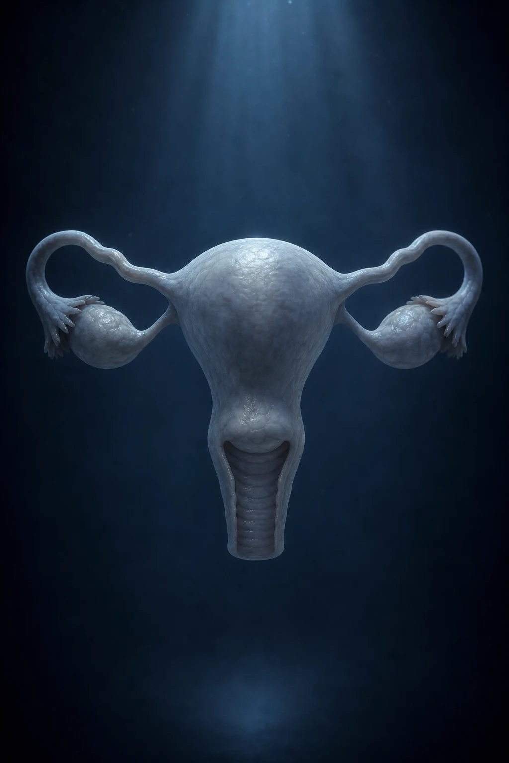
Amenorrhoea is classified anatomically by the compartment at fault, because the compartment determines the workup, the hormone pattern, and the treatment. Working from the top of the axis downwards, the four compartments are the hypothalamus/CNS, the pituitary, the ovary, and the uterus/outflow tract. Pregnancy sits outside this scheme and is excluded before any compartmental thinking begins.[1]
Hypothalamus / CNS
- Functional hypothalamic amenorrhoea: low energy availability, weight loss, exercise, stress, eating disorder
- Kallmann syndrome: isolated GnRH deficiency PLUS anosmia/hyposmia
- Constitutional delay of growth and puberty
- Chronic systemic illness, structural CNS lesion (craniopharyngioma), infiltrative disease
- Hormone signature: low/normal FSH and LH, LOW oestradiol — hypogonadotropic hypoestrogenism
Pituitary
- Hyperprolactinaemia: prolactinoma or drug-induced
- Sheehan syndrome: postpartum pituitary necrosis
- Other pituitary tumours; empty sella; Cushing disease; acromegaly
- Hormone signature: low/normal FSH and LH, LOW oestradiol — hypogonadotropic hypoestrogenism
Ovary
- Premature ovarian insufficiency (POI): raised FSH before age 40
- Turner syndrome (45,X): streak ovaries, short stature
- Swyer syndrome (46,XY gonadal dysgenesis)
- Iatrogenic: chemotherapy, pelvic radiotherapy, oophorectomy
- Hormone signature: HIGH FSH (and LH), LOW oestradiol — hypergonadotropic hypoestrogenism
Uterus / outflow
- Anatomical defect with NORMAL hormones
- Asherman syndrome: intrauterine adhesions after D&C
- Mullerian agenesis (MRKH): absent uterus and upper vagina
- Imperforate hymen, transverse vaginal septum: cyclic pain with retained menses
- Cervical stenosis
- Hormone signature: NORMAL FSH, LH, oestradiol; abnormal only the outflow
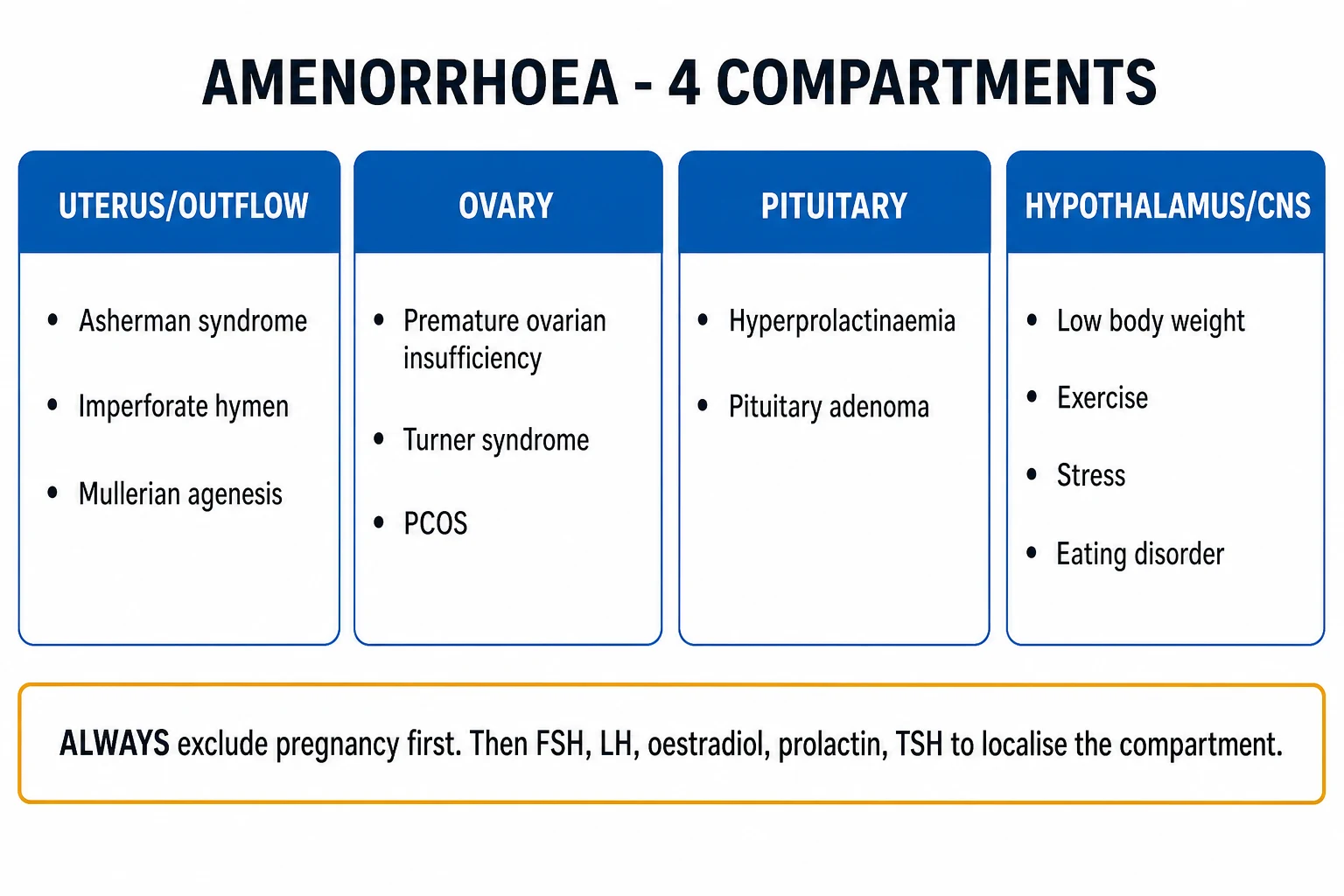
An alternative, equally valid classification splits causes by oestrogen status and gonadotrophin status: hypogonadotropic hypogonadism (hypothalamic or pituitary — low FSH/LH), hypergonadotropic hypogonadism (ovarian — high FSH), eugonadism with normal anatomy problems (PCOS — adequate oestrogen but chronic anovulation), and eugonadism with anatomical obstruction (Asherman, MRKH, imperforate hymen — normal hormones but mechanical defect). This hormonal framing is what the investigations section operationalises, and it is the framework examiners reward in a viva.[1]
Epidemiology & Risk Factors
Secondary amenorrhoea affects about 3 to 4 percent of reproductive-age women in the general population and is far commoner in clinical practice than primary amenorrhoea. In defined subgroups the prevalence rises sharply: up to half of competitive female athletes and a substantial fraction of dancers, gymnasts and military recruits have menstrual dysfunction, and amenorrhoea is essentially universal in anorexia nervosa at low body mass.[1][4]
- The commonest cause of secondary amenorrhoea is pregnancy — hence the universal rule to test first; pregnancy accounts for the majority of secondary cases in reproductive-age women.
- After pregnancy, the commonest cause is hypothalamic dysfunction (functional) — low energy availability, weight loss, exercise, stress — followed closely by polycystic ovary syndrome, then hyperprolactinaemia, premature ovarian insufficiency, thyroid disease and Asherman syndrome.
- Primary amenorrhoea is uncommon (a few percent of adolescents referred), and the yield of significant pathology is high: Turner syndrome (45,X) is the commonest chromosomal cause in a girl with short stature and absent puberty, while Mullerian agenesis (MRKH) and androgen insensitivity dominate the group with normal breast development but absent uterus.[2]
Amenorrhoea — key numbers
Risk factors and the compartment they favour: low body mass index, recent weight loss, strenuous exercise and eating disorders all push toward functional hypothalamic amenorrhoea. Obesity, hirsutism, acne and a family history of type 2 diabetes push toward PCOS. Galactorrhoea, antipsychotic or antiemetic drug use, and headache push toward hyperprolactinaemia. A recent pregnancy complicated by haemorrhage raises Sheehan syndrome. Prior dilation and curettage, postpartum or post-abortion instrumentation, or pelvic tuberculosis raise Asherman syndrome. Chemotherapy and pelvic radiotherapy raise iatrogenic ovarian failure. A family history of delayed puberty or anosmia raises Kallmann syndrome or constitutional delay. Short stature in a girl raises Turner syndrome.[1]
Pathophysiology
Normal cyclic menstruation requires three things working together: an intact HPO axis generating a regulated hormonal rhythm, a patent outflow tract, and a responsive endometrium that proliferates under oestrogen and bleeds on progesterone withdrawal. A break at any single point abolishes menses.[1]
- Hypothalamus — the arcuate (infundibular) nucleus releases gonadotrophin-releasing hormone (GnRH) in a pulsatile fashion, roughly every 60 to 90 minutes in the follicular phase. Pulse frequency governs the gonadotrophin mix: faster pulses favour luteinising hormone (LH), slower pulses favour follicle-stimulating hormone (FSH). GnRH pulsatility is exquisitely sensitive to energy availability, body fat, stress, weight change and systemic illness — this is the biological basis of functional hypothalamic amenorrhoea.
- Pituitary — GnRH drives the gonadotrophs to secrete FSH (recruits follicles, drives aromatisation of androgens to oestradiol) and LH (triggers ovulation and supports the corpus luteum). Prolactin, cortisol and thyroid hormones all modulate gonadotrophin secretion, which is why pituitary, adrenal and thyroid disease disturb the cycle.
- Ovary — granulosa cells produce oestradiol (proliferative phase); after ovulation the corpus luteum produces progesterone (secretory phase). A finite follicle pool is laid down in utero and declines; when it is depleted or dysfunctional, oestradiol falls, negative feedback on FSH is lost, and FSH rises — the mechanism of ovarian (hypergonadotropic) amenorrhoea.
- Uterus and outflow — oestradiol-driven endometrial proliferation followed by progesterone-supported maturation sets up a withdrawal bleed. If the endometrium is scarred (Asherman) or absent (MRKH), or the outflow is blocked (imperforate hymen, transverse septum, cervical stenosis), no bleed occurs despite normal hormones. [1]
A break at any level abolishes menses. The characteristic patterns, and the hormone signature each produces, are the core of the topic:[1][4]
- Hypothalamic — low energy availability, weight loss, stress or a structural lesion slows or abolishes pulsatile GnRH, lowering FSH and LH and hence oestradiol. The picture is hypogonadotropic and hypoestrogenic. Prolactin excess acts here too: prolactin inhibits GnRH pulse frequency, which is the mechanism of hyperprolactinaemic amenorrhoea (and the reason dopamine agonists restore cycles).
- Pituitary — direct destruction of gonadotrophs (Sheehan necrosis, tumour mass effect, infiltration) removes FSH/LH production; again low FSH/LH, low oestradiol.
- Ovarian — depletion or dysfunction of the follicle pool (POI, Turner, chemo/radiotherapy, surgery) removes oestradiol production and the negative feedback on FSH; the picture is hypergonadotropic and hypoestrogenic — high FSH, low oestradiol.
- Uterine/outflow — the HPO axis and hormones are entirely normal; the defect is mechanical (Asherman scarring, congenital absence, obstruction). The hormone panel is normal; the problem is revealed only by imaging or the challenge test. [1]
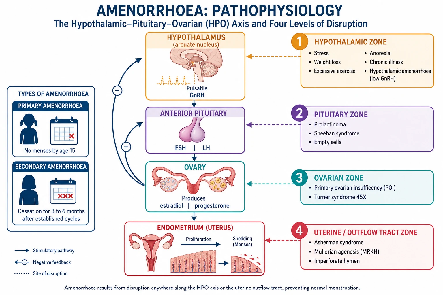
This compartment-and-signature logic is why a single hormone panel (FSH, LH, oestradiol, prolactin, TSH) localises almost every case: the height of FSH tells you whether the break is above or below the ovary, and the oestradiol level tells you whether the patient is hypoestrogenic (and therefore needs bone protection).[1]
Clinical Presentation
The presentation is dictated by the cause, so the history and examination are designed to localise the compartment rather than to confirm amenorrhoea itself. The absent periods are usually the presenting complaint; the rest of the interview hunts for pointers.[1]
History — the structured enquiry: establish pubertal timing and the prior cycle pattern (when did periods start, how regular, when did they stop); the onset and duration of amenorrhoea; weight change, diet, and exercise volume (hours per week, sport, recent intensification); psychological stress; drug history (antipsychotics, metoclopramide, domperidone, opiates, hormonal contraception, glucocorticoids, chemotherapy); galactorrhoea, headache, visual change (pituitary); hot flushes, night sweats, vaginal dryness, dyspareunia, low libido (hypoestrogenism); acne, hirsutism, voice deepening, central obesity (hyperandrogenism); cyclic pelvic pain (outflow obstruction); fertility wishes; and — for primary amenorrhoea — sense of smell (Kallmann), family history of delayed puberty, and parental growth trajectory. Always ask about a possibility of pregnancy, explicitly and without judgement. [1]
Symptom clusters that point to a compartment: [1]
- Hypoestrogenism (hypothalamic, pituitary, POI) — hot flushes, night sweats, vaginal dryness, dyspareunia, low libido, mood change, sleep disturbance. The patient describes menopausal-type symptoms out of keeping with her age.
- Hyperandrogenism (PCOS, congenital adrenal hyperplasia, androgen-secreting tumour) — hirsutism (Ferriman-Gallwey score over 4 to 6), acne, central obesity, acanthosis nigricans, male-pattern hair loss. Onset is usually peripubertal and slowly progressive.
- Hyperprolactinaemia — galactorrhoea, bilateral or unilateral; headache; bitemporal visual field loss from optic chiasm compression by a macroadenoma; reduced libido.
- Outflow obstruction — cyclic pelvic or lower abdominal pain in a girl who has never menstruated (cryptomenorrhoea — retained menses behind an imperforate hymen or transverse septum); a pelvic mass (haematocolpos/haematometra). Crucially, MRKH causes NO cyclic pain because there is no uterus to generate menses.
- Systemic clues — fatigue, cold intolerance (hypothyroidism); heat intolerance, palpitations (hyperthyroidism); weight gain, striae, bruising (Cushing); polyuria, polydipsia (diabetes insipididus from a pituitary lesion); failure to lactate postpartum (Sheehan).[1]
Phenotypes of primary amenorrhoea (exam favourites):[2]
- Turner syndrome (45,X) — short stature, webbed neck, shield chest with widely spaced nipples, low posterior hairline, cubitus valgus, lymphoedema; cardiac anomalies (coarctation of the aorta, bicuspid aortic valve) and renal anomalies (horseshoe kidney); streak ovaries with absent puberty and primary amenorrhoea.
- Kallmann syndrome — isolated GnRH deficiency with anosmia or hyposmia (failure of olfactory and GnRH neurons to migrate from the olfactory placode); primary amenorrhoea with absent or arrested puberty; normal female karyotype and normal pelvic anatomy; sometimes midline defects (cleft lip/palate) or unilateral renal agenesis.
- Mullerian agenesis (MRKH) — normal breasts, normal pubic and axillary hair, normal female karyotype (46,XX) and normal hormones; absent or rudimentary uterus and upper vagina; no cyclic pain; associated renal and skeletal anomalies in a minority.
- Complete androgen insensitivity (CAIS) — normal breast development (from aromatised testosterone), absent or scant pubic and axillary hair, no uterus (Mullerian structures regress due to testicular anti-Mullerian hormone), blind-ending vagina; karyotype 46,XY with male-range testosterone; presentation is primary amenorrhoea in an otherwise phenotypically female adolescent.
- Imperforate hymen / transverse vaginal septum — cyclic pelvic pain in a girl with normal secondary sex characteristics and no menarche; a bulging bluish membrane on inspection (imperforate hymen); a pelvic mass from retained menses. [1]
Atypical and red-flag presentations that examiners test deliberately: the athlete with recurrent stress fractures or stress-related bone injury (female athlete triad / Relative Energy Deficiency in Sport, RED-S); the postpartum woman who fails to lactate, becomes hypothyroid and hypoglycaemic (Sheehan syndrome — a hypopituitary emergency); the severely underweight patient with bradycardia, hypothermia and electrolyte derangement (anorexia nervosa with refeeding risk — never start hormones before nutritional rehabilitation); and the adolescent with primary amenorrhoea and hypertension (Turner with coarctation).[4][8]
Differential Diagnosis
The differential is compartment-driven. The skill is to rank by frequency for secondary amenorrhoea and to group by anatomy for primary amenorrhoea, and then to distinguish with the hormone panel.[1]
Secondary amenorrhoea — ranked by frequency (always exclude pregnancy first): [1]
- Pregnancy — always first; the cardinal rule.
- Functional (hypothalamic) amenorrhoea — low energy availability, weight loss, exercise, stress, eating disorder, chronic illness.
- Polycystic ovary syndrome — hyperandrogenic chronic anovulation with adequate oestrogen.
- Hyperprolactinaemia — prolactinoma or drug-induced.
- Premature ovarian insufficiency — raised FSH before age 40.
- Thyroid disease — both hypo- and hyperthyroidism.
- Asherman syndrome — intrauterine adhesions, post-instrumentation.
- Iatrogenic — chemotherapy, pelvic radiotherapy, hormonal contraception (post-pill amenorrhoea is a diagnosis of exclusion).
- Adrenal — non-classic congenital adrenal hyperplasia, Cushing disease, Addison disease. [1]
Primary amenorrhoea — grouped by uterus and secondary sex characteristics:[2]
| Uterus present? | Secondary sex characteristics? | Hormone pattern | Think of |
|---|---|---|---|
| Present | Present | Normal FSH/LH, normal oestradiol | Imperforate hymen, transverse vaginal septum, cervical stenosis (outflow obstruction — cyclic pain) |
| Present | Absent | Low FSH/LH | Constitutional delay; Kallmann (anosmia); hypothalamic/pituitary causes |
| Present | Absent | High FSH | Turner (45,X); 46,XX gonadal dysgenesis; POI |
| Absent | Present | Normal or male-range testosterone | MRKH (46,XX, normal T); androgen insensitivity (46,XY, male-range T, scant pubic hair) |
| Absent | Absent | High FSH | 46,XY gonadal dysgenesis (Swyer); Turner with Mullerian absence |
Key distinctions examiners probe: [1]
- MRKH versus androgen insensitivity — both have breast development and an absent uterus, but MRKH is 46,XX with normal female testosterone and normal pubic hair, whereas complete androgen insensitivity is 46,XY with male-range testosterone and absent or scant pubic hair. The karyotype and testosterone level settle it.
- Imperforate hymen versus MRKH — imperforate hymen has a uterus and therefore cyclic pain (menses retained behind the membrane); MRKH has no uterus and no cyclic pain. Pelvic inspection and ultrasound distinguish them.
- PCOS versus functional hypothalamic amenorrhoea — PCOS has adequate oestradiol with a raised LH:FSH ratio (over 2 to 3) and clinical or biochemical hyperandrogenism; functional hypothalamic has low oestradiol with low/normal gonadotrophins and is often underweight. They are near-opposites hormonally.
- POI versus functional hypothalamic — both are hypoestrogenic, but POI has a high FSH (ovarian) while functional hypothalamic has a low FSH (central). The FSH level is the single most discriminating second-line test after a negative pregnancy test.
- PCOS versus non-classic congenital adrenal hyperplasia — both present with hirsutism and anovulation; 17-hydroxyprogesterone is elevated in NCCAH (21-hydroxylase deficiency), normal in PCOS.[1][5]
Clinical & Bedside Assessment
The focused examination is built to find the physical stigmata of each compartment and to screen for pregnancy and its complications.[1]
General and growth: height, weight and body mass index against growth centiles (low BMI points to functional hypothalamic; high BMI to PCOS, Cushing); Tanner staging of breasts and pubic hair (hypoplastic in hypoestrogenism; well developed in MRKH and androgen insensitivity). [1]
Stigmata of Turner: short stature, webbed neck, shield chest with widely spaced nipples, low posterior hairline, cubitus valgus, lymphoedema; check the blood pressure in all four limbs (coarctation) and listen for murmurs. [1]
Pelvic inspection and examination: inspect the vulva and hymen (imperforate hymen is a bluish bulging membrane; assess vaginal length — a blind-ending short vagina suggests MRKH or androgen insensitivity); look for clitoromegaly (androgen excess); palpate for an abdominal or pelvic mass (haematocolpos, pregnancy); note pubic hair distribution (absent in androgen insensitivity). [1]
Endocrine screen: squeeze the nipples for galactorrhoea; test visual fields by confrontation for a bitemporal hemianopia (optic chiasm compression by a pituitary macroadenoma); examine the thyroid for goitre; score acne and hirsutism with the modified Ferriman-Gallwey score (over 4 to 6 in a Caucasian or over 2 to 3 in an Asian population suggests hyperandrogenism); look for acanthosis nigricans (insulin resistance in PCOS). [1]
Screen for systemic and psychiatric disease: signs of anorexia nervosa (lanugo, bradycardia, hypothermia, low BMI, eroded dental enamel if purging), Cushing (central obesity, striae, bruising, proximal myopathy), thyroid disease, diabetes, and chronic systemic illness. [1]
Why pregnancy testing is mandatory and first: beta-hCG is cheap, instant, and reshapes the entire workup. A positive result ends the diagnostic hunt and initiates antenatal care; any subsequent hormone interpretation or pelvic imaging is meaningless — indeed misleading — until pregnancy is excluded, because pregnancy hormones suppress FSH and LH and the endometrium decidualises. This is the single highest-yield step in the whole topic.[1]
Investigations
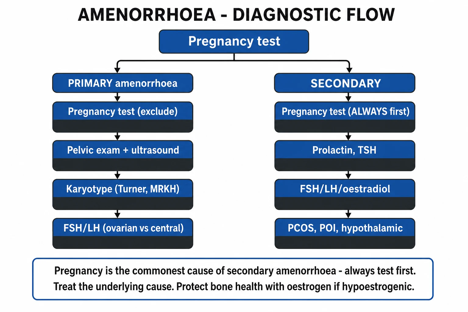
The workup is algorithmic and sequential. It moves from the universal first test (pregnancy) to the hormone panel that localises the compartment, then to dynamic tests and targeted imaging/genetics.[1]
Step 1 — pregnancy test (serum or urine beta-hCG) in EVERY patient, regardless of reported sexual activity and contraception. If positive, stop the amenorrhoea workup and arrange antenatal care (or, if ectopic is suspected, urgent ultrasound). [1]
Step 2 — the hormone panel: FSH, LH, oestradiol, prolactin and TSH, with total testosterone and sex-hormone-binding globulin (to calculate the free androgen index) added if there are signs of hyperandrogenism. Pattern interpretation is the heart of the workup:[1]
| FSH / LH | Oestradiol | Prolactin / TSH | Localisation and prototype cause |
|---|---|---|---|
| Low / low-normal | Low | Normal | Hypothalamic (functional, Kallmann, constitutional delay) or pituitary (Sheehan, tumour) — hypogonadotropic hypogonadism |
| High (FSH over 25 IU/L) | Low | Normal | Ovarian — POI, Turner, gonadal dysgenesis, iatrogenic — hypergonadotropic hypogonadism |
| Normal, raised LH:FSH ratio (over 2 to 3) | Normal or high | Normal | PCOS (with hyperandrogenism) — adequate oestrogen, chronic anovulation |
| Normal | Normal | Prolactin raised | Hyperprolactinaemia — prolactinoma or drug-induced (then MRI pituitary) |
| Normal | Normal | TSH abnormal | Thyroid disease — hypo- or hyperthyroidism |
| Normal | Normal | Normal, all hormones normal | Uterine/outflow — Asherman, MRKH, imperforate hymen (anatomy problem, normal axis) |
Step 3 — dynamic (challenge) tests when the panel is ambiguous or to confirm a mechanical cause:[1]
- Progestogen challenge test — medroxyprogesterone acetate 10 mg orally daily for 5 to 10 days (alternatives: norethisterone 5 mg daily, micronised progesterone 200 to 300 mg nightly). A withdrawal bleed within 2 to 7 days of stopping means anovulation with adequate endogenous oestrogen and a patent outflow tract (e.g. PCOS). No bleed means either hypoestrogenism (insufficient oestrogen to proliferate the endometrium) or outflow obstruction — the two are then separated by the combined test.
- Combined oestrogen-progestogen challenge — give oestrogen (e.g. conjugated oestrogens or oestradiol for 21 days) then add a progestogen. A bleed after this means the endometrium responds and the outflow is patent, so the problem was hypoestrogenism (hypothalamic/pituitary/ovarian). Still no bleed means outflow obstruction (Asherman, cervical stenosis). This test confirms Asherman when the hormone panel is normal.
- Repeating the FSH: POI is defined by a raised FSH on TWO samples at least one month apart — a single high value is not enough, because spontaneous fluctuation and even ovulation can occur.[3]
Step 4 — targeted imaging and genetic tests: pelvic ultrasound (uterus and ovaries present or absent, polycystic ovarian morphology, retained menses, haematocolpos); karyotype if primary amenorrhoea or POI under age 30 (Turner, gonadal dysgenesis, androgen insensitivity) plus fragile-X (FMR1) premutation screen in POI; pituitary MRI if prolactin is raised (to distinguish micro- from macroadenoma and to look for mass effect) or hypopituitarism is suspected; DEXA scan for bone mineral density in any hypoestrogenic amenorrhoea lasting over 6 to 12 months; hysteroscopy or saline-infusion sonohysterography (gold standard for Asherman syndrome); and 17-hydroxyprogesterone to exclude non-classic congenital adrenal hyperplasia in hyperandrogenic cases.[1]
Step 5 — additional endocrine tests as the picture demands: 17-hydroxyprogesterone (NCCAH), DHEA-S and androstenedione (adrenal vs ovarian androgen source — a very high DHEA-S or rapid virilisation raises an androgen-secreting tumour), ACTH/cortisol or overnight dexamethasone suppression (Cushing), IGF-1 (acromegaly), and a full pituitary panel (cortisol, free T4, IGF-1) if hypopituitarism is suspected (e.g. Sheehan).[7]
Named criteria — reproduced verbatim
Rotterdam PCOS diagnostic criteria (2003/2004) — diagnose PCOS if 2 of the following 3 are present, after excluding other causes of androgen excess or anovulation (congenital adrenal hyperplasia, androgen-secreting tumours, Cushing syndrome):[5]
- Oligo-ovulation or anovulation (oligomenorrhoea or amenorrhoea).
- Clinical and/or biochemical signs of hyperandrogenism (hirsutism, acne, raised total or free testosterone).
- Polycystic ovaries on ultrasound (the 2018 International Guideline updated morphology to 20 or more follicles per ovary and/or an ovarian volume over 10 mL, using a transducer at 8 MHz or higher). [1]
Premature ovarian insufficiency (POI) — amenorrhoea (or oligomenorrhoea) with a serum FSH over 25 IU/L on two samples taken at least one month apart, before age 40 (ESHRE 2016). The two-sample rule prevents overdiagnosis from transient fluctuation; spontaneous ovulation and even pregnancy can still occur in up to a quarter of women, so POI is not equivalent to sterility.[3]
Management — Resuscitation & Urgent Referral
Most amenorrhoea is investigated electively in the outpatient clinic, but several presentations need urgent specialist referral and a few are true emergencies:[1][7]
- Pituitary macroadenoma with visual field compromise (bitemporal hemianopia) — urgent endocrinology and neurosurgical referral. A dopamine agonist (cabergoline) is first-line even for macroprolactinomas causing chiasm compression and typically shrinks the tumour and relieves pressure within days to weeks; surgery (transsphenoidal) is reserved for medical failure, intolerance, or a non-prolactin-secreting mass lesion.
- Severe anorexia nervosa with bradycardia, hypothermia or electrolyte derangement — medical admission, careful nutritional rehabilitation with refeeding monitoring (phosphate, potassium, magnesium), and never start oestrogen replacement before weight restoration.
- Haematocolpos / haematometra from imperforate hymen or transverse septum with severe pain — urgent surgical decompression to prevent retrograde flow, endometriosis and infection.
- Confirmed pregnancy — the priority shifts entirely to antenatal care (or ruling out ectopic if symptomatic). Amenorrhoea is no longer the diagnosis.
- Suspected Sheehan syndrome (postpartum failure to lactate, hypotension, hypoglycaemia, hypothyroid) — a hypopituitary emergency: urgent endocrinology, stress-dose glucocorticoids, and full pituitary axis replacement.[8]
Bone-density protection is time-critical, not elective, in any hypoestrogenic state. Bone loss is rapid in the first 6 to 12 months of amenorrhoea and is only partially reversible. Order a DEXA scan for amenorrhoea with low oestradiol lasting over 6 to 12 months, and address the underlying cause plus give oestrogen replacement.[3][4]
Management — Definitive & Stepwise
Treatment is cause-directed. The principles below map onto the four compartments, and the spine of any exam answer is "name the cause, then give its specific treatment".[1][4]
Functional (hypothalamic) amenorrhoea — Endocrine Society 2017:[4]
- First-line is energy restoration — increase caloric intake, reduce training volume, aim for weight gain (a 2 to 3 kg gain often restores cycles in a previously underweight woman), and treat any eating disorder with multidisciplinary care and cognitive behavioural therapy (CBT has RCT evidence for cycle resumption).
- Hormone therapy if amenorrhoea persists despite 8 to 12 months of lifestyle change — transdermal oestradiol (50 to 100 microgram patch) plus cyclic oral progestogen (e.g. micronised progesterone 200 mg nightly for 12 days each month) to protect bone. The Endocrine Society explicitly states the combined oral contraceptive pill does NOT restore bone mineral density in functional hypothalamic amenorrhoea and should not be relied on for this purpose.
- No routine role for leptin, bisphosphonates, or teriparatide; they are investigational or for exceptional cases. The cycle usually returns with energy restoration; fertility, if needed, requires the cycles to return or gonadotrophin/GnRH therapy under specialist supervision.[4]
Polycystic ovary syndrome: lifestyle (a 5 to 10 percent weight loss improves all features — cycle regularity, hyperandrogenism, insulin resistance, fertility); combined oral contraceptive pill for cycle control, endometrial protection and hyperandrogenism; metformin (typically 1500 to 2000 mg daily in divided doses) for metabolic indications and cycle regulation; letrozole (first-line) or clomiphene for ovulation induction when fertility is desired; gonadotrophins and IVF for resistant disease. Endometrial protection with a progestogen is essential in any woman with chronic anovulation to prevent endometrial hyperplasia and cancer.[5][6]
Hyperprolactinaemia / prolactinoma: withdraw offending drugs where possible; treat a prolactinoma with a dopamine agonist — cabergoline first-line (e.g. 0.25 to 1 mg once or twice weekly, titrated to a normal prolactin), which is more efficacious and better tolerated than bromocriptine and typically shrinks macroadenomas; reserve transsphenoidal surgery for medical failure, drug intolerance, or a non-prolactin-secreting mass lesion (e.g. craniopharyngioma, non-functioning adenoma); radiotherapy is third-line. Monitor prolactin and visual fields and re-image to confirm shrinkage.[1][7]
Premature ovarian insufficiency (ESHRE 2016): combined HRT (oestradiol plus a progestogen) until the average age of natural menopause (about 51) for symptom relief, bone and cardiovascular protection; the COCP is not the optimal HRT formulation (higher thrombotic risk, less physiological dosing than transdermal oestradiol). Counsel that spontaneous ovulation may resume in up to a quarter — so POI is not a sterility diagnosis; oocyte donation IVF is the principal fertility route for women who want to conceive; screen for associated autoimmune disease (thyroid, adrenal) and the fragile-X (FMR1) premutation.[3]
Asherman syndrome: hysteroscopic adhesiolysis to cut the intrauterine adhesions, followed by a course of oestrogen therapy (e.g. oestradiol for several weeks) and a cycle of progestogen to regenerate the endometrium, often with an intrauterine device or Foley balloon stent left in situ to prevent re-adhesion. Prevent recurrence by avoiding unnecessary uterine instrumentation. Antibiotic cover may be used peri-procedurally.[1]
Mullerian agenesis (MRKH): counselling first — normal chromosomes, normal female identity and hormones, absent uterus (infertility, but genetic motherhood is possible via oocyte retrieval and gestational surrogacy); vaginal dilation (first-line) or surgery (vaginoplasty) for sexual function; psychological support is central given the impact on identity, body image and fertility.[2]
Imperforate hymen / transverse vaginal septum: surgical cruciate incision (hymen) or excision and reconstruction (septum) to relieve obstruction and prevent retrograde menstruation, endometriosis and infection; ideally performed before extensive haematocolpos develops.[2]
Kallmann syndrome / isolated hypogonadotropic hypogonadism: oestrogen replacement to induce puberty and protect bone, then combined HRT for long-term replacement. For fertility, pulsatile GnRH (via a pump) or gonadotrophins (FSH/LH) induce ovulation, with good success; the underlying defect is hypothalamic, so the ovary is intact.[2]
Turner syndrome: recombinant growth hormone in childhood (typically from early childhood) to optimise final adult height; oestrogen replacement initiated at an appropriate age (around 11 to 12, gradually increasing) to induce puberty and protect bone; lifelong cardiac surveillance (echocardiography/MRI for aortic root dilation and coarctation), renal and auditory screening; oocyte donation for fertility.[2]
Swyer syndrome (46,XY gonadal dysgenesis): gonadectomy (the streak gonads carry a high risk of malignant transformation — gonadoblastoma/dysgerminoma), then oestrogen replacement to induce puberty and maintain bone, with oocyte donation for fertility.[2]
Specific Subtypes & Scenarios
- Functional hypothalamic amenorrhoea / female athlete triad / RED-S — the classic triad is low energy availability (with or without disordered eating), menstrual dysfunction, and low bone mineral density; RED-S broadens this to the multisystem consequences of relative energy deficiency (metabolic, cardiovascular, immune, gastrointestinal). Seen in runners, dancers, gymnasts, figure skaters, rowers and military recruits. Restore energy first; the cycle and bone recover only when energy balance is positive. Anticipate the female athlete triad in any sportswoman with stress fractures.[4]
- PCOS — defined by Rotterdam criteria; carries metabolic syndrome (insulin resistance, type 2 diabetes, dyslipidaemia), endometrial hyperplasia/cancer risk from unopposed oestrogen, infertility, sleep apnoea and mood disorders. Management is lifelong and multidisciplinary. Letrozole is first-line for ovulation induction (PPCOS II — higher live birth than clomiphene).[5][6]
- Premature ovarian insufficiency (POI) — autoimmune (often with thyroid/adrenal autoimmunity), Turner mosaic, fragile-X premutation, iatrogenic (chemo/radiotherapy/surgery), idiopathic. HRT until 51, fertility via oocyte donation, and surveillance for associated autoimmune disease and osteoporosis. Not a sterility diagnosis — spontaneous ovulation recurs in a minority.[3]
- Hyperprolactinaemia / prolactinoma — distinguish microadenoma (under 10 mm) from macroadenoma (10 mm or over); macroadenomas threaten the optic chiasm. Macroprolactinaemia (biologically inactive prolactin complexes) is a benign cause of an isolated high prolactin with no symptoms — exclude it with PEG precipitation if the picture does not fit. Drug causes (antipsychotics, metoclopramide, domperidone, opiates, methyldopa, verapamil) must be excluded.[7]
- Primary amenorrhoea with absent uterus — MRKH (46,XX, normal T, normal pubic hair) versus androgen insensitivity (46,XY, male-range T, scant pubic hair) versus 46,XY gonadal dysgenesis (Swyer — absent puberty, high FSH, gonadal tumour risk). Karyotype, testosterone and pelvic imaging settle it; a 46,XY karyotype mandates gonadectomy (Swyer) or counselling (CAIS).[2]
- Asherman syndrome — intrauterine adhesions after dilation and curettage (especially postpartum or post-abortion), endometrial tuberculosis, or pelvic irradiation. Presents with secondary amenorrhoea or hypomenorrhoea, sometimes cyclic pain and infertility. Hysteroscopic adhesiolysis is the treatment; prevention by avoiding unnecessary uterine instrumentation is key.[1]
- Sheehan syndrome — postpartum pituitary necrosis from hypovolaemic shock at delivery; presents with failure to lactate (the classic early sign), persistent amenorrhoea, hypothyroidism, adrenal insufficiency and hypoglycaemia. A hypopituitary emergency needing stress-dose glucocorticoids then lifelong full pituitary replacement. Now uncommon where obstetric care is good, but still seen in resource-limited settings.[8]
- Kallmann syndrome and constitutional delay — both cause hypogonadotropic hypogonadism with delayed/absent puberty. Kallmann adds anosmia and is permanent (needs HRT, then GnRH/gonadotrophins for fertility); constitutional delay is a normal variant (eventual spontaneous puberty) and is a diagnosis of exclusion, supported by a bone age lag and a family history of late bloomers.[2]
Complications & Pitfalls
Long-term complications of untreated hypoestrogenic amenorrhoea (functional hypothalamic, POI, pituitary disease):[3][4]
- Osteoporosis and fragility fracture — bone loss is rapid early and may be only partially reversible; this is the principal and most clinically important long-term harm. Stress fractures in athletes, vertebral fractures in older POI.
- Cardiovascular disease — premature loss of ovarian oestrogen raises cardiovascular risk; HRT until 51 in POI mitigates this.
- Urogenital atrophy — vaginal dryness, dyspareunia, recurrent urinary tract infections, psychosexual dysfunction.
- Infertility and subfertility — central to the patient's concern; management is cause-specific.
- Psychological — body image distress, anxiety, depression, identity issues (especially in MRKH, Turner, androgen insensitivity). [1]
Complications of PCOS: endometrial hyperplasia and endometrial cancer (unopposed oestrogen — the most important preventable cancer risk), type 2 diabetes and metabolic syndrome, dyslipidaemia, non-alcoholic fatty liver disease, obstructive sleep apnoea, infertility, and mood disorders. Long-term metabolic surveillance is part of management.[5]
Classic pitfalls examiners trap:[1]
- Failing to exclude pregnancy first — the cardinal error; never interpret hormones or image the pelvis before a beta-hCG.
- Mislabelling PCOS without excluding other causes of hyperandrogenism (NCCAH, Cushing, androgen-secreting tumour) — Rotterdam requires exclusion first.
- Treating functional hypothalamic amenorrhoea with the COCP instead of energy restoration — the COCP does not restore bone density, masks the underlying energy deficit, and delays recovery.
- Using the COCP as HRT for bone protection in POI or FHA — it is not the optimal formulation; use transdermal oestradiol plus progestogen.
- Forgetting endometrial protection in chronic anovulation (PCOS) — unopposed oestrogen drives endometrial hyperplasia and cancer.
- Missing a prolactinoma behind a nonspecific "hormone imbalance" — always check prolactin and examine visual fields.
- Missing Sheehan syndrome in a postpartum woman with fatigue and failure to lactate.
- Overdiagnosing POI on a single FSH — the definition requires two raised samples a month apart.
- Confusing MRKH with androgen insensitivity — both have breasts and no uterus, but the karyotype, testosterone and pubic hair differ fundamentally.[2]
Prognosis & Disposition
Prognosis tracks the cause and the speed of correction:[1][4]
- Functional hypothalamic amenorrhoea — often resolves with energy restoration and weight gain (a 2 to 3 kg gain frequently restores cycles), but established bone loss may recover only partially; a minority become chronic. Early lifestyle intervention has the best outcome.
- PCOS — chronic but manageable; cycles and metabolic features improve with weight loss and the COCP; fertility is usually achievable with letrozole or ART. Lifelong metabolic and endometrial surveillance is needed.
- Premature ovarian insufficiency — irreversible ovarian failure but treated with HRT until 51; bone and cardiovascular outcomes normalise with timely HRT; fertility via oocyte donation IVF. Spontaneous ovulation recurs in up to a quarter, so POI is not contraception.
- Hyperprolactinaemia / prolactinoma — medical therapy with cabergoline is highly effective; microadenomas may not grow and macroadenomas usually shrink; withdrawal of dopamine agonist is sometimes possible after prolonged normoprolactinaemia.
- Asherman — prognosis depends on severity; mild adhesiolysis restores menses and fertility in most, severe disease recurs and reduces fertility.
- MRKH / Turner / Kallmann / androgen insensitivity — chronic conditions requiring lifelong care; fertility options (surrogacy, oocyte donation, gonadotrophins) and comorbidity surveillance (cardiac in Turner, gonadal tumour risk in 46,XY) dominate long-term management. [1]
Referral: endocrinology (POI, functional hypothalamic, prolactinoma, hypopituitarism, Cushing); gynaecology / reproductive medicine (PCOS with fertility needs, Asherman, MRKH, fertility preservation); genetics (Turner, karyotype abnormalities, fragile-X); eating-disorder or psychiatric services (anorexia nervosa — multidisciplinary, never hormones before nutrition); neurosurgery (macroadenoma refractory to medical therapy, non-prolactin pituitary mass); urology/nephrology (Turner renal anomalies); and cardiology (Turner aortic surveillance). [1]
Special Populations
- Adolescents with primary amenorrhoea — assess puberty stage precisely (Tanner), send karyotype and FSH/LH, image the pelvic anatomy, and offer psychological support; do not dismiss as a "late bloomer" once the age-15 (or age-13-without-thelarche) threshold is met. Constitutional delay is a diagnosis of exclusion and is supported by lagging bone age and a family history.[2]
- Athletes / dancers / military recruits — the female athlete triad (low energy availability with or without disordered eating, menstrual dysfunction, low bone mineral density) and RED-S (broader multisystem energy-deficiency consequences). Treat the energy deficit; restore menses through nutrition, not hormones.[4]
- Eating-disorder patients (anorexia nervosa) — multidisciplinary care; refeeding risk (phosphate, potassium, magnesium monitoring); bradycardia and electrolyte derangement may need admission; never start HRT before nutritional rehabilitation, because oestrogen does not restore bone while the patient remains energy-deficient.
- Postpartum women — Sheehan syndrome (failure to lactate, hypothyroid, hypoglycaemic, hypotensive postpartum) is a pituitary emergency needing stress-dose steroids and full replacement; pregnancy-related amenorrhoea is normal during lactation (lactational amenorrhoea method) but not indefinitely.[8]
- Women seeking fertility — ovulation induction (letrozole first-line for PCOS), ART/gonadotrophins for resistant disease, oocyte donation IVF for POI and Turner, gestational surrogacy for MRKH (genetic motherhood with the patient's oocytes and a surrogate uterus), and pulsatile GnRH or gonadotrophins for Kallmann/hypogonadotropic hypogonadism.
- Cancer survivors (post-chemo/radiotherapy) — iatrogenic POI; counsel on fertility preservation (oocyte or embryo cryopreservation) before gonadotoxic therapy where possible; HRT until 51 thereafter.
Evidence, Guidelines & Regional Differences
PPCOS II — Legro 2014 (NEJM)
Key finding
In 750 women with PCOS, letrozole gave a significantly higher live-birth rate (27.5 percent) than clomiphene (19.1 percent), establishing letrozole as first-line oral ovulation induction in PCOS.
Endocrine Society 2017 — Functional Hypothalamic Amenorrhea:[4] energy restoration is first-line; CBT has RCT evidence; HRT (transdermal oestradiol plus cyclic progestogen) if amenorrhoea persists beyond 8 to 12 months of lifestyle change; the COCP does NOT restore bone mineral density and should not be relied on for bone protection; no routine role for leptin, bisphosphonates, or teriparatide.
ESHRE 2016 — Premature Ovarian Insufficiency:[3] HRT is recommended until the age of natural menopause (about 51) for symptom relief and bone/cardiovascular protection; the COCP is not the optimal HRT formulation; oocyte donation is the principal fertility route; associated autoimmune disease and the fragile-X premutation should be screened for; spontaneous ovulation may resume.
Rotterdam 2003/2004 — PCOS diagnostic criteria:[5] two of three (oligo/anovulation, hyperandrogenism, polycystic ovaries on ultrasound) after exclusion of other causes — broader than the NIH 1992 criteria (hyperandrogenism plus oligo-ovulation) and the AE-PCOS Society 2006 criteria (hyperandrogenism plus ovarian dysfunction). The 2018 International Evidence-based PCOS Guideline (Misso/Teede) updated ultrasound morphology to 20 or more follicles per ovary or raised ovarian volume, using high-frequency (8 MHz or higher) transducers, and reorganised the diagnostic algorithm.
Prolactinoma management:[7] a 2023 JAMA review confirms cabergoline as first-line for both micro- and macroprolactinomas, with transsphenoidal surgery reserved for medical failure or non-prolactin lesions; dopamine agonists shrink macroadenomas and relieve chiasm compression, often within weeks.
Regional deltas: [1]
- India (FOGSI): PCOS prevalence is high (around 10 percent regionally, higher in some studies) and metabolic management (weight, metformin, glucose tolerance testing) is emphasised; insulin resistance and progression to type 2 diabetes are major concerns given the South Asian metabolic phenotype.
- Tuberculosis-endemic regions (parts of India, South Asia, Africa): genital tuberculosis is an important and under-recognised cause of both Asherman-type intrauterine adhesions and tubal/ovarian damage, and contributes more to secondary amenorrhoea and infertility than autoimmune POI in these populations. Endometrial biopsy with TB-PCR and cultures should be considered when the setting fits.
- Europe (ESHRE): HRT rather than the COCP for POI and FHA is strongly preferred; transdermal oestradiol is the default.
- US (Endocrine Society / AAFP): energy restoration first for FHA; letrozole first-line for PCOS fertility per the PPCOS II data.
- UK (NICE/RCOG): similar PCOS and POI frameworks; emphasises endometrial protection and fertility pathways.
- Access differences: Turner and congenital causes dominate primary amenorrhoea referrals everywhere, but access to growth hormone therapy, genetics, cardiac surgery and fertility services varies markedly by region — relevant to prognosis and counselling.[2]
Exam Pearls
Amenorrhoea — the COMPARTMENTS mnemonic
UP-OH
anatomical defect with NORMAL hormones — Asherman (post-D&C adhesions), imperforate hymen, Mullerian agenesis (MRKH)
hyperprolactinaemia (prolactinoma), Sheehan (postpartum necrosis) — low/normal FSH and LH with low oestradiol
premature ovarian insufficiency (raised FSH before 40), Turner (45,X, streak ovaries), iatrogenic chemo/radiotherapy
functional (low energy, exercise, stress), Kallmann (anosmia), chronic illness — low GnRH, low FSH and LH
- ALWAYS exclude pregnancy first — the single most-tested rule in amenorrhoea; never interpret hormones or image the pelvis before a beta-hCG.
- Primary amenorrhoea algorithm — is the uterus present? Are secondary sex characteristics present? The answers branch the differential; then karyotype and FSH/LH settle it.
- Short stature plus primary amenorrhoea equals Turner (45,X) until proven otherwise — check the karyotype, the heart (coarctation, bicuspid valve) and the kidneys.
- Anosmia plus primary amenorrhoea equals Kallmann syndrome — isolated GnRH deficiency; normal karyotype, normal pelvic anatomy.
- Normal breasts, absent uterus, 46,XX, normal testosterone equals MRKH; breasts, absent/scant pubic hair, no uterus, 46,XY, male-range testosterone equals androgen insensitivity.
- Postpartum amenorrhoea plus failure to lactate equals Sheehan syndrome — a hypopituitary emergency.
- Raised FSH before age 40 equals POI — define it on two samples at least a month apart; treat with combined HRT until 51.
- The COCP does NOT protect bone density in functional hypothalamic amenorrhoea — energy restoration does; use transdermal oestradiol plus progestogen if HRT is needed.
- Cyclic pelvic pain plus amenorrhoea equals obstructed outflow (imperforate hymen, transverse septum); MRKH has NO cyclic pain because there is no uterus.
- Cabergoline is first-line dopamine agonist for hyperprolactinaemia — more efficacious and better tolerated than bromocriptine.
- Letrozole is first-line ovulation induction in PCOS (PPCOS II, Legro 2014) — better live birth than clomiphene.
- Progestogen challenge: bleed equals adequate oestrogen plus patent outflow (PCOS); no bleed equals hypoestrogenism or obstruction; the combined test separates them. [1]
Exam application bank (NEET-PG / INICET)
One-line answer
Amenorrhoea is the absence of menstruation — a symptom, never a diagnosis. Primary amenorrhoea (no menses by age 15 with secondary sex characteristics, or by age 13 with no thelarche) is caused by constitutional delay, Turner syndrome, Kallmann syndrome, Mullerian agenesis (MRKH), androgen insensitivity and outflow obstruction. Secondary amenorrhoea (cessation for 3 or more months) is caused by pregnancy (always exclude first), functional hypothalamic (low BMI, exercise, stress, eating disorder), polycystic ovary syndrome, hyperprolactinaemia, premature ovarian insufficiency, thyroid disease and Asherman syndrome. Evaluation uses the four compartments — uterus/outflow, ovary, pituitary, hypothalamus — guided by a pregnancy test first, then FSH, LH, oestradiol, prolactin, TSH and testosterone. Management is cause-directed; oestrogen replacement protects bone in any hypoestrogenic state.
Worked stems (answer without another resource)
Stem 1 — Classic presentation. Map symptoms to mechanism; name the first investigation and first treatment step with dose/route if drug therapy is standard. [1]
Stem 2 — Unstable / complicated. List red flags that force immediate resuscitation, theatre, ICU, antidote, or reperfusion — and what you do in the first 15 minutes. [1]
Stem 3 — Atypical group. Elderly, pregnancy, child, or immunocompromised: how presentation and thresholds change. [1]
Stem 4 — Differential trap. Name the three closest mimics and one discriminator for each. [1]
Stem 5 — Disposition. Who goes home with safety-netting, who is admitted, who needs HDU/ICU/theatre, and what follow-up is mandatory. [1]
Rapid viva checklist
- Definition + classification
- Pathophysiology chain
- Bedside signs / criteria
- Score with exact components (if any)
- Emergency bundle
- Definitive therapy with doses
- Complications of disease and of treatment
- Special populations
- Guideline/trial name if classic
- Three exam traps
Coverage self-check
If you cannot answer any stem above from this page alone, re-read the matching section — the page is intended to be self-sufficient for final-prof and NEET-PG/INICET questions on Amenorrhoea (Primary & Secondary).
References
- [1]Klein DA, Paradise SL, Reeder RM. Amenorrhea: A Systematic Approach to Diagnosis and Management Am Fam Physician, 2019.PMID 31259490
- [2]Seppä S, Kuiri-Hänninen T, Holopainen E, et al. MANAGEMENT OF ENDOCRINE DISEASE: Diagnosis and management of primary amenorrhea and female delayed puberty Eur J Endocrinol, 2021.PMID 33687345
- [3]ESHRE Guideline Group on POI (Webber L, Davies M, Anderson R, et al.). ESHRE Guideline: management of women with premature ovarian insufficiency Hum Reprod, 2016.PMID 27008889
- [4]Gordon CM, Ackerman KE, Berga SL, et al. Functional Hypothalamic Amenorrhea: An Endocrine Society Clinical Practice Guideline J Clin Endocrinol Metab, 2017.PMID 28368518
- [5]The Rotterdam ESHRE/ASRM-Sponsored PCOS Consensus Workshop Group. Revised 2003 consensus on diagnostic criteria and long-term health risks related to polycystic ovary syndrome Fertil Steril, 2004.PMID 14711538
- [6]Legro RS, Brzyski RG, Diamond MP, et al. Letrozole versus clomiphene for infertility in the polycystic ovary syndrome N Engl J Med, 2014.PMID 25006718
- [7]Tritos NA, Miller KK. Diagnosis and Management of Pituitary Adenomas: A Review JAMA, 2023.PMID 37097352
- [8]Karaca Z, Kelestimur F. Sheehan syndrome: a current approach to a dormant disease Pituitary, 2025.PMID 39863703