Endocrinology · Endocrinology
Diabetic Ketoacidosis and Hyperosmolar Hyperglycaemic State (DKA / HHS)
Also known as Diabetic ketoacidosis · DKA · Hyperosmolar hyperglycaemic state · HHS · Hyperglycaemic hyperosmolar non-ketotic state · HONK · Diabetic coma
Diabetic ketoacidosis (DKA) is the triad of hyperglycaemia, ketosis and metabolic acidosis arising from absolute insulin deficiency (most often new or known type 1 diabetes). Hyperosmolar hyperglycaemic state (HHS) is severe hyperglycaemia with high osmolality and dehydration but minimal ketosis, classically in older type 2 patients. Both are medical emergencies triggered by infection, missed insulin, infarction or new diabetes. Treatment pillars are IV fluids first, fixed-rate IV insulin 0.1 units/kg/hr, careful potassium replacement, and treat the precipitant. The killing complications are hypokalaemia (the leading preventable death during treatment) and cerebral oedema (chiefly in children).
On this page & tools
Your progress
Saved locally on this device.
Exam tags
Red flags
Overview & Definition
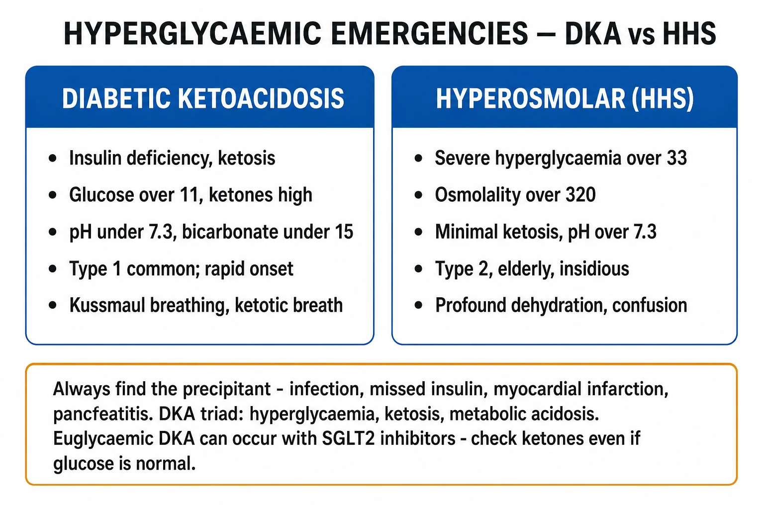
Diabetic ketoacidosis (DKA) and hyperosmolar hyperglycaemic state (HHS) are the two hyperglycaemic emergencies of diabetes. They share a single core defect — absolute or relative insulin deficiency combined with an excess of counter-regulatory hormones (glucagon, catecholamines, cortisol, growth hormone) — and lie on a continuum, but differ in degree of ketogenesis, acidosis, dehydration and the typical patient.[1][3]
- DKA is defined by the triad of hyperglycaemia, ketosis and metabolic acidosis. It arises from absolute insulin deficiency (most often type 1 diabetes) and is characterised biochemically by high anion-gap metabolic acidosis from accumulation of ketoacids (beta-hydroxybutyrate, acetoacetate).
- HHS (formerly hyperosmolar non-ketotic coma, HONK) is defined by severe hyperglycaemia, hyperosmolality and profound dehydration without significant ketoacidosis. It occurs in older type 2 diabetics who still secrete enough insulin to suppress lipolysis (and therefore ketogenesis) but not enough to control hepatic glucose output. [1]
The two can overlap ("mixed DKA-HHS"): a type 2 patient may present with HHS-range hyperosmolality yet also have ketosis. The therapeutic implication is that HHS has more water to replace, less acidosis to correct, and a higher thromboembolic and mortality risk.[1]
DKA remains the commonest cause of death in children and young adults with type 1 diabetes and is largely preventable. The clinical skill lies in (1) recognising the diagnosis (any unwell diabetic, or any comatose/acidotic patient, gets a venous gas and a ketone measurement); (2) executing the standard fluid–insulin–potassium bundle without shortcuts; (3) finding and treating the precipitant; and (4) anticipating complications — hypokalaemia, cerebral oedema, thromboembolism, and the recurrence of ketosis if insulin is stopped too early.[3]
Classification
By biochemical category (the ADA / Kitabchi consensus definitions):[3]
| Parameter | DKA | HHS |
|---|---|---|
| Plasma glucose (mmol/L) | over 11 (over 200 mg/dL) | over 33 (over 600 mg/dL) |
| Arterial / venous pH | below 7.30 | over 7.30 |
| Serum bicarbonate (mmol/L) | below 18 | over 15 |
| Beta-hydroxybutyrate (mmol/L) | over 3 | trace |
| Urine / serum ketones | positive (moderate or large) | trace / small |
| Anion gap | raised (over 12) | variable, often normal |
| Effective osmolality | variable | over 320 mOsm/kg |
| Ketonuria, Kussmaul breathing | present | absent |
| Mental state | often alert; coma less common | stupor / coma common |
| Typical patient | type 1, younger | type 2, older |
Severity grading of DKA (ADA / Kitabchi) — used to triage location (ward vs HDU vs ICU):[3]
- Mild DKA — pH 7.25 to 7.30, bicarbonate 15 to 18 mmol/L, alert, able to tolerate oral intake sometimes; anion gap raised.
- Moderate DKA — pH 7.00 to 7.24, bicarbonate 10 to under 15 mmol/L, alert or drowsy.
- Severe DKA — pH below 7.00, bicarbonate below 10 mmol/L, stupor or coma; manage in HDU/ICU. [1]
Euglycaemic DKA — the ketosis/acidosis triad with glucose below 11 mmol/L — is a subcategory increasingly seen with SGLT2 inhibitors, pregnancy, partial treatment, and chronic alcoholism.[2]
DKA
- Absolute insulin deficiency (type 1); ketogenesis prominent
- Glucose over 11 mmol/L, pH below 7.3, bicarbonate below 18, ketones over 3 mmol/L
- High anion-gap metabolic acidosis; Kussmaul breathing; ketotic (pear-drop) breath
- Onset hours to 1 to 2 days; younger patient; mortality 0.5 to 5 percent
- Fixed-rate insulin 0.1 unit/kg/hr; potassium-primed from the outset
HHS
- Relative insulin deficiency (type 2); enough insulin to suppress ketogenesis
- Glucose over 33 mmol/L, osmolality over 320, pH over 7.3, ketones trace
- Profound dehydration (loss of 9 to 12 L water), often coma or focal deficit
- Onset days to weeks; older patient; mortality 10 to 20 percent
- Slower saline, low-dose insulin 0.05 unit/kg/hr, prophylactic LMWH mandatory
Epidemiology & Risk Factors
Incidence and burden [1]
- DKA accounts for the commonest cause of death in type 1 diabetes under 30 years, and is the first presentation in roughly 25 to 30 percent of new type 1 diabetics (a figure that is rising as incidence of type 1 in children climbs globally).[1]
- HHS accounts for under 1 percent of diabetic admissions but carries a far higher mortality.
- Hospitalisation rates for DKA are rising across all age groups, attributed partly to wider use of SGLT2 inhibitors, pumps (catheter failure), insulin cost and adherence issues.
Mortality [1]
- DKA mortality under 5 percent in adults (over 50 percent where facilities are limited), 0.5 to 2 percent in children in centres with modern protocols.
- HHS mortality 10 to 20 percent — driven by older age, comorbidity, thromboembolism and the precipitant (infarction, sepsis), not by the biochemistry itself. [1]
Precipitants of DKA / HHS (the four I's and an S): [1]
- Infection (commonest, 30 to 50 percent) — pneumonia, urinary tract infection, gastroenteritis, COVID-19, influenza, cellulitis, sepsis.
- Insulin omission / pump failure — missed injections (especially in adolescents with eating disorders, "diabulimia"), faulty pen, insulin pump catheter dislodgement, expired insulin.
- Infarction / inflammation — myocardial infarction, stroke, pancreatitis, trauma, surgery, burns.
- Introduction of new drugs — SGLT2 inhibitors (euglycaemic DKA), corticosteroids, thiazides, sympathomimetics, atypical antipsychotics, pentamidine.
- Substance / other — cocaine, alcohol, pregnancy, severe stress, eating disorders. [1]
Specific DKA-provoking drugs (high-yield):[2]
- SGLT2 inhibitors (dapagliflozin, empagliflozin, canagliflozin) — glucosuria lowers the glucose, so euglycaemic DKA results; risk highest with surgery, fasting, low-carb diet, alcohol, type 1 diabetes (boxed warning).
- Corticosteroids — gluconeogenesis, insulin resistance.
- Atypical antipsychotics (olanzapine, clozapine) — hyperglycaemia.
- Thiazides, beta-agonists, pentamidine (beta-cell toxicity → insulinopenia). [1]
Risk factors for cerebral oedema (the killer in paediatric DKA):[5] younger age (especially under 5), severe acidosis (low pCO2, low pH), high urea at presentation, severe hypocapnia, failure of corrected sodium to rise during therapy, large-volume hypotonic fluid, and rapid insulin bolus. Blackouts from a higher baseline sodium and the use of bicarbonate have also been implicated.
Pathophysiology
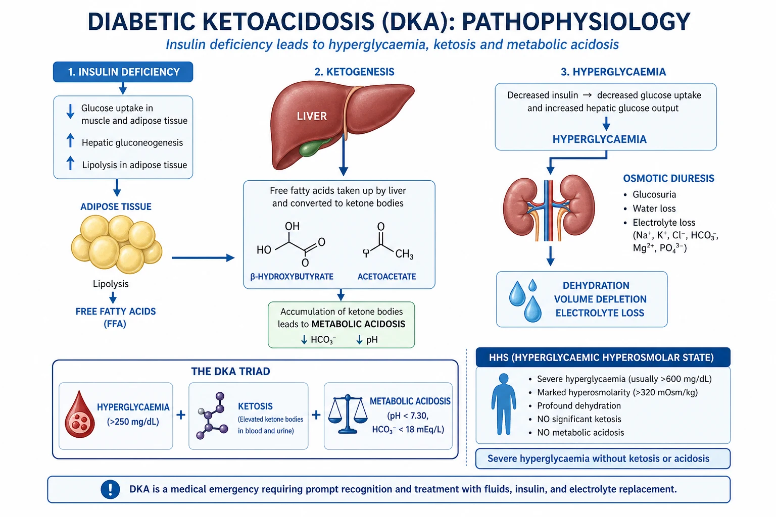
The single unifying defect in DKA and HHS is insulin deficiency (absolute in DKA, relative in HHS) combined with a surge of counter-regulatory hormones (glucagon, catecholamines, cortisol, growth hormone) released by the precipitant (infection, stress, missed dose). The downstream consequences are:[3]
1. Hyperglycaemia — three mechanisms
- Reduced glucose uptake by muscle and adipose tissue (insulin-dependent GLUT4 transporter is under-active).
- Unrestrained hepatic gluconeogenesis and glycogenolysis — glucagon drives the liver to churn out glucose.
- Reduced insulin-mediated suppression of gluconeogenesis (the futile cycle between glucose-6-phosphate and free glucose is wide open). [1]
The result is plasma glucose rising rapidly, exceeding the renal threshold (~10 mmol/L) and producing glucosuria → osmotic diuresis. Each gram of glucose spilled drags roughly 5 to 7 mL of water with it, plus sodium, potassium and phosphate. Water loss exceeds sodium loss — explaining the hypernatraemia / hyperosmolality of HHS and the free-water deficit that can reach 9 litres in HHS and 6 litres in DKA. [1]
2. Ketogenesis — why DKA is "keto"
In adipose tissue, insulin normally suppresses hormone-sensitive lipase. With insulin absent and catecholamines/cortisol abundant, lipase goes into overdrive: triglyceride → free fatty acids + glycerol. Free fatty acids travel to the liver, where the mitochondrial carnitine shuttle (CPT-1) ferries them in for beta-oxidation to acetyl-CoA. Insulin normally stimulates acetyl-CoA carboxylase to make malonyl-CoA, which inhibits CPT-1 and shuts off ketogenesis. Insulin absence removes this brake; glucagon activates the cAMP cascade that further accelerates the pathway. Acetyl-CoA is shunted into ketogenesis via HMG-CoA, producing the two circulating ketoacids:[3]
- Acetoacetate — the parent compound; modest blood level.
- Beta-hydroxybutyrate (BOHB) — the predominant ketoacid in DKA (ratio 3:1 to 10:1 with acetoacetate). In severe acidosis almost all ketones are BOHB, which is why urine ketone strips (nitroprusside) only detect acetoacetate and underestimate severity — only blood beta-hydroxybutyrate is reliable.
- Acetone — volatile, excreted via lungs, responsible for the fruity "pear-drop" breath; not an acid, contributes little to the anion gap. [1]
3. High anion-gap metabolic acidosis
Ketoacids are strong acids that dissociate at physiological pH, releasing H+, which is buffered by bicarbonate — so for every mole of ketoanion produced, one mole of bicarbonate is consumed. The ketoanion (beta-hydroxybutyrate, acetoacetate) is the unmeasured anion that widens the anion gap. [1]
Anion gap = Na+ − (Cl− + HCO3−) — normal 8 to 12 mmol/L. In DKA it commonly reaches 20 to 35. [1]
The delta gap and corrected bicarbonate (corrected HCO3 = measured HCO3 + 0.5 × [anion gap − 12]) help detect a coexisting metabolic alkalosis (from vomiting) — corrected HCO3 above 24 indicates a coexisting metabolic alkalosis, common in DKA from prodromal vomiting. [1]
4. Potassium — the most dangerous electrolyte
This is a high-yield trap. Serum potassium is normal or raised at presentation, but total-body potassium is profoundly depleted (often deficit of 3 to 6 mmol/kg). The reason:[3]
- Absolute insulin deficiency removes insulin's normal drive to push K+ into cells.
- Acidosis shifts K+ out of cells in exchange for H+ (transcellular shift) — so serum K+ looks high.
- Osmotic diuresis dumps huge amounts of K+ in the urine. [1]
When you give insulin, K+ is driven back into cells; when you give saline, the K+ is diluted and excreted. Serum K+ therefore plummets during treatment — hypokalaemia is the leading preventable cause of death in treated DKA. Replace potassium aggressively and early. [1]
5. Sodium and the corrected sodium
Hyperglycaemia draws water out of cells, diluting serum sodium — measured Na+ looks low. The corrected sodium adjusts for this: [1]
Corrected Na+ = measured Na+ + 0.4 × (glucose [mmol/L] − 5.5) (or +1.6 per 5.5 mmol/L glucose above 5.5; some use +0.3 per 5.5). [1]
In HHS, the corrected sodium should rise during treatment as glucose falls; a falling corrected sodium is a warning sign for cerebral oedema.[5]
6. Water — why HHS is so much more dehydrated
HHS develops slowly (days to weeks), allowing maximal free-water loss via sustained osmotic diuresis, but enough residual insulin to suppress ketogenesis. The result is profound hyperosmolality and dehydration (often 10 to 12 percent of body weight, or 9 to 12 L deficit in a 70 kg adult), with little acidosis. The slow onset also allows cerebral adaptation, so patients tolerate very high osmolalities — until decompensation precipitates coma, focal deficits, seizures or thrombosis. [1]
Clinical Presentation
Typical DKA
Onset is rapid (hours to 1 to 2 days). The classical picture:[1]
- Symptoms of hyperglycaemia: polyuria, polydipsia, weight loss, nocturia (often preceded by days of worsening control).
- Gastrointestinal: nausea, vomiting, diffuse abdominal pain (especially in children — "pseudo-appendicitis"; resolves with treatment and should not be attributed to a surgical cause until DKA is corrected), anorexia.
- Respiratory: Kussmaul respiration — deep, sighing, rapid breathing, the respiratory compensation for metabolic acidosis (the body tries to blow off CO2 to raise pH).
- Breath: fruity / nail-varnish-remover / pear-drop smell of acetone (often missed; ask a colleague to smell).
- Dehydration: dry mucous membranes, reduced skin turgor, sunken eyes, tachycardia, hypotension, oliguria, postural drop in blood pressure.
- Neurological: lethargy, drowsiness, confusion; coma is uncommon at presentation in adults (under 10 percent) and should prompt a search for another cause (stroke, hypoxia, sepsis, alcohol, head injury).
- Features of the precipitant: fever, cough, dysuria, chest pain, focal deficit, source of infection, recent surgery. [1]
Typical HHS
Onset is insidious (days to weeks). The patient is typically older, with known type 2 diabetes (or newly diagnosed), often with comorbidity and polypharmacy. The classical picture:[1]
- Profound dehydration — loss of skin turgor, sunken eyes, dry tongue, hypotension, oliguria.
- Altered conscious level — confusion, lethargy, coma (proportional to osmolality; coma unusual if osmolality below 320).
- Neurological: seizures, focal deficits, hemiparesis, visual disturbance that may mimic stroke.
- Less abdominal pain, no Kussmaul breathing, no acetone breath (minimal ketosis).
- Features of the precipitant are prominent (sepsis, infarction, stroke). [1]
Euglycaemic DKA
Glucose below 11 mmol/L but ketones over 3 mmol/L and pH below 7.3. Suspect this whenever a patient on an SGLT2 inhibitor (or in the perioperative period, or pregnant) presents with vomiting, abdominal pain, malaise or breathlessness. The trap is that the near-normal glucose masks the diagnosis; always check ketones and venous gas in any unwell diabetic regardless of glucose.[2]
Atypical presentations
- Elderly with HHS: may present with falls, delirium, dehydration, "off legs", or a stroke mimic — glucose and osmolality must be checked.
- Pregnancy: DKA may present with lower glucose (fetal glucose utilisation) and progresses faster — fetal mortality is high (10 to 35 percent).
- Children: abdominal pain is common and may masquerade as appendicitis; never operate for "acute abdomen" until DKA is excluded.
- Cerebral oedema during treatment: headache, behavioural change, irritability, drowsiness, incontinence, bradycardia, rising blood pressure, abnormal posturing, coma — a neurosurgical emergency evolving over minutes. [1]
Differential Diagnosis
The student must distinguish DKA from the other high anion-gap acidoses (MUDPILES), from HHS, and from other causes of coma in a diabetic.[3]
DKA
- Hyperglycaemia over 11 mmol/L, ketones over 3, pH below 7.3, AG over 12
- Kussmaul breathing, ketotic breath, abdominal pain; known or new T1DM
- Beta-hydroxybutyrate markedly raised; glucose and osmolality high but lower than HHS
HHS
- Glucose over 33 mmol/L, osmolality over 320, pH over 7.3, ketones trace
- Older T2DM, profound dehydration, coma, seizures, stroke-like
- Minimal acidosis; large free-water deficit; high thromboembolic risk
Lactic acidosis
- High AG metabolic acidosis but glucose near-normal, ketones low
- Causes: sepsis, shock, metformin accumulation (CKD), severe hypoxia, malignancy
- Lactate over 5 mmol/L; no Kussmaul-only pattern, no ketotic breath
Alcoholic ketoacidosis
- Chronic alcohol misuse + binge + starvation; vomiting, dehydration
- Low or normal glucose, mild acidosis, ketones positive, lactate may be high
- Beta-hydroxybutyrate predominant (like DKA); resolves with dextrose-saline + thiamine, NO insulin needed
Starvation ketosis
- Pregnant, fasting, post-operative, low-carb diet; mild ketones (under 3)
- Glucose low/normal, pH near-normal (over 7.3), bicarbonate over 18
- Resolves with food or dextrose; no insulin deficiency
Uraemic acidosis
- CKD with high urea/creatinine; high AG, no ketones, glucose variable
- Often normal-anion-gap (high chloride) until late; treat dialysis
Toxin-induced acidosis
- Salicylate (tinnitus, hyperventilation, mixed alkalosis/acidosis, AG high)
- Methanol (blindness, formate), ethylene glycol (oxalate crystals, renal failure), paraldehyde
- Anion gap high with osmolar gap; treat toxin-specifically
Coma in a diabetic — the bedside differentials
- Hypoglycaemic coma — sudden onset, sweating, tremor, tachycardia, blood glucose below 4 mmol/L (point-of-care), no ketones, normal or shallow breathing. Treatment is empirical 25 g IV dextrose / 1 mg IM glucagon if in doubt.
- DKA / HHS — as above; ketones and venous gas are diagnostic.
- Lactic acidosis (metformin, sepsis) — high AG, low/normal glucose, lactate over 5.
- Other coma — stroke, post-ictal, head injury, alcohol, opiate (pinpoint pupils → naloxone), hepatic/uraemic encephalopathy. [1]
Key distinguishing points [1]
- Glucose + ketones + pH + bicarbonate + osmolality + lactate in any unwell or comatose diabetic settles most cases.
- Beta-hydroxybutyrate over 3 mmol/L confirms ketoacidosis of any cause; glucose over 11 mmol/L then points to DKA (over 33 and osmolality over 320 → HHS; under 11 → euglycaemic DKA, alcoholic or starvation).
- Osmolar gap distinguishes toxin (over 10 mOsm/kg) from keto/lactic.
- Always check salicylate, ethanol, and consider methanol/ethylene glycol when AG is high without ketones. [1]
Clinical & Bedside Assessment
The bedside assessment answers three questions: how sick is the patient, how dehydrated are they, and what is the precipitant?[1]
Severity — ABCDE
- Airway/Breathing: rate, depth, Kussmaul breathing (rapid AND deep, the cardinal sign of metabolic acidosis; rate rises by 1 for every 1 mmol/L fall in bicarbonate — Winter's formula predicts expected pCO2 = 1.5 × HCO3 + 8 ± 2). Oxygen saturation, respiratory distress, smell for acetone.
- Circulation: heart rate, blood pressure (lying and sitting — postural drop indicates over 10 percent volume loss), capillary refill, peripheral temperature, JVP (low), mottled skin suggests shock. Cool peripheries with hypotension = critical.
- Disability: GCS, pupils, blood glucose (point-of-care), ketones (capillary beta-hydroxybutyrate), focal deficits (especially in HHS — stroke mimic).
- Exposure / Everything else: source of infection (skin, feet, catheter, line), calf swelling (DVT — common in HHS), abdominal exam (peritonism — but in DKA "pseudo-appendicitis" resolves with treatment). [1]
Degree of dehydration ( bedside )
- Mild (under 3 percent): not clinically apparent.
- Moderate (3 to 5 percent): dry mucous membranes, reduced skin turgor.
- Severe (over 5 percent; in DKA usually 6 percent, in HHS up to 12 percent): sunken eyes, tachycardia, hypotension, oliguria, cool peripheries, postural drop, altered mental state. [1]
Targets that confirm adequate resuscitation
- Blood pressure restored (mean arterial pressure over 65 mmHg).
- Capillary refill under 2 seconds, warm peripheries.
- Urine output over 0.5 mL/kg/hr (catheterise if obtunded or unable to void; allows accurate balance in HHS).
- Falling ketones and rising pH on serial venous gases (every 1 to 2 hours).
- Resolution of tachycardia and tachypnoea. [1]
Reassessment for cerebral oedema
After starting therapy, reassess hourly for headache, behavioural change, irritability, incontinence, bradycardia, rising blood pressure, abnormal posturing, falling GCS, abnormal pupillary response. Any neurological deterioration during treatment of paediatric DKA is cerebral oedema until proven otherwise — an emergency.[5]
Investigations
The goals are to (1) confirm the diagnosis, (2) grade severity, (3) find the precipitant, and (4) monitor therapy. [1]
Confirming investigations
Venous blood gas (preferred over arterial — less painful, accuracy comparable in DKA; an arterial sample is reserved for shock or hypoxia):[3]
- pH — below 7.3 in DKA (mild 7.25 to 7.30, moderate 7.00 to 7.24, severe below 7.00).
- Bicarbonate — below 18 mmol/L.
- Base excess — negative, typically under −5.
- pCO2 — low (compensatory Kussmaul); should match Winter's formula: expected pCO2 = 1.5 × HCO3 + 8 ± 2. Higher-than-expected pCO2 = coexisting respiratory acidosis (exhaustion, aspiration) — intubate. [1]
Blood ketones — beta-hydroxybutyrate (POC) — the gold standard for diagnosis and monitoring. Over 3 mmol/L confirms DKA. Urine ketones (nitroprusside strips) are unreliable: they detect only acetoacetate (not BOHB, the predominant ketone in severe DKA), lag behind treatment, and can paradoxically rise as BOHB converts back to acetoacetate during recovery.[3]
Plasma glucose — over 11 mmol/L (DKA), over 33 (HHS); bedside glucose every hour. [1]
Serum biochemistry [1]
- Urea and creatinine — raised (dehydration, AKI); metformin suggests lactic acidosis.
- Sodium — measured and corrected (formula above).
- Potassium — normal or high at presentation despite total-body deficit.
- Chloride — to calculate anion gap and the delta ratio.
- Calculated osmolality = 2 × Na + glucose + urea (all in mmol/L). HHS over 320.
- Lactate — exclude lactic acidosis (metformin, sepsis).
- Phosphate — often low; routine replacement not needed (see below).
- Magnesium, calcium — disturbed by osmotic diuresis.
- FBC — leucocytosis (stress response or infection); left shift indicates infection.
- CRP, blood cultures, MSU, throat swab, CXR, ECG, troponin — hunt the precipitant.
- Amylase / lipase — pancreatitis is both a precipitant and a complication of DKA; mild asymptomatic rise is common.
- Beta-hCG — pregnancy in any woman of reproductive age.
- Salicylate, paracetamol, ethanol levels — if ingestion suspected. [1]
Reproduced diagnostic thresholds
[1] [1]Monitoring schedule (during treatment)
Monitoring frequency
Management — Resuscitation
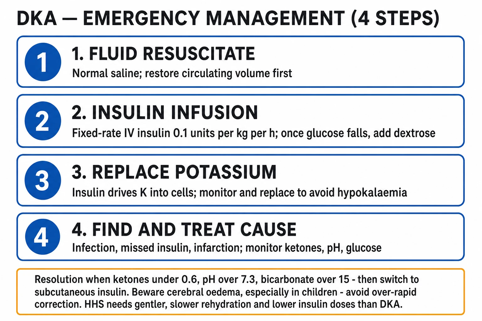
DKA and HHS are time-critical. The first hour is about circulation, not glucose. Resuscitation priorities, in order:[1][3]
ABCDE
- Airway: protect if GCS under 8 (rare in adult DKA at presentation); nurse in left lateral position if vomiting.
- Breathing: oxygen 15 L/min via non-rebreather mask if SpO2 under 94 %; intubate if respiratory exhaustion (rising pCO2 above Winter-predicted).
- Circulation: two large-bore IV cannulae, bloods drawn (VBG, ketones, FBC, U&E, glucose, lactate, CRP, amylase, troponin, blood cultures, beta-hCG, ECG).
- Disability: GCS, pupils, point-of-care glucose and ketones; check for focal deficit.
- Exposure: search for the precipitant (skin, feet, line, abdomen, calf). [1]
IV fluids — give BEFORE insulin
Why fluids first: restores intravascular volume, improves tissue perfusion (lowers counter-regulatory hormones), lowers glucose by dilution and improved renal excretion, and prevents the precipitous fall in K+ when insulin starts.[3]
Initial fluid (JBDS / ADA — adults): [1]
- 0.9 % sodium chloride 1 L stat over the first hour (faster if shock; in adults use 0.9 % saline).
- Then 0.9 % saline 1 L every 1 to 2 hours until intravascular volume restored, then reassess.
- Typical adult regimen (JBDS 2022): 1 L in hour 1, 1 L over 2 hours, 1 L over 2 hours, 1 L over 4 hours, 1 L over 6 hours, 1 L over 8 hours — roughly 6 L in first 12 hours, then 8-hourly thereafter. [1]
When glucose falls below 14 mmol/L, add 10 % glucose 125 mL/hr alongside the saline (or switch to a glucose-saline) to prevent hypoglycaemia while continuing insulin to suppress ketogenesis. The principle is: continue insulin until ketones are cleared, switching the fluid from saline to glucose as glucose normalises. [1]
Avoid hypotonic fluids initially (risk of rapid osmolality drop → cerebral oedema). Hartmann's contains lactate (metabolised to bicarbonate) and is sometimes used but JBDS continues to recommend 0.9 % saline. [1]
Potassium — replace before and with insulin
Insulin drives K+ into cells. Serum K+ must be checked before insulin is started, and the regimen chosen accordingly:[3]
| Serum K+ (mmol/L) | Action |
|---|---|
| Below 3.3 | HOLD insulin. Give 40 mmol KCl per litre of saline at 1 L/hr (with ECG monitoring) until K+ over 3.3. |
| 3.3 to 5.2 | Add 40 mmol KCl per litre of saline; start insulin at 0.1 unit/kg/hr. |
| Over 5.2 | Hold potassium; check K+ again in 2 hours; start insulin. Add K+ once K+ falls below 5.2. |
Maximum peripheral K+ concentration: 40 mmol/L (rate up to 20 mmol/hr); central line required for higher concentrations. [1]
When to defer insulin
- K+ below 3.3 mmol/L — hold insulin, replace K+ first (insulin would precipitate life-threatening hypokalaemia and arrhythmia).
- Sepsis with shock — resuscitate first; insulin still given once K+ is safe. [1]
Urinary catheter and central line
- Urinary catheter if obtunded, anuric, or in HHS (allows accurate fluid balance — urine output target 0.5 to 1 mL/kg/hr).
- Central line if prolonged therapy, high potassium concentration required, or for vasopressors in shock.
- NG tube if vomiting or GCS reduced (aspiration risk). [1]
Management — Definitive & Stepwise
The three pillars: fluids, insulin, potassium — with treat the precipitant and prevent complications.[3]
Pillar 1 — Fixed-rate intravenous insulin infusion (FRIII)
Weight-based fixed-rate IV insulin has replaced variable sliding scales in modern protocols because it gives predictable insulin levels and faster ketone clearance.[3]
- Soluble human insulin (Actrapid) 0.1 unit/kg/hr IV (50 units made up to 50 mL with 0.9 % saline → 1 unit/mL; run at 0.1 mL/kg/hr).
- For obese or insulin-resistant patients, may use 0.15 unit/kg/hr; for HHS use 0.05 unit/kg/hr (lower — HHS is sensitive to insulin and aggressive insulin causes rapid osmolality/fluid shifts → cerebral oedema).
- Do NOT give an IV insulin bolus (associated with cerebral oedema in children).
- Target: ketones fall by at least 0.5 mmol/L/hr, glucose falls by 3 mmol/L/hr, bicarbonate rises by 3 mmol/L/hr.
- If response inadequate: double insulin infusion rate (re-check at 1 hour; check pump, line, insulin expiry).
- Continue FRIII until: ketones under 0.6 mmol/L, pH over 7.3, bicarbonate over 15, AND the patient is eating and on long-acting subcutaneous insulin. [1]
Pillar 2 — Fluids (continued)
Follow the JBDS regimen above. Switch fluid when glucose below 14: [1]
- 10 % glucose 125 mL/hr alongside the saline (this is critical — insulin must keep running to clear ketones, even when glucose is normal).
- If sodium falls or corrected sodium is low, use 0.45 % saline cautiously. [1]
Pillar 3 — Potassium
Add 40 mmol KCl per litre of saline from the second bag, monitored by 2-hourly U&E and continuous ECG in HDU/ICU. Reduce if K+ rises, increase if K+ falls. [1]
Pillar 4 — Find and treat the precipitant
- Antibiotics (e.g. co-amoxiclav 1.2 g IV 8-hourly or per local sepsis policy) for infection; blood cultures first.
- MI: dual antiplatelet, heparin, cardiology; stroke: CT, thrombolysis pathway if eligible.
- Pancreatitis: nil-by-mouth, analgesia, IV fluids.
- Pump failure: reconnect or convert to MDI.
- Stop SGLT2 inhibitors in euglycaemic DKA. [1]
Pillar 5 — Bicarbonate: usually avoided
Bicarbonate is NOT routinely given in DKA. It worsens hypokalaemia (intracellular K+ shift), impairs tissue oxygen delivery (left-shifts Hb-O2 curve), lowers CSF pH paradoxically (CO2 crosses blood-brain barrier faster than bicarbonate), and has been associated with cerebral oedema in children.[3]
Indications (controversial, exceptional): [1]
- pH below 6.9 with haemodynamic instability refractory to fluids/vasopressors — give isotonic (1.26 %) bicarbonate 100 mL/hr until pH over 7.0, with potassium cover.
- Hyperkalaemia with ECG changes. [1]
Pillar 6 — Phosphate, magnesium
- Phosphate: routine replacement not recommended (no outcome benefit; risk of hypocalcaemia). Give IV potassium phosphate 20 to 30 mmol only if serum phosphate under 0.4 mmol/L and respiratory muscle weakness or cardiomyopathy.
- Magnesium: replace if under 0.5 mmol/L. [1]
Pillar 7 — Conversion from IV to subcutaneous insulin
Resolution criteria for DKA:[3]
- Ketones under 0.6 mmol/L (or urine ketones negative).
- pH over 7.3, bicarbonate over 15.
- Patient able to eat and drink. [1]
Conversion protocol (avoid rebound ketosis): [1]
- Give a long-acting basal insulin (e.g. glargine 0.25 unit/kg SC or patient's usual basal) at least 1 to 2 hours BEFORE stopping the IV insulin — the basal-on-board prevents a gap.
- Continue the IV insulin and glucose until the next meal; give rapid-acting analogue with the meal (e.g. aspart/lispro 4 to 6 units or 0.1 unit/kg).
- Stop the IV insulin at the meal; check ketones at 1 hour post-meal — if rising, restart FRIII.
- For new-onset type 1 diabetes, start a basal-bolus regimen at total daily dose 0.5 unit/kg/day (half basal, half bolus split across meals). [1]
If insulin is stopped before subcutaneous is established — rebound DKA within hours, because the half-life of IV insulin is only 5 minutes. Always overlap. [1]
HHS — differences from DKA
HHS differs
- More dehydration (9 to 12 L): slower saline repletion, 0.9 % then 0.45 %
- Lower insulin dose: 0.05 unit/kg/hr (insulin-sensitive, cerebral-oedema risk)
- Do NOT aim to suppress ketones (none present) — aim to lower glucose by 3 to 5 mmol/L/hr
- Glucose falls rapidly with fluids alone — insulin may not be needed at all in the first few hours
- Mandatory prophylactic LMWH (e.g. enoxaparin 40 mg SC OD): very high thromboembolic risk
- Slower correction over 24 to 72 hours to avoid cerebral oedema and osmotic demyelination
- Look hard for stroke, MI, sepsis; revascularise or treat source
Specific Subtypes & Scenarios
Euglycaemic DKA[2]
- Recognise: glucose below 11 mmol/L with ketones over 3 and pH below 7.3 in a patient on SGLT2 inhibitor, perioperative, pregnant, alcoholic, low-carb diet, or type 1 with partial treatment.
- Manage as DKA but add dextrose earlier (because glucose is already low) — 10 % glucose at 125 mL/hr with FRIII 0.1 unit/kg/hr to keep suppressing ketogenesis while avoiding hypoglycaemia.
- Stop the SGLT2 inhibitor permanently in type 1 and during acute illness in type 2.
- Admit; do not be reassured by the "normal" glucose. [1]
Paediatric DKA (ISPAD 2018)[4][5]
- Cerebral oedema is the feared complication (0.5 to 1 % of episodes; 20 to 40 % mortality).
- Fluids are more conservative than adults: 10 to 20 mL/kg of 0.9 % saline over 1 to 2 hours, then 0.45 % saline + KCl. Avoid bolus over 30 mL/kg unless shock.
- Insulin only after fluids started: 0.05 to 0.1 unit/kg/hr.
- No bicarbonate (associated with cerebral oedema).
- The FLUID trial (Kuppermann 2018) — 1389 children in a 2×2 factorial of fast vs slow rehydration and 0.9 % vs 0.45 % saline — found NO difference in neurological outcomes, refuting the long-held belief that aggressive fluids cause cerebral oedema.[4] Nevertheless, ISPAD still recommends cautious rehydration.
- Monitor hourly for headache, behavioural change, bradycardia, rising BP — any deterioration = mannitol 0.5 to 1 g/kg IV or 3 % saline 5 mL/kg over 10 min, intubate if GCS under 8, transfer to PICU.
DKA in pregnancy
- Lower glucose threshold (fetal glucose utilisation; placental insulinase) — DKA can occur with glucose as low as 7 mmol/L.
- Faster onset, more severe acidosis, fetal mortality 10 to 35 %, maternal mortality low.
- Common in T2DM in pregnancy, gestational diabetes, on beta-agonists / steroids, or with hyperemesis.
- Manage as adult DKA; obstetric review, continuous CTG if viable; resolve DKA before delivery (DKA itself is not an indication for delivery). [1]
HHS in the elderly type 2 patient
- Insidious onset, often precipitated by sepsis, stroke, MI, or new drug (steroid, thiazide).
- Coma, seizures, focal deficits, dehydration; mimics stroke.
- Slower correction (over 24 to 72 hours), lower insulin dose (0.05 unit/kg/hr), mandatory LMWH, search for thrombosis.
- Mortality 10 to 20 % — driven by comorbidity. [1]
DKA with coexisting chronic kidney disease
- Potassium handling: presenting K+ may be very high (no urinary loss) — use slower insulin, monitor K+ hourly, may need calcium resonium or dialysis if refractory hyperkalaemia.
- Insulin clearance is reduced — smaller doses, longer duration.
- Fluid overload risk — central line, daily weights, diurese if needed. [1]
Complications & Pitfalls
Acute complications
- Hypokalaemia — the leading preventable cause of death during treatment
- Cerebral oedema — chiefly paediatric, 0.5 to 1 % of episodes, 20 to 40 % mortality
- Hypoglycaemia — from over-aggressive insulin or late dextrose
- Hypoxia / ARDS — rare, fluid overload in elderly
- Thromboembolism (DVT, PE, MI, stroke) — especially HHS, prophylax with LMWH
- Acute respiratory distress syndrome (ARDS) — rare
Treatment pitfalls
- Giving insulin before K+ checked (→ arrhythmic death)
- Stopping IV insulin before SC established (→ rebound DKA)
- Switching to glucose too late (→ hypoglycaemia)
- Bicarbonate given routinely (→ hypokalaemia, cerebral oedema, paradoxical CSF acidosis)
- Treating 'abdominal emergency' surgically before correcting DKA (true surgical cause rare)
- Forgetting to look for / treat the precipitant (DKA recurs)
Cerebral oedema — the paediatric killer[5]
Risk factors: under 5 years, severe acidosis (low pCO2, low pH), high urea at presentation, severe hypocapnia, failure of corrected sodium to rise during therapy, large-volume hypotonic fluid, rapid insulin bolus, bicarbonate use. [1]
Clinical features (during treatment, 4 to 12 hours in): headache, behavioural change, irritability, incontinence, bradycardia, rising blood pressure (Cushing's triad), abnormal posturing, falling GCS, pupillary changes, seizures. [1]
Emergency management: [1]
- Mannitol 0.5 to 1 g/kg IV over 15 minutes OR hypertonic 3 % saline 5 mL/kg over 10 minutes.
- Intubate and hyperventilate (lowers ICP), raise head of bed to 30 degrees.
- Reduce insulin and fluids, transfer to PICU/ICU, urgent CT.
- Avoid lumbar puncture. [1]
Why hypokalaemia is the leading cause of preventable death
Insulin + saline drive K+ into cells and into urine. A patient with a 5 mmol/kg deficit who is treated without potassium cover can drop from K+ 5.5 to 2.5 in 4 hours — ventricular ectopics, VT/VF, asystole. Prevention: check K+ before insulin; add 40 mmol/L KCl to every bag from hour 1 if K+ under 5.2; 2-hourly U&E; continuous ECG if severe DKA. Re-check at any arrhythmia. [1]
Thromboembolism in HHS
HHS is a markedly prothrombotic state (dehydration, high viscosity, endothelial dysfunction). Prophylactic LMWH (e.g. enoxaparin 40 mg SC once daily — reduce to 20 mg if eGFR under 30) is mandatory unless contraindicated. [1]
Stopping insulin too early
If IV insulin is stopped without subcutaneous insulin on board, ketosis recurs within 1 to 2 hours (insulin half-life 5 minutes) — "rebound DKA." Always overlap the basal insulin for at least 1 to 2 hours before stopping the infusion. [1]
Prognosis & Disposition
Mortality [1]
- DKA: under 5 % adults, under 1 % children in modern centres; higher with severe acidosis, old age, comorbidity, delay in treatment.
- HHS: 10 to 20 % — driven by age, comorbidity and the precipitant (MI, stroke, sepsis), not by the biochemistry.[1]
Where to manage [1]
- Mild DKA (pH 7.25 to 7.3, alert): can be managed on a diabetes ward with hourly ketone and glucose monitoring.
- Moderate DKA (pH 7.00 to 7.24): admission unit / diabetes ward with close monitoring; HDU if K+ borderline, elderly, cardiac.
- Severe DKA (pH below 7.0, bicarbonate below 10, GCS reduced, haemodynamically unstable): HDU or ICU.
- All paediatric DKA: paediatric ward with senior review; PICU if pH below 7.1, GCS reduced, or cerebral oedema.
- HHS: HDU/ICU in most elderly patients (comorbidity, fluid balance, anticoagulation). [1]
Determinants of outcome: rapidity of diagnosis, adequacy of resuscitation, finding the precipitant, prevention of hypokalaemia and cerebral oedema, and the underlying comorbidity. [1]
Recurrence risk: 25 to 50 % of DKA is recurrent — intermittent insulin omission, eating disorder, inadequate diabetes education. Education, psychological support, structured diabetes review and sick-day rules ("never stop insulin; check ketones when unwell; drink fluids; call for help if vomiting") reduce recurrence. [1]
Special Populations
Children (ISPAD 2018)[4]
- Conservative fluid bolus (10 to 20 mL/kg over 1 to 2 hours, not 30 mL/kg).
- Insulin only after fluids at 0.05 to 0.1 unit/kg/hr — never bolus.
- No bicarbonate.
- Hourly neuro check for cerebral oedema.
- New diagnosis: basal-bolus insulin 0.5 unit/kg/day, diabetes education, psychological support. [1]
Pregnancy
- Lower glucose threshold for DKA; check ketones in any unwell pregnant diabetic.
- Fetal mortality 10 to 35 %; resolve DKA before delivery.
- Beta-agonists and steroids precipitate — monitor closely in preterm labour.
- Manage with adult protocol; obstetric + diabetic team. [1]
Elderly with HHS
- Slower correction (24 to 72 hours) to avoid cerebral oedema / ODS.
- Lower insulin dose (0.05 unit/kg/hr).
- LMWH mandatory.
- Aggressive hunt for precipitant (stroke, MI, sepsis, drug).
- Watch fluid overload (heart failure, AKI). [1]
Renal failure
- Presenting K+ may be very high — slower insulin, hourly K+, may need dialysis.
- Reduced insulin clearance — smaller doses.
- Fluid balance tight — central line, daily weights. [1]
Evidence, Guidelines & Regional Differences
Landmark trials [1]
- Kitabchi 2009 (ADA consensus) — established the modern fluid–insulin–potassium protocol; FRIII preferred over sliding scale; bicarbonate not routine.[3]
- Glaser 2001 (NEJM) — case-control of 61 children with cerebral oedema: risk factors included severe acidosis (low pCO2), high urea, failure of corrected sodium to rise, bicarbonate use, low PCO2, intubation with hyperventilation.[5]
- Kuppermann 2018 — FLUID trial (NEJM) — randomised 1389 paediatric DKA episodes in a 2×2 factorial of fast vs slow rehydration and 0.9 % vs 0.45 % saline; no difference in neurological outcomes (GCS, memory, IQ at 2 to 6 months). Concluded that neither fluid rate nor tonicity caused cerebral injury in children when used within standard protocols.[4]
- Umpierrez 2004, 2007 (Diabetes Care) — subcutaneous analogues in mild-moderate DKA on the ward; reduced ICU admissions; FRIII vs sliding scale.
- Dhatariya JBDS-IP 2022/2023 update — fixed-rate insulin, 10 % glucose at 14 mmol/L, no routine bicarbonate, beta-hydroxybutyrate for diagnosis and monitoring.
Regional deltas [1]
[1] [1]Controversies [1]
- Bicarbonate: routine use abandoned; still used at pH below 6.9 with shock — evidence weak.
- Phosphate: routine replacement not beneficial.
- ICU vs ward: many centres manage mild-moderate DKA on admissions units to spare ICU; ICU reserved for severe/complicated.
- Subcutaneous analogue protocols for mild DKA (Umpierrez) — gaining ground for resource-limited settings but not standard.
- Cerebral oedema mechanism: vasogenic (fluid shifts) vs cytotoxic (ischaemia) — FLUID trial shifted thinking away from fluid rate as the cause. [1]
Exam Pearls
Anion-gap differential — MUDPILES
MUDPILES
methanol → formate; metformin → lactic acidosis (CKD)
renal failure with retained organic anions
also alcoholic & starvation ketosis
rare; paraldehyde → acetic/ formic acid
iron → lactic; INH → lactate
sepsis, shock, hypoxia, malignancy
ethylene glycol → oxalate (crystalluria); ethanol → ketoacidosis
salicylate → mixed alkalosis/acidosis with tinnitus
Precipitants of DKA — the four I's + S
IIIIS
commonest (30 to 50 %) — pneumonia, UTI, gastroenteritis, COVID-19
missed dose, faulty pen, dislodged pump catheter
MI, stroke, pancreatitis, trauma, surgery
SGLT2i (euglycaemic DKA), steroids, thiazides, atypicals
alcohol, cocaine, pregnancy, eating disorder, severe stress
The DKA bundle in one line
FIX-K
0.9 % saline 1 L stat, then 1 L q1–2h
weight-based, no bolus
infection, MI, missed insulin, pancreatitis
40 mmol/L if K+ 3.3 to 5.2; HOLD insulin if below 3.3
High-yield minutiae
- Corrected sodium shortcut: Na(measured) + 0.4 × (glucose − 5.5) mmol/L. Falling corrected sodium during treatment = warning for cerebral oedema.
- Anion gap = Na − (Cl + HCO3); normal 8 to 12. Delta-delta (delta ratio = ΔAG / ΔHCO3): under 0.4 = pure normal-anion-gap acidosis; 1 to 2 = pure high-anion-gap acidosis (DKA); over 2 = coexisting metabolic alkalosis (vomiting).
- Ketones: BOHB predominates in DKA (3:1 to 10:1 ratio); urine strips detect only acetoacetate and underestimate severity and lag recovery.
- Pear-drop (fruity) breath = acetone (volatile) — exhaled, not an acid.
- Kussmaul breathing = rapid AND deep; rate rises ~1/min per 1 mmol/L fall in bicarbonate.
- Winter's formula: expected pCO2 = 1.5 × HCO3 + 8 ± 2; higher than expected = respiratory acidosis (exhaustion → intubate).
- Insulin half-life IV = 5 min — never stop IV insulin before subcutaneous basal is on board (rebound DKA).
- Hypokalaemia is the leading preventable death during DKA treatment — check K+ before insulin.
- HHS mortality 10 to 20 % vs DKA under 5 %; HHS needs LMWH and slower, lower-dose insulin.
- Euglycaemic DKA — think SGLT2 inhibitor, pregnancy, perioperative, alcohol; treat with dextrose + FRIII.
- Cerebral oedema — headache/behaviour change/bradycardia/rising BP/falling GCS during treatment → mannitol or 3 % saline, intubate, PICU.
- Pseudo-appendicitis in paediatric DKA — abdominal pain resolves with treatment; do not operate until DKA corrected.
- Bicarbonate — not routine; if pH below 6.9 with shock, isotonic 1.26 %, with K+ cover.
- Phosphate — routine replacement not recommended.
- 10 % glucose at 14 mmol/L (UK JBDS) / 5 % dextrose at 11 mmol/L (US ADA) — switch when glucose falls.
- Recurrence — 25 to 50 % of DKA; address adherence, education, eating disorder, sick-day rules. [1]
Exam application bank (NEET-PG / INICET)
One-line answer
Diabetic ketoacidosis (DKA) is the triad of hyperglycaemia, ketosis and metabolic acidosis arising from absolute insulin deficiency (most often new or known type 1 diabetes). Hyperosmolar hyperglycaemic state (HHS) is severe hyperglycaemia with high osmolality and dehydration but minimal ketosis, classically in older type 2 patients. Both are medical emergencies triggered by infection, missed insulin, infarction or new diabetes. Treatment pillars are IV fluids first, fixed-rate IV insulin 0.1 units/kg/hr, careful potassium replacement, and treat the precipitant. The killing complications are hypokalaemia (the leading preventable death during treatment) and cerebral oedema (chiefly in children). [1]
Worked stems (answer without another resource)
Stem 1 — Classic presentation. Map symptoms to mechanism; name the first investigation and first treatment step with dose/route if drug therapy is standard. [1]
Stem 2 — Unstable / complicated. List red flags that force immediate resuscitation, theatre, ICU, antidote, or reperfusion — and what you do in the first 15 minutes. [1]
Stem 3 — Atypical group. Elderly, pregnancy, child, or immunocompromised: how presentation and thresholds change. [1]
Stem 4 — Differential trap. Name the three closest mimics and one discriminator for each. [1]
Stem 5 — Disposition. Who goes home with safety-netting, who is admitted, who needs HDU/ICU/theatre, and what follow-up is mandatory. [1]
Rapid viva checklist
- Definition + classification
- Pathophysiology chain
- Bedside signs / criteria
- Score with exact components (if any)
- Emergency bundle
- Definitive therapy with doses
- Complications of disease and of treatment
- Special populations
- Guideline/trial name if classic
- Three exam traps
Coverage self-check
If you cannot answer any stem above from this page alone, re-read the matching section — the page is intended to be self-sufficient for final-prof and NEET-PG/INICET questions on Diabetic Ketoacidosis and Hyperosmolar Hyperglycaemic State (DKA / HHS).
[1] [1]References
- [1]Fayfman M, Pasquel FJ, Umpierrez GE. Management of Hyperglycemic Crises: Diabetic Ketoacidosis and Hyperglycemic Hyperosmolar State Med Clin North Am, 2017.PMID 28372715
- [2]Long B, Lentz S, Koyfman A. Euglycemic diabetic ketoacidosis: Etiologies, evaluation, and management Am J Emerg Med, 2021.PMID 33626481
- [3]Kitabchi AE, Umpierrez GE, Miles JM, Fisher JN. Hyperglycemic crises in adult patients with diabetes Diabetes Care, 2009.PMID 19564476
- [4]Kuppermann N, Ghetti S, Schunk JE, et al. Clinical Trial of Fluid Infusion Rates for Pediatric Diabetic Ketoacidosis N Engl J Med, 2018.PMID 29897851
- [5]Glaser N, Barnett P, McCaslin I, et al. Risk factors for cerebral edema in children with diabetic ketoacidosis. The Pediatric Emergency Medicine Collaborative Research Committee of the American Academy of Pediatrics N Engl J Med, 2001.PMID 11172153