Endocrinology · Endocrinology
Hyperthyroidism
Also known as Thyrotoxicosis · Overactive thyroid · Graves disease (when autoimmune) · Plummer disease (toxic adenoma) · Thyroid storm (decompensated extreme)
Hyperthyroidism is the syndrome of excess thyroid-hormone synthesis and secretion by the thyroid gland; thyrotoxicosis is the broader clinical state of excess circulating hormone from any source. The commonest cause is Graves disease (TSH-receptor stimulating antibody), followed by toxic multinodular goitre and toxic adenoma; destructive thyroiditis and exogenous hormone produce low-uptake thyrotoxicosis. The biochemical hallmark is a suppressed TSH with raised free T4 and/or free T3. Treatment options are antithyroid drugs (carbimazole/methimazole; propylthiouracil in pregnancy first trimester and thyroid storm), radioactive iodine (I-131) and surgery. Thyroid storm is the decompensated, life-threatening extreme — thionamide then iodine one hour later, beta-blocker, hydrocortisone, cooling, and treatment of the precipitant.
On this page & tools
Your progress
Saved locally on this device.
Exam tags
Red flags
Overview & Definition
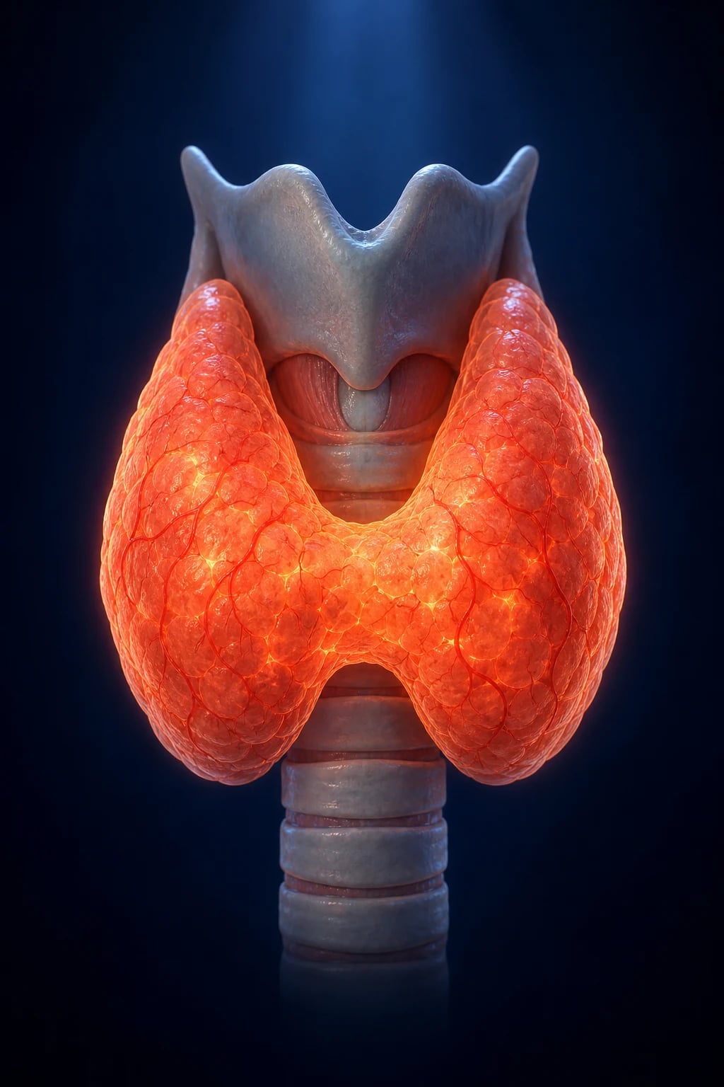
Hyperthyroidism is the clinical and biochemical syndrome caused by sustained overproduction and secretion of thyroid hormone (T4 and T3) by the thyroid gland itself, producing a hypermetabolic state at the tissue level.[4] The broader term thyrotoxicosis refers to the clinical state of excess circulating thyroid hormone from any source — endogenous overproduction (hyperthyroidism), hormone leak from a damaged gland (thyroiditis), or exogenous intake (factitious or iatrogenic).[2][4] The distinction matters because the source determines the investigation pathway (especially the radioactive uptake scan) and the treatment: antithyroid drugs work only when the gland is actively synthesising hormone, not when the hormone is leaking from a destroyed gland.[3]
The condition sits on a spectrum from subclinical (suppressed TSH with normal free hormones) through overt (suppressed TSH with raised free T4 and/or T3) to the decompensated extreme of thyroid storm — a medical emergency with mortality of 10 to 30%.[6] Thyroid hormone acts on virtually every tissue, so the presentation is heterogeneous, and hyperthyroidism enters the differential of unexplained weight loss, atrial fibrillation, anxiety, palpitations, osteoporosis, heat intolerance, diarrhoea and menstrual disturbance. The clinical skill is not the diagnosis (that is biochemical) but (a) identifying the cause (Graves vs toxic nodule vs thyroiditis vs factitious) — because it changes management — and (b) recognising thyroid storm and agranulocytosis, the two ways a thyrotoxic patient dies.[3]
Two thyroid physiology essentials underpin the whole topic. The axis: the hypothalamus secretes thyrotropin-releasing hormone (TRH), which drives pituitary thyrotrophs to release thyroid-stimulating hormone (TSH); TSH binds the TSH receptor (TSH-R), a G-protein-coupled receptor activating Gs and cAMP, which drives iodide uptake (sodium-iodide symporter, NIS), organification and coupling by thyroid peroxidase (TPO), and release of T4 (the prohormone) and a little T3. Peripheral deiodinases (D1, D2) convert T4 to the active T3; D3 inactivates to reverse T3. The feedback loop: circulating T4 and T3 exert negative feedback on TSH (pituitary) and TRH (hypothalamus). Because of this steep feedback, a suppressed TSH is the most sensitive marker of thyroid-hormone excess long before free T4 rises — which is why subclinical disease is defined by TSH alone.[2][4]
Classification
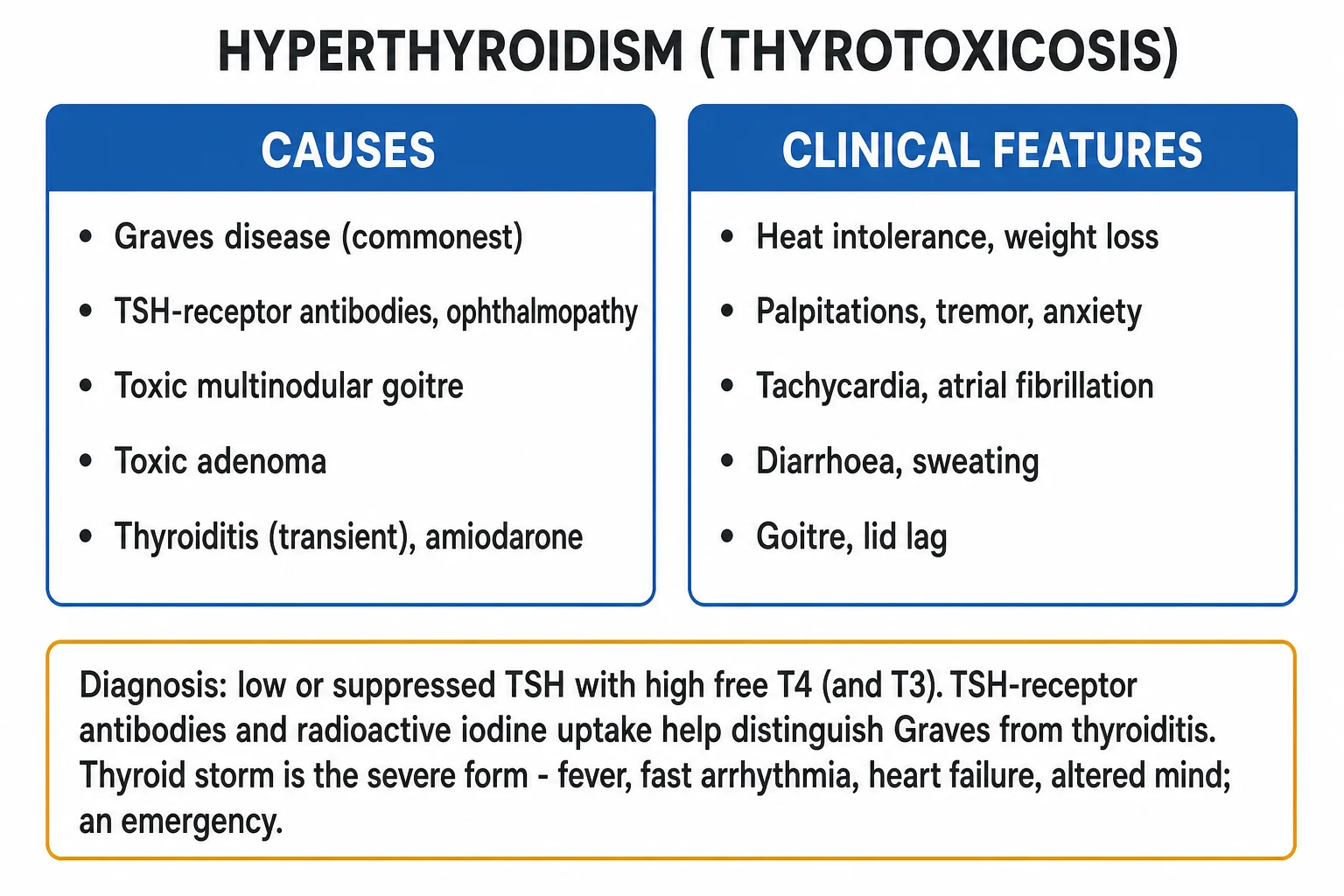
Thyrotoxicosis is classified along two axes — by source/uptake status (which drives aetiology and treatment) and by severity (overt vs subclinical). Both are needed to manage the patient.[2][3]
By source and radioactive uptake (the clinically decisive axis)
HIGH-uptake (true hyperthyroidism)
- The gland is actively OVER-synthesising hormone; the follicular cells are stimulated
- Radioactive iodine / Tc-99m pertechnetate uptake is raised or patchy
- Causes: GRAVES DISEASE (diffuse uptake), TOXIC MULTINODULAR GOITRE (patchy hot spots), TOXIC ADENOMA / PLUMMER DISEASE (single hot nodule with suppression of the rest), TSH-secreting pituitary adenoma, thyroid hormone resistance, hCG-mediated (molar)
- Treatment target: suppress synthesis (thionamides), ablate (RAI) or remove (surgery)
LOW-uptake (non-hyperthyroid thyrotoxicosis)
- The gland is NOT actively synthesising — hormone is LEAKING from damaged follicles or coming from OUTSIDE
- Radioactive uptake is low / suppressed
- Causes: SUBACUTE (de Quervain) thyroiditis, SILENT / painless / postpartum thyroiditis, AMIODARONE-INDUCED TYPE 2 (destructive), FACTITIOUS / exogenous hormone, iodine-induced (Jod-Basedow is high uptake however), STRUMA OVARII (ectopic thyroid tissue)
- Thionamides are largely useless; treat the underlying process and the symptoms (beta-blocker)
By severity
| Severity | TSH | Free T4 / T3 | Typical picture |
|---|---|---|---|
| Overt | Suppressed (under 0.1 mIU/L) | Raised | Symptomatic thyrotoxicosis |
| Subclinical | Suppressed (under lower limit, often under 0.4) | Normal | Usually asymptomatic; detected on screening |
| T3 toxicosis | Suppressed | Free T4 NORMAL, free T3 RAISED | Early or mild Graves; can be missed if only T4 measured |
| Thyroid storm | Suppressed | Markedly raised | Hyperpyrexia, tachyarrhythmia, altered mentation — emergency |
T3 toxicosis is a frequent exam point: in early or mild disease (and in iodine-deficient/toxic-nodule settings) the gland preferentially secretes T3, so free T4 may be normal while free T3 is raised — always measure free T3 when TSH is suppressed but free T4 is normal.[3]
Epidemiology & Risk Factors
The global prevalence of overt hyperthyroidism is about 0.2 to 1.4%, and subclinical hyperthyroidism affects a further 0.7 to 1.4%; the combined burden is roughly 2.5% of adults.[2] Key epidemiological facts:[1][2]
- Graves disease is by far the commonest cause in iodine-sufficient regions (60 to 80% of cases), with a global prevalence of about 2% in women and 0.5% in men.
- Sex: a strong female predominance of roughly 5 to 10:1, reflecting autoimmune predilection.
- Age: Graves peaks in the third to fifth decades (women of reproductive age); toxic multinodular goitre is a disease of older adults (over 50), arising in a long-standing multinodular goitre, often in iodine-deficient regions.
- Toxic adenoma (Plummer disease) typically presents in patients aged 30 to 50. [1]
Hyperthyroidism — key numbers
Host and environmental risk factors (an examiner staple):[1][3]
- Female sex and family history of autoimmune thyroid disease (Graves clusters with HLA-DR3, HLA-B8; CTLA-4 and PTPN22 polymorphisms).
- Other autoimmunity — type 1 diabetes, Addison disease, vitiligo, pernicious anaemia, coeliac disease (autoimmune polyglandular syndromes).
- Pregnancy and the postpartum period — immune reconstitution after delivery triggers postpartum thyroiditis and may unmask Graves.
- Smoking — a major risk factor for Graves orbitopathy (and worsens its response to treatment); the strongest modifiable risk factor for eye disease.
- Iodine load — amiodarone (each 200 mg tablet carries about 75 mg organic iodine, roughly 100 times the daily requirement), iodinated contrast, iodine supplementation in deficient patients (Jod-Basedow), kelp/seaweed supplements.
- Drugs — lithium, interferon-alpha, interleukin-2, alemtuzumab can precipitate or unmask thyroiditis or Graves.
- Stress — severe emotional or physical stress, infection, surgery, trauma and childbirth are classic precipitants of thyroid storm in a previously compensated patient. [1]
Pathophysiology
The shared cellular mechanism: why excess T3 causes every symptom
At the molecular level, T3 acts via the nuclear thyroid hormone receptor (TR-alpha, TR-beta), a ligand-activated transcription factor that upregulates genes including Na-K-ATPase, beta-adrenergic receptors, malic enzyme, SERCA and alpha-myosin heavy chain. In thyrotoxicosis:[2][4]
- Upregulated Na-K-ATPase drives a higher basal metabolic rate and thermogenesis (heat intolerance, sweating, weight loss despite increased appetite), and increases oxygen consumption by virtually every tissue.
- Increased beta-adrenergic receptor density (and direct T3 effects on the myocardium) sensitises the heart to catecholamines — producing tachycardia, high-output state, increased inotropy, fine tremor, anxiety, lid lag and hyperreflexia. This is the molecular basis for beta-blocker therapy working so effectively for symptoms.
- Increased bone turnover (osteoclastic resorption outstrips formation) → osteopenia/osteoporosis over time.
- Increased gut motility → diarrhoea, malabsorption of fat; increased hepatic glyconeogenesis and glycogenolysis → glucose intolerance.
- Upregulated Na-K-ATPase in kidney → increased renal iodide clearance. [1]
Mechanism of Graves disease (the prototype)
Graves is an organ-specific autoimmune disease. The immunological lesion:[1]
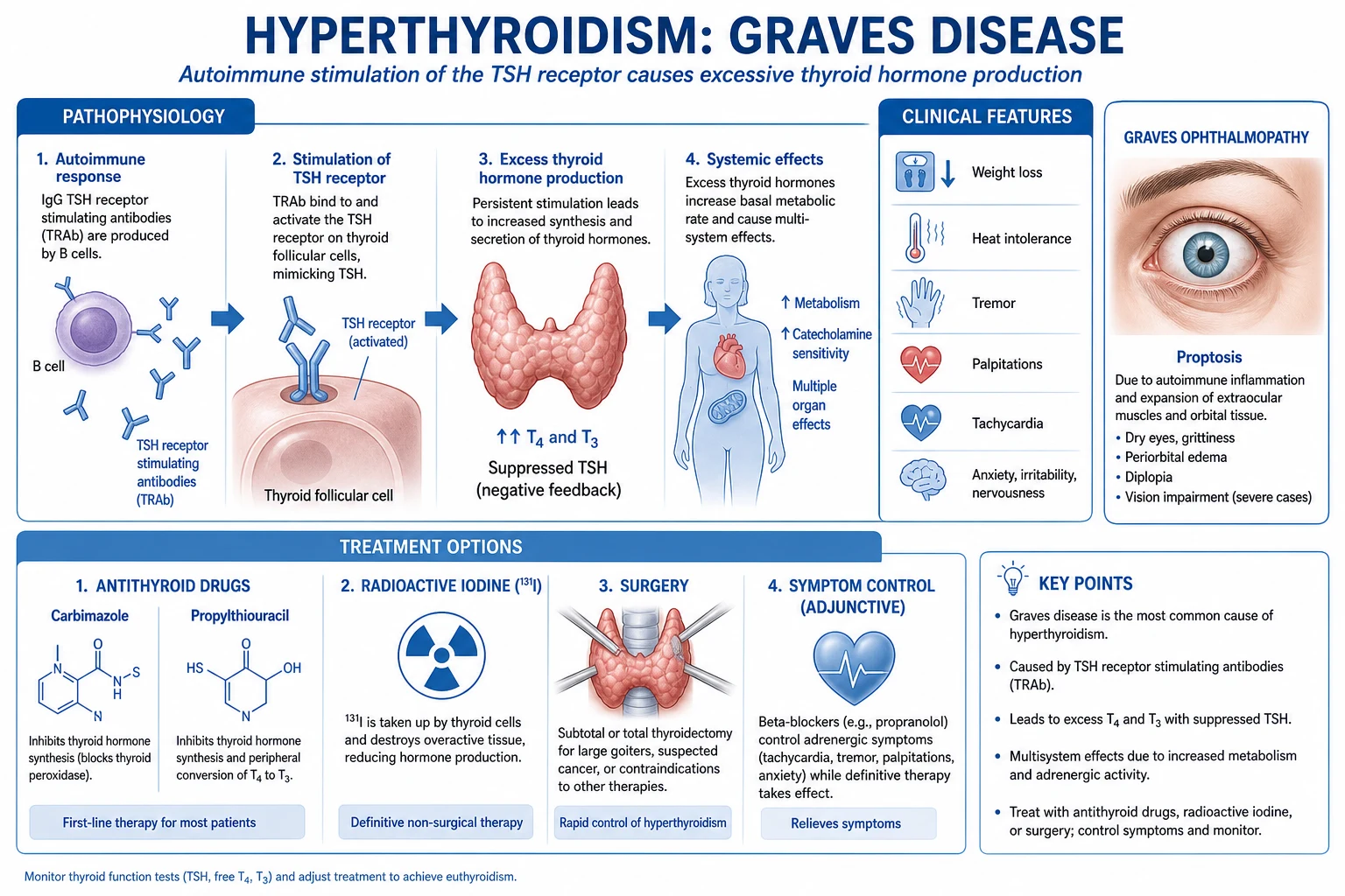
- Loss of self-tolerance to the TSH receptor (TSH-R), with production of thyrotropin-receptor antibodies (TRAb) — these are thyroid-stimulating immunoglobulins (TSI) that bind the TSH receptor and mimic TSH, activating the Gs–cAMP cascade.
- The result is autonomous, unregulated stimulation of every step of hormonogenesis — NIS iodide uptake, TPO organification and coupling, thyroglobulin synthesis and hormone release — producing a diffusely enlarged, hypervascular goitre (with an audible bruit) and overproduction of T4/T3.
- Because the excess hormone suppresses pituitary TSH, the gland is driven by the antibody, not by TSH — the feedback loop is broken. [1]
Why the eye and skin? TSH receptors and insulin-like growth factor-1 receptors (IGF-1R) are expressed on retro-orbital fibroblasts and preadipocytes. TRAb (acting with IGF-1R) activates orbital fibroblasts, which differentiate into adipocytes and secrete hydrophilic glycosaminoglycans (hyaluronic acid). The glycosaminoglycans retain water and expand the orbital contents within the rigid bony orbit → proptosis, oedema, inflammation and (in severe cases) optic nerve compression. The same process in the dermis of the pretibial region produces pretibial myxoedema (a localised, non-pitting, plaque-like infiltrate). Smoking strongly amplifies this orbital process.[1][5]
Mechanism of toxic multinodular goitre and toxic adenoma
These are non-autoimmune, autonomous causes. Over years, a multinodular goitre accumulates somatic gain-of-function mutations in the TSH-R signalling pathway or the Gs-alpha (GNAS) subunit, which constitutively activate cAMP independent of TSH.[3] As autonomous foci grow and iodine supply permits, they escape TSH control and overproduce hormone. When autonomous output exceeds the body's needs, TSH is suppressed and the surrounding normal thyroid tissue becomes quiescent. In a toxic adenoma (Plummer disease), a single dominant nodule carries the activating mutation; on a scan it is a single hot nodule with the rest of the gland suppressed.
Mechanism of thyroiditis-induced thyrotoxicosis (low-uptake)
In subacute (de Quervain) thyroiditis (typically post-viral), silent/painless thyroiditis, and postpartum thyroiditis, inflammatory destruction of follicles releases pre-formed T4 and T3 from stored colloid. The leaked hormone suppresses TSH, which switches off NIS-mediated uptake — hence the low radioactive uptake that distinguishes thyroiditis from Graves. The thyrotoxic phase is self-limiting (4 to 8 weeks, until the colloid store is exhausted), often followed by a transient hypothyroid phase and then recovery. Thionamides are ineffective because no new synthesis is occurring.[3][4]
Mechanism of amiodarone-induced thyrotoxicosis (type 1 vs type 2)
Amiodarone causes thyrotoxicosis by two distinct mechanisms that demand different treatment:[3][4]
- Type 1 (iodine-induced, high uptake): the enormous iodine load drives excess hormone synthesis in an underlying nodular goitre or latent Graves (Jod-Basedow phenomenon). Treat with thionamides (often high-dose, plus perchlorate).
- Type 2 (destructive, low uptake): amiodarone (or its iodine) causes a destructive thyroiditis with hormone leak. Treat with glucocorticoids (prednisolone 40 to 60 mg daily, tapering over weeks). Mixed forms occur; clinical discrimination (uptake, interleukin-6, colour-flow Doppler) guides therapy. [1]
Mechanism of factitious (exogenous) thyrotoxicosis
Ingestion of exogenous thyroid hormone (deliberate, for weight loss or performance, or accidental through meat/supplements) produces thyrotoxicosis with a suppressed TSH, low radioactive uptake, and a characteristically LOW thyroglobulin (the gland is quiescent — unlike endogenous overproduction or thyroiditis, in both of which thyroglobulin is raised). [1]
Clinical Presentation
Thyrotoxicosis produces a hyperadrenergic, hypermetabolic state. Symptoms and signs are best organised system by system.[2][4]
General:
- Heat intolerance and sweating, unintentional weight loss despite an increased appetite (a key clue — weight loss with a good appetite points to thyrotoxicosis, not malignancy), fatigue, insomnia, nervousness, irritability, anxiety (often misattributed to a primary anxiety disorder), proximal muscle weakness (thyrotoxic myopathy — difficulty climbing stairs or combing hair). [1]
Cardiovascular (a major source of morbidity and the exam favourite):
- Palpitations, sinus tachycardia, supraventricular ectopics, and — especially in the elderly — atrial fibrillation (found in 10 to 15% of thyrotoxic patients, and up to a third of older patients at presentation).
- High-output state with a hyperdynamic, bounding pulse, widened pulse pressure, systolic flow murmur; with longstanding disease, high-output (and later dilated) cardiac failure.
- Exertional dyspnoea. [1]
Neuromuscular:
- Fine tremor of the outstretched hands, brisk/hyperactive deep tendon reflexes (a brisk, snapping reflex — the opposite of the delayed relaxation in hypothyroidism), proximal myopathy, rarely thyrotoxic periodic paralysis (hypokalaemic, in Asian males, provoked by carbohydrate/exertion — a medical emergency treated with non-selective beta-blocker and cautious potassium). [1]
Skin and soft tissues:
- Warm, moist, velvety skin, palmar erythema, onycholysis (Plummer nails — distal nail separation), diffuse alopecia, hyperpigmentation (less common).
- Pretibial myxoedema (Graves dermopathy) — non-pitting, plaque-like induration on the anterior shins or dorsa of feet, pathognomonic of Graves. [1]
Gastrointestinal:
- Increased frequency of stool / frank diarrhoea, nausea, rarely malabsorption and steatorrhoea; hepatic dysfunction (raised transaminases and alkaline phosphatase). [1]
Reproductive:
- Oligomenorrhoea or amenorrhoea, anovulatory infertility, reduced libido; gynaecomastia in men (from increased aromatisation of androgens to oestrogens). [1]
Bedside signs that should trigger testing: warm moist palms, fine tremor, lid lag, hyperreflexia, tachycardia with bounding pulse. [1]
Graves-specific features (the triad that earns the diagnosis)
Diffuse goitre with BRUIT
- Soft, diffuse, symmetrically enlarged thyroid; a systolic/continuous bruit on auscultation reflects intense hypervascularity
- Pathognomonic for active Graves overproduction (other thyrotoxicoses are not vascular)
- Often with a palpable thrill
Graves ORBITOPATHY (thyroid eye disease)
- The hallmark extrathyroidal manifestation; present in about 25 to 50% of Graves patients
- Includes LID RETRACTION (Dalrymple sign), LID LAG (von Graefe sign), INFREQUENT BLINKING (Stellwag sign), PROPTOSIS / EXOPHTHALMOS, chemosis, conjunctival injection, diplopia (extraocular muscle involvement)
- CRITICAL: distinguish LID LAG (sympathetic, seen in ANY thyrotoxicosis) from PROPTOSIS (Graves-specific, due to orbital expansion)
- Sight-threatening: dysthyroid OPTIC NEUROPATHY (reduced colour vision, RAPD, visual loss) — an emergency
Pretibial MYXOEDEMA (dermopathy)
- Non-pitting, plaque-like, violaceous induration on the anterior lower legs or dorsa of feet
- Due to glycosaminoglycan (hyaluronic acid) deposition
- Graves-specific; coexists with orbitopathy and high TRAb
Thyroid storm (thyrotoxic crisis)
The decompensated extreme of thyrotoxicosis — a state in which the metabolic, thermoregulatory and cardiovascular compensatory mechanisms are overwhelmed.[6] Classic presentation:
- Hyperpyrexia (often 39 to 41 degrees C) out of proportion to other features, with profuse sweating.
- Severe tachyarrhythmia — atrial fibrillation with rapid ventricular response, or sinus tachycardia.
- Heart failure, pulmonary oedema, hypotension (late, pre-terminal).
- Altered mental status — agitation, delirium, psychosis, seizures, obtundation, coma.
- Gastrointestinal/hepatic — vomiting, diarrhoea, abdominal pain, jaundice (a grave sign). [1]
It is almost always precipitated in a known or undiagnosed thyrotoxic patient by infection, surgery, trauma, parturition, iodine load (contrast, amiodarone), radioiodine therapy, withdrawal of antithyroid drug, severe emotional stress, diabetic ketoacidosis, stroke or myocardial infarction. [1]
Atypical presentations
- Apathetic hyperthyroidism (elderly) — the classic sympathetic features are muted; instead, lethargy, apathy, depression, weight loss, atrial fibrillation, heart failure, unexplained osteoporosis or proximal myopathy. Often mistaken for malignancy or depression until the TSH returns suppressed. Lethargy + AF + weight loss in an older patient is apathetic hyperthyroidism until proven otherwise.[2]
- In pregnancy — symptoms overlap with normal pregnancy (heat intolerance, palpitations, fatigue); a persistent tachycardia, weight loss or failure to gain weight, and a goitre should prompt testing. HCG-mediated thyrotoxicosis (gestational transient thyrotoxicosis) in hyperemesis gravidarum or molar pregnancy mimics Graves but has negative TRAb and usually resolves with hydration and symptom control.[7]
- Thyrotoxic periodic paralysis — predominantly in young Asian males; acute, episodic hypokalaemic flaccid paralysis (often provoked by a carbohydrate load or exertion). Treat with non-selective beta-blocker (propranolol) and cautious potassium; do not over-correct potassium.
Differential Diagnosis
The differential depends on whether the question is "what is the cause of the thyrotoxicosis?" (the most useful) or "what else causes these symptoms?". [1]
Differential of the cause of thyrotoxicosis — high vs low uptake (examiner's core table)
| Cause | Uptake | TRAb | Goitre | Distinguishing features |
|---|---|---|---|---|
| Graves disease | High (diffuse) | Positive | Diffuse, soft, bruit | Orbitopathy, pretibial myxoedema; young women |
| Toxic multinodular goitre | High (patchy hot spots) | Negative | Multinodular, large | Older patient; long-standing goitre; no eye disease |
| Toxic adenoma (Plummer) | High (single hot nodule, rest suppressed) | Negative | Single palpable nodule | Single autonomous nodule; no eye disease |
| Subacute (de Quervain) thyroiditis | Low | Negative | Tender, firm, enlarged | Painful tender goitre, raised ESR/CRP, preceding viral illness |
| Silent / postpartum thyroiditis | Low | Negative | Firm, non-tender, mild enlargement | Painless; postpartum; autoimmune backdrop |
| Amiodarone type 2 | Low | Negative | Often normal | Destructive; steroid-responsive |
| Amiodarone type 1 / Jod-Basedow | Raised (or low in deficient glands) | Variable | Nodular | Iodine-induced synthesis; thionamide-responsive |
| Factitious (exogenous) | Low | Negative | Atrophic / normal | Low thyroglobulin; access to hormone; weight-loss motive |
| TSH-secreting pituitary adenoma | High | Negative | Diffuse | Inappropriately normal/raised TSH with raised free T4; pituitary mass on MRI; alpha subunit raised |
| Thyroid hormone resistance | High | Negative | Diffuse | Raised TSH with raised free T4/T3 (both pituitary and peripheral resistance, or isolated); family history; usually euthyroid clinically |
| hCG-mediated (molar, choriocarcinoma, hyperemesis) | High | Negative | Mild | High hCG (weak TSH agonist); pregnancy with hyperemesis; resolves with molar evacuation |
Differential of the clinical syndrome (when the TFT is not yet back)
- Anxiety/panic disorder — palpitations, tremor, agitation; but normal TSH, no weight loss with increased appetite, no goitre.
- Phaeochromocytoma — paroxysmal hypertension, palpitations, sweating, anxiety; but episodic severe hypertension (thyrotoxicosis is usually with a wide pulse pressure, not hypertensive crises), and a normal TSH. Check plasma metanephrines.
- Menopause — hot flushes, sweats, mood disturbance; normal TFT.
- Malignancy (occult) — weight loss; but appetite usually reduced, not increased; no goitre, no hyperreflexia.
- Caffeine / stimulant excess; withdrawal states. [1]
Differential of proptosis (when the orbit is the presenting feature)
Distinguish Graves orbitopathy (bilateral, lid retraction, exposure, restricted upgaze from inferior rectus involvement, TRAb positive) from orbital cellulitis (unilateral, fever, painful eye movement), orbital pseudotumour (idiopathic orbital inflammation) (painful, unilateral, responds rapidly to steroids), orbital/ocular tumour (unilateral, imaging shows a mass), and carotid-cavernous fistula (pulsatile proptosis, chemosis, bruit).[5]
Clinical & Bedside Assessment
A focused examination aims to (a) confirm the clinical suspicion, (b) characterise the goitre, (c) detect Graves-specific features, (d) assess cardiac complications, and (e) screen for thyroid storm.[1][4]
Vital signs and general: tachycardia, bounding pulse, wide pulse pressure, low-grade fever (storm if high), warm moist palms, fine tremor, hyperreflexia. [1]
Named bedside signs of thyrotoxicosis (examiner favourites):
- Lid lag (von Graefe sign) — when the patient looks down, the upper lid lags behind the globe, revealing sclera above the iris. Seen in any thyrotoxicosis (sympathetic overdrive to Muller's muscle).
- Lid retraction (Dalrymple sign) — upper lid retraction at rest, revealing sclera above the limbus ("stare").
- Stare / infrequent blinking (Stellwag sign).
- Joffroy sign — absence of forehead wrinkling on upward gaze.
- Mobius sign — convergence insufficiency (Graves orbitopathy).
- Graves orbitopathy signs (Graves-specific): proptosis/exophthalmos (measured by exophthalmometer), chemosis, conjunctival injection, restricted extraocular movements (especially elevation from inferior rectus involvement), diplopia.
- Plummer nails (onycholysis) — distal separation of the nail from the bed.
- Pretibial myxoedema — non-pitting plaque-like shin induration. [1]
The thyroid examination: inspect from the front (swelling, asymmetry, scars, distended veins — a pemberton sign on raising the arms suggests retrosternal goitre); palpate from behind the seated patient with both hands, using the thumbs to roll the tissue; assess size, consistency, nodularity, tenderness, mobility; ask the patient to swallow (a thyroid mass moves up with swallowing — distinguishes it from other neck lumps); palpate for cervical lymph nodes; auscultate over each lobe for a bruit (a systolic or continuous bruit indicates the intense hypervascularity of Graves).[1]
Characterising the goitre: [1]
| Cause | Goitre character | Distinguishing bedside feature |
|---|---|---|
| Graves | Diffuse, soft, smooth, symmetric; bruit | Orbitopathy, pretibial myxoedema |
| Toxic multinodular goitre | Multinodular, often large, asymmetrical | Older patient; no eye disease |
| Toxic adenoma | Single palpable nodule | Rest of gland impalpable/soft |
| Subacute thyroiditis | Tender, firm, diffuse | Pain on palpation; raised ESR |
| Silent/postpartum thyroiditis | Firm, non-tender, mildly enlarged | Painless; postpartum |
Cardiovascular assessment: listen for AF (irregularly irregular), a flow murmur, signs of heart failure (basal crackles, raised JVP, peripheral oedema). AF is the commonest cardiac complication and a frequent presentation in the elderly. [1]
Assess for thyroid storm at the bedside using the Burch-Wartofsky Point Scale (BWPS) — see Investigations. A patient with hyperpyrexia, tachyarrhythmia and altered mental status is in storm and treatment must not await biochemistry.[6]
Graves orbitopathy assessment: use the Clinical Activity Score (CAS) (spontaneous retrobulbar pain, pain on eye movement, eyelid erythema, eyelid oedema, conjunctival redness, chemosis, caruncle swelling — 1 point each; CAS of 3 or more indicates active disease amenable to immunosuppression) and assess severity for sight-threatening features (reduced visual acuity, colour-vision loss / red desaturation, relative afferent pupillary defect, visual field defect — any of which suggests dysthyroid optic neuropathy and mandates urgent treatment).[5]
Investigations
First-line thyroid function tests
- TSH — the single most sensitive first-line test; suppressed (under 0.1 mIU/L) in primary hyperthyroidism. A normal TSH in the absence of pituitary disease effectively excludes primary thyrotoxicosis.[2]
- Free T4 and free T3 — raised in overt disease. Always measure free T3 when TSH is suppressed but free T4 is normal (T3 toxicosis). Measure free (not total) hormone — total is confounded by thyroxine-binding globulin changes (pregnancy, oestrogen, nephrotic syndrome).
Interpreting the TFT pattern: [1]
| TSH | Free T4 | Free T3 | Diagnosis |
|---|---|---|---|
| Suppressed | Raised | Raised | Overt primary hyperthyroidism/thyrotoxicosis |
| Suppressed | Normal | Raised | T3 toxicosis (measure T3!) |
| Suppressed (mildly) | Normal | Normal | Subclinical hyperthyroidism |
| Suppressed | Low | Low | Consider central/pituitary disease, euthyroid sick, or recent treatment |
| Inappropriately normal / raised | Raised | Raised | TSH-secreting adenoma OR thyroid hormone resistance |
Confirming the cause
- TSH receptor antibody (TRAb / TSI) — confirms Graves disease (sensitivity and specificity both over 95% in overt disease). Positive TRAb obviates the need for a uptake scan in clear-cut Graves; also predicts relapse after thionamide withdrawal and is essential in pregnancy to assess risk of fetal/neonatal hyperthyroidism.[1][3]
- Thyroid uptake scan (radioiodine-123/131 or Tc-99m pertechnetate) — when the cause is unclear or a nodule is present. Diffuse high uptake = Graves; patchy = TMNG; single hot nodule = toxic adenoma; low/suppressed = thyroiditis/factitious.[3]
- Thyroid ultrasound — for nodular disease (characterise nodules, identify a dominant nodule, guide biopsy of suspicious nodules — a toxic nodule itself is rarely malignant, but coexisting non-functioning nodules need standard work-up).
- Thyroglobulin — low in factitious thyrotoxicosis (gland quiescent), high in endogenous overproduction and thyroiditis (rarely needed diagnostically).[4]
Reproducing the Burch-Wartofsky Point Scale (BWPS) for thyroid storm
The Burch-Wartofsky Point Scale is a bedside diagnostic aid for thyroid storm.[6] Score the following:
Thermoregulatory dysfunction (temperature, degrees C):
- 37.2 to 37.7 — 5 points
- 37.8 to 38.2 — 10 points
- 38.3 to 38.8 — 15 points
- 38.9 to 39.3 — 20 points
- 39.4 to 39.9 — 25 points
- 40.0 and above — 30 points [1]
Central nervous system disturbance:
- Absent — 0
- Mild (agitation) — 10
- Moderate (delirium, psychosis, extreme lethargy) — 20
- Severe (seizure, coma) — 30 [1]
Gastrointestinal-hepatic dysfunction:
- Absent — 0
- Moderate (diarrhoea, nausea/vomiting, abdominal pain) — 10
- Severe (unexplained jaundice) — 20 [1]
Tachycardia (beats per minute):
- 90 to 109 — 5
- 110 to 119 — 10
- 120 to 129 — 15
- 130 to 139 — 20
- 140 and above — 25 [1]
Congestive heart failure:
- Absent — 0
- Mild (pedal oedema) — 5
- Moderate (bibasilar crackles) — 10
- Severe (pulmonary oedema) — 15 [1]
Atrial fibrillation: absent — 0; present — 10 [1]
Precipitating event: absent — 0; present — 10 [1]
Interpretation: 45 and above — highly suggestive of thyroid storm; 25 to 44 — impending storm; under 25 — unlikely. Treatment of storm must not await laboratory confirmation; the BWPS is used to justify empiric treatment. [1]
Associated laboratory abnormalities (examiner favourites)
- Raised alkaline phosphatase and mild transaminitis (liver effects of excess T4).
- Hypercalciuria and mild hypercalcaemia (increased bone turnover).
- Low total/LDL cholesterol (T3 upregulates LDL receptor).
- Raised liver enzymes, raised glucose, glycosuria.
- Raised ESR/CRP — only in subacute (de Quervain) thyroiditis (a useful discriminator).
- Mild normocytic anaemia, raised red-cell mass.
- Hypokalaemia in thyrotoxic periodic paralysis. [1]
When to image
- Uptake scan when aetiology is uncertain or a nodule is present.
- CT or MRI of the orbits in moderate-to-severe Graves orbitopathy (assesses extraocular muscle enlargement, optic nerve compromise) — and urgent imaging with clinical features of optic neuropathy.
- Pituitary MRI if TSH-secreting adenoma suspected (inappropriately normal/raised TSH with raised free T4). [1]
Management — Resuscitation
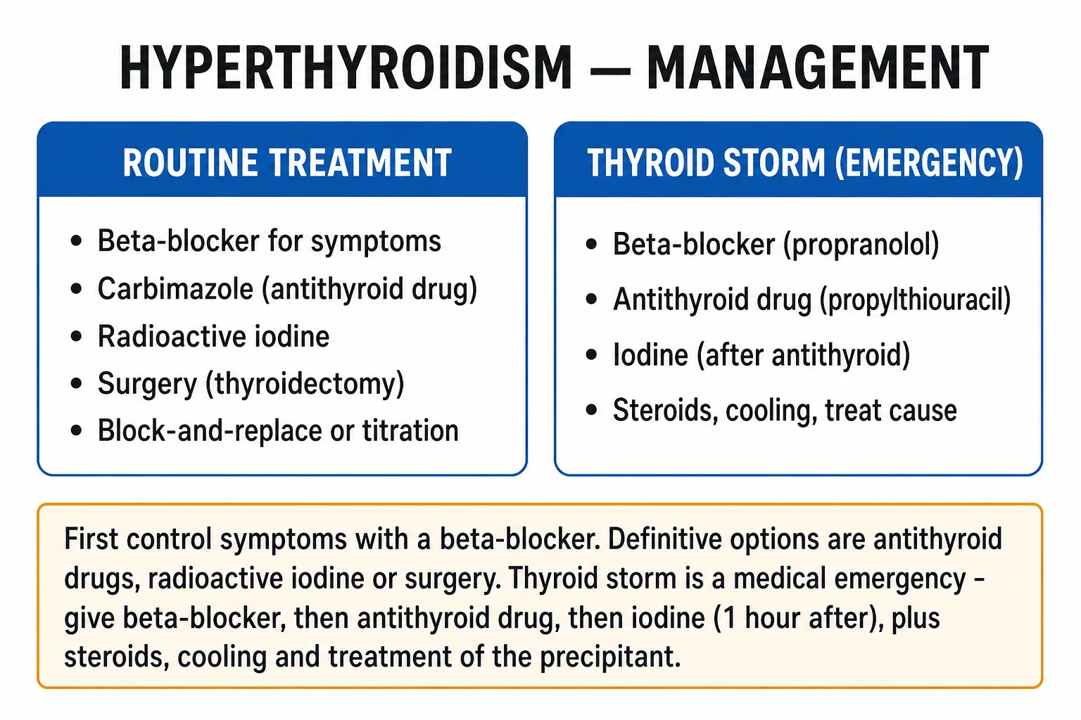
Thyroid storm is an endocrine emergency and treatment must not await TFT confirmation. The resuscitation bundle — apply simultaneously and in the correct pharmacological order:[6]
- Supportive care: ABCDE; oxygen; IV fluids (aggressive — patients are volume-depleted from vomiting, diarrhoea and fever); cooling with paracetamol/acetaminophen and cooling blankets. AVOID SALICYLATES — aspirin displaces T4 and T3 from thyroxine-binding globulin, acutely worsening the thyrotoxicosis, and interferes with radioiodine uptake.
- Beta-blockade — propranolol 60 to 80 mg orally every 4 hours, OR esmolol IV infusion (50 to 100 mcg/kg/min) in the unstable/ICU patient. Propranolol is preferred because at high doses it also inhibits peripheral T4-to-T3 conversion (D1 deiodinase inhibition). Caution in heart failure/asthma — use a cardioselective agent (bisoprolol) cautiously or avoid.
- Thionamide FIRST — then iodine ONE HOUR LATER. Give propylthiouracil (PTU) 500 to 1000 mg loading then 250 mg every 4 hours orally/per NG, OR carbimazole/methimazole 60 to 80 mg daily. PTU is preferred in storm because it inhibits both TPO and the D1 deiodinase (blocking peripheral T4-to-T3 conversion), giving faster biochemical control. The iodine is given one hour after the thionamide so it cannot be used as substrate for new hormone synthesis — it works by the Wolff-Chaikoff effect (acutely downregulating organification). Give Lugol's iodine 8 drops every 6 hours, or saturated potassium iodide (SSKI) 5 drops every 6 hours, or potassium iodide-iodine (Lugol's) orally.
- Glucocorticoid — hydrocortisone 100 mg IV every 8 hours (or dexamethasone 2 mg every 6 hours). It treats possible coexisting adrenal insufficiency (high turnover depletes cortisol), inhibits peripheral T4-to-T3 conversion, and modulates any autoimmune component. Taper as the patient recovers.
- Treat the precipitant — cultures and empirical antibiotics for sepsis (the commonest precipitant); treat DKA, MI, stroke; correct electrolytes.
- Refractory storm — plasmapheresis / plasma exchange (removes circulating T4/T3 and antibodies), cholestyramine (binds thyroid hormone in the gut, interrupting enterohepatic recirculation), and emergency thyroidectomy (after plasma exchange lowers hormone levels enough for safe anaesthesia).[6]
Thyroid storm bundle — I-AM WARM
WARM
infection, DKA, MI; antibiotics, fluids, cooling (paracetamol, NOT aspirin)
PTU 500 to 1000 mg load then 250 mg q4h (blocks TPO AND D1 deiodinase)
Lugol's/SSKI one hour after thionamide — Wolff-Chaikoff effect
propranolol (blocks T4-to-T3), hydrocortisone 100 mg IV q8h; ICU admission; plasmapheresis/cholestyramine/thyroidectomy if refractory
Management — Definitive & Stepwise
For overt hyperthyroidism of high-uptake type (Graves, TMNG, toxic adenoma), there are three definitive treatments: antithyroid drugs (thionamides), radioactive iodine (I-131), and surgery. The choice depends on cause, goitre size, comorbidity, pregnancy status, presence of Graves orbitopathy, and patient preference — and should be individualised and patient-centred.[2][3]
Beta-blockade for symptom control (all causes, immediate)
A non-selective beta-blocker is first-line for symptom control in any symptomatic thyrotoxic patient while definitive treatment is planned:
- Propranolol 20 to 40 mg orally every 6 to 8 hours (up to 80 mg, titrated to heart rate). Preferred because at high dose it also inhibits peripheral T4-to-T3 conversion.
- Use a cardioselective agent (bisoprolol, metoprolol) in asthma (but these do not block peripheral conversion).
- Beta-blockers can be withdrawn once the patient is biochemically euthyroid.[3]
Option 1 — Antithyroid drugs (thionamides)
Mechanism: thionamides inhibit thyroid peroxidase (TPO), blocking the organification and coupling of iodide to thyroglobulin, and therefore new hormone synthesis. PTU additionally inhibits the D1 deiodinase, reducing peripheral T4-to-T3 conversion (hence its preference in thyroid storm).[3][4]
Agents and dosing:
- Carbimazole (UK/India/Commonwealth) / methimazole (US) — first-line in most adults and children. Initial dose 15 to 40 mg daily (typically 20 to 30 mg) as a single daily dose; reduce as the patient becomes euthyroid. Two regimens:
- Titration regimen: start 15 to 40 mg daily, reduce to a maintenance of 5 to 15 mg daily once euthyroid; continue for 12 to 18 months. Biochemical monitoring every 4 to 6 weeks initially.
- Block-and-replace regimen: carbimazole 30 to 40 mg daily (to fully block synthesis) PLUS levothyroxine 100 mcg daily (to replace). Less monitoring needed; some studies show a slightly higher relapse rate, and it is avoided in pregnancy.
- Propylthiouracil (PTU) — 100 to 200 mg every 8 hours (three times daily dosing; shorter half-life). Reserved for first trimester of pregnancy (less teratogenic than carbimazole) and thyroid storm (D1 inhibition). Avoid long-term due to hepatotoxicity.[3]
Role, indications, duration:
- First-line for Graves in many regions (especially where RAI is declined or contraindicated, and in milder disease with a small goitre).
- Graves: a 12 to 18 month course produces remission in 40 to 50% (higher if TRAb becomes negative). Relapse predictors: large goitre, persistently positive TRAb, smoking, severe biochemical disease at presentation. Relapse usually occurs within the first year.
- Not for long-term use in toxic multinodular goitre or toxic adenoma — these are autonomous and do not remit; thionamides are used only to render euthyroid before definitive RAI or surgery.[3]
Adverse effects (examiner essentials):
- Agranulocytosis (0.2 to 0.5%) — occurs typically within the first 8 weeks. Counsel EVERY patient on starting a thionamide: any sore throat, fever or mouth ulcer — stop the drug and get an urgent full blood count. Treat with broad-spectrum antibiotics and G-CSF; do not rechallenge (cross-reactivity between carbimazole and PTU).[3]
- Hepatotoxicity — cholestatic (carbimazole/methimazole) or fulminant hepatic necrosis (PTU, idiosyncratic) — this is why PTU is reserved for first trimester and storm.
- ANCA-positive vasculitis (especially PTU) — renal, skin, respiratory involvement.
- Teratogenicity — carbimazole/methimazole in the first trimester associated with aplasia cutis congenita, choanal atresia, tracheo-oesophageal fistula and other anomalies (the "methimazole embryopathy"). Use PTU in the first trimester, switch to carbimazole in the second.[7]
- Milder effects: rash, pruritus, urticaria, arthralgia, transient mild leucopenia.
Option 2 — Radioactive iodine (I-131)
Mechanism: orally administered iodine-131 is taken up by the sodium-iodide symporter and concentrates in thyroid follicular cells, where its beta emission destroys thyroid tissue over weeks to months, reducing hormone output.[3]
Indications: definitive treatment for Graves (especially relapsed after thionamides, or where patient prefers), toxic multinodular goitre, and toxic adenoma; particularly suitable when surgery is high-risk. [1]
Contraindications:
- Absolute: pregnancy (must exclude with a pregnancy test beforehand — I-131 crosses the placenta and ablates the fetal thyroid), breastfeeding, inability to comply with radiation safety precautions.
- Relative/caution: active or moderate-to-severe Graves orbitopathy (RAI can worsen eye disease — give prophylactic oral prednisolone 0.4 to 0.5 mg/kg for 6 weeks if RAI is chosen and the patient has eye disease; prefer surgery or thionamides in active orbitopathy).[5]
- Large goitres with retrosternal extension or compressive symptoms respond less well to RAI (surgery preferred).
Pros and cons: highly effective and definitive; outpatient; most patients become permanently hypothyroid (anticipated, and managed with levothyroxine); onset slow (6 to 18 weeks to euthyroidism); transient worsening of thyrotoxicosis and (rarely) storm possible; avoid in pregnancy and active eye disease. Continue thionamides (stop 3 to 7 days before RAI, resume 3 to 7 days after, then taper).[3]
Option 3 — Surgery (thyroidectomy)
Indications: large goitre with compressive symptoms (dysphagia, dyspnoea, Pemberton sign), retrosternal goitre, suspicion or coexistence of malignancy, refractory hyperthyroidism or relapse after thionamides with RAI declined/contraindicated, pregnancy with poor control or allergy to thionamides, active Graves orbitopathy needing rapid control (RAI avoided), patient preference for definitive rapid cure. [1]
Pre-operative preparation (critical): the patient must be rendered biochemically euthyroid before surgery to avoid intra-operative/post-operative thyroid storm:
- Carbimazole/methimazole (or PTU) to euthyroid, plus beta-blocker.
- Lugol's iodine or saturated potassium iodide (SSKI) for 7 to 14 days before surgery — induces the Wolff-Chaikoff effect, reducing thyroid vascularity and hormone release, decreasing operative blood loss. Stop the iodine on the day of surgery.[3]
Extent: total or near-total thyroidectomy is preferred in Graves (lower recurrence than subtotal; lifelong levothyroxine anticipated). For a single toxic adenoma, a hemithyroidectomy (lobectomy) suffices. [1]
Complications (examiner favourites):
- Recurrent laryngeal nerve injury — hoarse voice (unilateral); airway compromise (bilateral — emergency).
- Hypoparathyroidism — hypocalcaemia (perioral tingling, Chvostek/Trousseau signs, tetany, seizures); check corrected calcium and PTH post-operatively, give calcium and calcitriol; often transient but may be permanent.
- Haematoma — airway compromise; a surgical emergency (open the wound at the bedside if airway threatened).
- Thyroid storm (if inadequately prepared), wound infection, seroma, keloid. [1]
Subclinical hyperthyroidism
Defined as suppressed TSH with normal free T4 and free T3. Management thresholds (ATA):[2][3]
- TSH persistently under 0.1 mIU/L — treat, especially if over 65 years (osteoporosis and AF risk), postmenopausal, cardiac disease, or symptomatic.
- TSH 0.1 to 0.4 mIU/L — consider treatment in over-65s and those with cardiac/ bone disease; otherwise observe, recheck in 6 months.
- Confirm persistence with a repeat TFT at 2 to 3 months before treating (transient suppression occurs in non-thyroidal illness).
Thyroiditis (low-uptake thyrotoxicosis) — symptom control only
Thionamides are ineffective (no active synthesis). Manage with beta-blocker for symptoms in the thyrotoxic phase; pain control (NSAIDs, sometimes steroids) in subacute thyroiditis; levothyroxine if a symptomatic hypothyroid phase follows; the condition is self-limiting in most cases. [1]
Specific Subtypes & Scenarios
Graves disease
The commonest cause of hyperthyroidism (60 to 80%). Young women, family history of autoimmunity, TRAb positive. Classic triad: diffuse vascular goitre with bruit, Graves orbitopathy, pretibial myxoedema. Diagnose on positive TRAb (or diffuse uptake on scan). Treat with thionamides (12 to 18 months, 40 to 50% remission), radioactive iodine, or surgery, individualised. Smoking cessation is critical for orbitopathy. Anticipate transient worsening of orbitopathy with RAI — give prophylactic steroids in active eye disease or avoid RAI.[1][5]
Toxic multinodular goitre
Older patients with a long-standing multinodular goitre (often in iodine-deficient regions). Gradual autonomous overproduction; no orbitopathy, no TRAb, no bruit. Uptake scan shows patchy hot spots. Thionamides do not produce remission — use them only to render euthyroid before definitive treatment with RAI or surgery. RAI is often preferred in older patients with comorbidity; surgery for large compressive goitres.[3]
Toxic adenoma (Plummer disease)
A single autonomous hyperfunctioning nodule (somatic TSH-R or Gs-alpha activating mutation), typically 3 cm or larger to cause thyrotoxicosis. Single palpable nodule; no orbitopathy; no TRAb. Uptake scan: single hot nodule with suppression of the rest of the gland. Definitive treatment with RAI or hemithyroidectomy (thionamides only as pre-treatment). Prognosis excellent; the suppressed normal tissue recovers after the nodule is ablated/removed.[3]
Subacute (de Quervain, granulomatous) thyroiditis
Painful, tender, firm goitre following a viral upper respiratory illness; raised ESR/CRP; a self-limiting thyrotoxic phase (4 to 8 weeks) followed by a euthyroid then transient hypothyroid phase, then recovery. Low radioactive uptake. Manage with NSAIDs (or prednisolone 40 mg tapering if severe); beta-blocker for thyrotoxic symptoms; levothyroxine if symptomatic in the hypothyroid phase (often transient).[4]
Silent (painless / lymphocytic) and postpartum thyroiditis
Painless, autoimmune-mediated, with a thyrotoxic then hypothyroid phase. Postpartum thyroiditis occurs within 6 months of delivery (anti-TPO positive; may recur in subsequent pregnancies; some progress to permanent hypothyroidism). Low uptake. Symptom control only; consider levothyroxine in the hypothyroid phase; long-term monitoring (annual TSH) for evolving hypothyroidism.[4]
Amiodarone-induced thyrotoxicosis
Check thyroid function before starting amiodarone and every 6 months during therapy. Two types (often mixed):[3][4]
- Type 1 (iodine-induced, Jod-Basedow): excess synthesis in an abnormal gland; raised or normal uptake; treat with thionamides (often high-dose) ± perchlorate.
- Type 2 (destructive thyroiditis): hormone leak from a normal gland; low uptake; treat with glucocorticoids (prednisolone 40 to 60 mg tapering over weeks). Mixed forms need combination therapy; definitive management may require continuing amiodarone if the cardiac indication demands.
Factitious (exogenous) thyrotoxicosis
Surreptitious or accidental ingestion of thyroid hormone (weight loss, performance, meat contamination, supplements). Suppressed TSH, low uptake, and characteristically LOW thyroglobulin (gland quiescent). Treat by withdrawing the exogenous hormone; psychiatric support if intentional. [1]
TSH-secreting pituitary adenoma and thyroid hormone resistance
Both show raised free T4/T3 with inappropriately normal or raised TSH (the TSH is not suppressed — the key discriminator from primary thyrotoxicosis).[4]
- TSHoma: a true pituitary adenoma; raised alpha-subunit, pituitary mass on MRI; treat with surgery ± radiotherapy and somatostatin analogue (octreotide).
- Thyroid hormone resistance (Refetoff syndrome): a beta-receptor mutation; usually euthyroid or hypothyroid clinically despite raised hormones; family history; managed conservatively (do not ablate the gland). Distinguish TSHoma from resistance using the TRH stimulation test and alpha-subunit/TSH molar ratio.
hCG-mediated hyperthyroidism
hCG is a weak agonist at the TSH receptor. Very high hCG (molar pregnancy, choriocarcinoma, hyperemesis gravidarum, multiple gestation) can drive thyrotoxicosis. TRAb negative, resolves with molar evacuation or as hCG falls in the second trimester. Manage with beta-blocker and hydration; antithyroid drugs generally not needed.[7]
Jod-Basedow phenomenon
Iodine-induced hyperthyroidism when iodine is given to a patient with an autonomous gland (nodular goitre, latent Graves) — after iodine supplementation, contrast, or amiodarone. High uptake (the gland uses the iodine substrate). Treat with thionamides. [1]
Complications & Pitfalls
Cardiovascular (the leading cause of morbidity and death)
- Atrial fibrillation in 10 to 15% (up to a third of elderly); carries an increased thromboembolic stroke risk — anticoagulate per stroke-risk scoring (CHA2DS2-VASc).
- High-output cardiac failure and thyrotoxic cardiomyopathy; pulmonary hypertension; angina worsened by tachycardia. Cardioversion usually fails until the patient is euthyroid.[2]
Bone
- Osteoporosis and increased fracture risk from sustained high bone turnover; partially reversible with treatment. Subclinical disease carries the same risk.[2]
Thyroid storm
- Mortality 10 to 30% despite treatment; worse with delayed presentation, severe hyperpyrexia, multi-organ failure. Survivors need definitive treatment of the underlying cause.[6]
Graves orbitopathy
- Dysthyroid optic neuropathy (sight-threatening), exposure keratopathy/corneal ulceration (from proptosis and lagophthalmos), disfiguring proptosis and diplopia. Smoking is the dominant modifiable risk factor and predicts progression and treatment failure.[5]
Treatment complications (examiner favourites)
- Thionamides: agranulocytosis (counsel for sore throat/fever — stop and check FBC), PTU hepatotoxicity, ANCA vasculitis (PTU), carbimazole embryopathy (first trimester — aplasia cutis, choanal atresia).
- RAI: worsening of Graves orbitopathy (give prophylactic steroids or avoid in active eye disease), transient thyrotoxic worsening, radiation thyroiditis, permanent hypothyroidism (anticipated).
- Surgery: hypoparathyroidism (hypocalcaemia), recurrent laryngeal nerve injury (hoarseness), haematoma (airway), storm if under-prepared, scarring/keloid.[3]
Classic pitfalls
- Missing T3 toxicosis by measuring only T4 when TSH is suppressed (always measure free T3).
- Treating thyroiditis with thionamides (no new synthesis — thionamides ineffective; beta-blocker and treat the underlying process).
- Giving iodine before the thionamide in thyroid storm (fuelling new hormone synthesis).
- Missing agranulocytosis — a patient on a thionamide presenting with sore throat/fever must have an urgent FBC.
- Giving carbimazole in the first trimester of pregnancy (teratogenic — use PTU).
- Giving RAI to a pregnant or breastfeeding woman (ablates the fetal/neonatal thyroid).
- Mislabelling a suppressed TSH as hyperthyroidism in a sick/hospitalised patient (euthyroid sick, glucocorticoids, dopamine, dopamine agonists all suppress TSH without thyrotoxicosis).
- Forgetting to exclude pregnancy before RAI.
Prognosis & Disposition
Treated Graves
With thionamides, a 12 to 18 month course produces remission in 40 to 50%; relapse usually within the first year (predictors: large goitre, persistent TRAb, smoking, severe disease). Relapse is managed with a second course, RAI, or surgery.[3]
After RAI or surgery
Permanent hypothyroidism is anticipated and managed with lifelong levothyroxine — a planned, favourable outcome (levothyroxine is far safer than recurrent hyperthyroidism). After thyroidectomy, levothyroxine starts immediately; check calcium/PTH for hypoparathyroidism.[3]
Thyroid storm
Mortality 10 to 30% despite treatment; survivors require lifelong definitive management of the underlying cause. Predictors of poor outcome: advanced age, severe hyperpyrexia, multi-organ failure, delayed presentation, underlying cardiac disease.[6]
Untreated and subclinical disease
Carries an increased risk of atrial fibrillation, heart failure, osteoporosis and fracture, and (in cohort studies) increased all-cause mortality — particularly in older patients with TSH persistently under 0.1 mIU/L.[2]
Monitoring and follow-up
- On thionamides: TFTs every 4 to 6 weeks initially, then 3-monthly once stable; FBC and LFTs at baseline and if symptomatic. Counsel every visit about agranulocytosis.
- After RAI: TFTs at 4 to 6 weeks, then periodically; most become hypothyroid by 6 months.
- After surgery: calcium/PTH on day 1; TFTs at 6 weeks; lifelong levothyroxine.
- Annual TFT for life after any definitive treatment.
When to refer to endocrinology
- Thyroid storm; pregnancy with thyrotoxicosis; Graves orbitopathy; amiodarone-induced thyrotoxicosis; refractory or relapsed disease; suspected TSHoma/resistance; complex comorbidity; surgical candidates.
Special Populations
Pregnancy and lactation
Hyperthyroidism in pregnancy is usually Graves (or gestational transient thyrotoxicosis from hCG). Poorly controlled hyperthyroidism risks pre-eclampsia, preterm labour, fetal loss, intrauterine growth restriction, maternal thyroid storm, and neonatal/fetal thyrotoxicosis (from transplacental TRAb).[7]
- Drug choice: propylthiouracil (PTU) in the FIRST trimester (carbimazole/methimazole is teratogenic — aplasia cutis, choanal atresia, tracheo-oesophageal fistula), switch to carbimazole in the SECOND and THIRD trimesters (PTU hepatotoxicity outweighs the small teratogenic risk after organogenesis). Use the lowest dose that controls symptoms (over-treatment causes fetal hypothyroidism).
- Targets: maintain free T4 at or just above the upper limit of the trimester-specific reference range; monitor every 4 weeks.
- Measure maternal TRAb (at diagnosis, and at 20 to 24 weeks if positive) — high TRAb predicts fetal/neonatal hyperthyroidism; monitor the fetal heart rate (tachycardia over 160 bpm is a clue) and the neonate's TFTs at birth.
- RAI is absolutely contraindicated in pregnancy and lactation. Surgery (if needed) is safest in the second trimester.
- Beta-blocker (propranolol) for short-term symptom control only (prolonged use associated with fetal growth restriction).
- Gestational transient thyrotoxicosis (hyperemesis) — supportive (hydration, antiemetics, beta-blocker); antithyroid drugs usually not needed; resolves as hCG falls.
- Lactation: both carbimazole (up to 20 mg/day) and PTU (up to 150 mg/day in divided doses) are considered compatible with breastfeeding (take the dose just after a feed).[7]
Neonatal/fetal hyperthyroidism
Caused by transplacental passage of maternal TRAb (mother may be euthyroid after prior RAI or surgery but still TRAb-positive). Presents with fetal tachycardia, goitre, growth restriction; neonatal irritability, tachycardia, poor weight gain, craniosynostosis. Treat the fetus by treating the mother with antithyroid drugs; the neonate needs a short course of antithyroid drugs (the antibody clears over weeks).[7]
The elderly — apathetic hyperthyroidism
The classic sympathetic features are muted; presentation is with lethargy, apathy, weight loss, atrial fibrillation, heart failure, osteoporosis or unexplained proximal myopathy. Often mistaken for malignancy or depression. Have a low threshold to check TSH in any older patient with AF, unexplained weight loss, or heart failure. Treat the underlying cause; beta-blockade with caution (risk of decompensating heart failure); consider RAI as definitive treatment (thionamide adverse effects more dangerous in the elderly).
Children and adolescents
Carbimazole/methimazole is first-line (weight-based: methimazole 0.25 to 1.0 mg/kg/day). PTU avoided routinely (hepatotoxicity) except first-trimester pregnancy/adolescent storm. RAI generally avoided in very young children; surgery by a high-volume surgeon if needed. Compliance and counselling about agranulocytosis are essential. [1]
Graves orbitopathy — special management
Smoking cessation is the single most important modifiable intervention. Maintain euthyroid (both hypo- and hyperthyroidism worsen eye disease). Selenium (sodium selenite 100 mcg twice daily for 6 months) improves mild active orbitopathy in selenium-deficient areas. Avoid RAI in active/moderate-to-severe disease (give prophylactic oral prednisolone 0.4 to 0.5 mg/kg for 6 weeks if RAI is unavoidable); prefer thionamides or surgery.[5] For active moderate-to-severe disease: first-line intravenous methylprednisolone (cumulative 4.5 g over 12 weeks; up to 8 g in severe cases) ± mycophenolate; second-line options include teprotumumab, rituximab, tocilizumab, orbital radiotherapy. Sight-threatening optic neuropathy: urgent high-dose IV methylprednisolone ± urgent orbital decompression. Inactive residual disease: rehabilitative surgery (decompression, squint, eyelid) in sequence.[5]
[1]Evidence, Guidelines & Regional Differences
ATA 2016 Hyperthyroidism Guideline (Ross et al)[3]
The American Thyroid Association's landmark 124-recommendation guideline covers the diagnosis and management of all causes of thyrotoxicosis. Key points: establish the aetiology before treatment (TRAb, uptake scan); methimazole is preferred over PTU except in first-trimester pregnancy and thyroid storm; RAI, surgery, and antithyroid drugs are all acceptable first-line for Graves — individualise; surgery preferred for large compressive goitres and suspected malignancy; prepare all patients for surgery by rendering euthyroid.
JAMA 2023 review (Lee & Pearce)[2]
Confirms the global prevalence (overt 0.2 to 1.4%, subclinical 0.7 to 1.4%), Graves as the dominant cause, and the principle that treatment of subclinical disease should target those at highest risk (over 65, persistent TSH under 0.1 mIU/L, osteoporosis, cardiac disease).
EUGOGO 2021 Graves orbitopathy guideline[5]
Establishes intravenous methylprednisolone (cumulative 4.5 g over 12 weeks) ± mycophenolate as first-line for active moderate-to-severe orbitopathy, with second-line teprotumumab, rituximab, tocilizumab and orbital radiotherapy; sight-threatening disease needs urgent IV steroid ± decompression. Smoking cessation and risk-factor control are central.
ATA 2017 Pregnancy & Postpartum Guideline[7]
Establishes trimester-specific free T4 targets, PTU in the first trimester then carbimazole, maternal TRAb testing to predict fetal/neonatal disease, and the absolute contraindication of RAI in pregnancy and lactation.
Regional differences
- India and the Commonwealth: carbimazole is the standard thionamide (methimazole is the US equivalent); RAI is available but underused (access, cost, radiation-safety); surgery remains common for large goitres. Iodine deficiency persists regionally, driving toxic multinodular goitre and Jod-Basedow after supplementation.
- US: methimazole first-line; RAI widely available and historically common (declining with Graves orbitopathy awareness).
- Europe (EUGOGO/ETA): orbitopathy management emphasised; thionamides first-line; surgery by high-volume centres preferred when chosen.
- NICE (UK): carbimazole first-line; PTU reserved for first-trimester pregnancy and storm.
Controversies
- Block-and-replace vs titration: titration is standard (slightly lower relapse with block-replace in some studies but more adverse effects); both are acceptable.
- Routine surgery in active Graves orbitopathy: increasingly preferred over RAI (which worsens eye disease) when definitive treatment is needed.
- Teprotumumab (IGF-1R inhibitor): a major advance for active moderate-to-severe orbitopathy, but cost and access limit use globally.
- Subclinical disease threshold for treatment (TSH under 0.1 vs under 0.4) remains debated in younger asymptomatic patients.
Exam Pearls
Hyperthyroidism symptoms — THYROTOXICOSIS
THYROID
outstretched hands; hyperreflexia
and sweating; weight loss with INCREASED appetite
nervousness, insomnia, irritability
palpitations, AF, high-output bounding pulse
infertility; gynaecomastia in men
increased motility; raised ALP, raised glucose
warm moist skin, palmar erythema, onycholysis (Plummer nails), pretibial myxoedema (Graves)
Thyroid storm precipitants — 6 I's
I I I I I I
the commonest precipitant (pneumonia, UTI, sepsis)
operative stress, burns, fractures
contrast, amiodarone, iodine supplementation
MI, stroke, DKA — any major medical stress
non-adherence or withdrawal before RAI
labour, delivery, the postpartum period
Exam application bank (NEET-PG / INICET)
One-line answer
Hyperthyroidism is the syndrome of excess thyroid-hormone synthesis and secretion by the thyroid gland; thyrotoxicosis is the broader clinical state of excess circulating hormone from any source. The commonest cause is Graves disease (TSH-receptor stimulating antibody), followed by toxic multinodular goitre and toxic adenoma; destructive thyroiditis and exogenous hormone produce low-uptake thyrotoxicosis. The biochemical hallmark is a suppressed TSH with raised free T4 and/or free T3. Treatment options are antithyroid drugs (carbimazole/methimazole; propylthiouracil in pregnancy first trimester and thyroid storm), radioactive iodine (I-131) and surgery. Thyroid storm is the decompensated, life-threatening extreme — thionamide then iodine one hour later, beta-blocker, hydrocortisone, cooling, and treatment of the precipitant.
Worked stems (answer without another resource)
Stem 1 — Classic presentation. Map symptoms to mechanism; name the first investigation and first treatment step with dose/route if drug therapy is standard. [1]
Stem 2 — Unstable / complicated. List red flags that force immediate resuscitation, theatre, ICU, antidote, or reperfusion — and what you do in the first 15 minutes. [1]
Stem 3 — Atypical group. Elderly, pregnancy, child, or immunocompromised: how presentation and thresholds change. [1]
Stem 4 — Differential trap. Name the three closest mimics and one discriminator for each. [1]
Stem 5 — Disposition. Who goes home with safety-netting, who is admitted, who needs HDU/ICU/theatre, and what follow-up is mandatory. [1]
Rapid viva checklist
- Definition + classification
- Pathophysiology chain
- Bedside signs / criteria
- Score with exact components (if any)
- Emergency bundle
- Definitive therapy with doses
- Complications of disease and of treatment
- Special populations
- Guideline/trial name if classic
- Three exam traps
Coverage self-check
If you cannot answer any stem above from this page alone, re-read the matching section — the page is intended to be self-sufficient for final-prof and NEET-PG/INICET questions on Hyperthyroidism.
References
- [1]Davies TF, Andersen S, Latif R, et al. Graves' disease Nat Rev Dis Primers, 2020.PMID 32616746
- [2]Lee SY, Pearce EN. Hyperthyroidism: A Review JAMA, 2023.PMID 37847271
- [3]Ross DS, Burch HB, Cooper DS, et al. 2016 American Thyroid Association Guidelines for Diagnosis and Management of Hyperthyroidism and Other Causes of Thyrotoxicosis Thyroid, 2016.PMID 27521067
- [4]De Leo S, Lee SY, Braverman LE. Hyperthyroidism Lancet, 2016.PMID 27038492
- [5]Bartalena L, Kahaly GJ, Baldeschi L, et al. The 2021 European Group on Graves' orbitopathy (EUGOGO) clinical practice guidelines for the medical management of Graves' orbitopathy Eur J Endocrinol, 2021.PMID 34297684
- [6]Kruithoff ML, Gigliotti BJ. Thyroid Emergencies: A Narrative Review Endocr Pract, 2025.PMID 40553957
- [7]Alexander EK, Pearce EN, Brent GA, et al. 2017 Guidelines of the American Thyroid Association for the Diagnosis and Management of Thyroid Disease During Pregnancy and the Postpartum Thyroid, 2017.PMID 28056690