Endocrinology · General Medicine
Hypocalcaemia
Also known as Hypocalcaemia · Low calcium · Hypoparathyroidism · Tetany · Carpopedal spasm
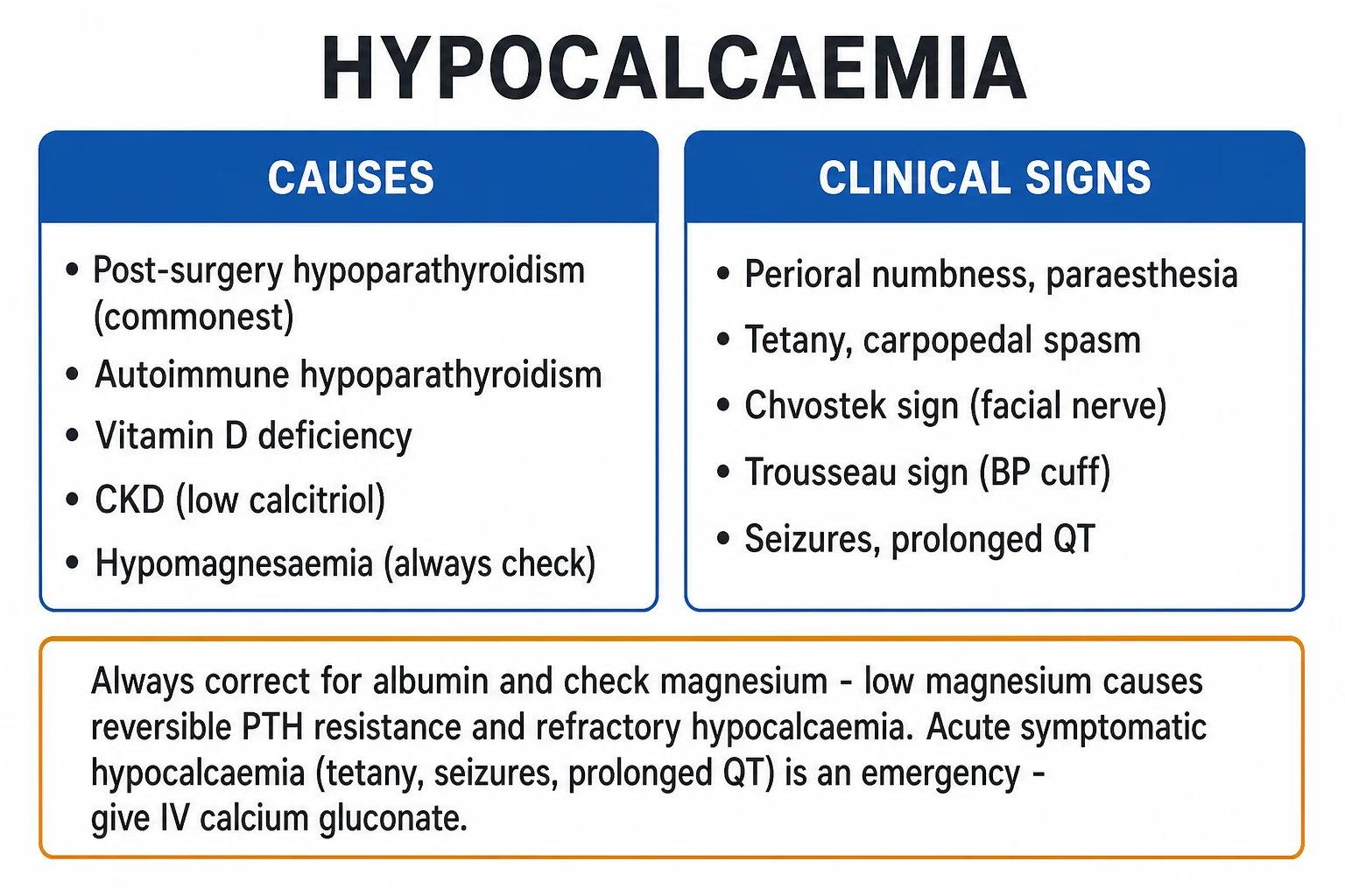
Hypocalcaemia (corrected calcium under 2.20 mmol/L or under 8.5 mg/dL) presents with neuromuscular irritability — perioral numbness and paraesthesia, tetany, carpopedal spasm, Chvostek and Trousseau signs, and in severe cases generalised seizures, laryngeal stridor and a prolonged QT interval. Causes are split by PTH: low or inappropriately normal PTH (post-surgical hypoparathyroidism — the commonest hospital cause, autoimmune / APS-1, DiGeorge 22q11.2 deletion, infiltrative, hypomagnesaemia, activating CaSR mutations, pseudohypoparathyroidism with end-organ resistance) versus high PTH with appropriate secondary hyperparathyroidism (vitamin D deficiency, CKD-MBD, malabsorption, hungry bone syndrome, bisphosphonates / denosumab / foscarnet, citrated massive transfusion). Always correct for albumin and check magnesium — hypomagnesaemia causes reversible PTH resistance and refractory hypocalcaemia. Acute severe symptomatic hypocalcaemia (tetany, seizures, prolonged QT) is treated with IV calcium gluconate 10 percent, 10 to 20 mL (1 to 2 g) over 10 to 20 minutes with cardiac monitoring; chronic management is oral calcium plus active vitamin D (calcitriol 0.25 to 1 mcg daily) and correction of the underlying cause.
On this page & tools
Your progress
Saved locally on this device.
Exam tags
Red flags
Overview & Definition
Hypocalcaemia is a corrected serum calcium below 2.20 mmol/L (8.5 mg/dL) — the lower limit of the adult reference range (total calcium 2.20 to 2.60 mmol/L, or 8.5 to 10.4 mg/dL). Because roughly forty per cent of circulating calcium is protein-bound (mainly to albumin) and only the ionised fraction (about 50 per cent) is physiologically active, every measured total calcium must be corrected for the patient's albumin before any clinical decision is made. The correction formula used in clinical practice is:[1][2]
Corrected Ca (mmol/L) = measured Ca + 0.02 × (40 − albumin in g/L) [1]
An equivalent US-style rule is: add 0.8 mg/dL to the measured calcium for every 1 g/dL that albumin is below 4.0 g/dL. Worked example: a patient with measured calcium 1.90 mmol/L and albumin 25 g/L has a corrected calcium of 1.90 + 0.02 × (40 − 25) = 1.90 + 0.30 = 2.20 mmol/L — i.e. borderline, not profoundly low. Failing to correct for a low albumin (e.g. in cirrhosis, nephrotic syndrome, sepsis) over-diagnoses hypocalcaemia and triggers unnecessary treatment; this is the classic pseudohypocalcaemia trap. When time or doubt exists, direct measurement of ionised calcium (normal 1.10 to 1.30 mmol/L) is the gold standard and is unaffected by albumin.[1]
Three severity bands drive management tempo. Mild hypocalcaemia (corrected Ca 1.90 to 2.20 mmol/L, or ionised Ca 0.9 to 1.1 mmol/L) is usually asymptomatic and managed orally. Moderate (corrected Ca 1.50 to 1.90 mmol/L) produces paraesthesia, a positive Chvostek or Trousseau sign, and usually requires parenteral calcium if symptomatic or rapidly evolving. Severe symptomatic hypocalcaemia (corrected Ca under 1.50 mmol/L, or any level with tetany, carpopedal spasm, seizures, laryngeal stridor, bronchospasm, a prolonged QT interval or hypotension) is a metabolic emergency requiring immediate IV calcium gluconate with cardiac monitoring.[1][2]
The pivotal diagnostic step — and the most frequently missed — is to measure PTH and magnesium simultaneously. PTH is the master regulator: a low or inappropriately normal PTH in the face of hypocalcaemia points to parathyroid gland failure (hypoparathyroidism), while a high PTH (appropriate secondary hyperparathyroidism) points to vitamin D deficiency, CKD, malabsorption, hungry bone or drug effect. Magnesium is the silent trap: severe hypomagnesaemia (serum Mg under 0.4 to 0.5 mmol/L) produces PTH resistance and suppressed PTH secretion, generating hypocalcaemia that is refractory to calcium until magnesium is replaced.[2][10]
Classification
Hypocalcaemia is classified along three independent axes that together determine the differential, the urgency and the treatment. [1]
By PTH response (the key axis)
- LOW or inappropriately NORMAL PTH (PTH-deficient): post-surgical, autoimmune, congenital, infiltrative, hypomagnesaemia, activating CaSR mutation, radiation
- HIGH PTH (PTH-resistant / vitamin-D-deficient state): vitamin D deficiency, CKD-MBD, malabsorption, hungry bone syndrome, drugs (bisphosphonate, denosumab, foscarnet)
- END-ORGAN resistance with HIGH PTH: pseudohypoparathyroidism (GNAS, type 1A with Albright hereditary osteodystrophy)
By tempo / onset
- ACUTE (hours): post-operative, massive transfusion citrate chelation, acute pancreatitis, foscarnet, fluoride poisoning, tumour lysis
- SUBACUTE (days to weeks): hungry bone syndrome, denosumab, severe vitamin D deficiency, sepsis
- CHRONIC (months to years): permanent hypoparathyroidism, pseudohypoparathyroidism, CKD-MBD, untreated malabsorption
By severity
- MILD (corrected Ca 1.90 to 2.20 mmol/L): usually asymptomatic
- MODERATE (1.50 to 1.90 mmol/L): paraesthesia, positive Chvostek / Trousseau
- SEVERE SYMPTOMATIC (under 1.50 mmol/L or any tetany/seizure/stridor/QT prolongation): emergency, IV calcium
Epidemiology & Risk Factors
The commonest cause of hypocalcaemia depends on where the patient is encountered. In the community and outpatient clinic, vitamin D deficiency dominates — subclinical insufficiency (25-OH-vitamin D below 50 nmol/L) is reported in over half of South Asian urban adults and produces a high-PTH hypocalcaemic tendency. In the hospital and post-operative ward, post-surgical hypoparathyroidism after total thyroidectomy is the single most common cause, followed by CKD, critical illness and acute pancreatitis.[3][8]
Post-surgical hypoparathyroidism is the best-characterised iatrogenic cause. A systematic review of more than 60 cohorts reports transient hypocalcaemia in roughly 20 to 40 percent of patients after total thyroidectomy (peaking at 24 to 48 hours) and permanent hypoparathyroidism in roughly 1 to 3 percent (some high-volume centres report under 2 percent; permanent defined as PTH dependence persisting beyond 6 to 12 months). The risk is highest with central neck dissection, re-operation, surgery for Graves' disease or large goitres, low preoperative vitamin D, surgical inexperience, and when parathyroid autotransplantation is not performed. Surgeon-volume and intraoperative nerve/parathyroid identification are the dominant modifiable factors.[8]
Hypocalcaemia — the numbers examiners ask
Beyond surgery, the high-risk groups the examiner expects you to name are: autoimmune polyglandular syndrome type 1 (APS-1 / APECED) — hypoparathyroidism with Addison's disease and chronic mucocutaneous candidiasis in childhood; DiGeorge syndrome (22q11.2 deletion) with parathyroid aplasia and neonatal hypocalcaemia; CKD and dialysis patients (low calcitriol, phosphate retention); malabsorption (coeliac disease, post-bariatric surgery, short bowel, pancreatic insufficiency); magnesium-wasting states (chronic PPI use, loop and thiazide diuretics, alcohol use disorder, Gitelman and Bartter syndromes, cisplatin, amphotericin B); critical illness and sepsis; acute pancreatitis; massive transfusion (citrate chelation, especially with hepatic dysfunction impairing citrate metabolism); tumour lysis syndrome and rhabdomyolysis (phosphate-driven calcium precipitation); and denosumab or bisphosphonate therapy, especially in CKD.[1][3]
Pathophysiology
Calcium homeostasis is governed by a tight PTH–vitamin D–calcium feedback loop. A fall in ionised calcium is sensed within seconds by the calcium-sensing receptor (CaSR) on parathyroid chief cells, triggering PTH release. PTH restores calcium through three coordinated actions: bone resorption (mobilising calcium and phosphate from osteocyte–osteoclast units within minutes), renal calcium reabsorption (in the distal convoluted tubule and thick ascending limb), and phosphate excretion (PTH down-regulates sodium-phosphate cotransport in the proximal tubule, so phosphate is dumped). Critically, PTH also up-regulates renal 1-alpha-hydroxylase, converting 25-hydroxyvitamin D to active 1,25-dihydroxyvitamin D (calcitriol), which then increases intestinal calcium and phosphate absorption. The whole loop closes on the parathyroid CaSR, which suppresses PTH when calcium normalises. Hypocalcaemia results whenever any node of this loop fails — the gland, the substrate (vitamin D), the kidney (calcitriol synthesis, phosphate excretion), or the target organ (bone, gut, kidney).[1][3]
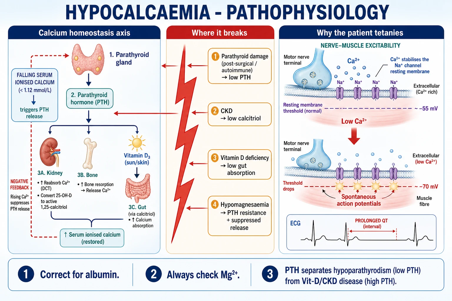
At the molecular level, PTH acts through the PTH1 G-protein-coupled receptor on renal tubular cells and osteoblasts, signalling via Gs-alpha (the GNAS gene product) → adenylate cyclase → cyclic AMP → protein kinase A. This cascade drives renal calcium reabsorption (up-regulating the apical TRPV5 calcium channel and the sodium-calcium exchanger in the distal convoluted tubule) and phosphate excretion (down-regulating the sodium-phosphate cotransporter NPT2a in the proximal tubule), and it switches on the CYP27B1 gene encoding 1-alpha-hydroxylase — the enzyme that converts 25-hydroxyvitamin D into the active 1,25-dihydroxyvitamin D (calcitriol). Calcitriol then binds the vitamin D receptor (VDR) in the gut to drive transcription of the calcium-transport proteins (calbindin-D9k, TRPV6) that absorb dietary calcium. Each of these steps is a potential failure point: GNAS mutation (pseudohypoparathyroidism), magnesium depletion (adenylate cyclase cofactor loss), CYP27B1 loss (CKD), and VDR substrate lack (vitamin D deficiency). The counter-regulatory hormone, calcitonin (from thyroid C-cells), modestly lowers calcium by inhibiting osteoclasts — relevant therapeutically in hypercalcaemia but a minor contributor to hypocalcaemia.[3][11]
The mechanisms of specific causes are exam classics. Hypomagnesaemia causes hypocalcaemia by two hits: magnesium is a cofactor for adenylate cyclase (the PTH signal-transduction enzyme in bone and kidney, producing PTH resistance) and, at severe levels (under 0.4 mmol/L), it suppresses PTH secretion from the parathyroid gland — so the patient has both a non-functioning gland and a resistant target organ, and IV calcium alone is futile until magnesium is replaced.[10] CKD lowers calcium through phosphate retention (hyperphosphataemia complexes circulating calcium), loss of renal 1-alpha-hydroxylase (low calcitriol → reduced gut absorption), skeletal resistance to PTH, and reduced intestinal absorption of calcium. Acute pancreatitis lowers calcium by saponification of peripancreatic fat (calcium–fatty-acid soaps) plus glucagon-stimulated calcitonin release; the historic use of a low calcium as a Ranson/prognostic marker reflects this. Hungry bone syndrome after parathyroidectomy for severe long-standing hyperparathyroidism produces rapid, sustained skeletal uptake of calcium and phosphate into demineralised bone now that the high PTH driving resorption has been removed — generating profound, prolonged hypocalcaemia with hypophosphataemia and hypomagnesaemia lasting days to weeks. Massive transfusion chelates calcium with citrate (the anticoagulant in packed red cells and FFP), which is normally metabolised by the liver within minutes but overwhelms in hepatic dysfunction, hypothermia, or rapid transfusion (over 1 unit every 5 minutes). Bisphosphonates and denosumab (anti-resorptive) acutely block bone efflux of calcium; denosumab discontinuation also causes a rebound hypercalcaemia in some patients but profound hypocalcaemia (sometimes a hungry-bone-like state) in others, especially in CKD.[1][3]
Clinical Presentation
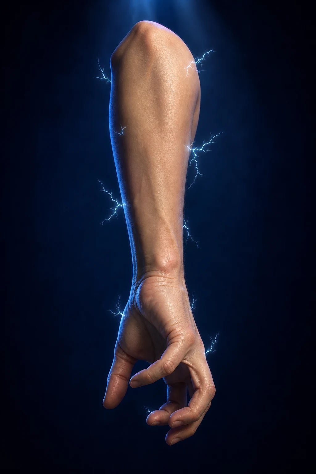
The clinical face of hypocalcaemia is neuromuscular irritability, and the tempo tracks severity. The earliest and most reliable symptom is perioral (circumoral) and distal (fingertips, toes) paraesthesia — a tingling that the patient often describes as "pins and needles". This progresses to muscle cramps, carpopedal spasm, laryngeal stridor and bronchospasm, and at the extreme to generalised tonic-clonic seizures and (rarely) focal neurological deficits that can masquerade as stroke. Carpopedal spasm has a classic shape — the main d'accoucheur (obstetrician's hand) posture with metacarpophalangeal flexion, interphalangeal extension and thumb adduction — and the same pattern occurs in the feet.[1][2]
The two bedside provocation signs are central to clinical assessment. Chvostek sign — tapping the facial nerve (CN VII) anterior to the ear (over the parotid, just below the zygomatic arch) elicits ipsilateral twitching of the facial muscles (upper lip, nasal ala, and sometimes the eye). It is sensitive but not specific: roughly one-quarter to one-half of normal adults have a faintly positive Chvostek, and it is absent in some genuinely hypocalcaemic patients. Trousseau sign — inflating a sphygmomanometer cuff above systolic pressure for 3 minutes — produces the characteristic carpal spasm (main d'accoucheur) within the ischaemic, hypocalcaemic hand. Trousseau is less sensitive but far more specific for hypocalcaemia, and is the sign examiners trust.[1][2]
The two bedside signs examiners distinguish
Cardiac involvement is the lethal dimension. Hypocalcaemia prolongs the QT interval (specifically the ST segment, with a normal T-wave morphology until late), predisposing to ventricular ectopy and torsades de pointes; severe cases cause bradycardia, hypotension, heart failure and reversible cardiomyopathy. Central nervous system features span anxiety, irritability, depression, confusion, frank psychosis and seizures (generalised, focal, or absence). Chronic hypocalcaemia — especially untreated hypoparathyroidism — produces extrapyramidal movement disorders from basal ganglia calcification, papilloedema (raised intracranial pressure), dental enamel hypoplasia, and subcapsular cataracts. The ectodermal phenotype of long-standing disease — dry scaly skin, brittle nails with transverse grooving, coarse brittle hair, alopecia and candidiasis (in APS-1) — is a high-yield stem clue.[1][3]
Atypical presentations are deliberately examined. The elderly patient may present with heart failure, cognitive decline or isolated falls rather than tetany. The post-thyroidectomy patient develops perioral and fingertip tingling within hours of surgery, sometimes progressing to stridor — routine post-operative calcium and PTH checks at 6, 12 and 24 hours are now standard. The breathless patient with laryngeal stridor can be mislabelled as asthma. The neonate may present with irritability, jitteriness, apnoea or seizures, and the pregnant patient on magnesium sulphate for pre-eclampsia can develop functional hypoparathyroidism in the puerperium. Pseudohypoparathyroidism type 1A is the one exam stem where the phenotype (Albright hereditary osteodystrophy — short stature, round face, short fourth metacarpals, subcutaneous ossifications) coexists with hypocalcaemia, high PTH and a normal parathyroid gland.[3][11]
Differential Diagnosis
The differential is best organised by the PTH-based framework, because the PTH result is the single most powerful branch point in the work-up. The key is to interpret PTH relative to the calcium — a "normal-range" PTH in the face of hypocalcaemia is inappropriately low and means parathyroid gland failure, not health.[1][3]
LOW or inappropriately normal PTH (gland failure)
- Post-surgical hypoparathyroidism (commonest) — look for a neck scar and recent thyroidectomy/parathyroidectomy
- Autoimmune: APS-1 / APECED — hypoparathyroidism with Addison's disease and chronic mucocutaneous candidiasis; AIRE gene; childhood onset
- Congenital: DiGeorge syndrome (22q11.2 deletion) — parathyroid aplasia, thymic aplasia (immunodeficiency), cardiac defects, cleft palate; neonatal hypocalcaemia
- Infiltrative: haemochromatosis, Wilson's disease, metastases, granulomatous (sarcoid), amyloidosis, manganese toxicity
- Hypomagnesaemia (under 0.4 mmol/L) — PTH resistance plus suppressed secretion; refractory until Mg replaced
- Activating CaSR or GNA11 mutation (autosomal dominant hypocalcaemia type 1/2) — 'spurious' hypocalcaemia with hypercalciuria and low PTH; do NOT treat with high-dose calcium
- Radiation to the neck; radioactive iodine therapy
HIGH PTH (appropriate secondary hyperparathyroidism)
- Vitamin D deficiency — malabsorption (coeliac, post-bariatric, short bowel, pancreatic insufficiency), sunlight and dietary lack; phosphate LOW
- Chronic kidney disease (CKD-MBD) — phosphate retention plus low calcitriol; phosphate HIGH
- Hungry bone syndrome after parathyroidectomy — rapid skeletal calcium uptake; phosphate LOW (and Mg LOW)
- Drugs: bisphosphonates, denosumab, foscarnet, ketoconazole; phosphate HIGH or LOW
- Massive transfusion / citrate chelation (especially hepatic dysfunction); acute and reversible
- Acute pancreatitis — saponification; phosphate usually normal or high
- Sepsis and critical illness — cytokine-mediated; multifactorial
- Tumour lysis and rhabdomyolysis — phosphate-driven calcium precipitation
Three mimics and pitfalls deserve explicit mention. Pseudohypocalcaemia (low total calcium with normal ionised calcium from hypoalbuminaemia) is corrected by the albumin formula; treating it with calcium is unnecessary and risks hypercalcaemia. Hypocalcaemia from respiratory alkalosis (hyperventilation, salicylate toxicity) lowers the ionised fraction by increasing albumin binding without changing total calcium — and the resulting tetany is identical to true hypocalcaemia. Gadolinium-based MRI contrast can spuriously lower the colorimetric calcium assay (in vitro pseudohypocalcaemia) — recheck after 12 to 24 hours. The prolonged QT differential — hypokalaemia, hypomagnesaemia, hypocalcaemia, drugs (antiarrhythmics, fluoroquinolones, antipsychotics), congenital long-QT syndromes — must always be considered, since torsades is the immediate fatal risk.[1][2]
The rare genetic end-organ resistance group is small but high-yield. Pseudohypoparathyroidism type 1A (maternally inherited GNAS mutation, inactivating the Gs-alpha signalling protein) presents with hypocalcaemia, high PTH, hyperphosphataemia, and the Albright hereditary osteodystrophy phenotype — short stature, round face, obesity, brachydactyly (short fourth metacarpals), subcutaneous ossifications, and often cognitive impairment. Type 1B has the biochemical phenotype without the somatic features (GNAS methylation defect). Type 2 has the biochemistry and a normal cyclic-AMP response to PTH (post-cAMP defect). Distinguishing pseudohypoparathyroidism from primary hypoparathyroidism is the PTH level: high in PHP, low in primary gland failure.[3][11]
Clinical & Bedside Assessment
The bedside assessment has three goals: confirm neuromuscular irritability, screen for the underlying cause, and detect the cardiac emergency. The focused examination begins with the hands — looking for carpopedal spasm, main d'accoucheur, and brachydactyly (short fourth metacarpals — the knuckle-knuckle sign of pseudohypoparathyroidism). Then the face — perioral twitching on tapping the facial nerve (Chvostek), and the cervical skin for a thyroidectomy/parathyroidectomy scar, goitre or neck irradiation marks. The skin, nails, hair and teeth — dryness, brittleness, dental enamel hypoplasia, alopecia, and the mucocutaneous candidiasis of APS-1. The eyes — for subcapsular cataracts and papilloedema. The CNS — for extrapyramidal signs (basal ganglia calcification), cognitive change and seizure activity. And the cardiovascular system — heart failure, bradycardia, and a baseline ECG.[1][3]
Chvostek sign is elicited by tapping the facial nerve just anterior to the ear lobe and below the zygomatic arch (over the parotid) with a reflex hammer; a positive sign is ipsilateral contraction of the facial muscles — most visibly the corner of the mouth, the ala of the nose and the orbicularis oculi. Grading varies (1+ = upper lip only; 2+ = upper lip and alae nasi; 3+ = eye closure; 4+ = whole hemiface). It is sensitive (present in most symptomatic hypocalcaemia) but poorly specific — up to a quarter of normocalcaemic adults have a low-grade positive Chvostek, and it disappears once the patient is treated. Trousseau sign is elicited by inflating a sphygmomanometer cuff to 20 mmHg above systolic pressure for up to 3 minutes; the ischaemia and the cuff pressure together provoke the carpal spasm (main d'accoucheur). A positive Trousseau is highly specific for hypocalcaemia and correlates with a corrected calcium below roughly 1.6 mmol/L. Examiners reward the distinction: Chvostek screens, Trousseau confirms.[1][2]
The ECG is mandatory in any symptomatic or severe hypocalcaemia. The classic finding is ST-segment prolongation causing QTc prolongation (a lengthened plateau of the action potential), with normal T-wave morphology (distinguishing it from congenital long-QT, which has a broad T-wave). T-wave inversion, ST elevation mimicking ischaemia, and torsades de pointes can occur in severe cases. The correction reverses within minutes to hours of IV calcium, providing a useful bedside confirmation of severity.[1]
In the post-thyroidectomy patient, the protocolised bedside assessment is part of the surgical pathway: calcium (and where available PTH) at 6, 12 and 24 hours, voice and stridor check, wound inspection for haematoma, and Chvostek/Trousseau on the ward round. A PTH drawn within 4 hours of completion of surgery that is above 15 pg/mL strongly predicts normocalcaemia and allows safe same-day discharge; a value below 10 pg/mL predicts hypocalcaemia and triggers oral calcium and calcitriol prophylaxis.[8]
Investigations
The first-line panel for any hypocalcaemia is short, focused, and answers the differential in one blood draw. All of the following should be sent before the first dose of IV calcium where possible (treatment must not be delayed in an emergency):[1][3]
First-line hypocalcaemia panel — CAMP-D
CAMP-D
confirm the level; corrected = measured + 0.02 × (40 − albumin g/L)
required for the correction; also screens nephrotic/liver disease
always — under 0.4 mmol/L causes refractory hypocalcaemia
the decoder: low PTH + high phosphate = gland failure; high PTH + low phosphate = vitamin D deficiency; high PTH + high phosphate = CKD
25-OH-D is the nutritional store (target over 50 nmol/L); eGFR for CKD-MBD
The PTH-phosphate decoder is the single most examinable pattern in the work-up and should be reproduced verbatim. Low (or inappropriately normal) PTH with high phosphate = hypoparathyroidism (post-surgical, autoimmune, congenital, infiltrative, hypomagnesaemia, activating CaSR). High PTH with low phosphate = vitamin D deficiency, malabsorption, hungry bone syndrome, post-bisphosphonate/denosumab (PTH is appropriately elevated, phosphate is wasted). High PTH with high phosphate = CKD-MBD (kidney cannot excrete phosphate, cannot make calcitriol). Low PTH with low phosphate is rare and suggests magnesium deficiency, hungry bone syndrome early, or vitamin D deficiency with secondary gland suppression — recheck with clinical context. Magnesium must be measured in every case; severe hypomagnesaemia (under 0.4 to 0.5 mmol/L) produces refractory hypocalcaemia by impairing PTH release and action.[1][10]
Second-line tests are guided by the differential and are not routine. 1,25-dihydroxyvitamin D (calcitriol) is occasionally useful in CKD, granulomatous disease (sarcoid, TB) and suspected activating CaSR/CTBP1 mutations; it is not needed for routine vitamin D deficiency (25-OH-D is the test). Alkaline phosphatase is high in vitamin D deficiency (osteomalacia), Paget's disease and metastatic bone disease. Amylase or lipase screens acute pancreatitis. Morning cortisol and ACTH screen Addison's disease in suspected APS-1; autoantibodies (anti-NALP5, anti-21-hydroxylase) support the diagnosis. Genetic testing (22q11.2 fluorescent in-situ hybridisation or microarray for DiGeorge; AIRE sequencing for APS-1; GNAS methylation and sequencing for pseudohypoparathyroidism; CASR, GNA11 for autosomal dominant hypocalcaemia) is targeted. ECG is mandatory in any symptomatic or severe case (QTc). Head CT or MRI is reserved for symptomatic basal ganglia calcification, papilloedema or seizures with atypical features.[1][3]
Urinary calcium (24-hour, or spot calcium:creatinine ratio) is part of chronic hypoparathyroidism monitoring — the goal is to keep 24-hour urinary calcium under roughly 300 mg (7.5 mmol); persistent hypercalciuria drives nephrocalcinosis and stones and is the trigger to lower calcium/calcitriol, add a thiazide, or escalate to a PTH analogue.[4][6]
Self-test — A 62-year-old woman three days after total thyroidectomy has corrected calcium 1.55 mmol/L, phosphate 1.9 mmol/L (high), PTH 0.8 pg/mL (low), magnesium 0.45 mmol/L. What is the diagnosis and first action?
Post-surgical hypoparathyroidism (low PTH, high phosphate, recent thyroidectomy). First action: IV calcium gluconate 10 percent 10 to 20 mL (1 to 2 g) over 10 to 20 minutes with cardiac monitoring, then a continuous infusion; magnesium is borderline (0.45 mmol/L) — replace with MgSO4 2 g IV over 10 to 20 minutes to prevent refractory hypocalcaemia. The high phosphate reflects loss of PTH's phosphaturic effect and does not need phosphate binders acutely. Once stable, transition to oral calcium plus calcitriol and a post-operative PTH-based risk-stratification pathway.
Management — Resuscitation
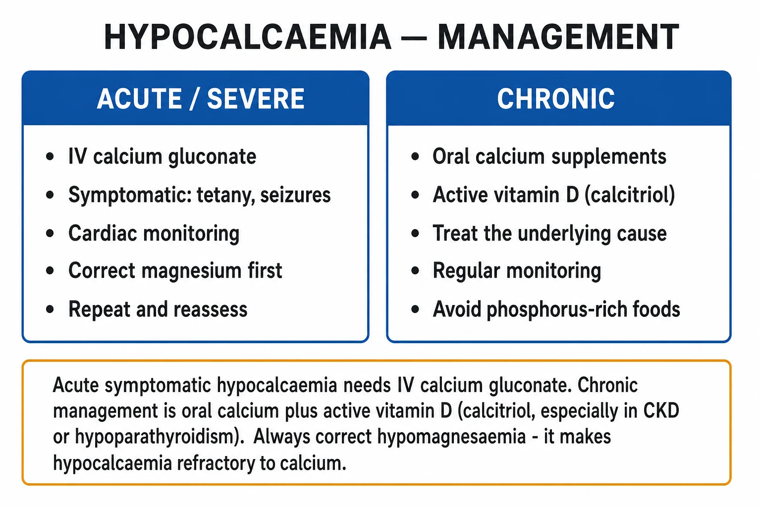
The acute severe symptomatic patient — tetany, carpopedal spasm, laryngeal stridor, bronchospasm, seizures, a prolonged QT interval, or haemodynamic instability — needs immediate IV calcium with continuous cardiac monitoring, secured IV access, and a checked potassium and magnesium drawn before the first dose where possible. The drug of choice is IV calcium gluconate 10 percent, 10 to 20 mL (1 to 2 g of calcium gluconate, equivalent to 90 to 180 mg of elemental calcium), diluted in 50 to 100 mL of 5 percent glucose and given over 10 to 20 minutes through a large peripheral line (calcium gluconate is far less irritant than calcium chloride and is preferred for peripheral use; calcium chloride requires a central line and is reserved for cardiac arrest). Each bolus raises the ionised calcium for only 1 to 2 hours, so a continuous infusion is started immediately afterwards: a typical regimen is 10 ampoules (100 mL) of 10 percent calcium gluconate added to 500 to 900 mL of 5 percent glucose or normal saline (yielding roughly 1 mg elemental calcium per mL), infused at 0.5 to 1.5 mg elemental calcium/kg/hour (about 30 to 100 mL/hour in a 70 kg adult), titrated to symptoms and serial calcium every 4 to 6 hours. For ongoing refractory cases, up to 2 mg/kg/hour may be required, particularly in hungry bone syndrome.[1][2]
Management — Definitive & Stepwise
The chronic management ladder is built around two principles: replace calcium (oral) and the active form of vitamin D (calcitriol or alfacalcidol), and keep the corrected calcium in the low-normal range (about 2.0 to 2.2 mmol/L) to minimise hypercalciuria — the dominant long-term complication of conventional therapy. Native cholecalciferol (vitamin D3) is sufficient for vitamin D deficiency but is inadequate in hypoparathyroidism or CKD, where there is no PTH to drive renal 1-alpha-hydroxylation; in those settings an activated vitamin D (calcitriol 0.25 to 1 mcg/day, or alfacalcidol 1 to 3 mcg/day) is mandatory.[3][6]
Chronic hypoparathyroidism (oral therapy)
- Oral elemental calcium 1 to 2 g/day in divided doses (calcium carbonate 40 percent elemental; calcium citrate 21 percent — preferred in PPI users or achlorhydria)
- Active vitamin D: calcitriol 0.25 to 1 mcg/day (OR alfacalcidol 1 to 3 mcg/day, especially in CKD) — native cholecalciderol is NOT enough
- Target: corrected Ca in low-normal range (2.0 to 2.2 mmol/L) — symptom-free but NOT mid-normal, to avoid hypercalciuria
- Magnesium repletion (oral Mg oxide 400 to 800 mg/day, or IV if severe) as needed
- Thiazide diuretic (hydrochlorothiazide 25 to 50 mg/day or chlorthalidone) to REDUCE urinary calcium — paradoxical use in hypoparathyroidism
- Low-phosphate diet if phosphate is high; separate calcium from iron, levothyroxine, quinolones by at least 2 hours
Refractory / PTH analogue
- Indicated when conventional therapy fails: symptomatic hypocalcaemia despite maximally tolerated calcium/calcitriol, recurrent hypercalciuria or nephrocalcinosis, or intolerable supplement burden
- Palopegteriparatide (TransCon PTH): long-acting PTH analogue, 0.4 to 1.2 mcg/kg/day subcutaneous; PaTHway trial showed sustained Ca normalisation and reduced supplement dose at 52 weeks
- Recombinant human PTH(1-84): 25 to 100 mcg subcutaneous daily; reduces calcium/calcitriol requirements and lowers urinary calcium
- Teriparatide PTH(1-34) — off-label but used; reduces hypercalciuria and supplement burden
- Monitor calcium, phosphate, 24-h urinary calcium, renal function and bone density regularly
- Black-box concerns: osteosarcoma in rat studies (historic), cost, and injection burden limit routine first-line use
The post-thyroidectomy patient deserves a protocolised pathway. The 2021 Edafe systematic review and modern endocrine-surgery consensus support a risk-stratified approach using post-operative PTH (drawn within 4 to 6 hours of surgery). PTH above 15 to 20 pg/mL predicts normocalcaemia — the patient can be discharged without routine supplementation. PTH under 10 pg/mL predicts symptomatic hypocalcaemia — start oral calcium 2 to 3 g elemental/day in divided doses plus calcitriol 0.5 mcg twice daily prophylactically. Intermediate PTH (10 to 15 pg/mL) warrants calcium monitoring at 6, 12 and 24 hours, with supplementation if symptomatic or if corrected Ca falls below 2.0 mmol/L. Most transient cases recover within weeks as stunned or ischaemic parathyroids recover; permanent hypoparathyroidism is defined as PTH dependence persisting beyond 6 to 12 months.[8]
Monitoring targets for chronic therapy: corrected calcium every 3 to 6 months once stable (more often during dose titration or intercurrent illness), 24-hour urinary calcium yearly (target under 300 mg/day; if rising, lower calcium, add a thiazide, or consider PTH analogue), renal function and renal ultrasound yearly (nephrocalcinosis screening), and ophthalmology review for cataracts. Patients must be counselled to recognise early symptoms (perioral tingling, paraesthesia, cramps) and to take an extra dose of oral calcium — and to seek emergency care for seizures, stridor or syncope.[4][6]
Specific Subtypes & Scenarios
Post-surgical hypoparathyroidism is the prototype and the commonest cause in hospital. Mechanisms include direct gland excision, devascularisation, thermal injury, haematoma stripping, or stunning from pre-existing disease. Risk is highest with central neck dissection, re-operation, Graves' disease, large goitres, low vitamin D and surgeon inexperience. Transient hypoparathyroidism (recovery within 6 months) reflects reversible stunning; permanent (beyond 6 to 12 months) reflects irreversible gland loss. Parathyroid autotransplantation of one or more identified glands into the sternocleidomastoid or forearm reduces permanent rates substantially.[8]
Autoimmune hypoparathyroidism in APS-1 (autoimmune polyendocrinopathy-candidiasis-ectodermal dystrophy, APECED) is an autosomal recessive disorder caused by mutations in the AIRE gene on chromosome 21p22. The classic triad is chronic mucocutaneous candidiasis (classically the first manifestation, often under 5 years), hypoparathyroidism (usually before 10 years), and Addison's disease (usually before 15 years) — two of three make the diagnosis. Hypoparathyroidism is the most common endocrine component and is driven by autoantibodies against parathyroid tissue (NALP5 autoantibody). Management combines calcium/calcitriol with candidiasis treatment (azoles), glucocorticoid and mineralocorticoid replacement for Addison's, and screening for the broader spectrum (hypogonadism, hepatitis, malabsorption, pernicious anaemia, enamel hypoplasia, asplenia).[3]
DiGeorge syndrome (22q11.2 microdeletion) is the most common chromosomal microdeletion syndrome (roughly 1 in 4000 live births). The classic tetrad is congenital cardiac defects (conotruncal anomalies — truncus arteriosus, interrupted aortic arch, tetralogy of Fallot), thymic aplasia or hypoplasia (T-cell immunodeficiency), cleft palate, and hypoparathyroidism from parathyroid aplasia. Neonatal hypocalcaemia is the typical presentation; seizures in the first weeks of life warrant a calcium and 22q11 work-up. Calcium abnormalities can recur in adolescence or adulthood, including in pregnancy.[12]
Hypocalcaemia of CKD (CKD-MBD) arises from phosphate retention, reduced 1,25-dihydroxyvitamin D (loss of 1-alpha-hydroxylase), skeletal resistance to PTH and reduced gut calcium absorption. Management targets all four: phosphate binders (calcium acetate, sevelamer, lanthanum) taken with meals to trap dietary phosphate; active vitamin D (calcitriol or paricalcitol) to support calcium and suppress PTH; dialysis to remove phosphate; and cinacalcet (calcimimetic) to lower PTH in secondary hyperparathyroidism. Denosumab in dialysis patients can cause profound, prolonged hypocalcaemia (a hungry-bone-like state) — pre-dose calcium, vitamin D and phosphate optimisation, and post-dose monitoring are mandatory.[3]
Hungry bone syndrome follows parathyroidectomy for severe, long-standing hyperparathyroidism (especially with osteitis fibrosa cystica, high preoperative alkaline phosphatase, large adenomas or parathyroid carcinoma). The skeleton, starved of calcium under chronic high PTH, abruptly re-mineralises when PTH is removed, producing profound, prolonged hypocalcaemia with hypophosphataemia and hypomagnesaemia lasting days to weeks. Management requires high-dose IV calcium (often 1 to 2 mg elemental/kg/hour), calcitriol 1 to 2 mcg/day, magnesium replacement, and frequent monitoring of calcium, phosphate and magnesium every 4 to 6 hours. Pre-operative recognition (high ALP, high bone turnover markers) allows pre-loading with calcium and calcitriol.[1]
Hypomagnesaemia-induced hypocalcaemia is the silent cause of refractory hypocalcaemia. Common offenders are chronic PPI use, loop and thiazide diuretics, alcohol use disorder, cisplatin, amphotericin B, aminoglycosides, Gitelman and Bartter syndromes, extensive bowel resection, and refeeding syndrome. The mechanism is PTH resistance (Mg is a cofactor for adenylate cyclase) plus suppressed PTH secretion at severe levels (under 0.4 to 0.5 mmol/L). The rule: if hypocalcaemia is refractory, check and correct magnesium first — IV MgSO4 2 g over 10 to 20 minutes for symptomatic/severe, then oral maintenance; oral magnesium oxide 400 to 800 mg/day (or magnesium glycerophosphate in PPI users) for chronic repletion.[10]
Pseudohypoparathyroidism type 1A is the rare but exam-classic end-organ resistance state. Patients have hypocalcaemia, hyperphosphataemia and a HIGH PTH (the gland works, the kidney does not respond) with the Albright hereditary osteodystrophy (AHO) phenotype — short stature, round face, obesity, brachydactyly (especially short fourth metacarpals — the missing-knuckle sign), subcutaneous ossifications and variable cognitive impairment. The defect is maternally inherited inactivation of Gs-alpha (the GNAS gene product); paternal inheritance produces the AHO phenotype without the biochemistry (pseudo-pseudohypoparathyroidism). Management is oral calcium and calcitriol as for primary hypoparathyroidism; PTH analogues are usually not needed because the bone still responds.[3][11]
Drug-induced hypocalcaemia rounds out the high-yield scenarios. Bisphosphonates (especially zoledronate) and denosumab acutely block bone calcium efflux — profound in CKD, with denosumab carrying the additional risk of a hungry-bone-like state on discontinuation. Foscarnet (an antiviral used for CMV and HSV) chelates ionised calcium directly. Cisplatin causes renal magnesium and calcium wasting. Fluoride toxicity (chronic exposure in endemic regions, or acute poisoning) forms calcium-fluoride complexes and inhibits parathyroid function. Citrated blood products (massive transfusion, plasmapheresis, leukodepletion) chelate calcium — most relevant in hepatic dysfunction, hypothermia, or rapid transfusion. Loop diuretics increase renal calcium excretion; calcitonin, mithramycin, plicamycin, gallium nitrate (rarely used) lower calcium. EGF-inhibitors and some kinase inhibitors occasionally cause hypomagnesaemia-driven hypocalcaemia.[1][3]
Complications & Pitfalls
The acute life-threatening complications of severe hypocalcaemia are laryngeal stridor, bronchospasm, generalised seizures, ventricular arrhythmia (including torsades de pointes from prolonged QT), hypotension, and reversible heart failure or cardiomyopathy. These are the indications for immediate IV calcium. The central nervous system complications of chronic untreated disease — basal ganglia calcification with extrapyramidal movement disorder, subcapsular cataracts, papilloedema from raised intracranial pressure, dental enamel hypoplasia, osteosclerosis and (in children) impaired neurodevelopment — are largely preventable with adequate calcium and active vitamin D replacement.[1][3]
The complications of treatment are dominated by hypercalciuria (the unavoidable consequence of replacing calcium without PTH, which normally reclaims urinary calcium). Sustained hypercalciuria drives nephrocalcinosis, nephrolithiasis and progressive renal impairment — the reason the chronic target is low-normal calcium, not mid-normal. Over-treatment produces hypercalcaemia (nausea, vomiting, polyuria, constipation, confusion, shortened QT). The pitfall of treating without correcting magnesium is refractory hypocalcaemia with escalating, futile calcium doses, extravasation tissue necrosis, and delayed recovery.[2][10]
The surgical pitfall of missing post-operative hypocalcaemia is delayed discharge or readmission with stridor, seizure or syncope — modern protocols using early post-operative PTH and prophylactic calcium/calcitriol in high-risk patients have substantially reduced this. The digoxin-toxicity pitfall is IV calcium precipitating fatal arrhythmia — relative caution in patients on digoxin. The hyperphosphataemia pitfall is tissue calcium-phosphate precipitation when large calcium boluses are given in CKD or tumour lysis — lower phosphate first. And the overshoot pitfall in hungry bone syndrome is giving too little calcium too late, allowing symptomatic hypocalcaemia to persist for days — early, aggressive replacement guided by frequent monitoring is the answer.[1][8]
Prognosis & Disposition
Acute hypocalcaemia resolves rapidly with IV calcium, magnesium correction and treatment of the underlying cause (pancreatitis resolves, citrate is metabolised, drugs are withdrawn). Post-surgical transient hypoparathyroidism recovers in most patients within weeks as stunned parathyroids recover; about 1 to 3 percent become permanent and require lifelong therapy. Chronic hypoparathyroidism is the only classical endocrine deficiency not historically treated with the missing hormone — conventional calcium/calcitriol therapy controls biochemistry but does not restore renal calcium conservation, leaving patients with reduced quality of life, recurrent symptoms and a real risk of renal complications. The introduction of PTH analogues (palopegteriparatide, PTH 1-84) has shifted this for refractory patients.[3][7]
Disposition follows severity. Severe symptomatic hypocalcaemia (tetany, seizures, stridor, prolonged QT, haemodynamic instability) goes to a monitored bed with continuous ECG, IV calcium infusion, and ICU if there is ongoing seizure, stridor, arrhythmia or shock. Mild to moderate asymptomatic or chronically compensated hypocalcaemia is managed orally as an outpatient with calcium, active vitamin D, magnesium as needed, and endocrinology follow-up. The post-operative patient is managed by protocol: PTH-based risk stratification, prophylactic calcium/calcitriol in high-risk patients, and 24-hour observation where indicated. Endocrinology referral is indicated for chronic or complex disease, refractory cases, suspected APS-1/DiGeorge/pseudohypoparathyroidism, pregnancy, or when a PTH analogue is being considered.[1][6]
Special Populations
Neonatal hypocalcaemia is divided by timing. Early-onset (first 72 hours) reflects prematurity, maternal diabetes, perinatal asphyxia, or maternal hyperparathyroidism (transient suppression of the neonatal parathyroids). Late-onset (after 72 hours, peaking at 5 to 7 days) classically follows high-phosphate feeds (whole cow's milk), maternal vitamin D deficiency, hypomagnesaemia, or DiGeorge syndrome. Presentation is non-specific — irritability, jitteriness, tremor, feeding difficulty, apnoea, seizures. Treatment is oral calcium glubionate or gluconate, with IV calcium gluconate for seizures or stridor (slow, with ECG monitoring, due to risk of arrhythmia and tissue necrosis from small veins).[1]
Pregnancy and lactation demand increased calcium and vitamin D. Hypoparathyroid patients usually need an increased dose of calcium and calcitriol during pregnancy (placental calcium drain, increased glomerular filtration) and a reduction post-partum if lactating (breast milk calcium) — close monitoring each trimester is essential. DiGeorge syndrome can present for the first time in pregnancy with hypocalcaemia. Pre-eclampsia treated with magnesium sulphate produces functional hypoparathyroidism from magnesium-induced PTH suppression, and the neonate can develop hypocalcaemia — neonatal calcium monitoring is warranted. Calcium and calcitriol are safe in pregnancy and lactation; bisphosphonates and denosumab are contraindicated.[5][12]
The elderly often present atypically — cognitive decline, falls, heart failure or isolated arrhythmia rather than classical tetany; a low threshold for a calcium level in any confused, falling or breathless older patient is warranted, particularly in care-home residents. Common drivers are vitamin D deficiency, malabsorption, polypharmacy (PPIs, loop and thiazide diuretics, bisphosphonates), CKD, post-surgical disease and immobilisation. The CKD and dialysis patient needs active vitamin D (calcitriol or paricalcitol — the failing kidney cannot perform 1-alpha-hydroxylation), phosphate binders (calcium acetate, sevelamer, lanthanum, sucroferric oxyhydroxide) taken with meals, careful cinacalcet use (a calcimimetic that lowers PTH but can worsen hypocalcaemia), and denosumab caution — denosumab in dialysis patients can precipitate profound, prolonged hypocalcaemia that is hard to reverse and may warrant pre-dose calcium, vitamin D and phosphate optimisation plus inpatient monitoring. After renal transplant, hypocalcaemia can occur from hungry-bone recovery as secondary hyperparathyroidism resolves and the graft restores calcitriol synthesis — usually transient but occasionally needing short-term calcium/calcitriol. Patients on denosumab or bisphosphonates, especially with CKD (eGFR under 30), can develop profound, prolonged hypocalcaemia that mimics hungry bone syndrome; pre-treatment calcium/vitamin D optimisation and post-dose monitoring with serial calcium for at least two weeks are standard. The anticoagulated patient on warfarin or a DOAC is at additional bleeding risk if invasive procedures are needed and calcium/calcitriol dosing is unaffected, but IV cannulae must be secured to avoid haematoma. The immunocompromised patient (HIV, post-transplant) is predisposed to hypomagnesaemia from tenofovir, amphotericin B and tacrolimus, driving refractory hypocalcaemia — magnesium monitoring is essential.[1][3]
Evidence, Guidelines & Regional Differences
The Endocrine Society 2016 and the European Society of Endocrinology 2022 and 2025 guidelines establish the modern framework for chronic hypoparathyroidism management. The 2025 ESE revision reaffirms the low-normal calcium target (to minimise hypercalciuria), the 24-hour urinary calcium ceiling of roughly 300 mg/day, and PTH analogues as second-line for refractory disease or recurrent hypercalciuria. The PaTHway trial (2025) of palopegteriparatide (TransCon PTH) — a long-acting PTH prodrug given as a once-daily subcutaneous injection (0.4 to 1.2 mcg/kg/day) — showed sustained normocalcaemia at 52 weeks with reduced calcium and calcitriol supplementation and improved quality of life in adults with chronic hypoparathyroidism, establishing it as the leading second-line agent. Recombinant PTH(1-84) and teriparatide remain alternatives.[4][9]
The Endocrine Society (US), European Society of Endocrinology (ESE) and UK NICE Clinical Knowledge Summary converge on the core principles: albumin-corrected calcium, always check magnesium and PTH, IV calcium for severe symptomatic disease, oral calcium plus activated vitamin D for chronic hypoparathyroidism and CKD, low-normal target to avoid hypercalciuria, and PTH analogue for refractory disease. The differences are largely in access and cost.
Controversies and evolving evidence include the optimal vitamin D target (most guidelines endorse 25-OH-D over 50 nmol/L, with 75 nmol/L preferred in CKD/pregnancy), the role of routine post-operative PTH (now widely adopted but variably implemented), the place of PTH analogues (cost, route, long-term skeletal safety, and the historic black-box for osteosarcoma in rat studies), and the management of denosumab discontinuation — between rebound hypercalcaemia and hungry-bone-like hypocalcaemia, the net effect varies and monitoring is essential.[3][7]
Exam Pearls
Causes of hypocalcaemia — CHOMP
CHOMP
low calcitriol (loss of 1-alpha-hydroxylase) plus phosphate retention; high PTH, high phosphate
PTH resistance plus suppressed secretion — refractory; ALWAYS check and fix magnesium first
post-surgical hypoparathyroidism — the commonest hospital cause; look for the neck scar
coeliac, post-bariatric, short bowel, sunlight and dietary lack; high PTH, low phosphate
saponification; autoimmune/DiGeorge; GNAS Albright hereditary osteodystrophy; pre-eclampsia/Mg; bisphosphonate, denosumab, foscarnet, citrate
Exam application bank (NEET-PG / INICET)
One-line answer
Hypocalcaemia (corrected calcium under 2.20 mmol/L or under 8.5 mg/dL) presents with neuromuscular irritability — perioral numbness and paraesthesia, tetany, carpopedal spasm, Chvostek and Trousseau signs, and in severe cases generalised seizures, laryngeal stridor and a prolonged QT interval. Causes are split by PTH: low or inappropriately normal PTH (post-surgical hypoparathyroidism — the commonest hospital cause, autoimmune / APS-1, DiGeorge 22q11.2 deletion, infiltrative, hypomagnesaemia, activating CaSR mutations, pseudohypoparathyroidism with end-organ resistance) versus high PTH with appropriate secondary hyperparathyroidism (vitamin D deficiency, CKD-MBD, malabsorption, hungry bone syndrome, bisphosphonates / denosumab / foscarnet, citrated massive transfusion). Always correct for albumin and check magnesium — hypomagnesaemia causes reversible PTH resistance and refractory hypoca [1]
Worked stems (answer without another resource)
Stem 1 — Classic presentation. Map symptoms to mechanism; name the first investigation and first treatment step with dose/route if drug therapy is standard. [1]
Stem 2 — Unstable / complicated. List red flags that force immediate resuscitation, theatre, ICU, antidote, or reperfusion — and what you do in the first 15 minutes. [1]
Stem 3 — Atypical group. Elderly, pregnancy, child, or immunocompromised: how presentation and thresholds change. [1]
Stem 4 — Differential trap. Name the three closest mimics and one discriminator for each. [1]
Stem 5 — Disposition. Who goes home with safety-netting, who is admitted, who needs HDU/ICU/theatre, and what follow-up is mandatory. [1]
Rapid viva checklist
- Definition + classification
- Pathophysiology chain
- Bedside signs / criteria
- Score with exact components (if any)
- Emergency bundle
- Definitive therapy with doses
- Complications of disease and of treatment
- Special populations
- Guideline/trial name if classic
- Three exam traps
Coverage self-check
If you cannot answer any stem above from this page alone, re-read the matching section — the page is intended to be self-sufficient for final-prof and NEET-PG/INICET questions on Hypocalcaemia.
References
- [1]Pepe J, Colangelo L, Biamonte F, et al. Diagnosis and management of hypocalcemia Endocrine, 2020.PMID 32367335
- [2]Cooper MS, Gittoes NJ. Diagnosis and management of hypocalcaemia BMJ, 2008.PMID 18535072
- [3]Mannstadt M, Bilezikian JP, Thakker RV, et al. Hypoparathyroidism Nat Rev Dis Primers, 2017.PMID 28857066
- [4]Bollerslev J, Rejnmark L, Marcocci C, et al. Revised European Society of Endocrinology Clinical Practice Guideline: Treatment of Chronic Hypoparathyroidism in Adults Eur J Endocrinol, 2025.PMID 41231236
- [5]Bollerslev J, Rejnmark L, Cohen A, et al. European Expert Consensus on Practical Management of Specific Aspects of Parathyroid Disorders in Adults and in Pregnancy: Recommendations of the ESE Educational Program of Parathyroid Disorders Eur J Endocrinol, 2022.PMID 34863037
- [6]Khan AA, Koch CA, Van Uum SHM, et al. Management of Hypoparathyroidism J Bone Miner Res, 2022.PMID 36161671
- [7]Khan S, Jit M, Mannstadt M. Hypoparathyroidism: diagnosis, management and emerging therapies Nat Rev Endocrinol, 2025.PMID 39905273
- [8]Edafe O, Antakia R, Laskar N, et al. Systematic review of incidence, risk factors, prevention and treatment of post-laryngectomy hypoparathyroidism Eur Arch Otorhinolaryngol, 2021.PMID 32700234
- [9]Clarke BL, Kay Berg C, Fox J, et al. Efficacy and Safety of TransCon PTH in Adults With Hypoparathyroidism: 52-Week Results From the Phase 3 PaTHway Trial J Clin Endocrinol Metab, 2025.PMID 39376010
- [10]Bobrowicz M, Glinicki B, Skrzypa M, et al. Hypomagnesaemia leading to parathyroid dysfunction, hypocalcaemia, and hypokalaemia as a complication of long-term treatment with a proton pump inhibitor - a literature review Endokrynol Pol, 2024.PMID 39279305
- [11]Wang Y, Wang R, Zhang Y, et al. Variable Bone Phenotypes in Patients with Pseudohypoparathyroidism Curr Osteoporos Rep, 2023.PMID 37014531
- [12]Shah MK, Wijeratne DT, Al Khalili Y, et al. Management of hypoparathyroidism during pregnancy following late maternal diagnosis of DiGeorge syndrome: a case report J Med Case Rep, 2026.PMID 41527007