Endocrinology · General Medicine
Hypopituitarism
Also known as Hypopituitarism · Panhypopituitarism · Pituitary insufficiency · Sheehan syndrome · Pituitary apoplexy
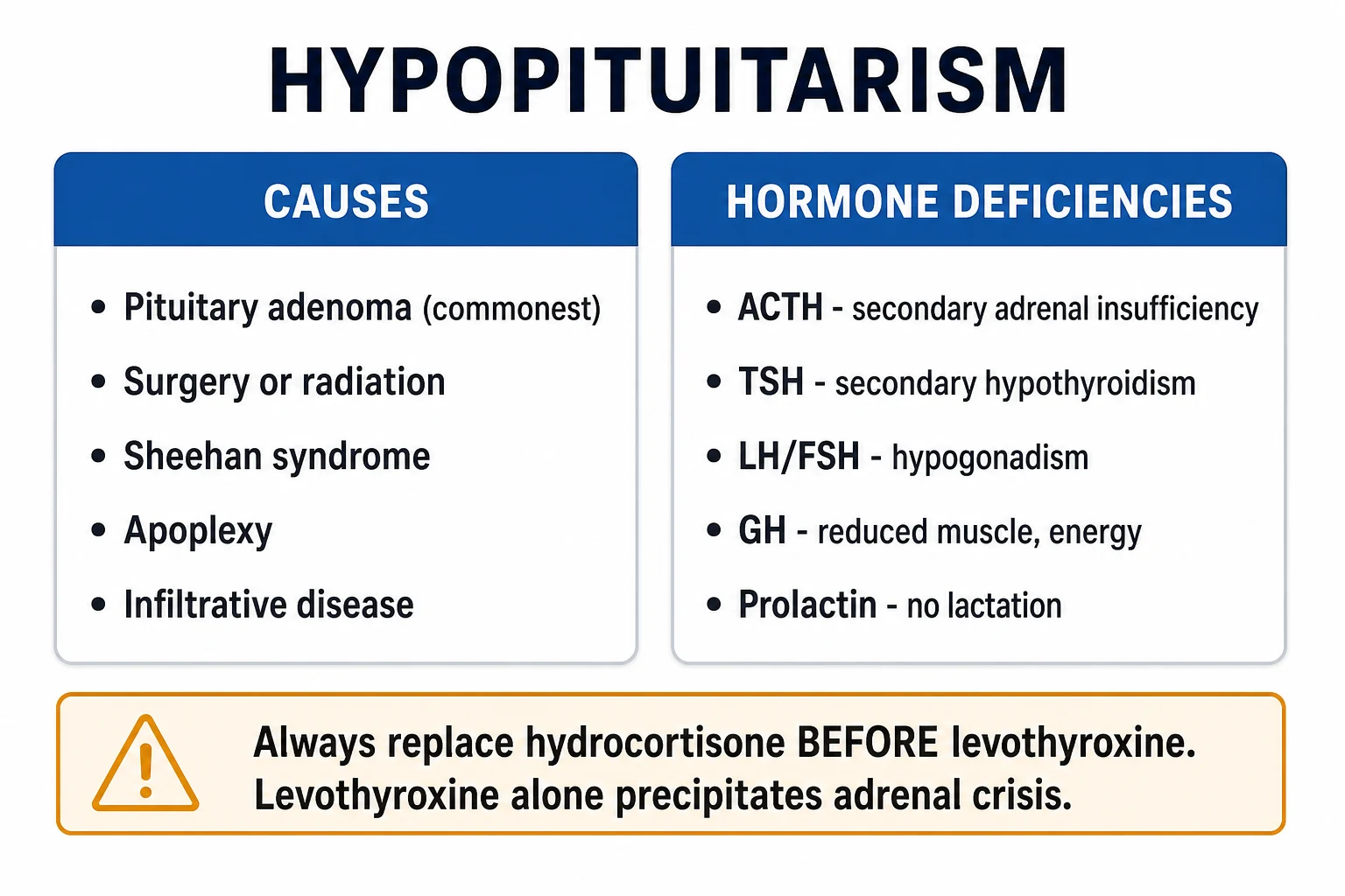
Hypopituitarism is the partial or complete deficiency of one or more anterior pituitary hormones (GH, PRL, ACTH, TSH, LH/FSH); loss of all anterior hormones is panhypopuitarism. It is caused most commonly by a pituitary adenoma, surgery or radiation, Sheehan syndrome (postpartum ischaemic necrosis), pituitary apoplexy, infiltrative disease (sarcoidosis, haemochromatosis, Langerhans cell histiocytosis), autoimmune hypophysitis (including immune-checkpoint inhibitors), genetic mutations (PROP1, POU1F1), or traumatic brain injury. Each hormone deficiency produces distinct features: ACTH deficiency causes secondary adrenal insufficiency (fatigue, postural hypotension, hypoglycaemia, hyponatraemia — but no hyperpigmentation and no hyperkalaemia, distinguishing it from primary Addison disease); TSH causes secondary hypothyroidism; LH/FSH causes hypogonadism (loss of libido, amenorrhoea, erectile dysfunction, infertility); GH causes reduced muscle mass, central adiposity and, in children, growth failure; prolactin causes failure of lactation. Large lesions cause bitemporal hemianopia and headache. Diagnosis shows low target hormones with low or inappropriately normal trophic hormones, confirmed by pituitary MRI. Management is hormone replacement — hydrocortisone FIRST (always before levothyroxine, to avoid precipitating adrenal crisis), then levothyroxine, sex steroids, growth hormone, and desmopressin for diabetes insipidus.
On this page & tools
Your progress
Saved locally on this device.
Exam tags
Red flags
Overview & Definition
Hypopituitarism is the partial or complete deficiency of one or more anterior pituitary hormones — growth hormone (GH), prolactin (PRL), adrenocorticotrophic hormone (ACTH), thyroid-stimulating hormone (TSH), luteinising hormone (LH) and follicle-stimulating hormone (FSH). The complete loss of all anterior hormones is termed panhypopituitarism. The posterior pituitary — which releases antidiuretic hormone (ADH/vasopressin) and oxytocin — is usually spared in primary pituitary destruction (because these hormones are synthesised in hypothalamic nuclei and only stored posteriorly), but is lost with stalk or hypothalamic lesions, producing diabetes insipidus.[1]
The anterior pituitary (adenohypophysis) and posterior pituitary (neurohypophysis) differ in embryology and physiology, and this distinction matters clinically. The anterior lobe arises from Rathke's pouch (oral ectoderm) and is driven by hypothalamic releasing hormones (GHRH, TRH, CRH, GnRH, dopamine) that reach it via the hypophyseal portal venous system. The posterior lobe is a neural downgrowth that stores hormones synthesised in the supraoptic and paraventricular nuclei of the hypothalamus. A mass or infiltrative lesion that destroys the anterior gland therefore tends to spare ADH, while a lesion of the stalk or hypothalamus (e.g. craniopharyngioma, metastasis) interrupts both portal input and the neural tract, producing panhypopituitarism plus diabetes insipidus.[1][2]
The clinical face of hypopituitarism ranges from an insidious, years-long decline (vague fatigue, weight loss, amenorrhoea, loss of body hair) to a rapid, life-threatening presentation in pituitary apoplexy or adrenal crisis. The two clinical rules that save lives are absolute: always replace hydrocortisone before levothyroxine (levothyroxine alone increases metabolic clearance of cortisol and precipitates crisis in uncorrected ACTH deficiency), and recognise pituitary apoplexy as a neurosurgical emergency.[1][3]
[1]Classification
Hypopituitarism is classified along four axes that examiners test deliberately.[1][2]
By number of axes involved: isolated (single-hormone deficiency, e.g. isolated GH deficiency, isolated ACTH deficiency), multiple (two or more axes), or panhypopituitarism (all anterior hormones). Isolated deficiencies are usually genetic (e.g. PROP1 lineage) or autoimmune; multiple and panhypopituitarism are typically mass or vascular. [1]
By anatomical site: primary (pituitary destruction — adenoma, Sheehan, apoplexy, infiltrate, surgery) versus secondary (hypothalamic or stalk disease — craniopharyngioma, metastasis, sarcoidosis — where the releasing-hormone drive is lost). Secondary causes are more likely to involve diabetes insipidus and a mild prolactin rise from loss of dopamine (the prolactin-inhibiting hormone) via the stalk effect. [1]
By onset: congenital (genetic: PROP1, POU1F1, HESX1 mutations; septo-optic dysplasia) versus acquired; and acute (pituitary apoplexy) versus chronic (slowly growing macroadenoma, radiotherapy). [1]
By aetiology — the classification examiners reward most: [1]
| Category | Examples |
|---|---|
| Neoplastic | Pituitary adenoma (commonest), craniopharyngioma (commonest in children/young adults), meningioma, metastases (breast, lung), Rathke cleft cyst |
| Iatrogenic | Transsphenoidal surgery, radiotherapy (progressive over months to years) |
| Vascular | Sheehan syndrome (postpartum ischaemic necrosis), pituitary apoplexy (haemorrhage or infarction into an adenoma) |
| Infiltrative / inflammatory | Sarcoidosis, haemochromatosis, Langerhans cell histiocytosis, IgG4-related disease, autoimmune hypophysitis (including immune-checkpoint inhibitors — ipilimumab, nivolumab, pembrolizumab) |
| Infective | Tuberculosis, syphilis, fungal (Aspergillus, Cryptococcus), pituitary abscess |
| Traumatic | Traumatic brain injury (TBI), subarachnoid haemorrhage, shearing of the stalk |
| Genetic / developmental | PROP1, POU1F1, HESX1 mutations; septo-optic dysplasia; primary empty sella |
| Idiopathic | Idiopathic hypopituitarism (a diagnosis of exclusion) |
Epidemiology & Risk Factors
Hypopituitarism has a prevalence of roughly 45 to 80 per 100,000 and an incidence of about 4 per 100,000 per year, with a slight female predominance driven by Sheehan syndrome and autoimmune hypophysitis. The commonest cause in adults is a pituitary adenoma (especially non-functioning macroadenoma); in children and adolescents, craniopharyngioma and genetic combined pituitary hormone deficiency predominate. Mortality is increased two- to three-fold compared with the general population, driven by cardiovascular and cerebrovascular disease (untreated GH and gonadal deficiency) and preventable adrenal crises.[2]
Risk factors cluster by mechanism and should be sought actively in the history: [1]
Mass / compressive
- Pituitary adenoma (commonest), craniopharyngioma, meningioma, metastasis
- Parasellar tumours; Rathke cleft cyst
- Produces hormone loss in order GH → LH/FSH → TSH → ACTH → PRL
Iatrogenic
- Transsphenoidal surgery (often transient DI, variable anterior deficits)
- Pituitary radiotherapy — progressive over 5 to 10 years
- Always screen all axes pre- and post-operatively
Vascular
- Sheehan: severe postpartum haemorrhage and shock
- Pituitary apoplexy: anticoagulation, pregnancy, DM, hypertension
- Sickle cell disease; increased intracranial pressure
Infiltrative / autoimmune
- Sarcoidosis, haemochromatosis, Langerhans cell histiocytosis
- IgG4-related disease
- Immune-checkpoint inhibitors (ipilimumab > anti-PD1)
Hypopituitarism — headline numbers
Sheehan syndrome is now rare where obstetric care is available, but remains common in regions with home or difficult delivery, where severe postpartum haemorrhage and shock produce low-flow ischaemic necrosis of the enlarged pregnant pituitary — the gland enlarges up to two-fold in pregnancy while its low-pressure portal vascular supply is vulnerable to hypoperfusion. Pituitary apoplexy occurs in roughly 2 to 12 percent of pituitary adenomas, often as the first presentation of an undiagnosed tumour; risk is increased by anticoagulation, pregnancy, bromocriptine initiation, head trauma, hypertension and diabetes.[4]
Pathophysiology
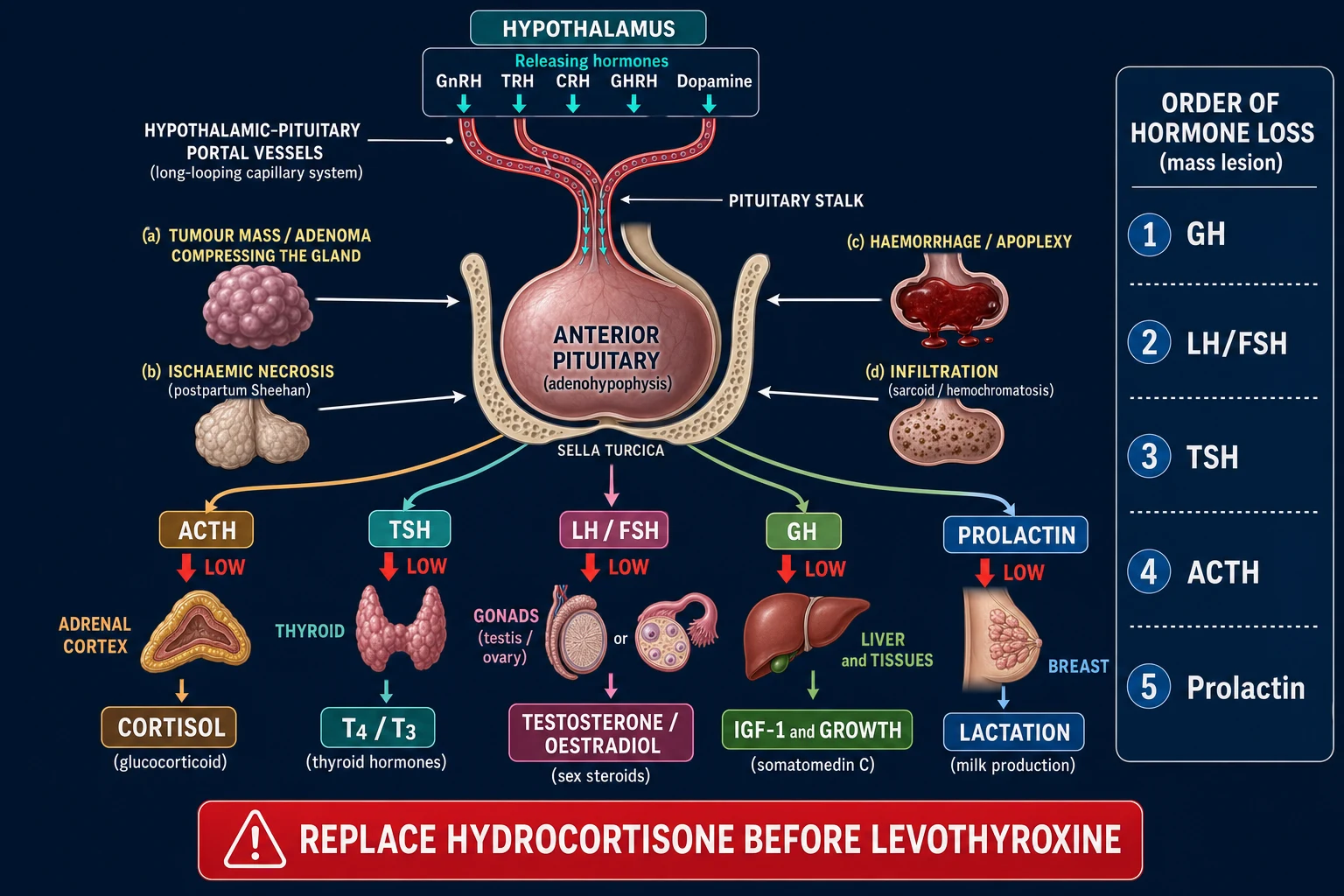
The anterior pituitary is driven by hypothalamic releasing hormones (GHRH, TRH, CRH, GnRH) that reach it via the hypophyseal portal venous system, a low-pressure capillary network that is uniquely vulnerable to ischaemia. The trophic hormones — ACTH, TSH, LH/FSH, GH and prolactin — then act on downstream target glands (adrenal cortex, thyroid, gonads, liver/bone/tissues, breast). Anything that destroys the gland, compresses it, interrupts the stalk, or replaces normal tissue removes that drive, and the target glands then atrophy through lack of trophic stimulation.[1]
Mass lesions cause hormone loss in a predictable order, reflecting the spatial distribution of cell types and the vulnerability of the portal supply: GH first, then LH/FSH, then TSH, then ACTH, then prolactin. Prolactin is often preserved or even elevated in stalk compression, because loss of hypothalamic dopamine (the prolactin-inhibiting hormone) removes tonic inhibition — the stalk effect. This explains why a patient with a non-functioning macroadenoma may present with galactorrhoea and amenorrhoea despite a normal lactotroph population: the prolactin rise is mechanistic, not neoplastic.[2]
Order of hormone loss in a pituitary mass — GO-LAT-TH
GO-LAT
lost FIRST — most vulnerable; reduced muscle, growth failure in children
gonadotrophins second — amenorrhoea, low libido, erectile dysfunction
thyroid axis third; secondary hypothyroidism
fourth — the LETHAL one; cortisol loss gives adrenal crisis
PRL last; but PRL often RISES via the stalk effect
Why ACTH deficiency is the lethal axis: cortisol loss impairs vascular tone (permissive effect on catecholamines), gluconeogenesis, and the stress response, producing adrenal crisis — hypotension, hypoglycaemia, hyponatraemia, abdominal pain, and shock. Crucially, aldosterone is spared because it is driven by the renin–angiotensin–aldosterone system (RAAS), not ACTH — so there is no hyperkalaemia and no salt-wasting (unlike primary Addison disease, where zona glomerulosa destruction removes aldosterone). This single physiological fact — aldosterone spared — is the discriminator that decides whether the patient needs fludrocortisone (no) and predicts the absence of hyperkalaemia.[2]
The hyponatraemia of ACTH deficiency is multifactorial: loss of cortisol's tonic inhibition of ADH release (cortisol normally suppresses vasopressin), reduced free-water clearance, and a contribution from hypothyroidism if TSH is also deficient. It is hypotonic and normokalaemic, and it corrects with hydrocortisone alone — distinguishing it from Addison (hyperkalaemic) and SIADH (requires fluid restriction). [1]
The levothyroxine trap: giving levothyroxine before glucocorticoid in a hypopituitary patient increases metabolic clearance of the already-deficient cortisol and precipitates adrenal crisis. The rule is absolute: hydrocortisone first, always. The reverse (giving hydrocortisone before levothyroxine) is safe and correct.[3]
Sheehan = the enlarged pregnant pituitary has a limited, low-pressure vascular supply; severe postpartum haemorrhage and shock produce low-flow ischaemic necrosis, with the infarcted gland eventually replaced by scar (a partially empty sella on later imaging). Pituitary apoplexy = sudden haemorrhage or infarction into a pre-existing adenoma, causing acute gland swelling that compresses the optic chiasm (visual loss) and cavernous sinus (cranial nerves III, IV, V1, V2, VI) and abruptly abolishes ACTH secretion (adrenal crisis).[4]
Clinical Presentation
Presentation reflects which axes are deficient plus any mass effect from the underlying lesion. The tempo is usually slow and insidious (months to years), except in pituitary apoplexy, which is abrupt and dramatic.[1][2]
Hormone-specific features
ACTH deficiency (secondary adrenal insufficiency) is the most dangerous axis to lose. It produces fatigue, weakness, postural hypotension, hypoglycaemia (especially in children), hyponatraemia, weight loss, anorexia, nausea, vomiting, and abdominal pain. The two features that are absent — and that distinguish it from primary Addison disease — are hyperpigmentation (because ACTH/pro-opiomelanocortin is low, not high) and hyperkalaemia (because aldosterone, driven by RAAS, is intact). There is no salt-craving and no salt-wasting. [1]
TSH deficiency (secondary hypothyroidism) produces fatigue, cold intolerance, dry skin, bradycardia, constipation, weight gain, delayed reflex relaxation, and cognitive slowing. It is often milder than primary hypothyroidism because some residual thyroid function persists; importantly, TSH is unreliable for diagnosis and monitoring (it is low or inappropriately normal despite low free T4). [1]
LH/FSH deficiency (hypogonadotropic hypogonadism) produces loss of libido, amenorrhoea or oligomenorrhoea, erectile dysfunction, infertility, loss of axillary and pubic hair (loss of adrenal and gonadal androgens), gynaecomastia in men, and osteopenia/osteoporosis from chronic sex-steroid deficiency. In women, breast atrophy and vaginal dryness develop over time. [1]
GH deficiency in adults produces reduced muscle bulk and exercise capacity, central adiposity, reduced bone mineral density, social isolation, fatigue, impaired quality of life, and an adverse lipid profile (increased total and LDL cholesterol). In children, growth failure (decreased height velocity, delayed bone age) is the dominant feature — and may be the first sign of a pituitary mass. [1]
Prolactin deficiency is clinically silent except in the postpartum period, where it causes failure of lactation (agalactia) — the hallmark of Sheehan syndrome. [1]
ADH deficiency (diabetes insipidus) — only with stalk or hypothalamic disease — produces polyuria (dilute, low-osmolality urine), polydipsia, nocturia, and hypernatraemia if water intake is inadequate. [1]
Mass effect (macroadenoma over 1 cm)
A macroadenoma causes bitemporal hemianopia (compression of the optic chiasm — the decussating nasal retinal fibres), headache (stretching of the diaphragma sellae dura), cranial nerve palsies (cavernous sinus — III, IV, V1, V2, VI), and a stalk-effect rise in prolactin with galactorrhoea and amenorrhoea. Very large or invasive tumours can cause CSF rhinorrhoea (erosion through the sphenoid sinus floor) and hypothalamic dysfunction (disturbed temperature, appetite, sleep). [1]
Mass-effect red flags
Acute presentation — pituitary apoplexy
Pituitary apoplexy presents with sudden thunderclap headache (often mistaken for subarachnoid haemorrhage), nausea and vomiting, altered consciousness, rapid visual loss (chiasmal compression), ophthalmoplegia (most commonly a cranial nerve III palsy — ptosis, divergent strabismus, fixed dilated pupil), meningism (blood in the subarachnoid space), and features of acute adrenal crisis (hypotension, hypoglycaemia) from abrupt ACTH loss. This is an endocrine and neurosurgical emergency.[4]
Atypical presentations
The elderly may present only with confusion, falls, cognitive decline or hyponatraemia mistaken for SIADH or dementia; the postoperative patient turns up hypotensive and hyponatraemic days to weeks after pituitary surgery; the postpartum woman simply cannot lactate, and her amenorrhoea is dismissed as lactational; the patient on immune-checkpoint inhibitors presents weeks to months into therapy with headache, fatigue and visual symptoms (immune-checkpoint-inhibitor hypophysitis).[7]
Differential Diagnosis
A patient with fatigue, hypotension and hyponatraemia is not always hypopituitary. The key is to distinguish primary from secondary adrenal insufficiency, primary from secondary hypothyroidism, and true hypopituitarism from functional mimics.[2]
Primary Addison disease
- HIGH ACTH → hyperpigmentation, salt-craving
- LOW aldosterone → HYPERKALAEMIA, salt-wasting, postural drop
- Needs fludrocortisone; autoimmune or TB adrenal; positive 21-OH antibodies
- Synacthen test: NO rise in cortisol
Secondary (hypopituitarism)
- LOW/normal ACTH → NO hyperpigmentation (pale)
- Aldosterone SPARED (RAAS intact) → NORMAL potassium, no salt-wasting
- Fludrocortisone NOT needed; pituitary cause on MRI
- Synacthen test: may be normal EARLY (adrenal not yet atrophied)
Primary versus secondary hypothyroidism — primary disease has a high TSH with low free T4; secondary (pituitary) has a low or inappropriately normal TSH with low free T4. The TSH level is the discriminator: in hypopituitarism it is never appropriately elevated. [1]
Anorexia nervosa, severe illness, and euthyroid-sick syndrome — all can produce low gonadotrophins, low T3, and apparent hypopituitarism. The history (weight loss, psychiatric features, recent severe illness) and a normal IGF-1 and normal dynamic testing distinguish them. Haemochromatosis deserves special mention: iron deposition in the pituitary causes hypogonadotropic hypogonadism as the dominant feature, with a high ferritin and abnormal iron studies pointing to the diagnosis. [1]
Stalk-effect hyperprolactinaemia — a non-functioning macroadenoma raises prolactin only mildly (usually under 2000 mU/L) via stalk compression; a true prolactinoma raises it markedly higher (often over 6000 mU/L). The mildly raised prolactin in stalk compression responds to nothing but decompression; the high prolactin of a macroadenoma is not a prolactinoma and should not be treated with dopamine agonists as first-line (the tumour is not lactotroph). [1]
Hyponatraemia mimics — SIADH, primary adrenal insufficiency, hypothyroidism, diuretics, vomiting and diarrhoea. Hypopituitary hyponatraemia is hypotonic with normal potassium (vs the hyperkalaemia of Addison), is associated with other hormone deficits, and responds to glucocorticoid alone.[2]
Cranial diabetes insipidus versus primary polydipsia — both cause polyuria and polydipsia, but primary polydipsia (psychogenic) has low serum sodium and a normal response to desmopressin (water excretion preserved); cranial DI has high-normal sodium and responds dramatically to desmopressin. A water deprivation test or hypertonic saline infusion distinguishes them. [1]
Clinical & Bedside Assessment
Vital signs reveal postural hypotension (ACTH deficiency — a drop of greater than 20 mmHg systolic on standing), bradycardia and hypothermia (TSH deficiency), and signs of dehydration (DI). General inspection: a pale (not pigmented) complexion (anaemia of chronic disease plus loss of ACTH/MSH), thin dry skin, loss of axillary and pubic hair (loss of gonadal and adrenal androgens), reduced muscle bulk with central adiposity (GH and gonadal deficiency), fine perioral wrinkles (GH deficiency), and delayed relaxation of reflexes (TSH deficiency).[1]
Bedside manoeuvres: [1]
- Visual field testing by confrontation — sit opposite the patient, ask them to cover one eye, and bring a moving target (or your wiggling fingers) in from the four quadrants. A bitemporal hemianopia (loss of the temporal field in each eye) indicates optic chiasm compression by a macroadenoma. This is the single most important bedside sign in pituitary disease.
- Cranial nerve examination (III, IV, V1, V2, VI) for cavernous-sinus involvement — ptosis, divergent strabismus and a fixed pupil suggest a CN III palsy in apoplexy or a laterally invasive tumour.
- Secondary sexual characteristics and breast/gonadal examination — breast atrophy, testicular atrophy, gynaecomastia, loss of body hair, and (in women) vaginal dryness. Galactorrhoea suggests a stalk-effect or prolactinoma.
- Skin and pigmentation — the absence of pigmentation in a hypotensive, hyponatraemic patient is itself diagnostic: it tells you the ACTH is low (secondary), not high (primary Addison). [1]
Clinical triad of Sheehan: failure of lactation (agalactia) plus failure to resume menses plus symptoms of hypopituitarism (fatigue, hypotension, weight loss) following severe postpartum haemorrhage. Ask every hypotensive, hyponatraemic postpartum woman about lactation — the question takes five seconds and the diagnosis is often missed.[1]
Clinical triad of pituitary apoplexy: sudden thunderclap headache plus rapid visual loss or ophthalmoplegia (CN III palsy) plus features of acute adrenal crisis (hypotension, hypoglycaemia, altered consciousness). Treat immediately — do not wait for imaging.[4]
Investigations
The defining biochemical pattern is a LOW target hormone with a LOW or INAPPROPRIATELY NORMAL trophic hormone. This pattern — the inverse of primary target-gland failure — is the cornerstone of diagnosis.[1][2]
Baseline panel (9 am where relevant)
- Cortisol low with low/normal ACTH; free T4 low with low/normal TSH; testosterone (men) or oestradiol (women) low with low/normal LH/FSH; IGF-1 low (with low GH on stimulation); prolactin (low in Sheehan; mildly raised in stalk effect; markedly raised in prolactinoma).
- U&Es — hyponatraemia with normal potassium (aldosterone spared). FBC — normocytic anaemia of chronic disease. Lipids — dyslipidaemia from GH/gonadal deficiency. Bone density (DEXA) — osteopenia/osteoporosis from gonadal and GH deficiency.
- Glucose — hypoglycaemia in ACTH deficiency (especially children). [1]
Dynamic tests
The short Synacthen (ACTH stimulation) test — give 250 micrograms of synthetic ACTH (Synacthen) IV/IM and measure cortisol at 0 and 30 minutes — is the standard first-line test for adrenal insufficiency. A normal response is a 30-minute cortisol greater than 500 to 550 nmol/L. In primary Addison it is abnormal (the atrophied adrenal cannot respond). In early secondary adrenal insufficiency, however, it can be falsely normal because the adrenal, though under-stimulated chronically, still has acute reserve; this is the classic pitfall. The Synacthen test is reliable only when hypopituitarism has been present long enough for the adrenal cortex to atrophy.[3]
The insulin tolerance test (ITT) is the gold standard for confirming ACTH and GH deficiency: give insulin 0.1 to 0.15 units/kg IV to drop blood glucose below 2.2 mmol/L (adequate stress), then measure cortisol and GH. A peak cortisol over 500 nmol/L excludes ACTH deficiency; a peak GH over 3 nanograms/mL (immunochemiluminescent assay) excludes adult GH deficiency. It is contraindicated in epilepsy, ischaemic heart disease, and the elderly (risk of severe hypoglycaemia), and requires medical supervision.[3][6]
The glucagon stimulation test is a safer alternative to the ITT (no risk of neuroglycopaenia), useful when ITT is contraindicated; it stimulates ACTH and GH via a non-insulin pathway. [1]
Imaging and visual assessment
Pituitary MRI is the gold standard — it shows the adenoma (micro under 1 cm, macro over 1 cm), apoplexy (blood within the tumour — T1 hyperintense), empty sella, stalk lesion, infiltrate, or craniopharyngioma (cystic with calcification). CT is used if MRI is contraindicated. Formal visual-field perimetry (Humphrey) quantifies any chiasmal compression and is repeated after treatment to document recovery.[1]
[1]Primary Addison
- Synacthen: NO cortisol rise (adrenal destroyed)
- ACTH HIGH; renin HIGH; aldosterone LOW
- 21-OH antibodies positive in autoimmune form
- Hyperkalaemia, hyperpigmentation
Secondary (pituitary)
- Synacthen: NORMAL early (adrenal not atrophied); abnormal late
- ACTH LOW; renin normal; aldosterone NORMAL (RAAS intact)
- MRI shows pituitary lesion
- Normal potassium, no hyperpigmentation
Management — Resuscitation
ABCDE first. If adrenal crisis is suspected, treat immediately — do not wait for cortisol results. A delay of hours can be fatal.[2][3]
Acute adrenal crisis in hypopituitarism — resuscitation bundle
Recognise
Hypotension, hyponatraemia, hypoglycaemia, abdominal pain, altered consciousness in any patient with known or suspected pituitary disease
IV hydrocortisone 100 mg stat
Then 50 mg IV every 6 hours, or 200 mg per 24 hours as a continuous infusion. DO NOT delay for results.
Aggressive 0.9% sodium chloride
1 litre stat, then 1 litre over 4 to 6 hours, titrated to blood pressure and sodium. Avoid hypotonic fluids.
Correct hypoglycaemia
IV 10 percent or 50 percent dextrose as needed; monitor capillary glucose
Identify and treat the precipitant
Infection, pituitary apoplexy, surgery, missed steroid dose, vomiting. Send cultures before antibiotics if septic.
Withhold levothyroxine until cortisol covered
Giving levothyroxine alone precipitates crisis. Once hydrocortisone running, levothyroxine can be added.
Monitor in HDU/ITU
Continuous observations, hourly glucose, 4-hourly sodium, urine output. Reassess for apoplexy (urgent MRI) if headache or visual signs.
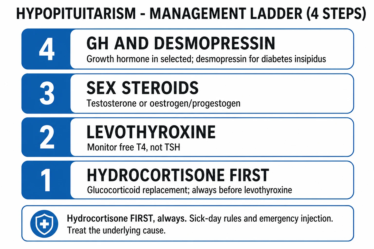
Order rule (non-negotiable): always replace glucocorticoid before thyroid hormone. Levothyroxine alone increases cortisol clearance and precipitates adrenal crisis. In crisis, give hydrocortisone first and withhold levothyroxine until the cortisol axis is covered. Fludrocortisone is NOT required in acute or chronic secondary adrenal insufficiency — aldosterone, driven by RAAS, is intact.[3]
Pituitary apoplexy resuscitation (per UK guidelines 2011): immediate IV hydrocortisone 100 mg, urgent MRI (or CT if MRI unavailable), fluid resuscitation, and urgent neurosurgical consultation. Surgical decompression (transsphenoidal) is indicated if there is visual deterioration, declining consciousness, or severe ophthalmoplegia — ideally within 7 to 14 days. Conservative management with high-dose steroids is acceptable if vision is stable and improving. Apoplexy is not a contraindication to surgery in the acute phase when vision is threatened.[4]
Management — Definitive & Stepwise
Long-term management mimics normal physiology with hormone replacement, plus treatment of the underlying cause. The replacement ladder is hydrocortisone first, then levothyroxine, then sex steroids, then growth hormone, then desmopressin for diabetes insipidus.[1][3][6]
Hypopituitarism — replacement doses
Glucocorticoid replacement (ALWAYS FIRST)
Hydrocortisone 15 to 25 mg/day in divided doses — for example 10 mg on waking, 5 mg at midday, 5 mg early evening — mimics the normal circadian cortisol rhythm. Titrate clinically, as there is no reliable biochemical monitor (cortisol day curves are used in specialist centres but are imperfect). Alternative: prednisolone 3 to 5 mg once daily (longer-acting, fewer doses, but harder to titrate). Fludrocortisone is NOT needed because aldosterone, driven by RAAS, is intact — this is a key difference from primary Addison.[3]
Thyroid replacement (ONLY AFTER CORTISOL COVERED)
Levothyroxine 1.6 micrograms/kg/day (typically 100 to 150 micrograms once daily), started only after hydrocortisone is established to avoid precipitating adrenal crisis. Monitor by free T4, not TSH — TSH is unreliable in pituitary disease (it may remain low even with adequate replacement). Target free T4 in the upper half of the reference range.[3]
Sex-steroid replacement
Men: testosterone — transdermal gel (e.g. 50 mg testosterone gel once daily) or intramuscular testosterone enanthate 250 mg every 2 to 3 weeks (or undecanoate 1000 mg every 10 to 14 weeks). Monitor morning testosterone, haematocrit, and prostate-specific antigen. [1]
Premenopausal women: oestrogen-progestogen HRT or a combined oral contraceptive, to maintain bone density, cardiovascular health, and quality of life. No progestogen is needed if the uterus was removed. [1]
Fertility: both sexes need gonadotrophin induction (hCG plus FSH) or pulsatile GnRH for hypogonadotropic hypogonadism; this is highly specialised and requires a reproductive endocrinologist. [1]
Growth hormone replacement
Somatropin (recombinant GH) 0.2 to 0.5 mg/day subcutaneously, titrated to IGF-1 in the age-adjusted reference range, for adults with proven GH deficiency (peak GH under 3 ng/mL on ITT or glucagon test). Benefits include improved body composition, lipid profile, bone density, and quality of life. Contraindications: active malignancy, proliferative retinopathy, intracranial hypertension. In children, GH is essential for linear growth.[6]
Desmopressin for diabetes insipidus
Desmopressin (1-deamino-8-D-arginine vasopressin) — oral 100 to 200 micrograms 2 to 3 times daily, intranasal 10 to 40 micrograms daily, or sublingual — for cranial DI (typically post-surgical or with stalk/hypothalamic disease). Titrate to control polyuria; avoid hyponatraemia. Triphasic DI (early polyuria → SIADH phase at 5 to 7 days → permanent DI) is common after pituitary surgery and requires close monitoring. [1]
Sick-day rules and emergency preparedness
Teach every patient on hydrocortisone these rules — they are life-saving: [1]
- Mild to moderate illness (fever, urinary tract infection, cold): double the oral hydrocortisone for 2 to 3 days.
- Severe illness, vomiting, or surgery: triple the dose, or switch to IV/IM hydrocortisone (50 to 100 mg every 6 to 8 hours).
- Vomiting or diarrhoea: oral absorption is unreliable — use the emergency injection (hydrocortisone 100 mg IM).
- Carry an emergency hydrocortisone 100 mg IM/SC injection kit at all times; teach the patient and family to administer it.
- Wear a steroid emergency card and a MedicAlert bracelet.
- Never stop steroids abruptly. [1]
Perioperative protocol:
- Major surgery: hydrocortisone 100 mg IM/IV at induction, then 50 mg IV every 8 hours for 24 to 72 hours, then taper to the usual dose.
- Minor surgery or dental work: hydrocortisone 50 mg IM/IV at induction, then resume the usual oral dose. [1]
Treat the underlying cause
- Transsphenoidal surgery for a symptomatic macroadenoma (visual compromise), craniopharyngioma, or apoplexy with visual deterioration.
- Dopamine agonist (cabergoline, bromocriptine) if the underlying lesion is a prolactinoma — these often shrink dramatically and surgery is avoided.
- Radiotherapy (stereotactic or conventional) rarely, for residual or recurrent tumour after surgery; effects on hormone function develop over years (progressive loss).
- Immune-checkpoint-inhibitor hypophysitis: high-dose glucocorticoids for symptomatic or severe cases; usually improves on cessation of the agent; some permanent deficiency remains. [1]
Specific Subtypes & Scenarios
Sheehan syndrome (postpartum pituitary necrosis)
Sheehan syndrome is ischaemic necrosis of the anterior pituitary following severe postpartum haemorrhage and shock. The enlarged pregnant pituitary — dependent on a low-pressure portal supply — is uniquely vulnerable to hypoperfusion. The classic presentation is the Sheehan triad: agalactia (failure to lactate), failure to resume menses (persistent amenorrhoea), and symptoms of hypopituitarism (fatigue, weight loss, hypotension, loss of body hair) following a complicated delivery. It may present acutely (postpartum collapse with hypotension and hypoglycaemia) or insidiously years later (chronic fatigue, amenorrhoea, secondary adrenal insufficiency). The posterior pituitary is usually spared (its blood supply is systemic, not portal), so DI is uncommon. Empty sella is seen on later imaging as the infarcted gland is replaced by CSF. Replace all deficient axes; the condition is preventable with good obstetric care and remains common only in regions with limited access to safe delivery.[1][5]
Pituitary apoplexy
Pituitary apoplexy is acute haemorrhage or infarction into a pre-existing pituitary adenoma, producing sudden swelling that compresses the optic chiasm and cavernous sinus and abruptly abolishes ACTH secretion. Presentation is dramatic: thunderclap headache, rapid visual loss, ophthalmoplegia (CN III palsy), meningism, vomiting, altered consciousness, and acute adrenal crisis (hypotension, hypoglycaemia). It is often the first presentation of an undiagnosed adenoma. Risk factors: anticoagulation, pregnancy, dopamine agonist initiation, hypertension, diabetes, head trauma. [1]
Management (UK Pituitary Apoplexy Guidelines 2011):
- Immediate IV hydrocortisone 100 mg then 50 mg every 6 hours, with fluid resuscitation.
- Urgent MRI (or CT) to confirm.
- Urgent neurosurgical consultation.
- Surgical decompression (transsphenoidal) within 7 to 14 days if: visual deterioration, declining consciousness, or severe ophthalmoplegia.
- Conservative management (high-dose steroids, close monitoring) is acceptable if vision is stable and improving.
- Some pituitary function recovers after decompression; long-term replacement is still often needed.[4]
Empty sella syndrome
Cerebrospinal-fluid herniation into the sella turcica flattens the pituitary against the floor. Primary empty sella (idiopathic, common in obese multiparous women, associated with benign intracranial hypertension) usually has no hormone deficiency — it is an incidental MRI finding. Secondary empty sella (after surgery, apoplexy, Sheehan, or radiotherapy) may have variable deficiency requiring replacement. The diagnosis is radiological; hormone testing decides who needs treatment.[2]
Traumatic brain injury and subarachnoid haemorrhage
Post-traumatic hypopituitarism is increasingly recognised and frequently missed. Mechanism: direct gland contusion, shearing of the stalk, or vascular injury. Deficiencies (most commonly GH, gonadotrophins, and ACTH) develop over 3 to 12 months after severe TBI. Screen every patient with severe TBI (GCS under 13), prolonged ICU stay, or diffuse axonal injury at 3 and 12 months with baseline axes; some recover, some are permanent. Subarachnoid haemorrhage carries similar risk.[2]
Genetic and congenital forms
PROP1 and POU1F1 mutations cause combined pituitary hormone deficiency (GH, PRL, TSH, later gonadotrophins) with short stature and a small or normal pituitary on MRI. HESX1 mutations cause septo-optic dysplasia (de Morsier syndrome — optic nerve hypoplasia, absent septum pellucidum, pituitary hypoplasia). Investigate the child with failure to thrive, growth failure, delayed puberty, or midline defects; check bone age, IGF-1, and a full pituitary panel, and arrange genetic workup and surveillance.[1]
Immune-checkpoint-inhibitor hypophysitis
Immune-checkpoint inhibitors (ICIs) — especially ipilimumab (anti-CTLA4), but also nivolumab and pembrolizumab (anti-PD1) — cause autoimmune hypophysitis. Ipilimumab tends to cause a mass-like presentation (headache, visual symptoms, enlarged pituitary on MRI) with ACTH, TSH and gonadotrophin loss and sometimes a mild prolactin rise; anti-PD1 agents cause a milder, predominantly ACTH-deficient picture without mass effect. Onset is typically weeks to months into therapy. Management: high-dose glucocorticoids (prednisolone 1 to 2 mg/kg or hydrocortisone 100 mg/day) for symptomatic or severe cases; the ICI may be held; hormone replacement (especially hydrocortisone) is usually permanent even after the inflammation resolves. Distinguish from brain metastases and pituitary metastasis.[7]
Iatrogenic
After transsphenoidal surgery: transient or permanent anterior deficits and diabetes insipidus are common; assess all axes pre- and post-operatively; cortisol is the priority. After radiotherapy: progressive hormone loss over 5 to 10 years (GH first, then the rest); annual screening for life. Cranial irradiation (for childhood leukaemia, brain tumours) carries the highest risk. [1]
Complications & Pitfalls
Adrenal crisis — hypotension, shock, hypoglycaemia, hyponatraemia, abdominal pain, and collapse; fatal if untreated. Precipitants: infection, surgery, trauma, a missed dose, vomiting (inability to absorb oral steroids), or giving levothyroxine without steroid cover. Every patient on hydrocortisone must carry an emergency injection kit and a steroid card.[2][3]
Permanent visual loss if a compressive macroadenoma or apoplexy is not decompressed promptly. Visual recovery after decompression is good if surgery is within 7 to 14 days but incomplete if delayed. [1]
Hypopituitary coma — severe deficiency (usually combined ACTH and TSH) compounded by stress (cold, infection, sedatives) produces hypothermia, bradycardia, hypoventilation, and coma. Treat with IV hydrocortisone first, then levothyroxine, rewarming, and supportive care. [1]
Long-term complications:
- Reduced bone mineral density and osteoporotic fracture (untreated hypogonadism and GH deficiency).
- Dyslipidaemia and atherosclerotic cardiovascular disease (untreated GH and gonadal deficiency) — drives the excess mortality.
- Impaired fertility (hypogonadotropic hypogonadism) — correctable with gonadotrophin induction.
- Impaired quality of life (GH deficiency, fatigue, altered body composition). [1]
Over-replacement itself harms: Cushingoid features, osteoporosis, and glucose intolerance from excess hydrocortisone; atrial fibrillation and osteoporosis from over-replaced levothyroxine; prostatic/haematocrit effects from excess testosterone. Titrate to clinical and biochemical targets.[3]
[1]Prognosis & Disposition
With appropriate replacement and treatment of the cause, patients have a near-normal life expectancy and good quality of life, with normal fertility achievable through gonadotrophin induction. Excess mortality (two- to three-fold) comes from cardiovascular and cerebrovascular disease (untreated GH and gonadal deficiency driving atherosclerosis) and preventable adrenal crises (inadequate patient education, missed sick-day dose, lack of emergency injection). Modern lower-dose hydrocortisone regimens (15 to 20 mg/day) reduce the metabolic burden compared with the older 30 mg/day doses.[2]
Disposition: discharge on lifelong hormone replacement with comprehensive sick-day education, an emergency hydrocortisone injection kit, and MedicAlert identification. Endocrinology follow-up — annual review with monitoring of all axes (free T4, IGF-1, sex steroids, morning cortisol off hydrocortisone if reassessing, bone density every 2 to 5 years), dose adjustment, and reproductive counselling for fertility. Post-surgical patients need early re-testing of all axes and visual fields at 3 months. Patients with craniopharyngioma need lifelong surveillance for recurrence and progressive hormone loss. [1]
Special Populations
Pregnancy
Fertility in hypogonadotropic hypogonadism requires gonadotrophin induction (hCG plus FSH) or pulsatile GnRH; pregnancy rates are good with specialist input. During pregnancy: increase hydrocortisone in the third trimester (by 50 percent) and in labour (intravenous hydrocortisone 100 mg at onset, then 50 mg every 8 hours). The pituitary normally enlarges in pregnancy — beware apoplexy and visual field deterioration. Levothyroxine requirements increase by 25 to 50 percent — monitor free T4 each trimester and titrate. Sheehan syndrome is the postpartum emergency: any woman with severe postpartum haemorrhage, hypotension, or failure to lactate must be assessed for anterior pituitary failure. Desmopressin for pre-existing DI is safe in pregnancy.[5]
Paediatrics
Growth failure and delayed puberty dominate the paediatric presentation. Weight-based GH replacement (essential for linear growth), levothyroxine, hydrocortisone, and (at the appropriate age) sex steroids for puberty induction. Congenital forms need genetic workup (PROP1, POU1F1, HESX1) and lifelong surveillance. Assess bone age and growth velocity every 6 to 12 months. Children on chronic steroids need bone-density monitoring and adequate calcium/vitamin D. [1]
Elderly
May present with confusion, falls, cognitive decline, or hyponatraemia attributed to SIADH or dementia. Use lower hydrocortisone doses (older patients are sensitive to steroid excess; aim for 15 mg/day) to avoid Cushingoid complications. High osteoporosis risk — monitor bone density and supplement calcium/vitamin D. Lower threshold to screen after falls, head trauma, or unexplained hyponatraemia. [1]
Post-pituitary surgery
Transient or permanent anterior deficits and diabetes insipidus are common; assess all axes pre- and post-operatively, with cortisol as the priority (the most dangerous acute loss). Watch for the triphasic DI pattern (early DI → SIADH at days 5 to 7 → permanent DI). Re-test at 3 months and annually thereafter, as deficits may recover or develop late (especially after radiotherapy). [1]
Patients on immune-checkpoint inhibitors
Screen for hypophysitis with baseline pituitary axes before starting therapy, and a low threshold to re-check if headache, fatigue, visual symptoms, or hypotension develop. Most cases present 4 to 16 weeks into therapy. Hydrocortisone replacement is usually permanent even after the ICI is held and inflammation resolves.[7]
Evidence, Guidelines & Regional Differences
The Endocrine Society 2016 guideline ("Hormonal Replacement in Hypopituitarism in Adults," Fleseriu et al.) is the reference standard for adult replacement, recommending hydrocortisone first-line (15 to 25 mg/day), free T4 for thyroid monitoring (not TSH), GH replacement in proven adult deficiency, and transdermal rather than oral testosterone in men. The Endocrine Society adult GH deficiency guideline (Molitch et al. 2006, reaffirmed) defines GH deficiency as a peak GH under 3 ng/mL on the insulin tolerance or glucagon stimulation test, and supports somatropin replacement titrated to IGF-1.[3][6]
The UK Pituitary Apoplexy Guidelines (Rajasekaran et al. 2011) standardise the acute management: immediate steroids, urgent imaging, early neurosurgical consultation, and surgical decompression within 7 to 14 days for visual deterioration. The UK Society for Endocrinology emergency hydrocortisone guidance (2019) advises a 100 mg IM hydrocortisone emergency injection for all adrenal-insufficient patients, with sick-day and SOS-injection education.[4]
UK and ANZ: hydrocortisone first-line; emergency 100 mg IM hydrocortisone kit standard; MedicAlert bracelets widely available; national pituitary apoplexy guidelines. [1]
US (Endocrine Society): same replacement principles; greater use of modified-release hydrocortisone (Chronocort) in research settings; insurance barriers to adult GH replacement. [1]
India and low-resource settings: Sheehan syndrome and tuberculosis remain common causes (unlike the autoimmune or idiopathic predominance in high-income countries); access to MRI and lifelong replacement can be limited; MedicAlert bracelets are less available, so patient education and family training in the emergency injection carry even more weight. The cost of adult GH replacement is often prohibitive. In areas with high TB prevalence, pituitary tuberculosis and tuberculoma must be in the differential of a sellar mass.
Controversies and evolving areas:
- Hydrocortisone regimen — once-daily modified-release (Chronocort, Plenadren) versus twice-daily versus thrice-daily; aim for physiologic exposure without over-replacement.
- Who benefits from adult GH replacement — cost, IGF-1 titration, and long-term cardiovascular outcomes remain debated; the Endocrine Society supports it for proven deficiency with symptoms.
- Pituitary apoplexy — conservative versus surgical — surgery for visual deterioration; conservative (high-dose steroids) is acceptable for stable vision; the UK guidelines provide the framework.
- Immune-checkpoint-inhibitor hypophysitis — whether to give high-dose steroids or physiologic replacement only; whether to rechallenge the ICI; long-term outcomes still emerging.
- Post-TBI screening — the optimal timing and cost-effectiveness of screening all severe TBI survivors.[2][7]
Exam Pearls
- No hyperpigmentation and no hyperkalaemia in secondary adrenal insufficiency (ACTH is low) — the single best discriminator from primary Addison, which has both.
- Always give hydrocortisone before levothyroxine — levothyroxine alone precipitates a fatal adrenal crisis by increasing cortisol clearance.
- Aldosterone is spared (RAAS intact) — so no fludrocortisone and no hyperkalaemia in secondary adrenal insufficiency.
- Order of hormone loss with a mass lesion: GH, then LH/FSH, then TSH, then ACTH, then prolactin (PRL often rises via the stalk effect from loss of dopamine inhibition).
- Sheehan triad: failure to lactate (agalactia), amenorrhoea, and hypopituitarism after severe postpartum haemorrhage.
- Pituitary apoplexy: thunderclap headache plus cranial nerve III palsy plus visual loss — emergency IV hydrocortisone plus neurosurgical decompression if vision compromised.
- Diagnosis: low target hormone with low or inappropriately normal trophic hormone; confirm with MRI; monitor levothyroxine by free T4, not TSH.
- The short Synacthen test can be falsely normal early in secondary adrenal insufficiency (the adrenal has not yet atrophied) — use an insulin tolerance test (gold standard) or glucagon stimulation test.
- Bitemporal hemianopia = optic chiasm compression by a macroadenoma; test visual fields by confrontation.
- Empty sella usually has no hormone deficiency in the primary form; the secondary form (after surgery, apoplexy, Sheehan) may.
- TBI, SAH, and immune-checkpoint inhibitors are increasingly recognised causes — screen survivors.
- A mildly raised prolactin (under 2000 mU/L) with a macroadenoma is a stalk effect, not a prolactinoma — do not treat with dopamine agonists as first-line; the tumour is non-lactotroph.
- Adult GH deficiency is defined as a peak GH under 3 ng/mL on the ITT; treat with somatropin titrated to IGF-1.
- Hyponatraemia in hypopituitarism is hypotonic with normal potassium and responds to hydrocortisone alone — distinguish from SIADH (fluid restriction) and Addison (hyperkalaemic). [1]
Exam application bank (NEET-PG / INICET)
One-line answer
Hypopituitarism is the partial or complete deficiency of one or more anterior pituitary hormones (GH, PRL, ACTH, TSH, LH/FSH); loss of all anterior hormones is panhypopuitarism. It is caused most commonly by a pituitary adenoma, surgery or radiation, Sheehan syndrome (postpartum ischaemic necrosis), pituitary apoplexy, infiltrative disease (sarcoidosis, haemochromatosis, Langerhans cell histiocytosis), autoimmune hypophysitis (including immune-checkpoint inhibitors), genetic mutations (PROP1, POU1F1), or traumatic brain injury. Each hormone deficiency produces distinct features: ACTH deficiency causes secondary adrenal insufficiency (fatigue, postural hypotension, hypoglycaemia, hyponatraemia — but no hyperpigmentation and no hyperkalaemia, distinguishing it from primary Addison disease); TSH causes secondary hypothyroidism; LH/FSH causes hypogonadism (loss of libido, amenorrhoea, erecti
Worked stems (answer without another resource)
Stem 1 — Classic presentation. Map symptoms to mechanism; name the first investigation and first treatment step with dose/route if drug therapy is standard. [1]
Stem 2 — Unstable / complicated. List red flags that force immediate resuscitation, theatre, ICU, antidote, or reperfusion — and what you do in the first 15 minutes. [1]
Stem 3 — Atypical group. Elderly, pregnancy, child, or immunocompromised: how presentation and thresholds change. [1]
Stem 4 — Differential trap. Name the three closest mimics and one discriminator for each. [1]
Stem 5 — Disposition. Who goes home with safety-netting, who is admitted, who needs HDU/ICU/theatre, and what follow-up is mandatory. [1]
Rapid viva checklist
- Definition + classification
- Pathophysiology chain
- Bedside signs / criteria
- Score with exact components (if any)
- Emergency bundle
- Definitive therapy with doses
- Complications of disease and of treatment
- Special populations
- Guideline/trial name if classic
- Three exam traps
Coverage self-check
If you cannot answer any stem above from this page alone, re-read the matching section — the page is intended to be self-sufficient for final-prof and NEET-PG/INICET questions on Hypopituitarism.
References
- [1]Yeliosof O, Gangat M. Diagnosis and management of hypopituitarism Curr Opin Pediatr, 2019.PMID 31082937
- [2]Iglesias P. An Update on Advances in Hypopituitarism: Etiology, Diagnosis, and Current Management J Clin Med, 2024.PMID 39458112
- [3]Fleseriu M, Hashim IA, Karavitaki N, et al. Hormonal Replacement in Hypopituitarism in Adults: An Endocrine Society Clinical Practice Guideline J Clin Endocrinol Metab, 2016.PMID 27736313
- [4]Rajasekaran S, Vanderpump M, Baldeweg S, et al. UK guidelines for the management of pituitary apoplexy Clin Endocrinol (Oxf), 2011.PMID 21044119
- [5]Chrisoulidou A, Boudina M, Karavitaki N. Pituitary disorders in pregnancy Hormones (Athens), 2015.PMID 25885105
- [6]Molitch ME, Clemmons DR, Malozowski S, et al. Evaluation and treatment of adult growth hormone deficiency: an Endocrine Society Clinical Practice Guideline J Clin Endocrinol Metab, 2006.PMID 16636129
- [7]Faje A, Reynolds K, Zubiri L, et al. Hypophysitis secondary to nivolumab and pembrolizumab is a clinical entity distinct from ipilimumab-associated hypophysitis Eur J Endocrinol, 2019.PMID 31176301