Endocrinology · Endocrinology
Hypothyroidism
Also known as Underactive thyroid · Myxoedema · Gull disease · Hashimoto thyroiditis (when autoimmune)
Hypothyroidism is the clinical and biochemical syndrome caused by deficiency of thyroid hormone (T4 and T3) at the tissue level. The commonest cause in iodine-sufficient regions is chronic autoimmune (Hashimoto) thyroiditis; worldwide, iodine deficiency dominates. The biochemical hallmark of primary disease is a raised TSH with low free T4. Treatment is lifelong oral levothyroxine at about 1.6 mcg/kg/day. Myxoedema coma is the decompensated, life-threatening extreme — IV levothyroxine plus hydrocortisone, passive rewarming and treatment of the precipitant.
On this page & tools
Your progress
Saved locally on this device.
Exam tags
Red flags
Overview & Definition
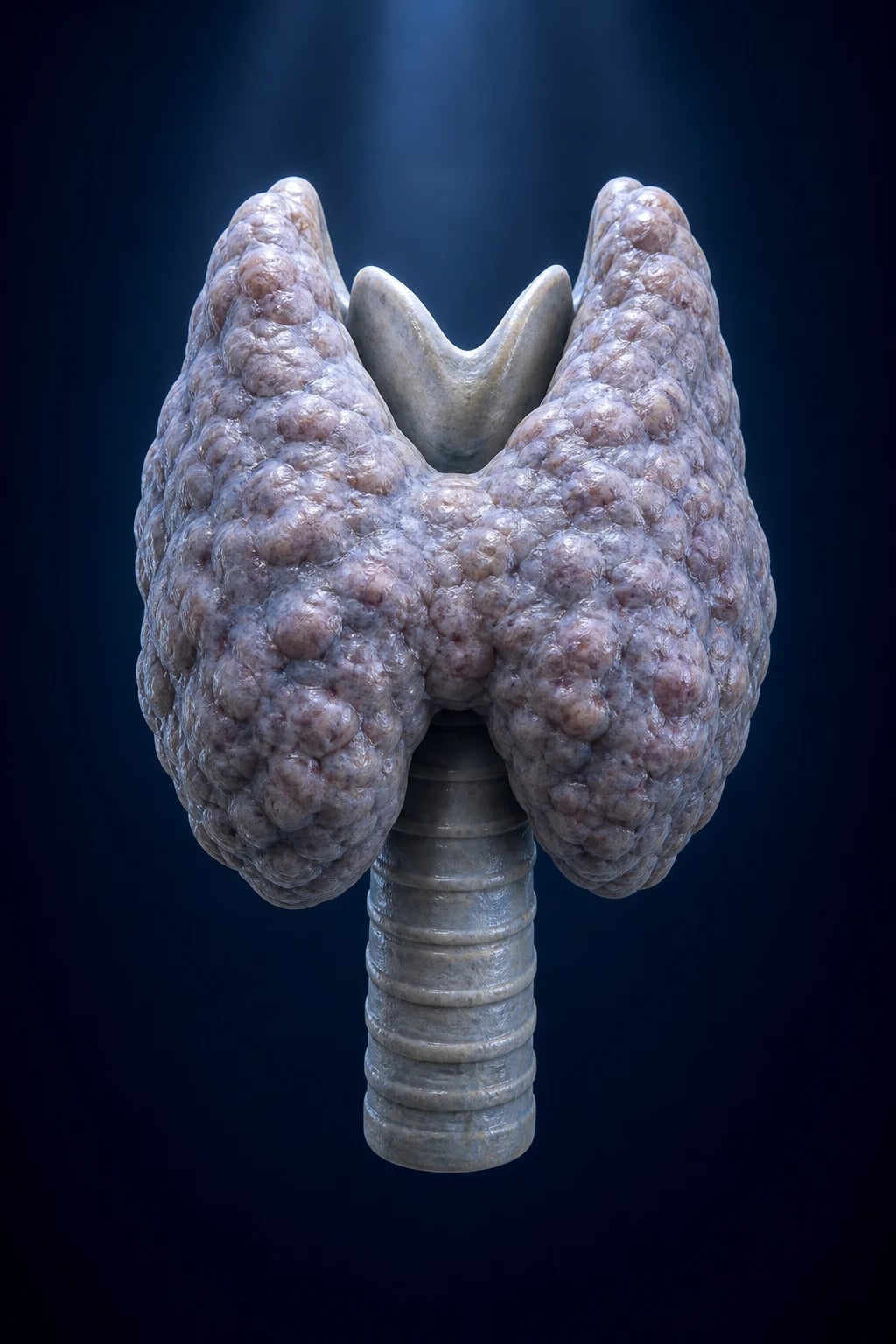
Hypothyroidism is the clinical and biochemical syndrome that results from deficient action of thyroid hormone (thyroxine, T4, and tri-iodothyronine, T3) on body tissues.[2] It is the commonest disorder of thyroid function and one of the most prevalent endocrine conditions in clinical practice, with a strong female preponderance and rising incidence with age.
The condition sits on a spectrum from subclinical (biochemical abnormality without symptoms) through overt (symptomatic hormone deficiency) to the decompensated extreme of myxoedema coma — a medical emergency. The clinical features reflect thyroid hormone's near-ubiquitous role in regulating basal metabolic rate, thermogenesis, lipid and carbohydrate metabolism, cardiac contractility, nervous-system myelination, and skeletal growth. Because thyroid hormone acts on virtually every organ, the presentation is heterogeneous, and hypothyroidism enters the differential diagnosis of a very wide range of presenting complaints — fatigue, constipation, menorrhagia, weight gain, depression, hyponatraemia, hyperlipidaemia, anaemia, and unexplained bradycardia. [1]
Two terms are commonly misused and should be distinguished: [1]
- Myxoedema refers to the characteristic non-pitting cutaneous and soft-tissue infiltration caused by the accumulation of hydrophilic glycosaminoglycans (chiefly hyaluronic acid and chondroitin sulfate) in the dermis. It is a sign of severe, long-standing hypothyroidism, not a synonym for the disease itself. (Confusingly, "myxoedema" was historically used as a synonym for hypothyroidism in general — the term was coined by William Ord in 1877 — but modern usage reserves it for the infiltrative skin change and the decompensated state.)
- Myxoedema coma is the life-threatening decompensation of hypothyroidism defined by altered mental status plus decompensated hypothyroidism plus a precipitating event, typically with hypothermia, hypoventilation, bradycardia and hyponatraemia.[7][8]
Thyroid physiology in brief: the hypothalamus secretes thyrotropin-releasing hormone (TRH), which stimulates the pituitary thyrotrophs to release thyroid-stimulating hormone (TSH). TSH binds the TSH receptor on thyroid follicular cells, driving iodide uptake (via the sodium-iodide symporter, NIS), organification of iodide onto tyrosyl residues of thyroglobulin (catalysed by thyroid peroxidase, TPO), coupling of iodotyrosines to form T4 and T3, and release of hormone. The thyroid secretes predominantly T4 (a prohormone), which is deiodinated in peripheral tissues (liver, kidney, and locally in the brain) by the deiodinase enzymes D1 and D2 to the active T3. Circulating T4 and T3 exert negative feedback on both the pituitary (TSH) and the hypothalamus (TRH). This elegant feedback loop is the basis of thyroid-function-test interpretation: a raised TSH is a sensitive marker of primary thyroid failure long before free T4 falls. [1]
Classification
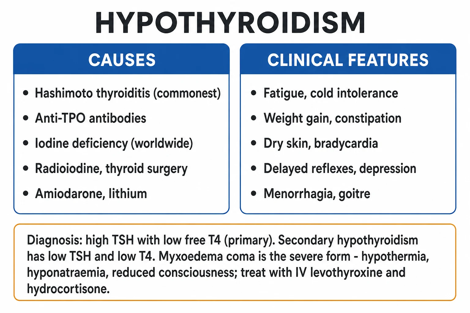
Hypothyroidism is classified along two axes — by anatomical level of the defect (primary, secondary, tertiary) and by severity (overt vs subclinical). These two classifications are not interchangeable and both are required to manage the patient.[2][3]
By level of the defect
Primary (thyroidal) — about 99% of cases
- Defect is in the thyroid gland itself; gland cannot produce enough T4 despite maximal TSH stimulation
- Biochemistry: TSH HIGH (often over 10 mIU/L) + free T4 LOW
- Causes: Hashimoto thyroiditis, iodine deficiency, iatrogenic (radioiodine, thyroidectomy), drugs (amiodarone, lithium), thyroiditis (transient)
- By far the commonest form — what is meant by 'hypothyroidism' in everyday practice
Secondary (pituitary / central)
- Defect is pituitary TSH deficiency; the thyroid is intrinsically normal but under-stimulated
- Biochemistry: TSH LOW or INAPPROPRIATELY NORMAL + free T4 LOW — the inappropriately normal TSH is the key: a normal TSH with a low free T4 is not normal
- Causes: pituitary adenoma, Sheehan syndrome (postpartum pituitary infarction), infiltrative disease (haemochromatosis, sarcoid, lymphocytic hypophysitis), pituitary surgery/radiation, trauma
- Always check cortisol before thyroid hormone: giving levothyroxine to a cortisol-deficient patient precipitates adrenal crisis
Tertiary (hypothalamic)
- Defect is TRH deficiency from hypothalamic disease
- Biochemistry identical to secondary (low/normal TSH + low free T4); distinguished by cause and on MRI
- Causes: hypothalamic tumours (craniopharyngioma), metastases, infiltrative disease, radiation, trauma
- Managed identically to secondary central disease
By severity
| Severity | TSH | Free T4 | Typical picture |
|---|---|---|---|
| Overt primary | Raised (often over 10 mIU/L) | Low | Symptomatic |
| Subclinical primary | Raised (above reference, typically 4.5 to 10 mIU/L) | Normal | Usually asymptomatic; may have mild symptoms or dyslipidaemia |
| Central (secondary/tertiary) | Low or inappropriately normal | Low | Variable; other pituitary deficits present |
Additional descriptors
- Goitrous vs atrophic: Hashimoto may present with either a goitre (early, lymphocytic infiltration) or an atrophic fibrotic gland (late). Iodine deficiency and dyshormonogenesis are goitrous; post-ablative and iatrogenic disease are non-goitrous.
- Congenital vs acquired: congenital hypothyroidism (cretinism) is detected on newborn screening and demands urgent replacement to protect neurodevelopment; acquired disease dominates in adults.
- Transient vs permanent: thyroiditis (subacute, postpartum, painless, amiodarone-induced) can cause transient hypothyroidism that recovers; autoimmune and iatrogenic causes are permanent. [1]
Epidemiology & Risk Factors
The global prevalence of hypothyroidism is about 1 to 2% of the adult population for overt disease, with subclinical hypothyroidism in a further 4 to 10%, rising sharply with age.[2] Key epidemiological facts:
- Sex: a strong female preponderance of roughly 5 to 10:1, reflecting the autoimmune predilection of Hashimoto disease.
- Age: incidence rises with age; the peak is in the 5th to 6th decade and beyond. Subclinical disease affects up to 10 to 15% of those over 65.
- Geography: the dominant cause worldwide is iodine deficiency, which affects an estimated 2 billion people and remains the commonest preventable cause of intellectual disability. In iodine-sufficient regions (most of North America, Europe, much of urban India post-iodisation), chronic autoimmune (Hashimoto) thyroiditis is the leading cause.[2]
Hypothyroidism — key numbers
Host risk factors for hypothyroidism:[1][2]
- Female sex — autoimmune thyroid disease is far commoner in women, modulated by oestrogen and the X chromosome.
- Age over 60 — both autoimmune and subclinical disease rise with age.
- Personal or family history of autoimmune disease — type 1 diabetes, coeliac disease, Addison disease, vitiligo, alopecia areata, pernicious anaemia, rheumatoid arthritis, systemic lupus erythematosus. A cluster of autoimmune disease in one patient should prompt screening of thyroid function.
- Other chromosomal syndromes — Turner syndrome, Down syndrome, Klinefelter syndrome all carry a markedly increased risk of autoimmune thyroiditis.
- Postpartum period — postpartum thyroiditis affects about 5 to 10% of women, often transient.
- Prior thyroid disease or treatment — history of Graves disease (even after radioiodine or surgery), thyroiditis, goitre, or thyroid nodule.
- Iodine intake — both deficiency and excess (the latter from contrast, kelp, amiodarone) cause hypothyroidism via the Wolff-Chaikoff effect (see pathophysiology). [1]
Iatrogenic and drug causes (an examiner staple):[2]
- Amiodarone — a high iodine load (each 200 mg tablet contains about 75 mg of organic iodine, 100x the daily requirement) disturbs thyroid function in up to 20% of patients; produces both hypothyroidism (type 1, iodine-induced/Wolff-Chaikoff, commoner in iodine-sufficient regions and in anti-TPO-positive patients) and thyrotoxicosis (type 2, destructive). Check thyroid function before and every 6 months during therapy.
- Lithium — inhibits thyroid hormone release; hypothyroidism in up to 20%, commoner in anti-TPO-positive women.
- Interferon-alpha, interleukin-2, immune-checkpoint inhibitors (ipilimumab, nivolumab, pembrolizumab) — precipitate autoimmune thyroiditis.
- Tyrosine kinase inhibitors (sunitinib, sorafenib, imatinib) — cause destructive thyroiditis with a hyperthyroid phase then hypothyroidism.
- Iodine excess — iodinated contrast, kelp supplements, the antiarrhythmic amiodarone (the Jod-Basedow phenomenon is the hyperthyroid counterpart).
- Post-radioiodine therapy (for Graves disease or thyroid cancer) — most patients become permanently hypothyroid within months to a year; lifelong levothyroxine is anticipated.
- Post-thyroidectomy — immediate lifelong replacement required. [1]
Pathophysiology
The hypothalamic-pituitary-thyroid axis
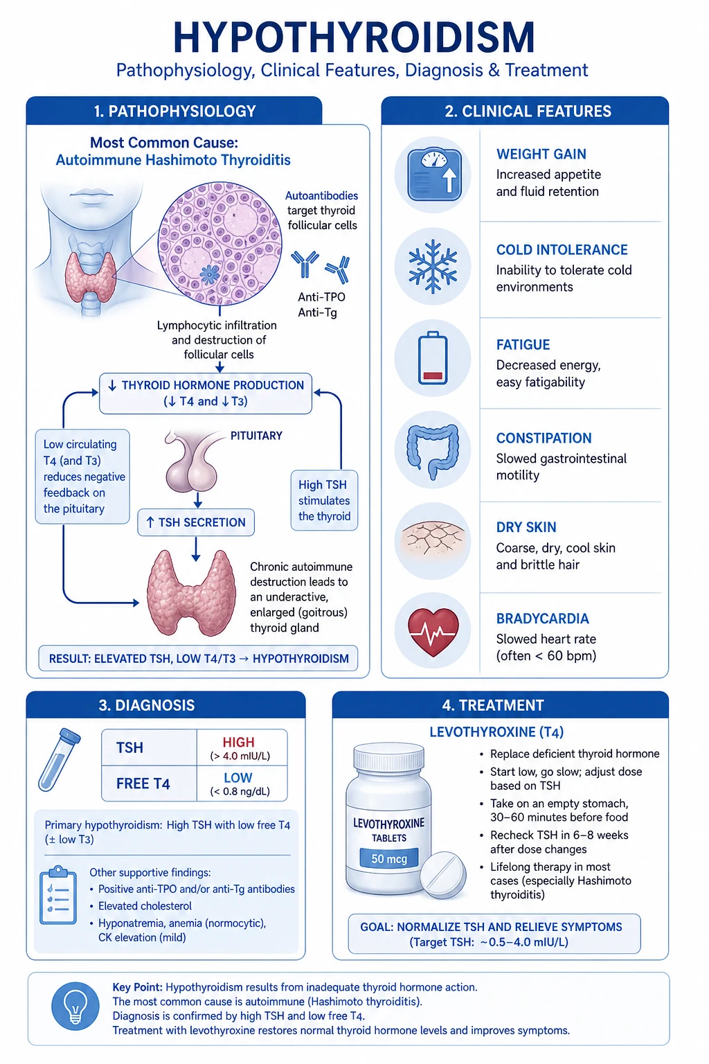
The axis operates as a classical negative-feedback loop. TRH, synthesised in the paraventricular nucleus of the hypothalamus, travels in the portal hypophyseal vessels to the anterior pituitary, where it stimulates thyrotrophs to synthesise and release TSH (a glycoprotein with an alpha subunit shared with FSH, LH and hCG, and a unique beta subunit). TSH binds the TSH receptor (TSH-R), a G-protein-coupled receptor on the basolateral membrane of the thyroid follicular cell, activating Gs and cAMP. Downstream it drives:[2]
- Iodide uptake via the sodium-iodide symporter (NIS) on the basolateral membrane (an "iodide trap" that achieves intracellular iodide concentrations 20 to 50 times plasma).
- Organification — iodide is oxidised by thyroid peroxidase (TPO) and hydrogen peroxide (generated by DUOX2) and coupled onto selected tyrosyl residues of thyroglobulin (Tg), a large glycoprotein backbone stored in the colloid of the follicular lumen. This produces monoiodotyrosine (MIT) and di-iodotyrosine (DIT).
- Coupling — TPO links two DIT molecules to form T4 (thyroxine) and one MIT + one DIT to form T3 (tri-iodothyronine).
- Release — under continued TSH stimulation, colloid is endocytosed, thyroglobulin is lysosomally degraded, and T4 and T3 are secreted into the blood. (A small amount of intrathyroidal T4 is converted to T3 by D1 deiodinase.)
- Negative feedback — circulating free T4 and free T3 act on the pituitary and hypothalamus (after local conversion to T3 by D2 deiodinase) to suppress TSH and TRH. This feedback is exquisitely sensitive: very small falls in free T4 produce measurable rises in TSH, which is why TSH is the most sensitive single marker of primary thyroid (dys)function. [1]
In the periphery, the deiodinase enzymes interconvert thyroid hormones: D1 and D2 convert T4 to the active T3; D3 inactivates T4 and T3 to reverse T3 (rT3) and T2. About 80% of circulating T3 is generated peripherally; only 20% is secreted directly by the thyroid. This is why levothyroxine (T4) alone is adequate replacement for almost all patients — peripheral conversion supplies T3. [1]
Most circulating T4 and T3 is protein-bound (to thyroxine-binding globulin, transthyretin and albumin); only the free fraction (free T4, free T3) is biologically active. Pregnancy, oestrogen and hepatitis raise thyroxine-binding globulin (TBG), raising TOTAL T4 but leaving FREE T4 unchanged — a key point in interpreting thyroid function in pregnancy (see Special Populations). [1]
Mechanism of Hashimoto (chronic autoimmune) thyroiditis
Hashimoto is an organ-specific autoimmune disease. The immunological lesion:[1]
- Loss of self-tolerance to thyroid antigens, with autoreactive CD4+ T-helper cells (predominantly a Th1 cytokine profile — IFN-gamma, TNF-alpha, IL-2) recruiting macrophages and cytotoxic CD8+ T cells into the gland.
- Anti-thyroid peroxidase (anti-TPO) antibodies are the serological hallmark (present in about 90%); anti-thyroglobulin (anti-Tg) antibodies are also common but less specific.
- The combination of cytotoxic T-cell killing, antibody-dependent cell-mediated cytotoxicity, complement activation and cytokine-mediated (Fas/FasL) apoptosis destroys thyroid follicular cells progressively.
- The gland shows dense lymphocytic infiltration with germinal centres, oxyphil (Hurthle) cell metaplasia of follicular epithelium, and variable fibrosis. Early disease may be goitrous (infiltration outpacing destruction); late disease is often atrophic (fibrosis).
- Function declines as follicular mass is lost: subclinical hypothyroidism (raised TSH, normal free T4) progresses over years to overt disease, particularly if anti-TPO positive (about 4% per year progression rate).
- Hashimoto carries a small but real risk of thyroid B-cell lymphoma (a rare MALT-type lymphoma arising in longstanding Hashimoto) and is associated with papillary thyroid carcinoma more often than chance. [1]
Mechanism of iodine-deficiency goitre
Without adequate iodide, organification of thyroglobulin is impaired and T4/T3 synthesis falls. The low circulating T4 releases the brake on TSH, which hyperstimulates the TSH receptor chronically. This drives follicular-cell hypertrophy and hyperplasia and colloid accumulation — the diffuse, soft, symmetric goitre of endemic iodine deficiency. With sustained deficiency, the gland enlarges progressively and may become multinodular. Iodine repletion reverses early goitre and prevents cretinism.[2]
Mechanism of central (secondary/tertiary) hypothyroidism
In central disease, the thyroid gland is intrinsically normal, but TSH (pituitary) or TRH (hypothalamic) drive is lost. Free T4 falls, but because the defect is upstream of the thyroid, TSH does not rise appropriately — it is low or in the "normal" range, which is inappropriately normal for the low free T4. The clinical clues are coexisting other anterior-pituitary deficits (secondary amenorrhoea, hypogonadism, adrenal insufficiency, growth failure), headache or visual field defects (mass effect of a macroadenoma on the optic chiasm), and a history of pituitary surgery, radiation, severe postpartum haemorrhage (Sheehan) or infiltrative disease. [1]
Mechanism of drug-induced hypothyroidism
- Amiodarone delivers a massive iodine load; in patients with underlying autoimmune thyroiditis (anti-TPO positive) the excess iodine cannot escape the Wolff-Chaikoff effect (a protective down-regulation of organification in response to high iodide) — the gland "fails to escape" and becomes transiently or permanently hypothyroid.
- Lithium inhibits TSH-stimulated hormone release and may exacerbate autoimmune thyroiditis.
- Cytokines (interferon, IL-2) and checkpoint inhibitors expose or precipitate autoimmune thyroiditis, often with a transient thyrotoxic phase followed by hypothyroidism. [1]
Cellular basis of the symptoms
At the molecular level, T3 acts via the nuclear thyroid hormone receptor (TR-alpha and TR-beta), a ligand-activated transcription factor that upregulates genes including Na-K-ATPase, beta-adrenergic receptors, malic enzyme, and SERCA. In hypothyroidism:[2]
- Reduced Na-K-ATPase activity lowers basal metabolic rate (bradyergia, cold intolerance, weight gain) and thermogenesis (hypothermia).
- Reduced beta-adrenergic receptor density and coupling produces bradycardia, reduced inotropy, and a fall in cardiac output (and the diastolic hypertension of hypothyroidism from increased systemic vascular resistance).
- Reduced SERCA slows diastolic relaxation of cardiac and skeletal muscle (delayed relaxation of reflexes, the slow-relaxing ankle jerk).
- Accumulation of hyaluronic acid and chondroitin sulfate glycosaminoglycans in the dermis traps water, producing the non-pitting myxoedematous puffiness; similar deposition in the vocal cords produces hoarseness.
- Reduced cholesterol clearance (downregulated LDL receptor) causes hypercholesterolaemia.
- Reduced free-water clearance and inappropriate ADH produce hyponatraemia.
- Reduced gut motility causes constipation and rarely myxoedema megacolon.
- Increased TRH (from loss of negative feedback) stimulates prolactin release, producing galactorrhoea and menstrual disturbance. [1]
Pathophysiology of myxoedema coma
Myxoedema coma is decompensated hypothyroidism — the patient's thermoregulation, ventilation and cognition fail. It is typically precipitated in a patient with longstanding untreated or under-treated hypothyroidism by an external insult: cold exposure, infection (pneumonia, UTI), sedatives and anaesthetics, opioids, stroke, myocardial infarction, gastrointestinal bleeding, or simply missed levothyroxine doses.[7][8] The hallmark physiology:
- Hypothermia from a profoundly reduced basal metabolic rate (core temperatures of 32 to 35 degrees C are typical; values below 30 degrees C carry a poor prognosis).
- Hypoventilation (a depressed central respiratory drive with hypercapnia and hypoxia) — the leading cause of death.
- Bradycardia and reduced cardiac output, sometimes with pericardial and pleural effusions.
- Hyponatraemia from impaired free-water clearance.
- Reduced consciousness / coma, sometimes with seizures.
- Frequently coexisting adrenal insufficiency (autoimmune Addison disease in Hashimoto, or ACTH deficiency in central disease) — the rationale for always giving hydrocortisone with IV levothyroxine. [1]
Clinical Presentation
Hypothyroidism presents insidiously. Symptoms develop over months, are often attributed to ageing or stress, and may be brought to attention only when a TFT is checked for an unrelated reason. The clinical features are best organised system by system:[2]
General:
- Fatigue, lethargy, somnolence, cold intolerance (the cardinal symptom — feeling cold when others are comfortable), unexplained weight gain (modest, 2 to 5 kg, from reduced metabolic rate and fluid retention rather than fat), sluggishness, slowed movement and speech. [1]
Skin and soft tissues:
- Dry, cool, pale, rough skin; brittle hair and slow-growing nails; lateral third of eyebrow thinning (madarosis, a classical but non-specific sign).
- Myxoedema — non-pitting, doughy puffiness of the face (periorbital), hands and feet; macroglossia (large tongue); broad, thickened lips; hoarse, croaking voice (vocal-cord glycosaminoglycan deposition).
- Carotenaemia — yellow-orange discoloration of the palms, soles and nasolabial folds from impaired hepatic conversion of beta-carotene to vitamin A (a striking and easily missed sign).
- Pallor may reflect anaemia or vasoconstriction; vitiligo and alopecia areata suggest coexisting autoimmune disease. [1]
Cardiovascular:
- Bradycardia, reduced pulse pressure, often diastolic hypertension (increased systemic vascular resistance).
- Distant heart sounds and mild cardiomegaly from pericardial effusion (a transudate, usually haemodynamically insignificant but may be large).
- Reduced exercise tolerance and, in advanced disease, heart failure (high-output to low-output transition; chest pain may be masked because the bradycardia lowers myocardial oxygen demand — treating hypothyroidism can unmask angina). [1]
Respiratory:
- Pleural effusions, reduced vital capacity, depressed respiratory drive, sleep-disordered breathing and obstructive sleep apnoea (worsened by macroglossia and myxoedematous pharyngeal infiltration). [1]
Gastrointestinal:
- Constipation (often severe), rarely adynamic ileus or myxoedema megacolon; ascites (rare, part of a generalised effusive tendency); malabsorption (modest).
- Gallstones are commoner (impaired gallbladder motility).
- Achlorhydria and pernicious anaemia may coexist (autoimmune gastritis). [1]
Neuromuscular:
- Slow-relaxing (hung-up) ankle reflexes — the classical bedside sign (delayed relaxation phase, not just a slow reflex).
- Muscle aching, stiffness, proximal weakness (myopathy); raised creatine kinase; muscle hypertrophy (Hoffmann syndrome) in severe long-standing disease, especially in children.
- Carpal tunnel syndrome (bilateral), sensorineural deafness, peripheral neuropathy, cerebellar ataxia (rare).
- Depression, cognitive slowing, poor concentration, memory impairment; rarely psychosis ("myxoedema madness") and seizures. [1]
Reproductive:
- Menorrhagia, oligomenorrhoea, amenorrhoea, anovulatory infertility in women; galactorrhoea from TRH-driven hyperprolactinaemia; loss of libido and erectile dysfunction in men.
- Increased risk of miscarriage, pre-eclampsia, preterm birth and impaired fetal neurodevelopment in pregnancy (see Special Populations). [1]
Haematological and metabolic:
- Normocytic or macrocytic anaemia (the latter from coexisting B12 deficiency/pernicious anaemia or from direct marrow effect); raised creatine kinase (myopathy); hypercholesterolaemia (raised LDL); hyponatraemia; hyperprolactinaemia. [1]
Bedside signs that should trigger testing: periorbital puffiness, slow-relaxing ankle reflexes, bradycardia, cold dry skin, hoarse voice. [1]
Atypical presentations
[1]- "Apathetic" presentation in the elderly — depression, cognitive decline, falls, failure to thrive, or simply "going off." Classic signs are muted; the diagnosis is made on TFT. Confused with dementia until tested.
- Diabetic patients — unexplained deterioration in glycaemic control, recurrent hypoglycaemia (reduced insulin clearance), or new dyslipidaemia.
- Pregnant women — fatigue, weight change, constipation and cold intolerance are common in normal pregnancy and may mask hypothyroidism; universal vs targeted screening is debated (see Evidence).
- Children — growth retardation, delayed bone age, delayed puberty, poor school performance; congenital disease (detected on newborn screen) presents with failure to thrive, prolonged jaundice, constipation, macroglossia, umbilical hernia, hypotonia if missed.
- Postoperative/ICU patients — unexplained hyponatraemia, hypoventilation, hypothermia, or failure to wean from ventilation.
- Myxoedema coma — the decompensated extreme (see Resuscitation). [1]
Differential Diagnosis
The differential depends on the presentation. The "tired, cold, constipated, weight-gaining" patient has a broad differential; the differential of the TFT pattern itself is narrower and more useful.[2][6]
Differential of the clinical syndrome
| Differential | Distinguishing features |
|---|---|
| Depression | Low mood with anhedonia; preserved appetite change (often reduced), normal TSH; screen TFT in any new depression because they overlap and coexist |
| Anaemia (iron-deficiency or pernicious) | Fatigue and pallor; check FBC and ferritin/B12; pernicious anaemia coexists with Hashimoto — test both |
| Chronic kidney disease | Fatigue, anaemia, fluid retention; raised creatinine; TFT normal (though CKD patients have higher prevalence of hypothyroidism) |
| Chronic fatigue syndrome / fibromyalgia | Widespread pain, unrefreshing sleep, normal TFT and other tests; a diagnosis of exclusion |
| Normal ageing | Insidious functional decline; normal TFT; the diagnosis is hypothyroidism until the TSH is checked |
| Heart failure | Exertional dyspnoea, oedema, raised JVP; pericardial effusion of hypothyroidism can mimic; echo distinguishes |
| Obstructive sleep apnoea | Daytime somnolence, snoring; coexists with hypothyroidism (macroglossia) — screen and treat both |
Differential of the TFT pattern — distinguishing primary from central, and from euthyroid sick
This is the examiner's most important differential. [1]
| Pattern | TSH | Free T4 | Interpretation | Next step |
|---|---|---|---|---|
| Overt primary hypothyroidism | High | Low | Thyroid failure | Anti-TPO; start levothyroxine |
| Subclinical hypothyroidism | High (4.5 to 10) | Normal | Early thyroid failure | Repeat in 3 months; treat per thresholds |
| Central hypothyroidism | Low or inappropriately normal | Low | Pituitary/hypothalamic disease | Check cortisol; pituitary MRI; replace cortisol first |
| Euthyroid sick (non-thyroidal illness) syndrome | Low, normal, or mildly high | Low or low-normal | Severe systemic illness; low or normal TSH with low free T3 and high reverse T3 | Treat the underlying illness; do NOT start levothyroxine; TFT will normalise on recovery |
Euthyroid sick syndrome is the classic trap: a critically-ill patient (sepsis, post-op, malignancy, ICU) has a low or normal TSH with low free T3 (and sometimes low free T4, with high reverse T3). It is an adaptive down-regulation of metabolism (D1 and D2 down, D3 up) and does not require thyroid-hormone replacement — it resolves as the underlying illness recovers. Mislabelling it as central hypothyroidism and giving levothyroxine is a recognised pitfall.[6]
Differential of myxoedema coma (the comatose, hypothermic patient)
| Differential | Distinguishing features |
|---|---|
| Sepsis | Fever (may be masked by hypothermia), source on examination and culture, raised lactate/WCC/CRP; sepsis and myxoedema coma coexist and the sepsis is often the precipitant |
| Adrenal crisis | Hypotension, hyperkalaemia, hyponatraemia, hypoglycaemia; may coexist with myxoedema coma — always give hydrocortisone |
| Hypoglycaemia | Bedside glucose; cold, clammy, reduced consciousness |
| Drug overdose / intoxication | History, drug levels; opioids and sedatives are common precipitants of myxoedema coma |
| Hypothermia (environmental) | History of cold exposure; check TFT in any hypothermic patient |
| Stroke | Focal signs; CT brain |
The key clinical point: any unexplained hypothermia with reduced consciousness warrants a TSH and free T4, because myxoedema coma is treatable and missing it is fatal. [1]
Differential of the goitre
A goitre with hypothyroidism is most often Hashimoto (firm, rubbery, diffuse) or iodine-deficiency (diffuse, soft). Distinguish from:
- Graves disease (diffuse, soft, bruit; thyrotoxic not hypothyroid, though a hypothyroid phase can follow treatment).
- Multinodular goitre (nodular, often asymptomatic; can later become toxic).
- Solitary thyroid nodule (single nodule; rule out malignancy with US-FNAC; a hyperfunctioning "hot" nodule causes hyperthyroidism, not hypothyroidism).
- Thyroid cancer (hard, fixed, rapidly enlarging nodule with lymphadenopathy).
- Subacute (de Quervain) thyroiditis (tender, painful goitre with a transient thyrotoxic then hypothyroid phase).
- Riedel thyroiditis (rock-hard, "woody" goitre that infiltrates surrounding structures; rare; fibrosing IgG4-related disease). [1]
Clinical & Bedside Assessment
A focused bedside examination aims to (a) confirm the clinical suspicion of hypothyroidism, (b) characterise the goitre, (c) identify the severity, and (d) look for associated autoimmune or pituitary disease.[1][2]
The thyroid examination:
- Inspect from the front — swelling, asymmetry, scars, scars of previous surgery, distended neck veins.
- Ask the patient to swallow (sip of water) — a thyroid swelling rises with swallowing (it is invested in pretracheal fascia).
- Palpate from behind the seated patient using both hands; assess size, consistency, surface (smooth vs nodular), mobility, tenderness, and lower extent (a goitre that extends below the sternoclavicular joint on swallowing is retrosternal).
- Percuss the manubrium for a dull note (retrosternal extension).
- Auscultate for a bruit (a continuous or systolic bruit suggests Graves hyperthyroidism from hypervascularity, not Hashimoto).
- Examine cervical and supraclavicular lymph nodes. [1]
Characterising the goitre in hypothyroidism: [1]
| Cause | Goitre character |
|---|---|
| Hashimoto (early) | Firm, rubbery, diffuse, symmetrical, mobile, non-tender; may have a pyramidal lobe |
| Hashimoto (late, atrophic) | No goitre — the gland is fibrotic and shrunken |
| Iodine deficiency | Diffuse, soft, smooth; later multinodular; may be very large |
| Subacute (de Quervain) thyroiditis | Tender, firm, painful |
| Riedel thyroiditis | Rock-hard, "woody", fixed, infiltrates strap muscles |
Bedside signs of hypothyroidism:
- Slow-relaxing (hung-up) ankle reflexes — demonstrate by tapping the Achilles tendon with the patient kneeling on a chair or with the foot dorsiflexed; the reflex is brisk to fire but delayed in the relaxation phase (the classic sign).
- Bradycardia, diastolic hypertension, distant heart sounds, signs of pericardial effusion.
- Periorbital and pedal puffiness, cool dry skin, carotenaemia, macroglossia, hoarse voice, slow speech.
- Hypothermia, hypoventilation, reduced consciousness (myxoedema coma).
- Proximal muscle weakness, thenar wasting and Tinel's sign (carpal tunnel). [1]
Look for associated autoimmune disease (vitiligo, alopecia areata, Addison pigmentation, palpable thyroid autoimmune goitre) and, in suspected central disease, features of pituitary mass (bitemporal hemianopia on confrontation, hypogonadism, hypopigmentation, postural drop from ACTH deficiency) and other anterior-pituitary deficits. [1]
Investigations
First-line thyroid function tests
- TSH is the single most sensitive first-line test for primary thyroid disease because of the steep negative-feedback relationship. A normal TSH in the absence of pituitary disease effectively excludes primary thyroid dysfunction.[2]
- Free T4 is added when TSH is abnormal, when central disease is suspected, or in pregnancy. Free (not total) T4 is essential, because total T4 is confounded by thyroxine-binding globulin changes (pregnancy, oestrogen, hepatitis, nephrotic syndrome).
- Free T3 is rarely needed in primary disease — it is reserved for suspected T3-toxicosis (a hyperthyroid pattern), monitoring T3 in special cases, and is never used to monitor routine levothyroxine therapy.
Interpreting the TFT pattern
| TSH | Free T4 | Diagnosis |
|---|---|---|
| High | Low | Overt primary hypothyroidism — treat |
| High | Normal | Subclinical primary hypothyroidism — treat per thresholds |
| Low/normal | Low | Central hypothyroidism — check cortisol, image pituitary |
| Normal | Normal | Euthyroid (or early/mild disease) |
Antibodies
- Anti-thyroid peroxidase (anti-TPO) antibodies — the serological marker of Hashimoto thyroiditis (positive in about 90%). Confirm the autoimmune aetiology and predict progression from subclinical to overt disease (about 4% per year if positive vs under 2% if negative).[1]
- Anti-thyroglobulin (anti-Tg) — supportive but less sensitive and specific; also measured as a tumour marker surveillance caveat after thyroid cancer.
- TSH-receptor antibodies (TRAb) — measure in suspected Graves disease (hyperthyroidism), not hypothyroidism (though blocking TRAb can cause atrophic thyroiditis, a rare cause of primary hypothyroidism without a goitre).
Confirming subclinical hypothyroidism
Subclinical disease is defined as a TSH above the reference range (typically above 4.0 to 4.5 mIU/L) with a normal free T4 in a stable, non-pregnant patient.[5][6] It must be confirmed by repeating the TFT at 3 months (transient TSH elevation is common after non-thyroidal illness). Treatment thresholds (per ATA/ETA):
- TSH over 10 mIU/L — treat (clear biochemical hypothyroidism; benefit on symptoms and lipids).
- TSH 4.5 to 10 mIU/L — individualise: treat if symptomatic, anti-TPO positive, goitre, pregnancy or IVF-planned, dyslipidaemia, or young age; otherwise monitor TFT every 6 to 12 months. [1]
Associated laboratory abnormalities (examiner favourites)
- Dyslipidaemia — raised LDL cholesterol (downregulated LDL receptor), sometimes raised triglycerides.
- Hyponatraemia — usually mild; from impaired free-water clearance and inappropriate ADH.
- Anaemia — normocytic (direct marrow effect) or macrocytic (coexisting B12-deficient pernicious anaemia, which is autoimmune and associated).
- Raised creatine kinase — from myopathy (can be very high; do not confuse with rhabdomyolysis or MI).
- Raised prolactin — TRH-driven; usually mild, resolves with replacement.
- Hypoglycaemia, hypocalcaemia, hyperkalaemia — only in severe disease or coexisting adrenal insufficiency. [1]
When to image
- Thyroid ultrasound — for a nodular goitre, a rapidly enlarging gland, a dominant nodule, or cervical lymphadenopathy (rule out malignancy). Not required for routine Hashimoto.
- Pituitary MRI with contrast — when central hypothyroidism is suspected (low/normal TSH + low free T4) to define a pituitary/hypothalamic lesion.
- CT chest/abdomen — not for the thyroid itself.
- Cardiac echo — if pericardial effusion or heart failure is suspected. [1]
Diagnosing myxoedema coma
Myxoedema coma is a clinical diagnosis supported by biochemistry; treatment must not await laboratory confirmation.[7][8] The diagnostic triad:
- Altered mental status (confusion, lethargy, obtundation, coma, seizures).
- Decompensated hypothyroidism biochemically (high TSH + low free T4 in primary disease; or low/normal TSH + low free T4 in central disease).
- A precipitating event (infection, cold exposure, sedatives, missed levothyroxine, stroke, GI bleed) and/or decompensation features: hypothermia, bradycardia, hypoventilation, hyponatraemia, hypoglycaemia. [1]
The Popoveniuc score (a clinical scoring tool) incorporates thermoregulatory, neurological, cardiovascular, gastrointestinal, and metabolic signs alongside precipitating factors to estimate the probability of myxoedema coma and guide empiric treatment. Always send cortisol and a Synacthen (ACTH-stimulation) test before giving steroids, then start treatment empirically. [1]
Management — Resuscitation
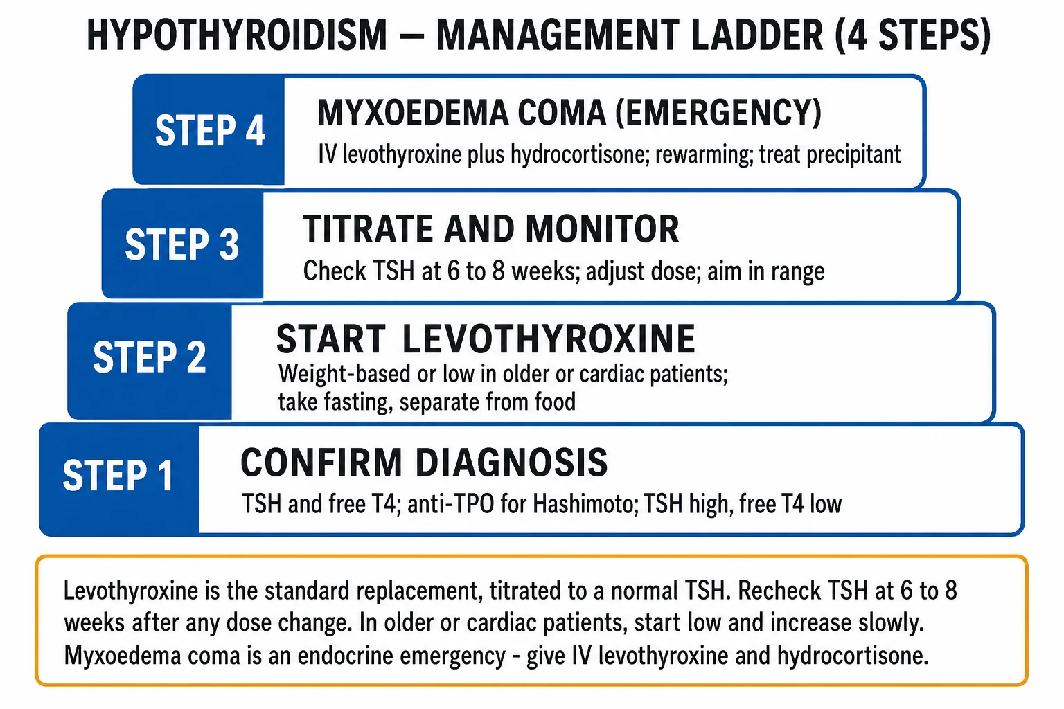
Myxoedema coma is an endocrine emergency. The resuscitation bundle (apply simultaneously, do not await TFT results):[7][8]
- Airway and breathing — secure the airway; give supplemental oxygen; the patient is often hypoventilating with CO2 retention — non-invasive or invasive ventilation may be required early. Hypercapnic respiratory failure is the leading cause of death.
- IV access — bloods for TSH, free T4, cortisol and ACTH (Synacthen test), FBC, U&E, CK, lipids, glucose, troponin, blood cultures, lactate, and a venous gas. CXR and ECG.
- IV hydrocortisone 100 mg stat, then 50 mg every 6 hours — given with or before thyroid hormone. Rationale: coexisting autoimmune adrenal insufficiency (Schmidt syndrome) or ACTH deficiency (central disease) is common, and giving levothyroxine to a cortisol-deficient patient precipitates adrenal crisis by increasing cortisol clearance and metabolic demand. Steroids can be tapered once adrenal insufficiency is excluded.
- IV levothyroxine — a loading dose of 200 to 500 mcg (typically 200 to 250 mcg in older/frailer patients, up to 500 mcg in younger, larger, severely ill patients) to replete the peripheral T4 pool, followed by 50 to 100 mcg IV daily. The oral route is unreliable in coma (gut hypomotility, oedema). IV liothyronine (T3) 10 to 20 mcg every 4 to 6 hours is added or substituted in some protocols (T3 is the active hormone and bypasses impaired peripheral conversion), particularly in profound coma — at the cost of a higher risk of precipitating angina and arrhythmia, so it requires cardiac monitoring.
- Passive rewarming — insulating blankets and a warm room. Active external rewarming (forced-air warmers, warm baths) is AVOIDED because peripheral vasodilation in a patient with profound bradycardia and reduced cardiac output can precipitate cardiovascular collapse ("rewarming shock"). Active core warming (warmed IV fluids, warmed humidified oxygen) is reasonable.
- Correct metabolic derangements — hyponatraemia is usually dilutional and corrects with thyroid replacement and free-water restriction; severe symptomatic hyponatraemia may need hypertonic saline cautiously (avoid rapid correction — osmotic demyelination). Hypoglycaemia is treated with IV dextrose. Restrict free water.
- Treat the precipitant — empirical broad-spectrum antibiotics after cultures (infection is the commonest precipitant and may be occult in the hypothermic, bradycardic patient — they may not mount a fever or leucocytosis). Stop offending drugs (sedatives, opioids, amiodarone, lithium). Treat cardiac ischaemia.
- Avoid sedatives, opioids, and unnecessary drugs — impaired clearance and reduced respiratory drive make these dangerous.
- ICU/HDU admission — these patients are critically ill and need ventilatory, cardiovascular and biochemical monitoring. [1]
Myxoedema coma bundle — H-A-I-R
HAIR
IV hydrocortisone 100 mg THEN IV levothyroxine 200 to 500 mcg loading (cortisol checked first)
Ventilatory support for hypercapnia; empirical antibiotics for the precipitant
Cautious fluids; correct hyponatraemia and hypoglycaemia; treat the precipitant
Passive (NOT active) rewarming; refrain from sedatives/opioids; ICU admission
The cortisol-first principle applies beyond myxoedema coma: in any patient with central hypothyroidism or suspected panhypopituitarism, always replace cortisol before thyroid hormone to avoid precipitating adrenal crisis. [1]
Management — Definitive & Stepwise
Chronic replacement therapy — levothyroxine (T4)
Levothyroxine is the standard of care for hypothyroidism. It is oral, once-daily, cheap, well tolerated, and effective as monotherapy in essentially all patients because peripheral deiodination supplies T3.[3][9]
Dosing:[3]
- Young, otherwise healthy adult: start at the full estimated replacement dose of about 1.6 mcg/kg/day (typically 100 to 150 mcg daily for an average adult). For a 60 kg woman, about 100 mcg daily.
- Elderly (over 65) or those with ischaemic heart disease / cardiac failure: start low and go slow — levothyroxine 25 mcg daily, increasing by 25 mcg every 2 to 4 weeks until TSH is in range. Rationale: raising metabolic rate and cardiac workload can precipitate angina, myocardial infarction or atrial fibrillation in compensated coronary disease.
- Pregnancy (see Special Populations): increase the existing dose by about 25 to 30% as soon as pregnancy is confirmed (often 2 extra tablets per week); use trimester-specific TSH targets.
- Children/adolescents: weight-based dosing, higher per kg in younger children (about 4 to 6 mcg/kg/day in young children, falling to adult-equivalent by adolescence).
- Congenital hypothyroidism (newborn): urgent high-dose levothyroxine (10 to 15 mcg/kg/day) to protect neurodevelopment — see Special Populations. [1]
How to take it:
- Once daily, on an empty stomach, 30 to 60 minutes before breakfast with water. Consistency of timing matters more than the exact interval.
- Separate from interacting drugs — calcium, iron (ferrous sulphate), proton-pump inhibitors, sucralfate, bile-acid sequestrants, oestrogen, raloxifene, and some foods (soy, coffee, high-fibre) impair absorption; separate by at least 4 hours where possible.
- Same brand/consistent formulation preferred (small bioequivalence differences can shift TSH). [1]
Key interactions:
- Absorption reduced: calcium carbonate, ferrous sulphate, PPIs, sucralfate, cholestyramine, aluminium antacids, coffee, soy, grapefruit juice.
- Increased requirement (raised TBG): oestrogen (HRT, oral contraceptives), pregnancy, tamoxifen, raloxifene, heroin/methadone, hepatitis.
- Increased clearance: phenytoin, carbamazepine, rifampicin, phenobarbital (enzyme inducers); sertraline (modest).
- Mechanism (raised TSH on a previously stable dose): first check adherence, then pregnancy, then interactions/absorption, then weight change, before assuming progression. [1]
Monitoring:[3]
- Recheck TSH at 6 to 8 weeks after any dose change (TSH has a half-life of about a week, so steady state takes 6 to 8 weeks). Adjust by 12.5 to 25 mcg and repeat.
- Once stable, recheck TSH every 6 to 12 months lifelong, and at any clinical change (pregnancy, new symptoms, new interacting drug, weight change).
- Monitor TSH, not free T4, in primary disease. In central disease, monitor free T4 (because TSH is unreliable) and target a free T4 in the upper half of the reference range.
- Target TSH: typically the lower half of the reference range (around 0.4 to 2.0 mIU/L) for symptomatic wellbeing, but within the laboratory reference is acceptable. Avoid over-replacement (TSH suppressed) — risk of atrial fibrillation and osteoporosis.
Subclinical hypothyroidism
Treatment thresholds (ATA/ETA):[5][6]
- TSH over 10 mIU/L — start levothyroxine (full but gradually introduced) and monitor.
- TSH 4.5 to 10 mIU/L — treat if symptomatic, anti-TPO positive, goitre, dyslipidaemia, pregnancy or planning conception/IVF, or young; otherwise monitor TFT every 6 to 12 months. Confirm with a repeat TFT at 3 months before committing to lifelong therapy.
- In pregnancy: treat any TSH above the trimester-specific reference (see Special Populations).
Central hypothyroidism
- Confirm with low/normal TSH + low free T4 in the context of known or suspected pituitary disease.
- Exclude and treat coexisting adrenal insufficiency first — check a morning cortisol and ACTH (Synacthen test); replace cortisol (oral hydrocortisone) before starting levothyroxine.
- Treat with levothyroxine, but monitor to free T4 (target upper half of the reference range), because TSH is unreliable in central disease.
- Investigate the underlying pituitary lesion (MRI, visual fields, full anterior-pituitary panel — prolactin, IGF-1, FSH/LH, testosterone/oestradiol, cortisol). [1]
Levothyroxine-liothyronine combination therapy
A minority of patients report persistent symptoms (fatigue, low mood, cognitive fog) despite a biochemically normal TSH on levothyroxine monotherapy. Levothyroxine-liothyronine (T4/T3) combination therapy is a controversial option.[9]
- The 2021 ATA/European Thyroid Association task force concludes there is insufficient evidence to support routine combination therapy, but a carefully monitored trial may be considered in symptomatic, biochemically well-replaced patients who have been adequately worked up for alternative causes.
- Not recommended in pregnancy, cardiac disease, the elderly, or osteoporosis.
- Liothyronine (T3) monotherapy is not recommended for routine hypothyroidism. [1]
lodine supplementation
- In iodine-deficient regions, universal salt iodisation (universal salt iodisation in India under the National Iodine Deficiency Disorders Control Programme) is the public-health cornerstone and prevents endemic goitre and cretinism.
- Do not advise iodine or kelp supplements to patients with autoimmune thyroiditis — excess iodine worsens the disease via the Wolff-Chaikoff effect. [1]
Patient education
- Take levothyroxine fasting, 30 to 60 minutes before breakfast; separate from calcium, iron and PPIs.
- Do not stop — replacement is lifelong for permanent disease.
- Carry a card/medical alert and seek review in illness, pregnancy, or new medications.
- Pregnancy planning — increase the dose at confirmation of pregnancy. [1]
Specific Subtypes & Scenarios
Hashimoto thyroiditis
The commonest cause of hypothyroidism in iodine-sufficient regions. Female, 30 to 60 years, family history of autoimmune disease. Presents with gradual hypothyroid symptoms and a firm, rubbery, diffuse goitre; some are euthyroid or subclinical at presentation.[1]
- Anti-TPO positive (about 90%); anti-Tg also positive in many.
- Histology: lymphocytic infiltration with germinal centres, Hurthle (oxyphil) cell metaplasia, fibrosis.
- May have a transient thyrotoxic phase ("hashitoxicosis") from follicular rupture releasing preformed hormone, distinguished from Graves by the absence of TRAb and a low (not suppressed) radioactive iodine uptake.
- Associations: type 1 diabetes, pernicious anaemia, Addison (autoimmune polyglandular syndrome type 2 / Schmidt syndrome), vitiligo, alopecia, coeliac disease, Sjogren, SLE, rheumatoid arthritis, Turner and Down syndromes.
- Complications: progressive hypothyroidism; B-cell lymphoma of the thyroid (MALT lymphoma) — a rare but classically-tested complication of longstanding Hashimoto presenting as a rapidly enlarging, asymmetric goitre; papillary thyroid carcinoma association. [1]
Post-ablative and iatrogenic hypothyroidism
- After radioiodine for Graves — most patients become permanently hypothyroid within 3 to 12 months; lifelong levothyroxine is anticipated and is preferable to recurrent hyperthyroidism.
- After thyroidectomy — immediate lifelong replacement (start within 24 to 48 hours).
- After external beam radiotherapy to the neck (for lymphoma, head-and-neck cancer) — late-onset hypothyroidism; monitor TFT annually. [1]
Drug-induced hypothyroidism
- Amiodarone — check TFT before and every 6 months during therapy. Hypothyroidism (commoner in iodine-sufficient regions and anti-TPO-positive patients) is treated with levothyroxine; amiodarone can usually be continued if the cardiac indication demands. Amiodarone-induced thyrotoxicosis (type 2) is a destructive thyroiditis treated with steroids (and/or carbimazole for type 1).
- Lithium — check TFT at baseline and 3 to 6 months; monitor. Treat with levothyroxine if hypothyroid; lithium need not always be stopped.
- Immune-checkpoint inhibitors (ipilimumab, nivolumab, pembrolizumab), interferon, interleukin-2 — can precipitate painless thyroiditis with a thyrotoxic phase then hypothyroidism; treat with levothyroxine and (if symptomatic) beta-blockers in the thyrotoxic phase. [1]
Subacute (de Quervain) and postpartum thyroiditis
Both can cause a transient hypothyroid phase following a thyrotoxic phase.
- Subacute (de Quervain) thyroiditis — viral, painful, tender goitre, raised ESR/CRP; NSAIDs and beta-blockers in the thyrotoxic phase; levothyroxine if symptomatic and hypothyroid (often transient, taper after 3 to 6 months).
- Postpartum thyroiditis — painless, occurs within 1 year postpartum in 5 to 10% of women, often with anti-TPO positivity; a thyrotoxic phase at 1 to 3 months postpartum (often mislabelled postpartum depression/anxiety) then a hypothyroid phase at 4 to 8 months; treat the hypothyroid phase with levothyroxine if symptomatic, taper postpartum; high recurrence in subsequent pregnancies and 20 to 40% progress to permanent hypothyroidism — long-term monitoring. [1]
Central (secondary/tertiary) hypothyroidism
- Cause: pituitary macroadenoma; Sheehan syndrome (postpartum pituitary infarction — failure to lactate, amenorrhoea, hypopituitarism); infiltrative disease (haemochromatosis, sarcoid, lymphocytic hypophysitis); surgery/radiation; trauma; genetic.
- Biochemistry: low/inappropriately normal TSH + low free T4.
- Always rule out coexisting cortisol deficiency first — replace cortisol, then levothyroxine; monitor to free T4 (not TSH).
- Imaging: pituitary MRI; visual-field testing for chiasmal compression. [1]
Congenital hypothyroidism (cretinism)
Detected on newborn screening (heel-prick TSH at 3 to 5 days of life). Untreated, it causes severe, irreversible intellectual disability, growth failure and the classic features — macroglossia, umbilical hernia, prolonged jaundice, constipation, hypotonia, large fontanelles, dry skin, hoarse cry. [1]
- Causes: thyroid dysgenesis (athyreosis, ectopic/sublingual gland — most common, sporadic); dyshormonogenesis (inborn errors of NIS, TPO, thyroglobulin, pendrin — usually autosomal recessive, often goitrous); maternal iodine deficiency; maternal antithyroid drugs or radioiodine; TSH receptor blocking antibodies transplacentally.
- Treatment is urgent — start levothyroxine 10 to 15 mcg/kg/day as soon as the diagnosis is confirmed (certainly by 2 weeks of age) to protect neurodevelopment; delay beyond 6 months of untreated disease causes irreversible cognitive impairment. TSH monitoring is intensive.
- Endemic cretinism (iodine deficiency) has two forms — neurological (deaf-mutism, spastic diplegia, intellectual disability) and myxoedematous (severe hypothyroid features, stunted growth); prevented by maternal iodine supplementation and universal salt iodisation. [1]
Complications & Pitfalls
Cardiovascular
- Dyslipidaemia (raised LDL) accelerates atherosclerosis — untreated hypothyroidism is a cardiovascular risk factor.
- Pericardial and pleural effusions (rarely tamponade).
- Heart failure (dilated cardiomyopathy in severe disease; reverses with treatment).
- Bradycardia, diastolic hypertension, increased systemic vascular resistance.
- Precipitation of angina, myocardial infarction or atrial fibrillation on starting levothyroxine too rapidly in the elderly or those with ischaemic heart disease. [1]
Obstetric and fetal
Untreated overt hypothyroidism in pregnancy carries significant risk:[4]
- Miscarriage, fetal loss, stillbirth.
- Pre-eclampsia, gestational hypertension, placental abruption, anaemia.
- Preterm birth, low birthweight.
- Impaired fetal neurodevelopment — maternal T4 is essential for early fetal brain development before the fetal thyroid is functional (around 12 to 14 weeks); maternal hypothyroidism in the first trimester reduces offspring IQ. Even subclinical hypothyroidism in pregnancy is associated with adverse outcomes.
Neurological
- Peripheral neuropathy, carpal tunnel syndrome, sensorineural deafness, cerebellar ataxia (rare).
- Myxoedema coma (the decompensated extreme — see Resuscitation).
- Dementia, depression, psychosis that may be misdiagnosed as primary psychiatric disease. [1]
Complications of over-treatment
Iatrogenic (subclinical) hyperthyroidism from over-replacement of levothyroxine causes:
- Atrial fibrillation (especially in the elderly).
- Osteoporosis and increased fracture risk.
- Tachycardia, palpitations, anxiety, tremor, heat intolerance, weight loss. [1]
This is why TSH monitoring is essential — a suppressed TSH on levothyroxine means over-treatment and warrants dose reduction. [1]
Classic pitfalls
- Missing central hypothyroidism — interpreting an "in-range" TSH as normal when the free T4 is low (a "normal" TSH with a low free T4 is not normal). Always request a free T4 when central disease is suspected.
- Giving levothyroxine before cortisol in panhypopituitarism or myxoedema coma — precipitates adrenal crisis. Always check cortisol and replace it first.
- Mislabelling euthyroid sick syndrome as hypothyroidism and treating it — TFTs normalise on recovery; do not treat.
- Under-dosing in pregnancy — the requirement rises 25 to 30%; failure to increase leads to adverse obstetric and fetal outcomes.
- Precipitating angina on initiation in ischaemic heart disease by starting at full dose — start 25 mcg and titrate slowly.
- Over-treatment (suppressed TSH) causing atrial fibrillation and osteoporosis, especially in the elderly.
- Missing drug interactions (calcium, iron, PPI, oestrogen) as a cause of a rising TSH on a previously stable dose.
- Failing to recognise myxoedema coma as a treatable cause of hypothermia and reduced consciousness. [1]
Prognosis & Disposition
Treated primary hypothyroidism
Excellent. With adequate levothyroxine replacement, life expectancy and quality of life are normal, and pregnancy outcomes are equivalent to the euthyroid population. Replacement is lifelong for permanent causes (autoimmune, iatrogenic, congenital); thyroiditis-related hypothyroidism may be transient and can be weaned after 3 to 6 months under TFT monitoring.[2][3]
Subclinical hypothyroidism
The natural history depends on anti-TPO status and age:
- About 2 to 5% per year progress to overt disease, higher if anti-TPO positive (about 4% per year) and with higher baseline TSH.
- Some revert to euthyroidism, especially if anti-TPO negative.
- Longstanding untreated subclinical disease is associated with cardiovascular and heart-failure risk (especially in younger patients) and dyslipidaemia. [1]
Myxoedema coma
High mortality — 20 to 60% despite appropriate treatment, especially in the elderly, those with very low core temperature (under 30 degrees C), sepsis, bradycardia, or delayed presentation.[7][8] Predictors of poor outcome: advanced age, hypothermia severity, sepsis, hypoventilation/hypercapnia, cardiovascular instability, and delay in initiating treatment. Survivors require lifelong levothyroxine.
Monitoring cadence
- After any dose change: TSH at 6 to 8 weeks.
- Once stable: TSH every 6 to 12 months lifelong.
- Pregnancy: TSH each trimester and 4 to 6 weeks postpartum. [1]
When to refer to endocrinology
- Central hypothyroidism (pituitary disease).
- Pregnancy (planning, pregnant, postpartum thyroiditis).
- Myxoedema coma (ICU with endocrine input).
- Children and congenital disease.
- Refractory biochemistry (rising TSH despite escalating doses, persistent symptoms despite biochemical euthyroidism, suspected malabsorption).
- Coexisting adrenal insufficiency (autoimmune polyglandular syndromes).
- Rapidly enlarging goitre, asymmetric goitre, or cervical lymphadenopathy (rule out lymphoma/cancer).
- Amiodarone-induced thyroid dysfunction (complex, mixed patterns). [1]
Special Populations
Pregnancy and lactation
Pregnancy substantially increases levothyroxine requirement:[4]
- Mechanism: rising oestrogen increases thyroxine-binding globulin (TBG), increasing total (but not initially free) T4; the placental D3 deiodinase degrades T4/T3; the fetal demand for maternal T4 in the first trimester (before the fetal thyroid functions); increased renal iodine clearance.
- Effect: requirement rises by 25 to 30% from weeks 4 to 6, peaking at 16 to 20 weeks.
- Action: at confirmation of pregnancy, increase the levothyroxine dose by about 25 to 30% (a practical rule: two extra tablets per week). Check TSH each trimester and 4 to 6 weeks postpartum, then reduce to the pre-pregnancy dose after delivery.
- Trimester-specific TSH targets (ATA 2017): first trimester under 2.5 mIU/L; second trimester under 3.0 mIU/L; third trimester under 3.0 mIU/L.
- Untreated overt hypothyroidism: miscarriage, pre-eclampsia, abruption, preterm birth, impaired fetal neurodevelopment.
- Subclinical hypothyroidism in pregnancy: treat to keep TSH within trimester-specific reference.
- Screening: the ATA recommends targeted screening (women with goitre, strong family history, prior thyroid disease, type 1 diabetes, autoimmune disease, infertility, prior preterm delivery, ICSI/IVF, miscarriage, age over 30, BMI over 40, amiodarone/lithium, recent contrast); the case for universal screening remains debated — both are defensible.
- Hypothyroidism is safe in lactation — levothyroxine crosses minimally into breast milk and is identical to endogenous T4.
Elderly
- Atypical presentation: apathy, depression, cognitive decline, falls, failure to thrive, hyponatraemia, constipation — often mistaken for dementia or normal ageing. Have a low threshold to test TSH.
- Start low and go slow: levothyroxine 25 mcg, titrate by 25 mcg every 2 to 4 weeks, because rapid replacement can precipitate angina, infarction or atrial fibrillation in occult ischaemic heart disease.
- Cardiac comorbidity: coordinate with cardiology; if angina occurs on initiation, reduce the dose and treat the angina first before re-titrating. [1]
Ischaemic heart disease
- Start levothyroxine 25 mcg daily, increase by 25 mcg every 2 to 4 weeks under symptom surveillance; do not start at full dose.
- Severe hypothyroidism with decompensated heart failure or myxoedema coma may still need IV levothyroxine — balance risk with cardiology/endocrine input. [1]
Coexisting adrenal insufficiency
- Always replace cortisol before thyroid hormone — in autoimmune polyglandular syndrome type 2 (Schmidt: Addison + Hashimoto + type 1 diabetes), central hypothyroidism, and myxoedema coma.
- Hydrocortisone (oral 15 to 25 mg/day in divided doses; or IV 100 mg stat then 50 mg every 6 hours in emergency) before levothyroxine. [1]
Children and adolescents
- Weight-based dosing is higher per kg than in adults — about 4 to 6 mcg/kg/day in young children, falling to adult-equivalent (1.6 mcg/kg/day) by adolescence.
- Growth and puberty are exquisitely sensitive to thyroid status — undertreatment causes growth retardation and delayed puberty, over-treatment accelerates bone age.
- Adherence in teenagers is a common problem — a rising TSH in a stable adolescent usually means non-adherence. [1]
Congenital / newborn
- Newborn screening (heel-prick TSH) detects disease before symptoms — the cornerstone of prevention.
- Start levothyroxine urgently (10 to 15 mcg/kg/day) by 2 weeks of age to protect neurodevelopment.
- Intensive TSH and free T4 monitoring (every 1 to 2 months in the first 6 months, every 3 months to age 3, then every 6 to 12 months). [1]
Evidence, Guidelines & Regional Differences
ATA/AACE 2014 Treatment Guidelines (US)[3]
- Levothyroxine monotherapy is the standard of care for primary hypothyroidism.
- Weight-based dosing (1.6 mcg/kg/day) for young healthy adults; low-and-slow in the elderly and cardiac.
- Monitor TSH at 6 to 8 weeks after any change, then annually.
- Do not routinely use liothyronine or combination therapy; an empirical trial may be considered in well-replaced but symptomatic patients.
- Target TSH in the lower half of the reference range in younger patients; within the reference range generally. [1]
ETA 2013 Subclinical Hypothyroidism Guideline (Europe)[5]
- Confirm raised TSH with a repeat at 3 months.
- Treat if TSH over 10 mIU/L (or symptomatic, goitrous, anti-TPO positive, pregnant, dyslipidaemic, young).
- Monitor otherwise every 6 to 12 months.
- L-thyroxine starting dose 25 to 50 mcg, titrated to a TSH in the reference range (often the lower half). [1]
ATA 2017 Pregnancy & Postpartum Guideline (US)[4]
- Trimester-specific TSH targets (under 2.5 mIU/L first trimester; under 3.0 second and third).
- Increase levothyroxine 25 to 30% at confirmation of pregnancy.
- Targeted (case-finding) screening recommended; universal screening remains debated.
- Treat overt and symptomatic subclinical hypothyroidism; manage women with isolated hypothyroxinaemia or thyroid autoantibodies on a case-by-case basis. [1]
Combination therapy evidence (ATA/ETA 2021)[9]
A monitored trial of T4/T3 combination may be considered in well-replaced symptomatic patients, but evidence does not support routine use. Not recommended in pregnancy, cardiac disease, osteoporosis, or the elderly. [1]
Regional picture
- India and the developing world: iodine deficiency remains the dominant cause of hypothyroidism and cretinism despite the National Iodine Deficiency Disorders Control Programme (NIDDCP) and universal salt iodisation; autoimmune disease is rising in urban populations as iodine intake improves. Subacute thyroiditis (de Quervain) is relatively common. Herbal and ayurvedic preparations containing undisclosed iodine can precipitate or worsen disease.
- Iodine-sufficient regions (North America, much of Europe): autoimmune Hashimoto dominates; iodine excess (contrast, supplements) is a more common cause than deficiency. [1]
Controversies
- Universal vs targeted screening in pregnancy — both defended; ATA favours targeted, many European centres screen universally.
- T4/T3 combination therapy — popular with patient-advocacy groups; evidence weak for routine use.
- The upper limit of the TSH reference range — some advocate lowering it to 2.5 to 3.0 mIU/L (accounting for the skewed distribution that includes subclinical disease), which would label many more people as hypothyroid; mainstream practice keeps the population-derived range (about 0.4 to 4.5 mIU/L). [1]
Exam Pearls
Hypothyroidism symptoms — HYPOTHYROID
HYPOTHYROID
vocal-cord glycosaminoglycan deposition
lethargy, fatigue, slowed cognition
non-pitting myxoedema
weight gain from low metabolic rate
cold intolerance is cardinal
and diastolic hypertension
impaired beta-carotene conversion
slow-relaxing ankle jerks
infertility, galactorrhoea
rarely megacolon
brittle hair, madarosis
Myxoedema coma precipitants — COLD SHOCK
COLD
the classic precipitant
depress respiratory drive and metabolism
non-adherence to replacement
any systemic illness is a common trigger
Exam application bank (NEET-PG / INICET)
One-line answer
Hypothyroidism is the clinical and biochemical syndrome caused by deficiency of thyroid hormone (T4 and T3) at the tissue level. The commonest cause in iodine-sufficient regions is chronic autoimmune (Hashimoto) thyroiditis; worldwide, iodine deficiency dominates. The biochemical hallmark of primary disease is a raised TSH with low free T4. Treatment is lifelong oral levothyroxine at about 1.6 mcg/kg/day. Myxoedema coma is the decompensated, life-threatening extreme — IV levothyroxine plus hydrocortisone, passive rewarming and treatment of the precipitant. [1]
Worked stems (answer without another resource)
Stem 1 — Classic presentation. Map symptoms to mechanism; name the first investigation and first treatment step with dose/route if drug therapy is standard. [1]
Stem 2 — Unstable / complicated. List red flags that force immediate resuscitation, theatre, ICU, antidote, or reperfusion — and what you do in the first 15 minutes. [1]
Stem 3 — Atypical group. Elderly, pregnancy, child, or immunocompromised: how presentation and thresholds change. [1]
Stem 4 — Differential trap. Name the three closest mimics and one discriminator for each. [1]
Stem 5 — Disposition. Who goes home with safety-netting, who is admitted, who needs HDU/ICU/theatre, and what follow-up is mandatory. [1]
Rapid viva checklist
- Definition + classification
- Pathophysiology chain
- Bedside signs / criteria
- Score with exact components (if any)
- Emergency bundle
- Definitive therapy with doses
- Complications of disease and of treatment
- Special populations
- Guideline/trial name if classic
- Three exam traps
Coverage self-check
If you cannot answer any stem above from this page alone, re-read the matching section — the page is intended to be self-sufficient for final-prof and NEET-PG/INICET questions on Hypothyroidism.
[1]References
- [1]Ralli M, Angeletti D, Fiore M, et al. Hashimoto's thyroiditis: An update on pathogenic mechanisms, diagnostic protocols, therapeutic strategies, and potential malignant transformation Autoimmun Rev, 2020.PMID 32805423
- [2]Taylor PN, Medici MM, Hubalewska-Dydejczyk A, et al. Hypothyroidism Lancet, 2024.PMID 39368843
- [3]Jonklaas J, Bianco AC, Bauer AJ, et al. Guidelines for the treatment of hypothyroidism: prepared by the american thyroid association task force on thyroid hormone replacement Thyroid, 2014.PMID 25266247
- [4]Alexander EK, Pearce EN, Brent GA, et al. 2017 Guidelines of the American Thyroid Association for the Diagnosis and Management of Thyroid Disease During Pregnancy and the Postpartum Thyroid, 2017.PMID 28056690
- [5]Pearce SH, Brabant G, Duntas LH, et al. 2013 ETA Guideline: Management of Subclinical Hypothyroidism Eur Thyroid J, 2013.PMID 24783053
- [6]Cooper DS, Biondi B. Subclinical thyroid disease Lancet, 2012.PMID 22273398
- [7]Kruithoff ML, Gigliotti BJ. Thyroid Emergencies: A Narrative Review Endocr Pract, 2025.PMID 40553957
- [8]Valerio Aguirre RA, Ocana Martinez KC, Martinez Rodriguez EU, et al. From the Popoveniuc Score to Therapeutic Protocols: A Comprehensive Review of Myxedema Coma Cureus, 2026.PMID 41658844
- [9]Jonklaas J, Bianco AC, Cappola AR, et al. Evidence-Based Use of Levothyroxine/Liothyronine Combinations in Treating Hypothyroidism: A Consensus Document Thyroid, 2021.PMID 33276704