Endocrinology · General Medicine
Osteoporosis
Also known as Osteoporosis · Low bone mass · Fragility fractures · Silent bone disease
Osteoporosis is a systemic skeletal disease of low bone mass and microarchitectural deterioration, increasing the risk of fragility fractures (hip, spine, wrist). Diagnosis is by DEXA T-score of minus 2.5 or less (osteopenia minus 1 to minus 2.5; a fragility fracture establishes the diagnosis regardless of score), and the FRAX score estimates 10-year fracture risk to guide treatment. Risk factors include age, female sex, postmenopausal status, family history, low BMI, glucocorticoids, smoking, hypogonadism and hyperthyroidism. It is often silent until a fracture. Management is lifestyle (weight-bearing exercise, no smoking, calcium and vitamin D) plus first-line bisphosphonates (alendronate, zoledronate), with denosumab and anabolic teriparatide for high-risk or refractory disease. After 3 to 5 years of a bisphosphonate, a drug holiday is considered. The goal is preventing the first fragility fracture, especially hip and vertebral, which carry high morbidity and mortality.
On this page & tools
Your progress
Saved locally on this device.
Exam tags
Red flags
Overview & Definition
Osteoporosis is the most common metabolic bone disease and one of the most important chronic diseases of ageing. It is defined as a progressive systemic skeletal disorder characterised by low bone mass and microarchitectural deterioration of bone tissue, leading to increased bone fragility and a consequent increase in fracture risk. In practice it is a disease whose clinical importance is entirely its fractures — most often of the hip, vertebrae and distal forearm — because the bone itself is painless until it breaks. Those fractures carry major morbidity, mortality and cost, especially in older adults, so the entire strategy of management is preventive: detect high-risk bone early with DEXA and FRAX, optimise lifestyle, calcium and vitamin D, and start bone-targeted therapy — most often a bisphosphonate — to prevent the first fracture, because a prior fracture strongly predicts another.[1]
The operational WHO diagnostic threshold is based on bone mineral density (BMD) measured by dual-energy X-ray absorptiometry (DEXA): osteoporosis is a T-score of minus 2.5 or less at the femoral neck, total hip or lumbar spine; osteopenia (low bone mass) is a T-score between minus 1.0 and minus 2.5; and normal is a T-score of minus 1.0 or higher. Crucially, a low-trauma (fragility) fracture of the hip or spine establishes the diagnosis of osteoporosis regardless of the T-score, so treatment is indicated even when the density falls in the osteopenic range. A defining biochemical feature is that serum calcium, phosphate and alkaline phosphatase are normal — a point that reliably separates osteoporosis from osteomalacia, primary hyperparathyroidism, multiple myeloma and Paget disease at the bedside.[1][2]
Classification
Two overlapping axes classify osteoporosis. The first is pathophysiological: primary osteoporosis is divided into type 1 (postmenopausal), driven by oestrogen deficiency and accelerated trabecular loss in women aged roughly 50 to 70, and type 2 (senile), an ageing-related loss of both cortical and trabecular bone in men and women over 70. The second, and more clinically useful, axis is aetiological: primary versus secondary. Roughly half of women and the majority of men with a fragility fracture have an identifiable secondary cause, so a structured search for secondary drivers is mandatory in every new diagnosis (see Differential Diagnosis and Investigations).[1]
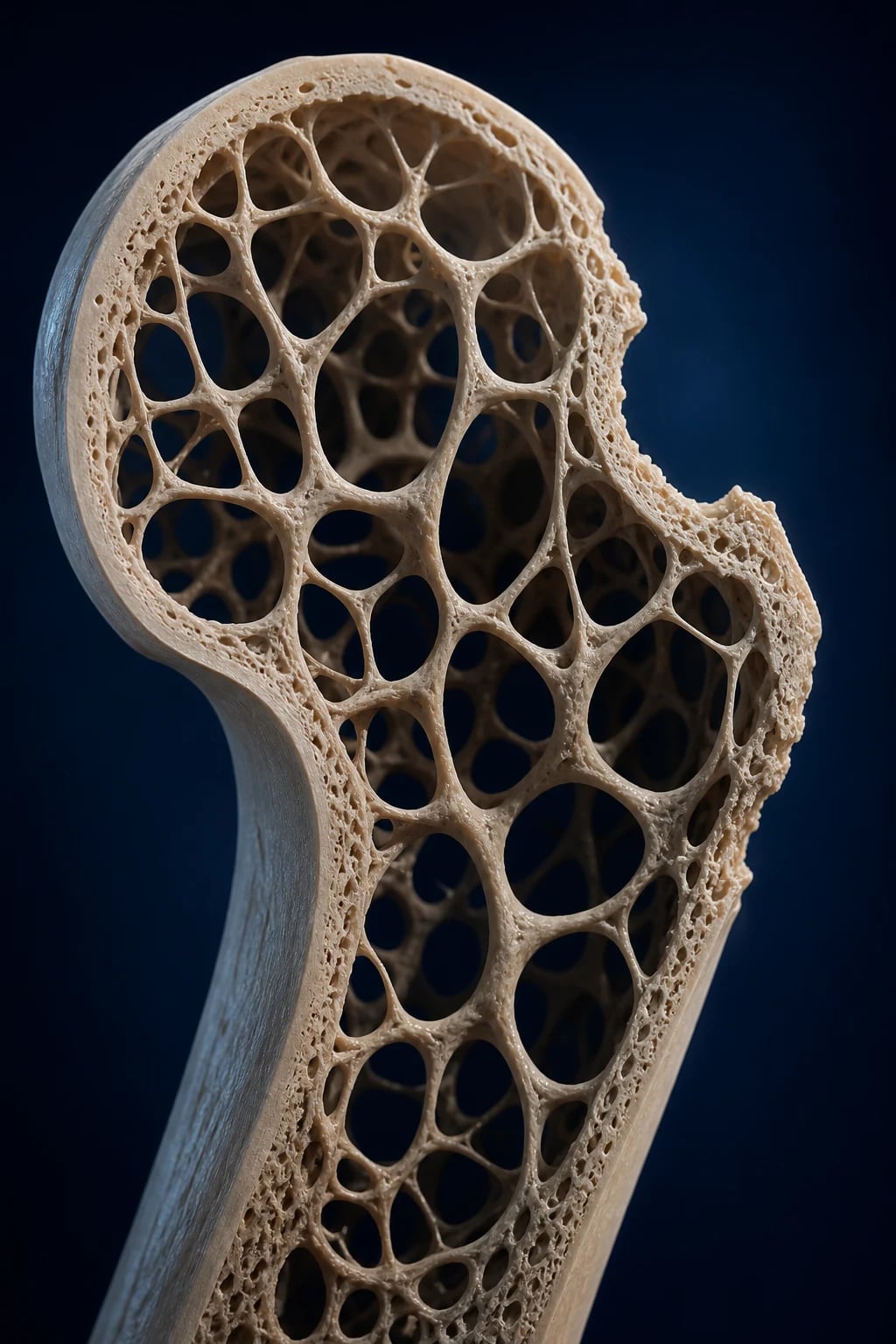
Type 1 (postmenopausal)
oestrogen deficiency
- Women, age 50 to 70
- High-turnover, osteoclast-driven
- Accelerated trabecular loss
- Vertebral and distal-radial fractures
Type 2 (senile)
ageing
- Men and women over 70
- Low-turnover, fewer osteoblasts
- Cortical and trabecular loss
- Hip, pelvic and proximal fractures
Secondary
an identifiable cause
- Glucocorticoids, hypogonadism
- Hyperthyroidism, hyperparathyroidism
- Malabsorption, CKD, RA
- Most men and many younger women
Epidemiology & Risk Factors
Osteoporosis affects an estimated 200 million people worldwide and is responsible for an osteoporotic fracture roughly every 3 seconds. Lifetime risk is strikingly asymmetric: about one in three women and one in five men over the age of 50 will sustain a fragility fracture. Hip-fracture incidence rises exponentially in women after age 50 and in men after age 70, and as populations age the global burden is projected to rise sharply, making osteoporosis a leading — and growing — cause of morbidity, mortality and health-care cost.[1]
Osteoporosis — the numbers that matter
The burden falls hardest on the hip. Worldwide there are an estimated 9 million osteoporotic fractures annually, of which about 1.6 million are hip fractures, and hip-fracture numbers are projected to roughly double by 2050 as populations age and urbanise — driven increasingly by ageing in Asia, where much of the future burden now lies. The economic cost is substantial: hip fracture consumes a major share of orthopaedic and rehabilitation budgets in every developed health system, and the loss of independence after a hip fracture is the single largest driver of new nursing-home admissions in older women. The demographic skew is consistent everywhere — women dominate the postmenopausal cohort, but the male case-fatality after a hip fracture is, if anything, higher because of comorbidity and later presentation. Vitamin D deficiency, low dietary calcium, limited sun exposure and physical inactivity are particularly prevalent in the housebound and in dark-skinned or covered populations living at high latitude, who develop osteomalacia and osteoporosis concurrently and are easily missed.[1]
Risk factors divide into those that are largely fixed (age, female sex, menopause, family history — particularly a parental history of hip fracture — low body mass index, ethnicity with white and Asian populations at highest risk) and those that are modifiable or iatrogenic. The modifiable and secondary drivers are the actionable list an examiner expects: oral glucocorticoids, hypogonadism (including premature menopause, anti-androgen therapy, hyperprolactinaemia), hyperthyroidism, primary hyperparathyroidism, chronic kidney disease, malabsorption (coeliac disease, post-gastrectomy, bariatric surgery), chronic liver disease, rheumatoid arthritis, low calcium and vitamin D, immobility and low body mass index. Several common medications accelerate bone loss and are exam favourites: glucocorticoids (the single most important), proton-pump inhibitors (long-term), selective serotonin re-uptake inhibitors, anti-epileptics (enzyme-inducers such as phenytoin and carbamazepine impair vitamin D metabolism), heparin, aromatase inhibitors, gonadotropin-releasing hormone agonists, thiazolidinediones and excess thyroid hormone replacement. Smoking, alcohol intake of 3 or more units daily, and high caffeine are independent risk factors. A prior fragility fracture is itself the strongest single predictor of a future fracture, roughly doubling the risk.[1][2]
Pathophysiology
Bone is not inert; it is a dynamic tissue that is continually remodelled throughout life in tightly coupled basic multicellular units. Two cell lineages do the work: osteoclasts (multinucleated, haematopoietic-derived cells of the monocyte-macrophage line) resorb old or damaged bone, and osteoblasts (mesenchymal-derived) lay down new osteoid that subsequently mineralises. Osteoporosis is fundamentally a remodelling imbalance: over any given cycle, resorption exceeds formation, producing net bone loss. Although the absolute bone turnover rate may rise or fall, the net balance is negative, so trabeculae thin and perforate, cortex becomes porous, and bone strength — which depends on density and quality (architecture, mineralisation, microdamage accumulation) — falls below the threshold at which everyday loads cause fracture.[1]
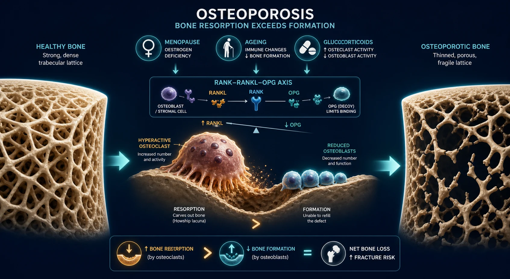
The molecular control of this balance centres on the RANK / RANKL / OPG pathway. RANKL (receptor activator of nuclear factor kappa-B ligand) is expressed by osteoblasts and stromal cells and binds RANK on the osteoclast precursor, driving osteoclast differentiation, activation and survival. The decoy receptor osteoprotegerin (OPG), also produced by osteoblasts, binds RANKL and blocks it, braking resorption. After the menopause, oestrogen deficiency removes its normal tonic restraint: production of RANKL and pro-resorptive cytokines (IL-1, IL-6, TNF-alpha) rises while OPG falls, so osteoclasts are recruited and activated and resorption accelerates. The result is high-turnover, type 1 (postmenopausal) osteoporosis, with selective loss of trabecular bone at the vertebrae and distal forearm — exactly the sites that fracture first.[1]
A single remodelling cycle moves through a stereotyped sequence — activation, resorption, reversal, formation, and mineralisation with resting — that normally ends in balanced repair. The cycle is choreographed by the osteocyte, the most abundant and long-lived cell in bone, which is exquisitely mechanosensitive: it senses load through its lacuno-canalicular network and, in response, turns remodelling on or off. When bone is underloaded (immobility, weightlessness, paralysis) osteocytes signal for resorption; when loaded, they promote formation. The key molecular link from mechanosensing to formation is Wnt signalling: osteocytes normally secrete inhibitors of this pathway, chiefly sclerostin (SOST) and Dickkopf-1 (DKK1), which restrain osteoblast activity. Suppress sclerostin and bone formation rises — exactly the mechanism exploited by romosozumab, a sclerostin-neutralising antibody. Coupling between resorption and formation is partly carried by ephrin to Eph signalling and by factors released from the resorbed matrix (TGF-beta, IGF-1) that recruit osteoblasts to refill each cavity. In osteoporosis this coupling falters: postmenopausal oestrogen loss both amplifies the resorptive signal (via RANKL) and impairs the osteoblast response, while in senescence the osteoblast progenitor pool shrinks and sclerostin rises, so each cycle underfills the cavity. Oestrogen therefore protects bone in two directions — it restrains resorption and sustains formation — which is why its loss at the menopause is so devastating to the skeleton, and why the same biology predicts that blocking resorption (bisphosphonates, denosumab) or stimulating formation (teriparatide, romosozumab) should each shift the balance back toward net gain.[1]
In parallel, ageing causes a different lesion: osteoblast formation declines and osteocytes become senescent and die, so each remodelling cycle underfills the resorption cavity. Sclerostin, an osteocyte-derived inhibitor of Wnt signalling and therefore of osteoblast activity, rises with age. Cortical bone becomes porous and trabecular connectivity is lost, producing low-turnover, type 2 (senile) osteoporosis and the characteristic hip and pelvic fractures of advanced age. The same final common pathway is amplified by secondary drivers: glucocorticoids both suppress osteoblast and osteocyte survival and increase resorption; hyperthyroidism and excess thyroid replacement accelerate turnover; hyperparathyroidism drives osteoclast-mediated resorption; hypogonadism removes sex-steroid braking of remodelling in either sex; vitamin D deficiency causes secondary hyperparathyroidism; immobility unloads the skeleton, which by Wolff's law then resorbs; and smoking impairs osteoblast function and accelerates menopause.[1]
Denosumab — the pathway in one drug
Fully human monoclonal antibody to RANKL (antiresorptive)
Dose
60 mg subcutaneously every 6 months
Clinical Presentation
Osteoporosis is the classic silent disease: it produces no symptoms until bone breaks. Many patients are first identified through a DEXA scan performed for risk factors, or — far too often — only after the first fracture. Clinically, presentation takes one of three fracture patterns, occasionally with chronic deformity from accumulated vertebral collapse.[1]
Vertebral (fragility) fractures are the most common osteoporotic fracture. Two-thirds are clinically silent and found incidentally on chest or abdominal imaging; the rest present with acute or subacute mid- to lower-thoracic and upper-lumbar back pain after minimal or no trauma (often just bending, lifting, or coughing). The pain is typically local and mechanical, eased by rest and worsened by sitting forward or weight-bearing. With multiple wedge-compression fractures the spine shortens and angulates, producing loss of height (more than 4 cm is significant), a progressive thoracic kyphosis (the so-called dowager's hump), a protuberant abdomen (rib margin meeting the pelvic brim), and in severe cases restrictive respiratory limitation and early satiety. Hip fractures present after a fall from standing height or less, with a shortened, externally rotated leg and inability to weight-bear; the patient is usually elderly and the fracture is a surgical emergency. Distal radial (Colles) fractures follow a fall on an outstretched hand, producing the classic dinner-fork deformity at the wrist and occurring most often in younger postmenopausal women (around 55 to 65). Proximal humerus, pelvic, rib and proximal femoral fractures are also typical of osteoporosis when low-trauma.[1]
Atypical presentation in the elderly and the frail. Pain response is blunted, so an osteoporotic vertebral fracture may be reported only as "I shrank" or new difficulty reaching shelves; a hip fracture in a cognitively impaired patient may present as a fall with refusal to walk rather than classic limb deformity. Always palpate the spine, measure standing height, and image the hip in any non-weight-bearing faller — the absence of severe pain does not exclude fracture.
A unifying clue across all presentations is that the fracture follows low-energy trauma — defined as a fall from standing height or less, or a force that would not break a healthy bone. Any fracture sustained in such circumstances, at any age, establishes osteoporosis until proven otherwise and triggers investigation. [1]
Differential Diagnosis
Because osteoporosis is biochemically silent and presents with fracture or low density, the differential is the set of conditions that also produce bone pain, fracture or low bone mass — and the discriminating feature is almost always a biochemical or radiological abnormality that osteoporosis lacks.[1]
The discriminator that an examiner wants stated explicitly: in osteoporosis the serum calcium, phosphate and alkaline phosphatase are normal. A raised calcium points to malignancy or hyperparathyroidism; a raised ALP points to osteomalacia, Paget disease, malignancy or liver disease; and a low calcium points to osteomalacia, vitamin D deficiency or hypoparathyroidism. Always consider multiple myeloma in any older patient with a fragility fracture — check the full blood count, renal function, calcium and, if suspicious, serum and urine protein electrophoresis. [1]
Clinical & Bedside Assessment
The focused assessment has three aims: confirm the fracture, assess fall and fracture risk, and hunt for a secondary cause.[1][2]
Measure height annually. Loss of more than 4 cm from the young-adult peak height is a sensitive trigger to image the thoracolumbar spine for occult vertebral fractures. Weigh and calculate body mass index — a BMI under 21 to 22 kg per square metre is an independent risk factor and a FRAX input. Examine for kyphosis (wall-to-occiput gap), proximal muscle weakness (a proximal myopathy suggests osteomalacia), focal bony tenderness over the spine (vertebral fracture or, if constant and progressive, malignancy), signs of thyrotoxicosis or hypothyroidism, features of Cushing syndrome (striae, bruising, central obesity, proximal myopathy), hypogonadism in men (loss of secondary sexual hair, small testes), and chronic liver or renal disease. Assess fall risk directly: gait (get-up-and-go), balance, vision, cognitive state, orthostatic blood pressure, and a careful medication review for sedatives, antihypertensives, hypnotics and polypharmacy. Ask about home hazards (loose rugs, poor lighting, absence of grab rails) and alcohol intake.[1]
A prior fragility fracture is both the strongest risk factor and a diagnostic event — examine the patient for old fractures (wrist, hip, spine, proximal humerus, pelvis) and corroborate with prior imaging. [1]
Investigations
Investigation answers four questions: how dense is the bone, what is the absolute fracture risk, is there an occult vertebral fracture, and is there a secondary cause.[2]
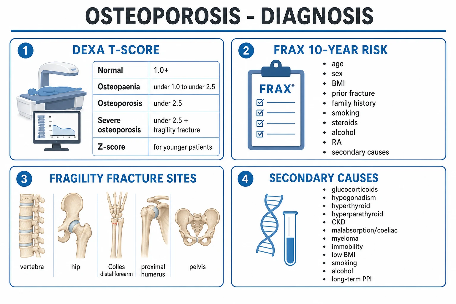
DEXA of the lumbar spine, total hip and femoral neck is the gold standard, reported as the T-score (the number of standard deviations the patient's BMD lies below the young-adult healthy mean) in adults over 50, and the Z-score (age- and sex-matched) in premenopausal women and men under 50. The WHO thresholds are reproduced exactly: osteoporosis = T-score of minus 2.5 or less; osteopenia = T-score minus 1.0 to minus 2.5; normal = T-score of minus 1.0 or higher; and severe (established) osteoporosis = T-score of minus 2.5 or less plus a fragility fracture. DEXA is reported per-site because arthritis, aortic calcification or vertebral fracture can artefactually raise the spine reading, so the lowest of femoral neck, total hip and spine is used clinically.[1][2]
DEXA T-score criteria (WHO operational thresholds)
FRAX integrates clinical risk factors — age, sex, BMI, prior fragility fracture, parental history of hip fracture, current smoking, glucocorticoid use, rheumatoid arthritis, secondary causes of osteoporosis, and alcohol 3 or more units daily — with or without femoral-neck BMD, to compute the 10-year probability of hip fracture and of a major osteoporotic fracture (clinical spine, hip, forearm, proximal humerus). FRAX is most useful in the osteopenic range, where it decides who to treat, and it is calibrated to national fracture epidemiology. Note that FRAX uses yes/no categorical inputs (it does not accumulate dose of smoking or steroids), so it can underestimate risk in very high-risk individuals.[10][2]
Vertebral fracture assessment (VFA) is performed on the DEXA machine at the time of BMD, or as a lateral thoracolumbar spine X-ray, to detect the morphometric vertebral fractures that are clinically silent but that, once present, establish the diagnosis and independently raise future fracture risk. Indications include height loss over 4 cm, historical height loss over 6 cm, recent non-traumatic back pain, and the combination of osteopenia with age over 70 (women) or 80 (men).[2]
Screen for secondary causes in every new diagnosis. A focused, cost-effective panel is: full blood count, ESR, serum calcium, phosphate, albumin and ALP, renal and liver function, TSH, 25-hydroxy vitamin D, and serum testosterone in men. When clinically indicated, add intact PTH, 24-hour urinary calcium (to detect hypercalciuria or malabsorption), serum and urine protein electrophoresis and serum free light chains (myeloma), coeliac serology (tissue transglutaminase IgA) in unexplained or low-BMI disease, a morning cortisol or overnight dexamethasone suppression test (Cushing), gonadotrophins (FSH, LH) to characterise hypogonadism, and a tissue transglutaminase if malabsorption is suspected. Bone turnover markers — serum CTX (resorption) and P1NP (formation) — are not diagnostic but are useful to confirm adherence and treatment response (a fall of at least one-third in CTX after 3 to 6 months of an antiresorptive indicates response) and to decide a drug holiday.[1][2]
Plain X-rays confirm symptomatic fractures but are not a measure of density; bone must lose roughly 30 to 40 percent of its mineral content before osteopenia is visible radiographically. Hence a normal-looking X-ray does not exclude osteoporosis — DEXA is required. [1]
Management — Resuscitation
There is no resuscitation for osteoporosis itself, but an acute fragility fracture is a time-critical surgical and orthogeriatric event.[1][5]
Acute hip fracture is a surgical emergency managed under a combined orthogeriatric model: prompt work-up, surgical fixation or arthroplasty within 36 hours (the standard in many national hip-fracture audits), early mobilisation, and prompt initiation of osteoporosis therapy — importantly, a zoledronic acid infusion 5 mg intravenously within 90 days of surgical repair reduces further clinical fractures by 35 percent and all-cause mortality by 28 percent. Acute painful vertebral fracture is managed with adequate, stepped analgesia (paracetamol, then a short course of an opioid; NSAIDs used sparingly in the elderly), early mobilisation to prevent the complications of immobility, and a brief period of external support; percutaneous vertebroplasty or kyphoplasty is reserved for persistent severe pain unresponsive to conservative care after several weeks. In all cases prevent the complications of immobility: venous thromboembolism prophylaxis, pressure-area care, and pneumonia prevention. The fracture is also the entry point to a Fracture Liaison Service that initiates the secondary-prevention pathway below.[5]
Management — Definitive & Stepwise
Definitive management is a stepwise ladder built on universal lifestyle measures and calcium and vitamin D, then pharmacotherapy stratified by risk.[1][2]
Step 1 — Universal baseline for all patients. Prescribe weight-bearing, impact and resistance exercise (walking, jogging, dancing, weights) three to five times weekly; smoking cessation; limit alcohol to less than 2 units per day; and a structured fall-prevention programme (home-hazard reduction, balance training, vision correction, medication review). Ensure an adequate calcium intake of 1000 to 1200 mg per day (diet first, supplement the remainder) and vitamin D 800 to 1000 IU per day, targeting a serum 25-hydroxy vitamin D of at least 50 to 75 nmol per litre. Calcium and vitamin D alone reduce fractures in institutionalised elderly patients who are deficient, but they are adjuncts — not substitutes — for specific therapy in established disease.[2]
Step 2 — First-line pharmacotherapy: a bisphosphonate for most patients with osteoporosis or with osteopenia plus a FRAX above treatment threshold. Bisphosphonates are synthetic analogues of pyrophosphate that bind hydroxyapatite and impair osteoclast function and survival, shifting the remodelling balance toward formation. Oral alendronate 70 mg once weekly (or risedronate 35 mg once weekly) is the workhorse; zoledronic acid 5 mg intravenously over at least 15 minutes once yearly is preferred when adherence or upper-gastrointestinal tolerability is a concern, in acute fracture, or in patients who prefer intermittent dosing. Ibandronate 150 mg orally once monthly (or 3 mg intravenously every 3 months) reduces vertebral but not robustly hip fracture. All bisphosphonates must be taken correctly: on rising, on an empty stomach, with a full glass of water, and the patient must stay upright for at least 30 minutes and not eat or take other medication in that window, to prevent oesophageal irritation; they are contraindicated in active upper-gastrointestinal disease, in inability to stay upright, and should be used with caution if the estimated glomerular filtration rate is below 30 to 35 mL per minute per 1.73 square metres (the IV route can cause acute-phase reactions and nephrotoxicity).[3][4][2]
Bisphosphonates — the first-line ladder
Antiresorptive; impair osteoclast function
Dose
Alendronate 70 mg PO weekly; risedronate 35 mg PO weekly; zoledronic acid 5 mg IV annually; ibandronate 150 mg PO monthly
Step 3 — Alternatives and anabolic therapy. Denosumab 60 mg subcutaneously every 6 months is a fully human monoclonal antibody against RANKL; it is as effective as a bisphosphonate, a useful option in renal impairment (it is not renally cleared), and preferred by many patients for its convenience — but it must never be stopped without a transition bisphosphonate, because stopping causes a rapid rebound rise in bone turnover and a cluster of multiple vertebral fractures. The anabolic agents teriparatide (PTH 1-34) 20 micrograms subcutaneously daily and abaloparatide 80 micrograms daily stimulate osteoblast activity and build new bone; they are reserved for severe or very-high-risk disease (multiple fractures, very low T-score, or fractures on therapy) and are limited to a maximum of 2 years (18 to 24 months lifetime), after which an antiresorptive must be given to consolidate the BMD gain. Romosozumab 210 mg subcutaneously monthly for 12 months is a monoclonal antibody to sclerostin with a unique dual action — it both increases bone formation and decreases resorption — and is used for severe postmenopausal osteoporosis at high fracture risk, again followed by an antiresorptive. Because a signal of serious cardiovascular adverse events was seen in one trial, romosozumab is avoided in patients with recent myocardial infarction or stroke.[6][7][8][9]
Step 4 — Other agents. Raloxifene 60 mg daily, a selective oestrogen-receptor modulator (SERM), is a weaker antiresorptive that reduces vertebral (not hip) fractures and reduces breast-cancer risk; it increases thromboembolism and hot flushes. Hormone replacement therapy (HRT) is effective for fracture prevention but is now reserved for menopausal symptom control in younger postmenopausal women, used at the lowest dose and for the shortest time because of breast-cancer, thromboembolic and cardiovascular risk. Calcitonin (salmon calcitonin 200 IU intranasal daily) is a weak antiresorptive now used mainly short-term for the analgesic effect in acute painful vertebral fracture, and has fallen out of long-term use because of a small associated cancer signal. Strontium ranelate 2 g orally at night (available in some countries, not US-approved) has mixed action and is restricted to severe disease because of an increased cardiovascular risk; it is contraindicated in ischaemic heart disease.[1][2]
Step 5 — Drug holiday. After 3 to 5 years of an oral bisphosphonate (or 3 years of IV zoledronate), reassess risk. In a stable, lower-risk patient with a T-score above minus 2.5 and no recent fractures, a drug holiday of 2 to 3 years exploits the drug's prolonged skeletal retention while reducing the rare risks of atypical femoral fracture and osteonecrosis of the jaw. In a high-risk patient (T-score below minus 2.5, prior hip or spine fracture, or fractures on therapy) treatment continues — up to 10 years has a favourable benefit-to-risk profile. The holiday is monitored with DEXA and bone-turnover markers, and therapy is resumed if risk rises, density falls, or a fracture occurs. Note that a drug holiday applies only to bisphosphonates — denosumab cannot be "holidayed", and anabolic therapy must always be followed by an antiresorptive.[1][2]
Treatment thresholds — who gets a drug?
Established osteoporosis
T-score, minus 2.5 or less; OR a fragility fracture (hip, spine) at any T-score. Treat.
Osteopenia plus high FRAX (US / NOF)
T-score minus 1.0 to minus 2.5 with 10-yr hip fracture probability at least 3% OR major-osteoporotic at least 20%. Treat.
Glucocorticoid-induced
Prednisolone over 7.5 mg per day for over 3 months. Treat with a bisphosphonate plus calcium and vitamin D (FRAX is adjusted upward for steroids).
Very high risk
Multiple or recent fractures, very low T-score, or fracture on therapy. Use anabolic-first sequencing (teriparatide or romosozumab), then an antiresorptive.
Osteoporosis management — FRACTURE
FRACTURE
weight-bearing exercise, smoking cessation, limit alcohol, fall prevention
DEXA T-score plus FRAX 10-year fracture probability
calcium 1000 to 1200 mg per day, vitamin D 800 to 1000 IU per day
glucocorticoids, hyperthyroidism, hypogonadism, low vitamin D, malabsorption
alendronate weekly, risedronate weekly, or zoledronate annually
teriparatide or romosozumab for very-high-risk, then an antiresorptive
consider a bisphosphonate drug holiday in stable, lower-risk patients
monitor every 1 to 3 years; never stop denosumab without a transition bisphosphonate
Specific Subtypes & Scenarios
Glucocorticoid-induced osteoporosis (GIOP) is the most common secondary osteoporosis and the most rapidly destructive — bone loss is fastest in the first 3 to 6 months of steroid therapy, and fracture risk rises within weeks. Every patient expected to need prednisolone over 5 mg per day (often cited over 7.5 mg per day) for over 3 months should be assessed and given prophylactic bone protection: a bisphosphonate plus calcium and vitamin D, with lifestyle measures and the lowest effective steroid dose for the shortest time. FRAX can be used in GIOP but underestimates risk, so it is adjusted upward (the equivalent of a one-decade increase in age, or multiplying the FRAX probability by about 1.15 for hip and 1.1 for major osteoporotic fracture at prednisolone doses of 7.5 mg per day or more). Teriparatide is preferred over a bisphosphonate in very-high-risk GIOP where available.[2]
Male osteoporosis is under-recognised. About one in five men over 50 will fracture, and secondary causes are found in roughly half to two-thirds of men with a fragility fracture — most commonly hypogonadism, steroid therapy (including for COPD and inflammatory disease), alcohol excess, smoking and hyperthyroidism. Always measure serum testosterone, with LH and FSH, in any man with low bone mass; testosterone replacement both treats hypogonadism and improves BMD. Bisphosphonates and denosumab are first-line in men, with teriparatide for severe disease.[1]
Post-fracture secondary prevention is delivered by a Fracture Liaison Service (FLS): every patient over 50 presenting with a fragility fracture is identified, investigated (DEXA, secondary-cause screen) and started on therapy. FLS coverage is the single most effective — and most neglected — intervention to close the treatment gap, because the highest-risk patient is the one who has just fractured.[1][5]
Very-high-risk and anabolic-first sequencing. Patients with multiple or recent fractures, a T-score below minus 3.0 or minus 3.5, or a fracture sustained while on antiresorptive therapy are candidates for an anabolic agent first (teriparatide, abaloparatide or romosozumab), followed by a potent antiresorptive to lock in the gain. This build-then-lock sequence produces larger BMD gains and fewer fractures than starting with an antiresorptive in this group.[8][9]
Pre-treatment and transplantation osteoporosis. Patients about to undergo androgen-deprivation therapy for prostate cancer, aromatase-inhibitor therapy for breast cancer, or solid-organ or stem-cell transplantation face rapid, predictable bone loss and a high fracture rate within the first year. The principle is anticipatory: measure a baseline DEXA, start calcium and vitamin D, and initiate a bisphosphonate (or denosumab) before or at the start of the gonadotropin-releasing-hormone agonist, aromatase inhibitor, or transplant immunosuppression — not after the bone is lost. Glucocorticoid-loaded transplant regimens, calcineurin inhibitors and hypogonadism combine to make the post-transplant year the period of fastest loss, so therapy is most effective when begun prophylactically and continued long-term.[1][2]
Self-test — a 58-year-old woman on long-term prednisolone
She takes prednisolone 10 mg per day for rheumatoid arthritis and has done so for 8 months. Her DEXA femoral-neck T-score is minus 1.8, she has never fractured, and her FRAX 10-year hip-fracture probability (entered without glucocorticoid adjustment) is 1.8 percent. [1]
Question: Does she warrant bone-protection therapy, and why? [1]
Answer: Yes. Glucocorticoid use itself is a FRAX input and the unadjusted FRAX underestimates her true risk; at prednisolone doses of 7.5 mg per day or more the probability is adjusted upward (roughly multiplied by 1.15 for hip and 1.1 for major osteoporotic fracture). On long-term steroids she meets the threshold for prophylactic bisphosphonate plus calcium and vitamin D regardless of the adjusted score, with teriparatide preferred if her risk is very high. This is exactly the scenario in which waiting for the T-score to cross minus 2.5 — or for a fracture — is a preventable error.
Complications & Pitfalls
Disease complications are dominated by the fractures themselves. Hip fracture carries a one-year mortality of about 20 to 30 percent, a loss of independence (half of previously independent patients need help with activities of daily living afterwards), and a high risk of complications of immobility — venous thromboembolism, pressure ulcers, pneumonia and urinary infection. Vertebral fractures cause acute and chronic back pain, kyphosis with reduced vital capacity and restrictive respiratory physiology, early satiety and weight loss from the reduced abdominal cavity, and a raised future fracture and mortality risk proportional to severity. Kyphosis itself reduces mobility and balance and so feeds back into further falls.[1]
Drug complications and pitfalls are exactly what an examiner probes. Long-term bisphosphonates rarely cause atypical femoral fracture (a transverse, non-comminuted fracture of the subtrochanteric or diaphyseal femur, often preceded by weeks of prodromal thigh pain) and osteonecrosis of the jaw (exposed non-healing bone, usually after dental extraction in patients on high-dose therapy for malignancy). Both are rare in osteoporosis dosing, but the risk accumulates with duration — the rationale for the drug holiday. Acute-phase reaction (fever, myalgia, arthralgia) occurs in roughly 10 to 15 percent after the first IV zoledronate infusion and is self-limiting; hypocalcaemia can follow both zoledronate and denosumab, so vitamin D deficiency must be corrected first. Atrial fibrillation was reported more with zoledronate in the pivotal trial and is monitored but not considered a contraindication in standard dosing. Oesophoritis and (rarely) oesophageal ulceration with oral bisphosphonates underlie the strict administration rules. Stopping denosumab without a transition bisphosphonate precipitates multiple rebound vertebral fractures within months — a critical, frequently-examined pitfall. Teriparatide carries a black-box warning for osteosarcoma based on animal data and is avoided after prior skeletal radiation or in patients with bone malignancy, although the human risk appears negligible.[1][2][9]
Prognosis & Disposition
Osteoporosis is preventable and treatable but not cured — therapy is typically long-term, sometimes lifelong. With effective antiresorptive or anabolic therapy the risk of vertebral fracture falls by 40 to 70 percent, hip fracture by 25 to 50 percent, and nonvertebral fracture by 20 to 40 percent, and a single zoledronic acid infusion after a hip fracture reduces all-cause mortality by 28 percent. A prior fragility fracture roughly doubles the future fracture risk, and the risk is highest immediately after the index fracture — the so-called "imminent risk" window that justifies prompt secondary prevention. Hip fractures carry a one-year mortality of 20 to 30 percent and major loss of independence; vertebral fractures also raise mortality, partly through respiratory restriction and partly as a marker of frailty. The main real-world barrier to good outcomes is non-adherence — roughly half of patients stop an oral bisphosphonate within a year — which is why IV zoledronate, denosumab and fracture-liaison follow-up improve real-world effectiveness. Disposition is community-based with a fracture-liaison or primary-care review, DEXA every 2 years (1 to 3 years depending on risk), and bone-turnover markers to confirm response.[1][5]
Special Populations
Pregnancy and lactation cause a transient, reversible loss of 3 to 10 percent of bone mass; bisphosphonates are contraindicated (teratogenic, with long skeletal half-life), so management is calcium, vitamin D and lifestyle, with rare pregnancy- and lactation-associated osteoporosis managed by a specialist. The elderly and frail are the highest-risk group and benefit most from fall-prevention programmes, vitamin D supplementation (which reduces falls in deficient individuals), home-hazard modification and IV zoledronate to overcome adherence problems — while avoiding sedating drugs and overtreatment that causes hypotension and falls. Chronic kidney disease: bisphosphonates are avoided below an eGFR of 30 to 35 mL per minute per 1.73 square metres (risk of nephrotoxicity and adynamic bone disease), where denosumab (not renally cleared, but monitor calcium closely) or specialist metabolic-bone input is preferred. Premature menopause (before 40) accelerates bone loss and warrants HRT at least until the average age of natural menopause. Younger adults (under 50) with low bone mass are reported by Z-score (not T-score) and almost always have a secondary cause that must be found and treated rather than empirically bisphosphonated.[1][2]
Diabetes and the "normal-density but fragile" bone. Type 2 diabetes is a paradox worth knowing: patients with diabetes have a higher than average bone mineral density yet a paradoxically increased fracture risk, because chronic hyperglycaemia, advanced glycation end-products and altered collagen cross-linking impair bone quality rather than quantity. DEXA therefore underestimates skeletal fragility in diabetes, and FRAX similarly underestimates risk, so a lower threshold to treat and attention to fall risk are warranted. Bone quality tools such as the trabecular bone score (TBS), which DEXA can derive, help capture this hidden risk. Ethnic and geographic variation: hip-fracture incidence is highest in white northern-European and south-Asian populations and substantially lower in black African populations, who have greater peak bone mass and wider femoral necks; nonetheless, mortality after a hip fracture is often higher in non-white populations, partly because of later presentation and comorbidity. FRAX is calibrated to national epidemiology precisely because these differences are large, and treatment thresholds are not directly transferable between regions.[1][10]
Evidence, Guidelines & Regional Differences
The strength of osteoporosis therapy rests on a series of large randomised placebo-controlled trials that collectively defined modern practice.[3][4][6][7]
Fracture Intervention Trial (FIT) — alendronate
Lancet, 1996
RCT of 2027 postmenopausal women with low BMD and an existing vertebral fracture, alendronate vs placebo over 3 years.
Key finding
Alendronate reduced new morphometric vertebral fractures by 47% (RR 0.53), clinical vertebral fractures by 55%, and hip fracture by 51%.
Practice change
Established oral bisphosphonates as first-line therapy for established postmenopausal osteoporosis.
HORIZON Pivotal Fracture Trial — zoledronic acid
N Engl J Med, 2007
RCT of 7765 postmenopausal women, once-yearly IV zoledronic acid 5 mg vs placebo over 3 years.
Key finding
Reduced morphometric vertebral fracture by 70%, hip fracture by 41%, and nonvertebral fracture by 25%; serious atrial fibrillation was more frequent.
Practice change
Once-yearly IV zoledronate became the standard antiresorptive where adherence or oral tolerability is an issue.
HORIZON Recurrent Fracture Trial — zoledronate after hip fracture
N Engl J Med, 2007
RCT of 1065 patients given zoledronic acid within 90 days of surgical repair of a hip fracture vs placebo.
Key finding
Reduced new clinical fracture by 35% and all-cause mortality by 28%.
Practice change
Mandated early zoledronate in secondary prevention after hip fracture and underpinned modern Fracture Liaison Services.
FREEDOM — denosumab
N Engl J Med, 2009
RCT of 7868 women with T-score minus 2.5 to minus 4.0, denosumab 60 mg SC 6-monthly vs placebo over 3 years.
Key finding
Reduced vertebral fracture by 68%, hip fracture by 40%, and nonvertebral fracture by 20%.
Practice change
Denosumab became a first-tier antiresorptive; its later withdrawal-safety problem defined the transition rule.
Neer et al — teriparatide (PTH 1-34)
N Engl J Med, 2001
RCT of 1637 postmenopausal women with prior vertebral fracture, PTH 1-34 (20 or 40 micrograms) vs placebo.
Key finding
20-microgram daily dose reduced new vertebral fracture by 65% and nonvertebral fracture by 53%; lumbar BMD rose 9 to 13%.
Practice change
Introduced anabolic therapy for severe osteoporosis, with a 2-year lifetime limit.
FRAME and ARCH — romosozumab
N Engl J Med, 2016 & 2017
FRAME: romosozumab 210 mg monthly vs placebo for 12 months then denosumab. ARCH: romosozumab vs alendronate in very-high-risk women.
Key finding
FRAME: 73% lower vertebral fracture risk at 1 year. ARCH: 48% lower vertebral and 38% lower hip fracture vs alendronate, but more positively-adjudicated serious cardiovascular events.
Practice change
Introduced a dual-action (anabolic plus antiresorptive) agent for severe disease, with a cardiovascular caution.
Regional guideline differences are examinable and reflect different health-system economics. The FRAX algorithm itself was developed on UK epidemiology.[10]
[1] [1]International Osteoporosis Foundation (IOF) / Europe — ESCEO. Treat any patient with a prior fragility fracture (hip or spine) regardless of BMD; treat osteoporosis by T-score (minus 2.5 or less); and treat osteopenia with a high FRAX (commonly cited as major-osteoporotic 20% or hip 3%, with European variations). IOF also drives the Capture the Fracture campaign standardising Fracture Liaison Services globally.
Monitoring & Prevention
Monitoring combines DEXA, bone-turnover markers and adherence. Repeat DEXA every 2 years (1 to 2 years in high risk, up to 3 years in stable disease) and look for the least significant change — a change exceeding roughly 3 to 5 percent at the spine and 3 to 6 percent at the hip to be real. A stable or rising T-score confirms response; a falling T-score or rising bone-turnover markers on therapy signal non-adherence (by far the commonest cause), an unrecognised secondary cause, or treatment failure and should trigger review. Serum CTX should fall by at least one-third within 3 to 6 months of an oral bisphosphonate or within days of IV zoledronate; a failure to fall means the drug was not taken or absorbed.[2]
Prevention is staged across life. Achieve a high peak bone mass in youth (adequate calcium, vitamin D and weight-bearing exercise; avoid smoking and excessive alcohol); maintain bone through midlife; and detect and treat risk after the menopause and in older age. Population strategies — vitamin D and calcium sufficiency, physical activity, smoking and alcohol moderation, fall prevention, and Fracture Liaison Service coverage of every fragility-fracture patient — are more effective at reducing the fracture burden than any single drug.[1]
Exam Pearls
Doses and thresholds you must know cold
Exam application bank (NEET-PG / INICET)
One-line answer
Osteoporosis is a systemic skeletal disease of low bone mass and microarchitectural deterioration, increasing the risk of fragility fractures (hip, spine, wrist). Diagnosis is by DEXA T-score of minus 2.5 or less (osteopenia minus 1 to minus 2.5; a fragility fracture establishes the diagnosis regardless of score), and the FRAX score estimates 10-year fracture risk to guide treatment. Risk factors include age, female sex, postmenopausal status, family history, low BMI, glucocorticoids, smoking, hypogonadism and hyperthyroidism. It is often silent until a fracture. Management is lifestyle (weight-bearing exercise, no smoking, calcium and vitamin D) plus first-line bisphosphonates (alendronate, zoledronate), with denosumab and anabolic teriparatide for high-risk or refractory disease. After 3 to 5 years of a bisphosphonate, a drug holiday is considered. The goal is preventing the first frag
Worked stems (answer without another resource)
Stem 1 — Classic presentation. Map symptoms to mechanism; name the first investigation and first treatment step with dose/route if drug therapy is standard. [1]
Stem 2 — Unstable / complicated. List red flags that force immediate resuscitation, theatre, ICU, antidote, or reperfusion — and what you do in the first 15 minutes. [1]
Stem 3 — Atypical group. Elderly, pregnancy, child, or immunocompromised: how presentation and thresholds change. [1]
Stem 4 — Differential trap. Name the three closest mimics and one discriminator for each. [1]
Stem 5 — Disposition. Who goes home with safety-netting, who is admitted, who needs HDU/ICU/theatre, and what follow-up is mandatory. [1]
Rapid viva checklist
- Definition + classification
- Pathophysiology chain
- Bedside signs / criteria
- Score with exact components (if any)
- Emergency bundle
- Definitive therapy with doses
- Complications of disease and of treatment
- Special populations
- Guideline/trial name if classic
- Three exam traps
Coverage self-check
If you cannot answer any stem above from this page alone, re-read the matching section — the page is intended to be self-sufficient for final-prof and NEET-PG/INICET questions on Osteoporosis.
References
- [1]Compston JE, McClung MR, Leslie WD. Osteoporosis Lancet, 2019.PMID 30696576
- [2]Camacho PM, Petak SM, Binkley N, et al. AMERICAN ASSOCIATION OF CLINICAL ENDOCRINOLOGISTS/AMERICAN COLLEGE OF ENDOCRINOLOGY CLINICAL PRACTICE GUIDELINES FOR THE DIAGNOSIS AND TREATMENT OF POSTMENOPAUSAL OSTEOPOROSIS-2020 UPDATE Endocr Pract, 2020.PMID 32427503
- [3]Black DM, Cummings SR, Karpf DB, et al. Randomised trial of effect of alendronate on risk of fracture in women with existing vertebral fractures. Fracture Intervention Trial Research Group Lancet, 1996.PMID 8950879
- [4]Black DM, Delmas PD, Eastell R, et al. Once-yearly zoledronic acid for treatment of postmenopausal osteoporosis N Engl J Med, 2007.PMID 17476007
- [5]Lyles KW, Colon-Emeric CS, Magaziner JS, et al. Zoledronic acid and clinical fractures and mortality after hip fracture N Engl J Med, 2007.PMID 17878149
- [6]Cummings SR, San Martin J, McClung MR, et al. Denosumab for prevention of fractures in postmenopausal women with osteoporosis N Engl J Med, 2009.PMID 19671655
- [7]Neer RM, Arnaud CD, Zanchetta JR, et al. Effect of parathyroid hormone (1-34) on fractures and bone mineral density in postmenopausal women with osteoporosis N Engl J Med, 2001.PMID 11346808
- [8]Cosman F, Crittenden DB, Adachi JD, et al. Romosozumab Treatment in Postmenopausal Women with Osteoporosis N Engl J Med, 2016.PMID 27641143
- [9]Saag KG, Petersen J, Brandi ML, et al. Romosozumab or Alendronate for Fracture Prevention in Women with Osteoporosis N Engl J Med, 2017.PMID 28892457
- [10]Kanis JA, Johnell O, Oden A, Johansson H, McCloskey E. FRAX and the assessment of fracture probability in men and women from the UK Osteoporos Int, 2008.PMID 18292978