Endocrinology · General Medicine
Paget Disease of Bone
Also known as Paget disease of bone · Osteitis deformans · Paget disease
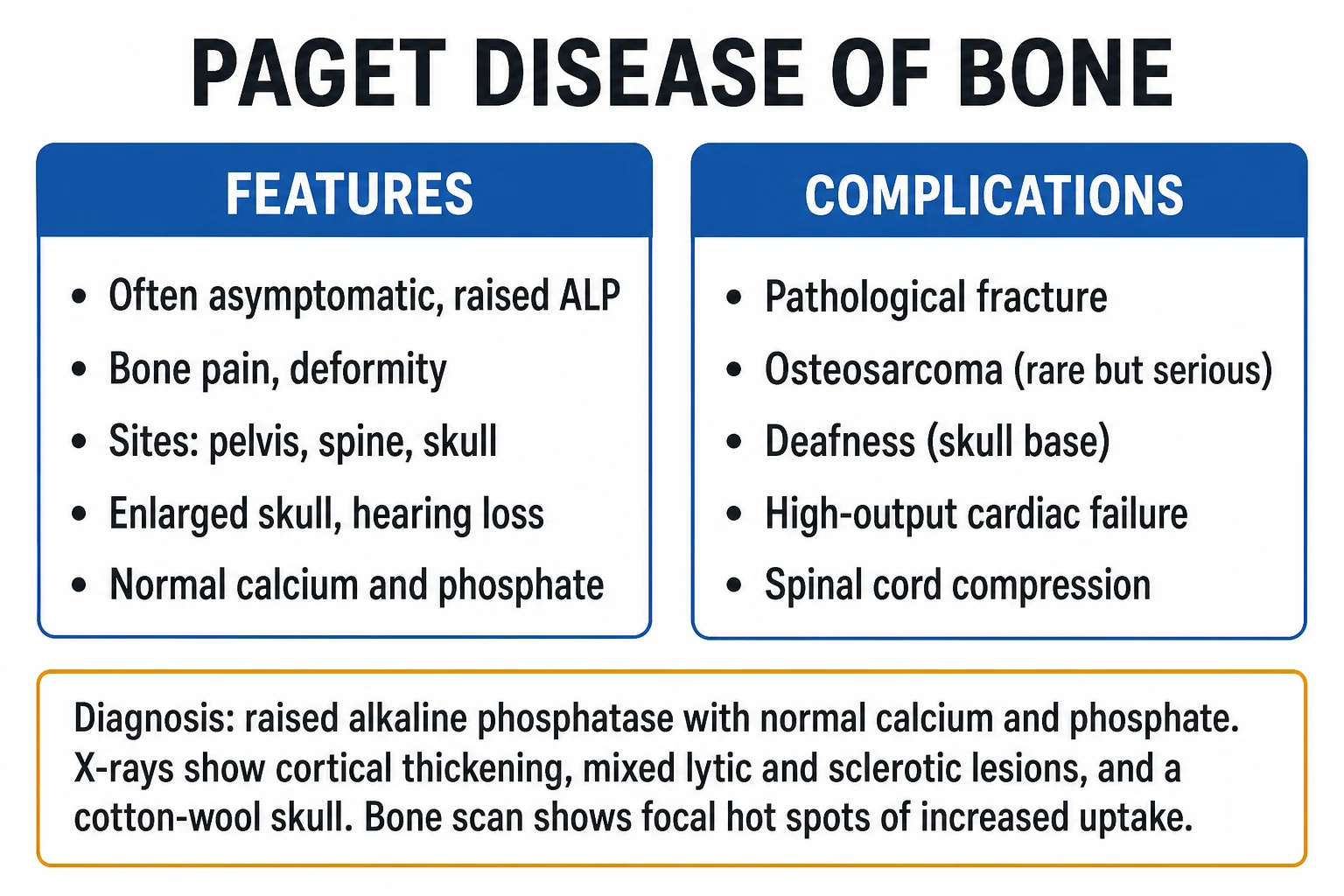
Paget disease of bone (osteitis deformans) is a chronic, focal disorder of adult bone remodeling in which excessive, disorganised osteoclast activity is followed by chaotic osteoblast repair, producing bone that is thick and hypervascular but mechanically weak (a disordered woven/mosaic pattern). It is usually asymptomatic and found on incidental raised alkaline phosphatase with normal calcium and phosphate; symptoms include bone pain (often nocturnal), bony deformity (sabre tibia, enlarging skull, kyphosis), sensorineural deafness and, rarely, neurological or cardiac complications. Common sites are the pelvis, spine, femur, skull and tibia. Diagnosis rests on raised ALP with normal Ca/PO4, characteristic X-rays (cortical thickening, mixed lytic-sclerotic lesions, cotton-wool skull, blade-of-grass, picture-frame vertebrae) and an isotope bone scan showing focal hot spots. The cornerstone treatment is a single 5 mg IV infusion of zoledronic acid (plus calcium and vitamin D) for symptomatic or complication-risk disease, with ALP monitoring. The feared complication is osteosarcoma transformation (under 1 percent), heralded by new or rapidly worsening pain.
On this page & tools
Your progress
Saved locally on this device.
Exam tags
Red flags
Overview & Definition
Paget disease of bone (historically osteitis deformans, described by Sir James Paget in 1877) is a chronic, focal — not systemic — disorder of adult bone remodeling. At an affected site, the normal coupled cycle of osteoclastic resorption followed by osteoblastic formation becomes pathologically accelerated and disordered: osteoclasts are abnormally large and multinucleated, resorb bone excessively, and the resulting osteoblast repair is chaotic, laying down coarse, disorganised woven bone in a characteristic "mosaic" pattern. The paradox of Paget disease is that the bone becomes bulkier, thicker and hypervascular yet is mechanically weak, prone to bowing, deformity and fracture.[1][2]
Because turnover is markedly increased at affected sites but confined to them, the biochemical signature is distinctive: alkaline phosphatase (a bone-formation marker) is markedly raised while serum calcium, phosphate and parathyroid hormone remain normal. Most patients are asymptomatic; the disease is discovered incidentally on blood tests or imaging. When symptoms occur, they reflect either the bone itself (pain, deformity), compression of adjacent structures (deafness, cranial-nerve or cord compression), or a complication (pathological fracture, osteosarcoma, high-output cardiac failure). Treatment with a potent bisphosphonate — most often a single zoledronic acid infusion — suppresses the disordered remodeling for months to years.[1][3]
Clinical distinctions and naming pitfalls
Paget disease of bone is entirely unrelated to Paget disease of the breast or vulva (an intra-epidermal adenocarcinoma sharing only the eponym). Juvenile Paget disease is a separate, genetically distinct entity caused by loss-of-function mutations in TNFRSF11B (encoding osteoprotegerin, OPG); it produces massive, widespread juvenile bone turnover with deafness and retinopathy and is not adult Paget disease. Another distinct entity, familial expansile osteolysis, and the IBMPFD syndrome (inclusion-body myopathy with Paget disease and frontotemporal dementia, due to VCP mutations) overlap genetically but are multisystem — examiners test the distinction.[2][4]
Classification
Paget disease is classified along three axes that the examiner will probe: [1]
- By number of sites — monostotic (single bone, ~one-third) versus polyostotic (multiple sites, ~two-thirds). Polyostotic disease with the classic pelvis–spine–skull pattern carries a higher complication burden.
- By pathological phase at a given site — every lesion evolves through three phases: an early lytic (osteoclastic, "hot") phase, an intermediate mixed phase, and a late sclerotic ("burnt-out") phase.
- By aetiology — sporadic (the vast majority) versus familial (autosomal dominant, SQSTM1 mutations, ~10–30 percent of familial cases and a smaller fraction of sporadic).[2][4]
Monostotic
single site
- About one-third of cases
- Often an incidental X-ray finding
- Pelvis, tibia or a single vertebra are typical
- Lower complication burden; may simply be observed
Polyostotic
multiple sites
- About two-thirds of cases
- Classic pelvis–spine–skull–femur pattern
- Higher risk of deafness, deformity, high-output cardiac failure
- Usually warrants bisphosphonate therapy
Familial (SQSTM1)
autosomal dominant
- Around 10–30 percent of familial cases carry SQSTM1/p62 mutations
- Earlier onset, more often polyostotic
- Screen first-degree relatives in middle age
- Distinct from juvenile Paget disease (TNFRSF11B/OPG)
Epidemiology & Risk Factors
Paget disease is a disease of older adults and of specific populations. The key epidemiological facts the examiner rewards: [1]
- Age — rare under 40; prevalence rises steeply with age, reaching roughly 2–3 percent of individuals over 55 in high-prevalence regions and higher still in the very elderly. Onset is essentially never in childhood.
- Geography and ancestry — highest prevalence in the United Kingdom, Western Europe, North America (especially among those of British descent), Australia and New Zealand. It is notably rare in Asia, Africa, Scandinavia and the Indian subcontinent, supporting a strong interplay of genetic susceptibility with an environmental trigger.
- Sex — slight male predominance (roughly 1.5:1).
- Familial clustering — a family history is obtained in 15–30 percent of cases; SQSTM1 mutations account for a substantial proportion of familial disease, with autosomal dominant inheritance and variable penetrance.[4]
- Declining incidence and severity — over recent decades the prevalence, severity and diagnostic age at onset have all fallen markedly in traditional high-prevalence countries. The decline is too rapid to be genetic and is attributed to environmental changes (improved nutrition, reduced childhood infections, declining domestic coal/air pollution), supporting a viral/environmental contribution to aetiology.[5]
Paget disease — epidemiological numbers
The practical consequence of this epidemiology is twofold. First, in an older patient of Anglo-Saxon ancestry, Paget disease is the leading explanation for an isolated raised ALP with normal calcium and phosphate. Second, a young patient, or one of non-European ancestry, with apparent Pagetoid change warrants careful thought about mimics (fibrous dysplasia, metastases) and the rare genetic syndromes above. [1]
Pathophysiology
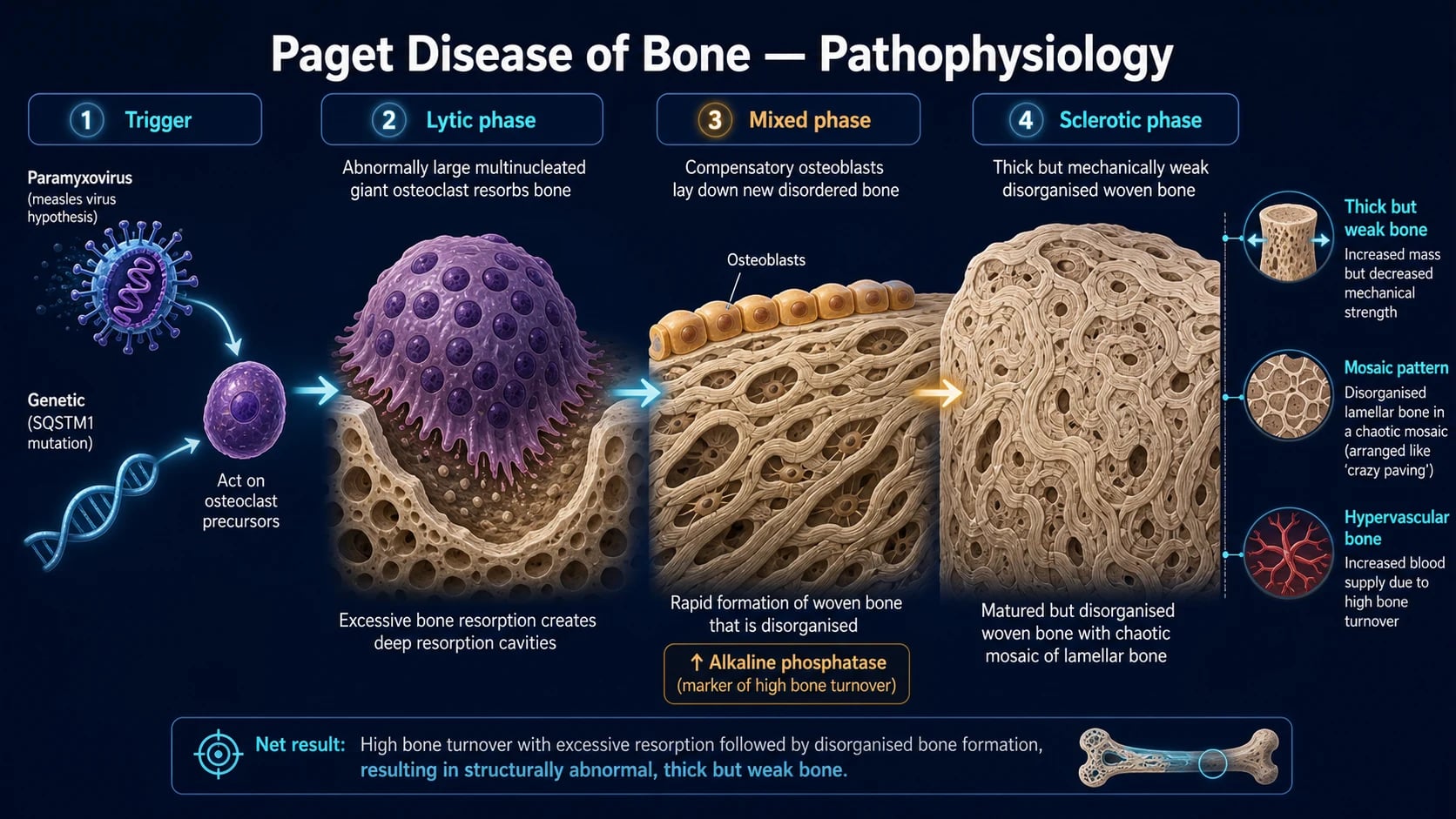
Paget disease is best understood as a two-hit process: a genetically susceptible host (germline SQSTM1/p62 or related mutations lowering the threshold for osteoclast activation) encounters an environmental trigger (a paramyxovirus-like infection of osteoclast precursors) that ignites focal, disordered remodeling. [1]
The osteoclast: the cell that drives the disease
Pagetic osteoclasts are abnormally large and hypernucleated (often containing 20–100 nuclei versus the normal 3–10) and frequently contain nuclear inclusion bodies with microcylindrical filaments resembling paramyxovirus nucleocapsids (measles and canine distemper have been implicated by in-situ hybridisation). These giant osteoclasts resorb bone rapidly and excessively through an upregulated RANK–RANKL pathway: RANKL (receptor activator of NF-κB ligand), expressed by osteoblasts and stromal cells, binds RANK on osteoclast precursors, driving their fusion and activation; osteoprotegerin (OPG), the decoy receptor that normally restrains this axis, is functionally impaired. SQSTM1 (sequestosome-1/p62) mutations — which encode an adaptor protein in the NF-κB signalling cascade — further sensitise osteoclasts to RANKL, lowering their activation threshold.[2][4]
The three pathological phases
At any affected site the disease evolves through three overlapping phases: [1]
- Lytic ("hot", osteoclastic) phase — the dominant activity is aggressive osteoclastic resorption. On X-ray this produces sharply demarcated lytic lesions: osteoporosis circumscripta of the skull vault, and the V-shaped "blade of grass" (or "flame-shaped") advancing lytic wedge in a long bone. Bone scan uptake is intense. The bone may be subtly expanded and hypervascular.
- Mixed phase — compensatory osteoblast overdrive kicks in to replace the resorbed bone, but the repair is disorganised and excessive. Bone is laid down as coarse woven bone rather than normal lamellar bone, producing the characteristic "mosaic" pattern of irregular cement lines under the microscope. X-rays show a mixture of lytic and sclerotic areas with cortical thickening, coarsened trabeculae and bone expansion.
- Sclerotic ("burnt-out") phase — turnover slows; the lesion is dominated by dense, thick, disordered sclerotic bone. X-rays show a thickened, expanded, sclerotic bone (e.g., a "cotton-wool" skull, an "ivory" vertebra) that is bulky but mechanically weak.[1][2]
Why the bone is "thick but weak" — and why ALP rises
The rapid, chaotic osteoblastic repair produces woven bone that lacks the organised lamellar architecture giving normal bone its tensile and compressive strength. The result is bone that is bulky, sclerotic and hypervascular on imaging, yet brittle — hence the characteristic bowing deformity of weight-bearing long bones and the propensity to pathological fracture. Because osteoblast activity (and therefore alkaline phosphatase) is markedly increased while the process is confined to focal sites, ALP rises but calcium and phosphate remain normal — the resorbed calcium is promptly redeposited by the overactive osteoblasts, so there is no net systemic mineral disturbance. This is the central biochemical insight that distinguishes Paget disease from systemic metabolic bone disease.[1][5]
The RANK–RANKL–OPG axis and the genetic basis of susceptibility
Normal bone remodeling is governed by a tightly coupled cytokine triad. RANKL, expressed on the surface of osteoblasts and marrow stromal cells (and also secreted in soluble form), binds its receptor RANK on osteoclast precursors, committing them to fusion, differentiation and bone-resorbing activation. The decoy receptor osteoprotegerin (OPG), encoded by TNFRSF11B, binds RANKL and prevents it from engaging RANK, braking osteoclast formation. In Paget disease this balance is tipped decisively toward RANK activation: pagetic osteoclasts and their precursors show exaggerated RANK responsiveness, and the SQSTM1/p62 mutations carried by familial cases encode an adaptor protein in the NF-κB signalling cascade downstream of RANK. By altering p62's sequestosome/ubiquitin-binding function, these mutations (the recurrent P392L substitution being the prototypical example) lower the threshold at which RANKL triggers osteoclastogenesis and reduce apoptosis of mature osteoclasts — producing the giant, hypernucleated, long-lived cells that define the disease histologically.[4]
This molecular framework explains both the focal nature of the disease (the genetic susceptibility is necessary but not sufficient; a local environmental trigger — most plausibly a paramyxovirus infection of an osteoclast precursor — must ignite the focus) and its response to therapy: nitrogen-containing bisphosphonates such as zoledronic acid are internalised by osteoclasts and inhibit farnesyl pyrophosphate synthase in the mevalonate pathway, preventing prenylation of small GTPases essential for osteoclast function, so the cells undergo apoptosis and stop resorbing. The result is a rapid fall in osteoblast-stimulating signals, a fall in ALP, and a switch from active lytic/mixed disease toward a quiescent, mechanically competent state.[1][3]
Clinical Presentation
The clinical hallmark of Paget disease is its heterogeneity: most patients are entirely asymptomatic, while a minority present with bone pain, deformity, deafness, a complication, or — very rarely — osteosarcoma. [1]
Asymptomatic (the majority)
Most cases are detected incidentally — either through routine blood tests showing an isolated raised ALP with otherwise normal chemistry (the single most common presentation in modern practice), or on an X-ray performed for another reason (e.g., a pelvis film for hip osteoarthritis revealing pagetoid change). The introduction of routine ALP measurement on biochemical panels means Paget disease is now most often biochemically, not clinically, diagnosed.[1]
Symptomatic bone disease
- Bone pain — classically a deep, aching pain at the affected site, often worse at night and unrelated to activity. Pain may arise from the pagetoid bone itself, from periosteal stretching, from microfractures, or from secondary osteoarthritis of an adjacent joint (e.g., hip or knee) caused by altered mechanical loading.
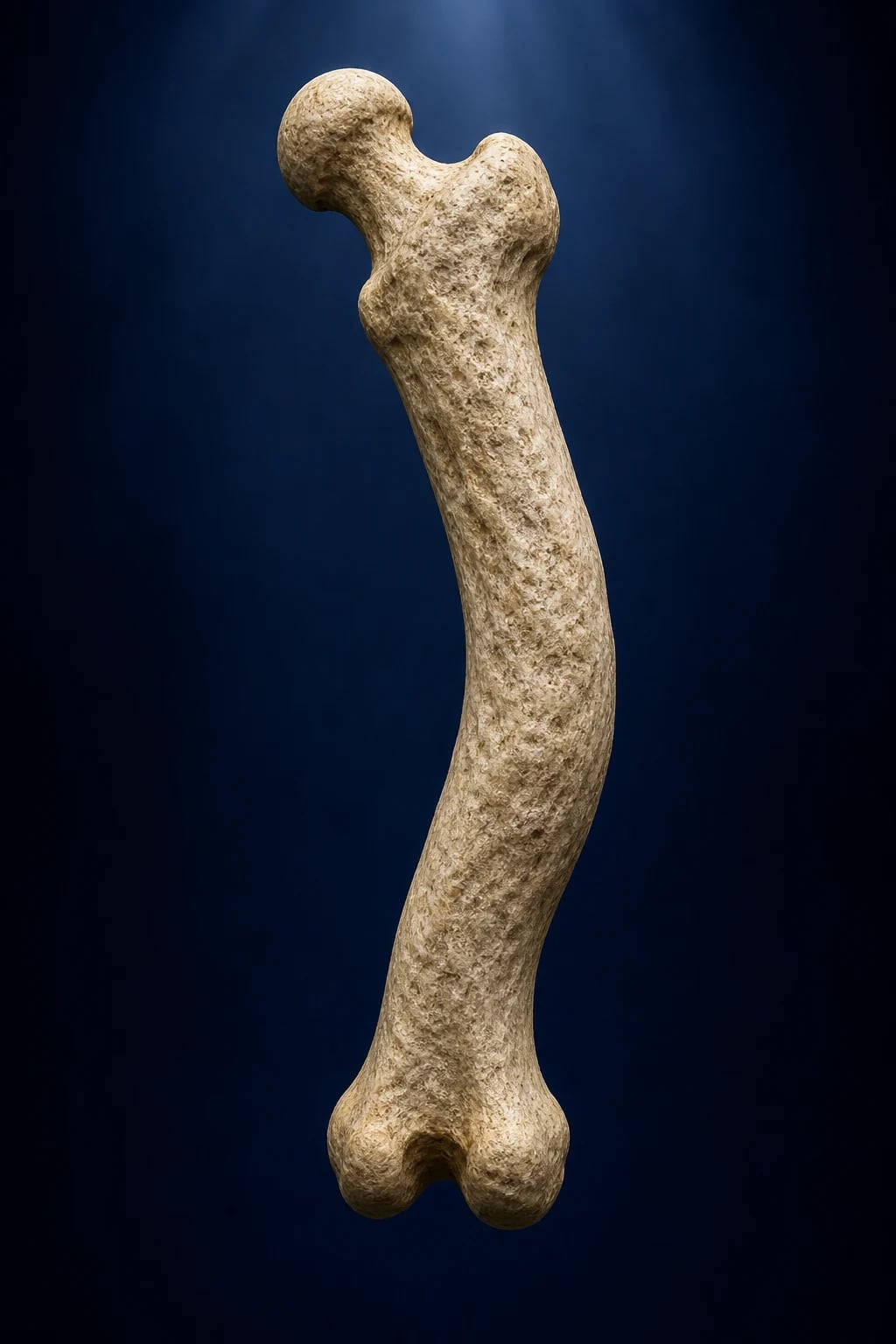
- Deformity — slow, progressive bowing of long bones (the classic sabre shin / sabre tibia from anterior tibial bowing, or a bowed femur); enlargement of the skull (the patient may report needing a progressively larger hat size, or frontal bossing); progressive kyphosis; and an asymmetric, broadened long-bone contour. Long-bone bowing produces a characteristic antalgic, broad-based gait.
- Warmth over the bone — the skin over an affected tibia or skull can feel noticeably warm because pagetoid bone is hypervascular; this is a useful bedside sign.
- Hearing loss — sensorineural deafness (and occasionally tinnitus or vertigo) from cochlear and eighth-nerve involvement at the pagetoid skull base. It develops slowly and is one of the most common symptomatic complications of skull disease.
- Dental and cranial-nerve features — rarely, cranial-nerve palsies (II, V, VII) from skull-base foramina, and hypercementosis / loosening of teeth with ill-fitting dentures. [1]
Complication-led presentation (the acute forms)
A subset present through a complication rather than chronic symptoms: [1]
- Pathological fracture — through weak pagetoid bone, classically of the femoral neck or subtrochanteric femur, the tibia, or the humerus; a long bone that was bowed and painful suddenly gives way with minimal trauma.
- New neurological deficit — spinal cord compression (paraparesis, sensory level, sphincter disturbance) from pagetoid vertebrae; basilar invagination / platybasia causing brainstem compression, lower cranial-nerve palsies, ataxia, or obstructive hydrocephalus.
- High-output cardiac failure — only with extensive polyostotic disease: the large hypervascular bone bed acts as a chronic arteriovenous shunt, raising cardiac output and precipitating heart failure in those with limited cardiac reserve.
- Sudden new or rapidly worsening pain at a pagetoid site — the red flag for osteosarcoma transformation (see Complications). [1]
Differential Diagnosis
The differential depends on whether the presentation is biochemical (isolated raised ALP), radiological (sclerotic or lytic bone lesions), or clinical (bone pain, deformity, deafness). Examiners test the discriminator for each. [1]
Paget disease
raised ALP, normal Ca and PO4
- Focal disordered remodeling — thick but weak woven bone
- ALP raised; calcium and phosphate both NORMAL
- Polyostotic sclerotic hot spots; cotton-wool skull
- First-line: single-dose zoledronate
Primary hyperparathyroidism
raised ALP, raised Ca, low PO4
- Systemic high-turnover from PTH excess
- ALP raised; calcium HIGH, phosphate LOW
- Subperiosteal resorption, brown tumours, salt-and-pepper skull
- Treat the parathyroid adenoma
Bone metastases
raised ALP, variable Ca
- Prostate (sclerotic), breast, lung, kidney, thyroid
- ALP raised; calcium normal or HIGH (malignancy)
- Asymmetric sclerotic or lytic deposits; primary tumour sought
- Treat the underlying malignancy; bisphosphonates/denosumab
Multiple myeloma
lytic lesions, ALP normal
- Punched-out lytic lesions, typically normal ALP
- Anaemia, raised ESR, monoclonal band, renal impairment
- Bone scan often COLD (purely lytic)
- Treat with myeloma-directed therapy
Fibrous dysplasia
young, unilateral, ground-glass
- McCune-Albright triad in polyostotic form
- Ground-glass expansile lesions in children/young adults
- ALP can be mildly raised; Ca and PO4 normal
- Activating GNAS mutation; treat fractures/deformity
The single highest-yield discriminator is the biochemical signature: Paget disease is the only common cause of a markedly raised ALP with simultaneously normal calcium, phosphate, renal function and (usually) liver function. If the gamma-GT is normal, the ALP is of bone origin and Paget moves straight to the top of the differential in an older patient. A normal ALP with a destructive skull or spine lesion points away from Paget toward myeloma, metastasis or a primary bone tumour. Any change in a known pagetoid lesion — new pain, cortical destruction, soft-tissue mass, rising ALP after a period of stability — mandates exclusion of osteosarcoma by MRI and biopsy.[1][6]
Clinical & Bedside Assessment
The focused examination in suspected or known Paget disease looks for deformity, hypervascularity, neurological compromise and cardiac involvement, and surveys the sites the disease favours. [1]
- General inspection — frontal bossing and skull enlargement (measure head circumference; ask about hat size); kyphosis; antalgic gait; sabre (anteriorly bowed) tibia; bowing of the femur; apparent shortening of a limb from deformity.
- The bone — palpate for bony enlargement, warmth and tenderness over the skull, spine, pelvis, femur and tibia. Compress the affected tibia for warmth. Listen for a bruit over a hypervascular lesion (rare but classical).
- Skull-base and cranial nerves — audiometry for sensorineural hearing loss (CN VIII is the most commonly affected); test the other cranial nerves for basal invagination signs (lower cranial-nerve palsies, ataxia, long-tract signs).
- Spine and nervous system — a full neurological examination looking for spinal cord or nerve-root compression (sensory level, paraparesis, sphincter disturbance) from pagetoid vertebrae.
- Joints — assess for secondary osteoarthritis of the hip, knee or sacroiliac joint adjacent to a pagetoid segment.
- Cardiovascular — examine for high-output cardiac failure (raised JVP, basal crackles, third heart sound, flow murmurs) in patients with extensive polyostotic disease.
- Functional impact — gait, mobility, hearing, and activities of daily living; weight loss or new systemic symptoms suggesting malignancy. [1]
Targeted history
Ask specifically about family history of Paget disease or unexplained fractures (suggests SQSTM1 familial disease), a change in hat or shoe size, progressive hearing decline, previous fractures, and any new or worsening pain in a known pagetoid site (the osteosarcoma red flag). Establish ethnic ancestry and age of onset.[1]
Investigations
The diagnosis of Paget disease is made by combining the biochemical signature with characteristic imaging; biopsy is reserved for suspected malignancy. [1]
Biochemistry
- Alkaline phosphatase (ALP) — markedly raised (often 3–10 times the upper limit of normal in active polyostotic disease); it is the principal marker of disease activity and treatment response. Importantly, the rise is proportionate to the burden of active disease, so monostotic or burnt-out disease may have only a mildly raised or even normal ALP.
- Calcium, phosphate, renal function, parathyroid hormone — all normal. This combination — raised ALP with normal Ca, PO4, eGFR and PTH — is the biochemical signature of Paget disease. (Calcium can rise with prolonged immobilisation, e.g., after a fracture, but is otherwise normal.)
- Liver function, especially gamma-GT — to confirm the ALP is of bone rather than liver origin; a normal gamma-GT with raised ALP points to bone.
- Bone turnover markers — bone-specific ALP and P1NP (total procollagen type 1 N-terminal propeptide, a formation marker) and urinary NTX or serum CTX (resorption markers) quantify turnover and are more sensitive than total ALP in mild disease, though they are not required for routine diagnosis.
- 25-hydroxyvitamin D — measure before bisphosphonate therapy; vitamin D deficiency must be corrected first to avoid treatment-induced hypocalcaemia.[1][5]
Interpreting the raised ALP — the discriminator table
An isolated raised ALP is one of the commonest abnormal results in adult medicine, and Paget disease is only one of several explanations. The fastest discriminator is to fractionate the ALP (or measure gamma-GT in parallel): a normal gamma-GT means the ALP is of bone origin, after which the calcium and phosphate decide whether the cause is focal (Paget) or systemic. The table below is the reasoning an examiner expects: [1]
Paget disease
ALP up, gamma-GT normal, Ca & PO4 normal
- Focal bone turnover; normal gamma-GT confirms bone origin
- Calcium and phosphate both normal — the discriminator
- Confirm with X-ray and isotope bone scan
- Treat symptomatic disease with zoledronic acid
Liver / biliary disease
ALP up, gamma-GT up
- Raised gamma-GT confirms hepatobiliary origin
- Look for cholestatic pattern, bilirubin, imaging of liver/bile ducts
- Not Paget — the bone is not the source
Primary hyperparathyroidism
ALP up, Ca up, PO4 down
- Systemic high-turnover from PTH excess
- Raised calcium and LOW phosphate define it
- Subperiosteal resorption, brown tumours
- Treat the parathyroid adenoma
Malignancy (bone mets / PTHrP)
ALP up, Ca often up
- Sclerotic (prostate) or lytic (breast, lung, myeloma) deposits
- PTHrP-mediated hypercalcaemia in squamous cancers
- Identify the primary; treat with bisphosphonate / denosumab
Osteomalacia
ALP up, Ca low/normal, PO4 low
- Vitamin D deficiency or renal phosphate wasting
- Low phosphate with elevated ALP
- Treat with cholecalciferol / vitamin D analogue
The clinical rule the examiner rewards: in an older patient of Anglo-Saxon ancestry with an isolated bone-source ALP and normal calcium and phosphate, Paget disease heads the differential — order a pelvic and skull X-ray and a bone scan. A raised gamma-GT redirects the work-up to the liver; a disturbed calcium or phosphate redirects to metabolic bone disease or malignancy.[1][5]
Investigation thresholds — what to check and why
Plain radiographs — the diagnostic X-ray features
Plain X-rays remain the primary imaging modality and carry named, examinable signs specific to the phase and site: [1]
- Skull — early osteoporosis circumscripta (a well-defined lytic defect, usually frontoparietal); later the "cotton-wool" appearance of mixed sclerotic patches on a lytic background; thickening of the vault and base; basilar invagination.
- Long bones (femur, tibia) — the advancing V-shaped "blade of grass" (also called a "flame-shaped") lytic wedge marking the active resorption front; cortical thickening, coarsened trabeculae, bone expansion and anterolateral bowing (sabre deformity).
- Pelvis — the most commonly affected site; thickening of the iliopectineal line, sclerosis, coarse trabeculae and asymmetry ("whiskering").
- Spine — the "picture-frame" vertebra (a square vertebral body with a thickened sclerotic rim and relatively lucent centre) and, in the burnt-out phase, the "ivory" vertebra (a uniformly dense, enlarged vertebral body).
- General — cortical thickening, bone expansion, loss of the corticomedullary distinction, and the mixed lytic-sclerotic texture that is the radiological hallmark.[1][2]
Isotope bone scan (technetium-99m)
The Tc-99m diphosphonate bone scan is the most sensitive test for defining the extent and number of sites — it identifies polyostotic disease that plain films miss and is performed at diagnosis to map the skeleton. Active pagetoid lesions appear as intensely avid "hot spots" reflecting high osteoblastic turnover. Bone scan does not characterise the lesion (lytic vs sclerotic, cortical destruction, soft-tissue mass) — that is the job of plain films and cross-sectional imaging.[1]
Cross-sectional imaging and biopsy
- MRI / CT — reserved for a pagetoid site with suspected osteosarcoma (new or escalating pain, cortical destruction, an extraosseous soft-tissue mass), for spinal disease with neurological compromise, and for basilar invagination with brainstem signs. MRI defines cord compression and tumour extent; CT defines cortical destruction.
- Bone biopsy — when imaging raises suspicion of sarcomatous transformation; histology confirms osteosarcoma, fibrosarcoma or chondrosarcoma arising in pagetoid bone.
- Audiometry — at baseline and for any symptomatic hearing loss in skull disease.
- ECG and echocardiogram — in extensive polyostotic disease to screen for high-output cardiac failure.[1][6]
Management — Resuscitation & Emergencies
Uncomplicated Paget disease is chronic and requires no acute resuscitation. Three scenarios, however, are time-critical emergencies that every candidate must be able to manage: [1]
- Spinal cord compression from pagetoid vertebrae — urgent MRI of the spine, IV dexamethasone to reduce oedema, and an immediate neurosurgical / spinal surgical opinion for decompression; bisphosphonate is started once the acute situation is addressed.
- Pathological or impending fracture — orthopaedic assessment for internal fixation, with pre-operative bisphosphonate therapy (where feasible) to reduce the marked hypervascularity and intra-operative bleeding typical of pagetoid bone.
- Suspected osteosarcoma transformation (new, severe, rapidly escalating pain, a palpable mass, or a sharp ALP rise after remission) — urgent MRI and biopsy; refer to a sarcoma / orthopaedic oncology service.
- High-output cardiac failure from extensive polyostotic disease — standard guideline-directed heart-failure therapy plus prompt bisphosphonate suppression of the hypervascular bone bed that is driving the high-output state.[1][6]
Management — Definitive & Stepwise
Who to treat — and who to watch
Treatment is not automatic at diagnosis. The decision balances symptom burden against complication risk: [1]
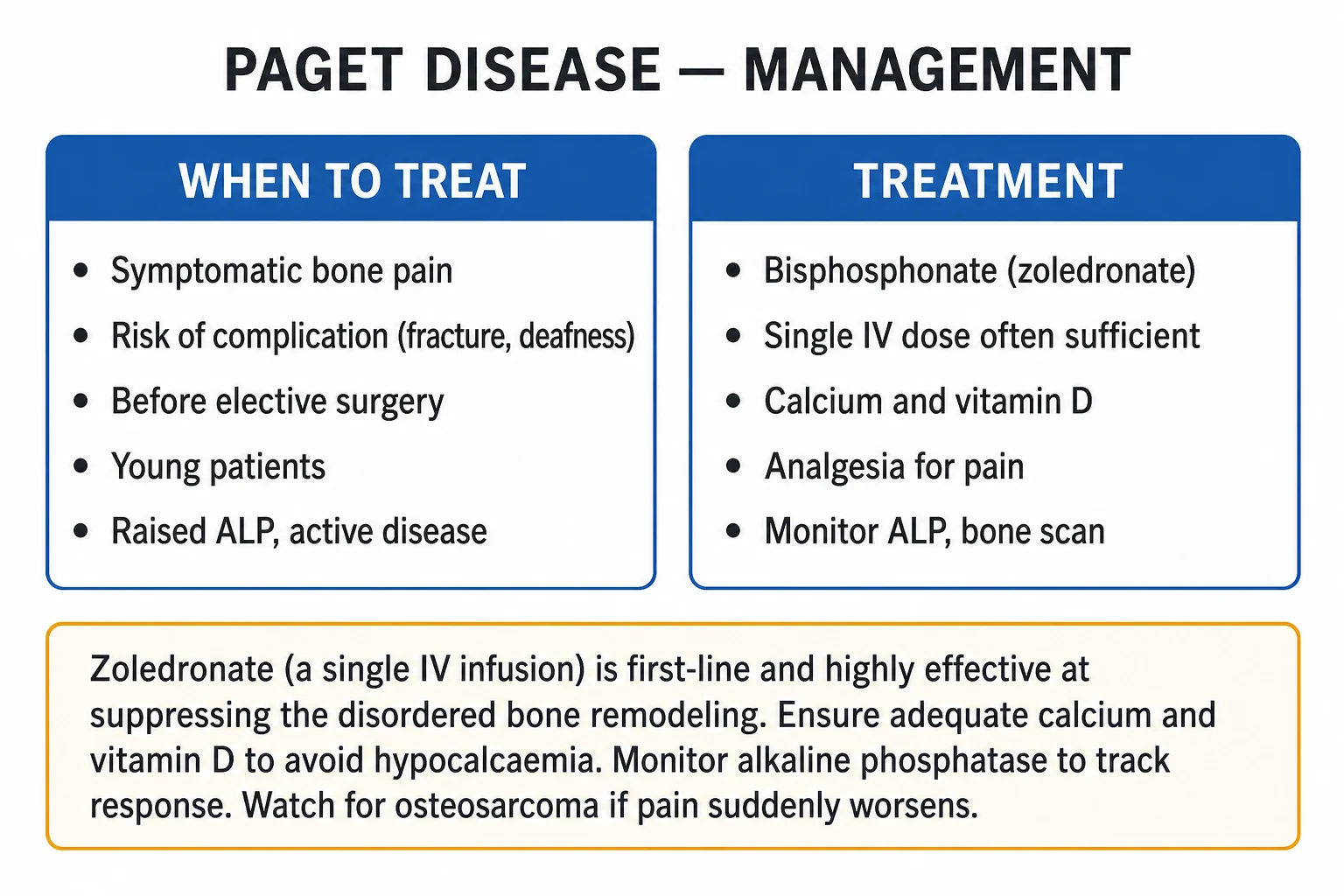
- Treat if — symptomatic bone pain; deformity or its imminent risk; complication risk (deafness, cord or nerve compression, basilar invagination, high-output failure); a lesion at a weight-bearing or fracture-prone site (femur, tibia); polyostotic / extensive active disease with a markedly raised ALP; young patients (who have decades of disease ahead); before elective orthopaedic surgery at a pagetoid site; and to suppress disease before joint replacement.
- Observe (do not treat) if — the disease is asymptomatic, monostotic, in a non-critical site (e.g., a single scapular lesion), biochemically quiescent, and there is no impending complication.[1][3]
First-line: a single zoledronic acid infusion
A single 5 mg intravenous infusion of zoledronic acid (given over at least 15 minutes in 100 mL of normal saline) is the first-line, most potent bisphosphonate for Paget disease. In the landmark randomised HORIZON trial, a single zoledronic acid infusion produced a more sustained biochemical remission than oral risedronate: it normalised ALP in the great majority of patients and maintained remission far longer, with most still in remission at one year.[3][1]
Oral and alternative bisphosphonate regimens
When oral therapy is preferred or IV access is impractical, oral risedronate is the standard alternative; older or less potent agents are largely historical or second-line: [1]
- Zoledronic acid — 5 mg IV infusion over at least 15 minutes, single dose; first-line; remission maintained for one to several years.[3]
- Risedronate — 30 mg orally daily for 2 months; a normal ALP is achieved in around two-thirds; an effective oral option.[1]
- Alendronate — 40 mg orally daily for 6 months (or, in some regimens, the osteoporotic 70 mg once-weekly dose for a longer course); less potent than zoledronate but widely available.
- Pamidronate — older IV option, given as multiple infusions (e.g., 60–90 mg IV on three occasions), largely superseded by zoledronic acid.
- Etidronate — avoided: it impairs bone mineralisation (causes osteomalacia) and is no longer recommended.[1]
Adjuncts and supportive care
- Calcium and vitamin D — replete before and after bisphosphonate therapy to avoid treatment-induced hypocalcaemia and secondary hyperparathyroidism; a typical regimen is elemental calcium 1000–1500 mg daily plus colecalciferol 800–1000 IU daily (titrated to a replete 25-hydroxyvitamin D).
- Analgesia — paracetamol first-line; NSAIDs for associated osteoarthritis; opiates briefly for severe pain or fracture.
- Calcitonin — 100 IU subcutaneously daily; rapidly lowers osteoclast activity and is occasionally used when bisphosphonates are contraindicated (severe renal impairment) or for rapid pain control, but is now rarely first-line.[5]
- Physiotherapy, occupational therapy and walking aids — to maintain mobility, prevent contractures around bowed limbs and reduce falls risk.
- Hearing aids — for symptomatic sensorineural deafness.
- Orthopaedic surgery — for fracture fixation, joint replacement (hip/knee for secondary osteoarthritis) and, rarely, corrective osteotomy for severe deformity. Pre-treat the pagetoid site with a bisphosphonate to reduce intra-operative bleeding from hypervascular bone.[1]
Monitoring
- Alkaline phosphatase every 6–12 months — the principal marker of response and relapse; a normalised ALP indicates biochemical remission.
- Re-treat on biochemical relapse (a sustained rise in ALP above the patient's remission baseline, typically above 1.25 times the upper limit of normal) or on symptomatic relapse.
- Lifelong surveillance for osteosarcoma: any new, focal, escalating pain, palpable mass, or unexpected ALP rise in a treated patient demands urgent imaging and biopsy.[1][6]
Worked example — from biochemistry to treatment
A 70-year-old man of British descent is referred for an asymptomatic ALP of 540 U/L (reference 40–120). Calcium 2.36, phosphate 1.05, eGFR 78, gamma-GT 22, PTH 4.1 (all normal). Pelvic X-ray shows thickening of the iliopectineal line with coarsened trabeculae; skull film reveals early osteoporosis circumscripta; bone scan demonstrates intense uptake in the right pelvis, L3 and the skull vault. The diagnosis is polyostotic Paget disease (the bone-source ALP with normal Ca/PO4 is the signature). Because the disease is polyostotic and biochemically active, with skull involvement risking deafness, treatment is indicated. After confirming a replete 25-hydroxyvitamin D, he receives a single 5 mg IV zoledronic acid infusion over 15 minutes with oral calcium and vitamin D supplementation. ALP is checked at 3 months (expecting a sharp fall), then 6–12 monthly; the goal is biochemical remission (normalised ALP). If, two years later, his ALP climbs and he develops new tibial pain, the priority is not to re-treat empirically but to image the tibia and biopsy to exclude osteosarcoma transformation before considering a second zoledronic acid dose.[1][3][6]
Specific Subtypes & Scenarios
- Monostotic versus polyostotic — about a third of cases involve a single bone (often found incidentally on an X-ray); the remainder involve multiple sites in the classic pelvis–spine–skull–femur distribution. Polyostotic disease carries a higher risk of deafness, deformity, basilar invagination and high-output cardiac failure and is more often an indication for treatment.
- The three phases — every lesion evolves from lytic (active, "hot", osteoporosis circumscripta, blade of grass) through mixed to sclerotic/burnt-out (cotton-wool skull, ivory vertebra). Treatment is most effective against the active, lytic/mixed phase; burnt-out lesions may show little biochemical response because turnover has already declined.
- Familial Paget disease (SQSTM1) — autosomal dominant with variable penetrance; earlier onset, more often polyostotic, and an indication to screen first-degree relatives with ALP in middle age.[4]
- IBMPFD (VCP mutation) — inclusion-body myopathy with Paget disease of bone and frontotemporal dementia; suspect when a patient with Paget disease also develops progressive myopathy or cognitive decline.
- Juvenile Paget disease (TNFRSF11B/OPG) — a separate entity: massive, widespread childhood bone turnover, deafness, retinopathy and vascular calcification from loss of osteoprotegerin; do not confuse with adult Paget disease.[2]
Complications & Pitfalls
- Pathological fracture — through mechanically weak pagetoid bone, most often the femoral neck or subtrochanteric femur, tibia or humerus; may be the presenting event.
- Osteosarcoma / sarcomatous transformation — the most feared complication; incidence around 0.7–1 percent of Paget patients (rising with extent and duration of disease). It presents with new, severe, rapidly worsening pain, a palpable soft-tissue mass, cortical destruction on imaging, and sometimes a sharp rise in ALP after a period of biochemical remission. Histology is most often osteosarcoma, occasionally fibrosarcoma or chondrosarcoma. Prognosis is poor; treatment is surgical (limb-salvage or amputation) with chemotherapy. Any change in a known pagetoid lesion mandates imaging and biopsy.[6]
- Deafness — sensorineural, from cochlear and eighth-nerve involvement; the commonest symptomatic complication of skull disease; rarely other cranial-nerve palsies.
- Secondary osteoarthritis — of the hip, knee or sacroiliac joint adjacent to a pagetoid segment, from altered mechanical loading; a common source of pain and a frequent indication for joint replacement.
- Spinal cord or nerve-root compression — from pagetoid vertebral expansion; an emergency requiring MRI, steroids and surgical decompression.
- Basilar invagination / platybasia — softening and flattening of the skull base allows the odontoid to invaginate, compressing the brainstem and lower cranial nerves and occasionally causing obstructive hydrocephalus.
- High-output cardiac failure — only in extensive polyostotic disease; the hypervascular bone bed acts as a chronic AV shunt.
- Hypercalcaemia of immobilisation — uncommon, but can occur after a fracture or prolonged bed rest when resorbed calcium is no longer redeposited into active bone.
- Nephrolithiasis — from the increased calcium load in active disease or immobilisation.
- Dental complications — hypercementosis, loosening of teeth, ill-fitting dentures; also bleeding risk during dental extraction in hypervascular bone, and osteonecrosis of the jaw as a rare bisphosphonate-related adverse effect.
The classic pitfalls are three: mistaking a raised ALP for liver disease without checking gamma-GT; failing to image and biopsy a changing pagetoid lesion (delaying the diagnosis of osteosarcoma); and giving a bisphosphonate without first repleting calcium and vitamin D, precipitating hypocalcaemia.[1][6]
Prognosis & Disposition
Paget disease is usually slowly progressive but highly manageable with modern bisphosphonate therapy. The prognosis splits along clinical lines: [1]
- Asymptomatic, monostotic disease — normal life expectancy; observation with periodic ALP is appropriate.
- Symptomatic or complication-risk disease — responds well to zoledronic acid, with biochemical remission (normalised ALP) sustained for months to years; quality of life, mobility and hearing are the principal functional outcomes.
- Osteosarcoma transformation — the dominant disease-specific cause of death; rare but carries a poor prognosis.
- Long-term trend — both the incidence and severity of Paget disease have been declining for decades, so contemporary disease tends to be milder and more often monostotic than historical series suggest.[1][5]
Disposition is outpatient in almost all cases, with shared care between primary care (ALP monitoring, analgesia, falls prevention, hearing aids) and a metabolic bone clinic (bisphosphonate therapy, response assessment). Admit only for fracture, cord compression, sarcoma work-up, or high-output cardiac failure. [1]
Follow-up, safety-net and patient education
Every Paget patient leaves the consultation with three explicit safety-net messages. First, an annual review of ALP (more often in the first year after treatment) is the cornerstone of follow-up — a normalised ALP after zoledronic acid predicts a durable remission, while a rising ALP after remission prompts re-imaging, not automatic re-treatment, to exclude sarcoma. Second, any new, focal, escalating pain at a pagetoid site, a palpable mass, or unexplained weight loss is treated as possible osteosarcoma until MRI and biopsy prove otherwise — patients are taught to report such changes urgently rather than waiting for their next routine appointment. Third, bone health measures apply as in any high-turnover bone disease: adequate calcium and vitamin D, weight-bearing exercise within the limits of deformity, smoking cessation, and falls prevention (home hazard reduction, vision and hearing correction, gait aids). Patients with skull disease should have baseline and periodic audiometry, and those with extensive polyostotic disease a baseline echocardiogram to detect early high-output failure. First-degree relatives of patients with SQSTM1-familial disease are offered ALP screening from middle age.[1][4][6]
Special Populations
- Before elective orthopaedic surgery at a pagetoid site — give a bisphosphonate (typically zoledronic acid) for 2–3 months beforehand to reduce the hypervascularity and intra-operative bleeding and to improve bone quality. Coordinate timing with the surgeon.[1]
- Young patients — tend to have more aggressive disease with a longer runway for complications; treat actively to prevent deformity, deafness and fracture.
- Extensive / polyostotic disease — higher complication burden (deafness, basilar invagination, high-output failure); treat and monitor closely, including echocardiography and audiometry.
- Renal impairment — avoid IV zoledronic acid in significant CKD (eGFR under 35 mL/min); consider calcitonin or a carefully chosen oral agent.
- Pregnancy — bisphosphonates cross the placenta and are avoided in pregnancy; weigh any need for treatment against risk, and prefer supportive measures until delivery.
- Anaesthetic considerations — positioning around fixed bony deformities, hypervascular bone increasing operative bleeding, and vigilance for hypercalcaemia with immobilisation.[1]
Evidence, Guidelines & Regional Differences
- Biochemical signature — the combination of a raised ALP with normal calcium, phosphate and PTH remains the diagnostic cornerstone; it separates focal Paget disease from systemic metabolic bone disease.[1]
- Bone scan vs X-ray — the isotope bone scan is the most sensitive test for extent and number of sites, whereas X-rays characterise the lesion (lytic vs sclerotic, cortical destruction, deformity); both are needed at diagnosis.[1]
- HORIZON trial (Reid 2005, NEJM) — a single 5 mg zoledronic acid infusion was superior to oral risedronate, producing a higher rate of ALP normalisation and a more durable remission; this established zoledronic acid as first-line.[3]
- Calcium and vitamin D first — must be repleted before bisphosphonate therapy to prevent hypocalcaemia and secondary hyperparathyroidism.[1]
- Sarcomatous transformation — the Deyrup clinicopathological series confirmed osteosarcoma as the dominant histology, with a poor prognosis; any change in a pagetoid lesion mandates biopsy.[6]
- Declining epidemiology — prevalence and severity are falling in traditional high-prevalence countries, consistent with an environmental (likely infective/pollution-related) contribution to aetiology on top of genetic susceptibility.[5]
International guidelines (the Endocrine Society / Bone and Mineral Research guideline of Ralston et al., endorsed in the UK, Australasia and North America) converge on zoledronic acid as first-line for symptomatic or complication-risk Paget disease, with calcium and vitamin D repletion and ALP monitoring. Regional practice is broadly uniform; minor variations exist in the choice of oral alternative (risedronate widely in the UK and Europe; alendronate used in some settings) and in the threshold ALP for re-treatment.
Exam Pearls
Paget disease features — PAGET
PAGET
deep aching bone pain, often nocturnal; fractures through weak Pagetoid bone
the biochemical signature of focal bone turnover
paramyxovirus / SQSTM1 driven; resorptive lytic phase
increasing hat size; sensorineural deafness from skull base
mosaic pattern; single 5 mg IV bisphosphonate first-line
X-ray signs by site — SITES
SITES
frontoparietal lytic defect early; patchy sclerosis late
thickened sclerotic vertebral rim with lucent centre
V-shaped advancing lytic wedge in long bones
coarse trabeculae, bowed sabre shin
most sensitive test for extent and number of sites
Treatment regimens — at a glance
Exam application bank (NEET-PG / INICET)
One-line answer
Paget disease of bone (osteitis deformans) is a chronic, focal disorder of adult bone remodeling in which excessive, disorganised osteoclast activity is followed by chaotic osteoblast repair, producing bone that is thick and hypervascular but mechanically weak (a disordered woven/mosaic pattern). It is usually asymptomatic and found on incidental raised alkaline phosphatase with normal calcium and phosphate; symptoms include bone pain (often nocturnal), bony deformity (sabre tibia, enlarging skull, kyphosis), sensorineural deafness and, rarely, neurological or cardiac complications. Common sites are the pelvis, spine, femur, skull and tibia. Diagnosis rests on raised ALP with normal Ca/PO4, characteristic X-rays (cortical thickening, mixed lytic-sclerotic lesions, cotton-wool skull, blade-of-grass, picture-frame vertebrae) and an isotope bone scan showing focal hot spots. The cornerstone
Worked stems (answer without another resource)
Stem 1 — Classic presentation. Map symptoms to mechanism; name the first investigation and first treatment step with dose/route if drug therapy is standard. [1]
Stem 2 — Unstable / complicated. List red flags that force immediate resuscitation, theatre, ICU, antidote, or reperfusion — and what you do in the first 15 minutes. [1]
Stem 3 — Atypical group. Elderly, pregnancy, child, or immunocompromised: how presentation and thresholds change. [1]
Stem 4 — Differential trap. Name the three closest mimics and one discriminator for each. [1]
Stem 5 — Disposition. Who goes home with safety-netting, who is admitted, who needs HDU/ICU/theatre, and what follow-up is mandatory. [1]
Rapid viva checklist
- Definition + classification
- Pathophysiology chain
- Bedside signs / criteria
- Score with exact components (if any)
- Emergency bundle
- Definitive therapy with doses
- Complications of disease and of treatment
- Special populations
- Guideline/trial name if classic
- Three exam traps
Coverage self-check
If you cannot answer any stem above from this page alone, re-read the matching section — the page is intended to be self-sufficient for final-prof and NEET-PG/INICET questions on Paget Disease of Bone.
Quick self-test: a 70-year-old man with ALP 520 U/L, Ca 2.36, PO4 1.05, gamma-GT normal — what is the biochemical diagnosis, and what two tests next?
The pattern of a markedly raised ALP with normal calcium, phosphate and gamma-GT is the biochemical signature of Paget disease of bone (an isolated bone-source ALP). The two next tests are a plain X-ray of the pelvis/skull/spine (looking for cortical thickening, mixed lytic-sclerotic change, cotton-wool skull, picture-frame vertebrae) and a Tc-99m isotope bone scan to map the extent and number of pagetoid sites.[1]
References
- [1]Ralston SH, Corral-Gudino L, Cooper C, et al. Diagnosis and Management of Paget's Disease of Bone in Adults: A Clinical Guideline J Bone Miner Res, 2019.PMID 30803025
- [2]Gennari L, Rendina D, Falchetti A, et al. Paget's Disease of Bone Calcif Tissue Int, 2019.PMID 30671590
- [3]Reid IR, Miller P, Lyles K, et al. Comparison of a single infusion of zoledronic acid with risedronate for Paget's disease N Engl J Med, 2005.PMID 16135834
- [4]Laurin N, Brown JP, Morissette J, Raymond V. Recurrent mutation of the gene encoding sequestosome 1 (SQSTM1/p62) in Paget disease of bone Am J Hum Genet, 2002.PMID 11992264
- [5]Cundy T. Paget's disease of bone Metabolism, 2018.PMID 28780255
- [6]Deyrup AT, Montag AG, Inwards CY, et al. Sarcomas arising in Paget disease of bone: a clinicopathologic analysis of 70 cases Arch Pathol Lab Med, 2007.PMID 17550323