Endocrinology · General Medicine
Primary Aldosteronism (Conn Syndrome)
Also known as Primary aldosteronism · Conn syndrome · Hyperaldosteronism · Aldosterone-producing adenoma
Primary aldosteronism is autonomous aldosterone secretion that is independent of renin (high aldosterone, suppressed renin), causing sodium retention with hypertension, and potassium and hydrogen loss with hypokalaemic metabolic alkalosis. It is the commonest cause of secondary hypertension, affecting 5 to 10 percent of all hypertensives and over 20 percent of those with resistant hypertension, yet is frequently missed because most patients are normokalaemic. Causes are bilateral idiopathic adrenal hyperplasia (commonest), a unilateral aldosterone-producing adenoma (Conn syndrome), unilateral adrenal hyperplasia, and familial hyperaldosteronism types I to IV. Screen at-risk patients with the aldosterone-to-renin ratio (ARR), confirm autonomy with a suppression test, then localise with CT and adrenal venous sampling (AVS). Treat unilateral disease with laparoscopic adrenalectomy (curative) and bilateral disease with a mineralocorticoid receptor antagonist (spironolactone or eplerenone).
On this page & tools
Your progress
Saved locally on this device.
Exam tags
Red flags
Overview & Definition
Primary aldosteronism is a group of disorders in which the adrenal zona glomerulosa secretes aldosterone autonomously — that is, in excess of, and independent of, the prevailing renin-angiotensin system activity. The biochemical signature is therefore inappropriately high aldosterone with suppressed renin, producing the high aldosterone-to-renin ratio (ARR) that is the cornerstone of screening. The downstream phenotype is the triad of hypertension (typically resistant), hypokalaemia and metabolic alkalosis, although a normal serum potassium is now the rule rather than the exception, which is precisely why the disease is so often missed.[1]
Recognising primary aldosteronism matters for three independent reasons. First, it is far more common than once believed — about 5 to 10 percent of all hypertensive patients and over 20 percent of those with resistant hypertension — making it the single commonest specifically treatable cause of secondary hypertension. Second, targeted treatment (adrenalectomy for unilateral disease, a mineralocorticoid receptor antagonist for bilateral disease) controls blood pressure far better than escalating standard antihypertensives and cures the hypertension in a substantial proportion of unilateral cases. Third, and most importantly, aldosterone excess causes cardiovascular and renal damage that is disproportionate to the blood pressure itself — patients with primary aldosteronism have higher rates of stroke, atrial fibrillation, left ventricular hypertrophy, heart failure and chronic kidney disease than age-, sex- and blood-pressure-matched patients with essential hypertension — so finding and treating it changes outcome, not just a number.[1][6]
The diagnostic discipline, which examiners repeatedly probe, is a deliberately staged four-step algorithm: screen with the ARR in an at-risk patient (after correcting interfering drugs), confirm autonomy with a suppression test, localise and lateralise with CT and adrenal venous sampling, then treat by subtype. The cardinal error — operating on an abnormal CT without first proving autonomy or lateralisation — is what the algorithm exists to prevent.[2][3]
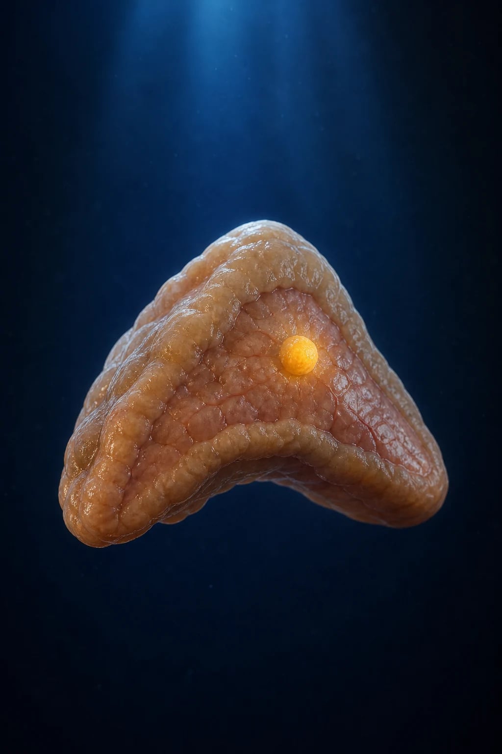
Classification
The single most important classification decision is unilateral versus bilateral disease, because that distinction alone separates curative surgery from lifelong medical therapy. Within that split sit the anatomical subtypes and, separately, the familial forms that demand a genetic answer before any operating list.[1]
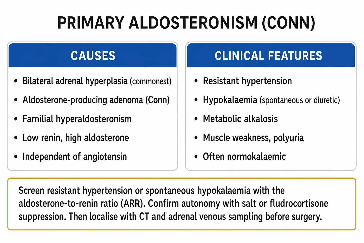
Bilateral idiopathic adrenal hyperplasia (BAH)
commonest, ~60%
- Bilateral zona-glomerulosa hyperplasia — the COMMONEST cause overall
- Diffuse bilateral aldosterone excess; no single tumour to remove
- Treated MEDICALLY with a mineralocorticoid receptor antagonist (spironolactone or eplerenone)
- Adrenalectomy does NOT cure bilateral disease — never operate on BAH alone
- Predominant in older patients; biochemistry and severity usually milder than Conn adenoma
Aldosterone-producing adenoma (Conn)
unilateral, ~30%
- Single unilateral cortisol-coexisting adenoma — the classic Conn syndrome
- SURGICALLY CURABLE by unilateral laparoscopic adrenalectomy
- Lateralisation must be PROVEN by adrenal venous sampling (AVS) before surgery
- Predominant in younger women; classically more severe biochemistry (low K, high aldosterone)
- Somatic KCNJ5 mutation is the commonest driver mutation
Unilateral adrenal hyperplasia
unilateral, ~2%
- Unilateral micronodular or macronodular hyperplasia with a normal-appearing contralateral gland
- Behaves like Conn — surgically curable if AVS proves unilateral lateralisation
- Easily missed on CT because no discrete mass; AVS is essential
Familial hyperaldosteronism (FH I to IV)
young, under 40, or family history
- FH-I (glucocorticoid-remediable): chimeric CYP11B1/CYP11B2 gene places aldosterone synthase under ACTH control — cured by LOW-DOSE GLUCOCORTICOID
- FH-II: familial non-glucocorticoid-remediable, often KCNJ5 germline
- FH-III: severe, KCNJ5 germline (often bilateral macronodular hyperplasia)
- FH-IV: CACNA1H germline; test ALL young patients (under 40) or those with a family history BEFORE surgery
- FH-I screened with the dexamethasone suppression test and confirmed by the CYP11B chimeric gene (long-range PCR)
Two rarer entities round out the anatomical list and are worth knowing for viva: an aldosterone-producing adrenocortical carcinoma (a large, irregular mass — over 4 cm — with mixed steroid excess, managed surgically as for any adrenal malignancy), and ectopic aldosterone secretion (ovary, kidney, gut — exceptionally rare, and a reason biochemical cure can follow non-adrenal resection).[1]
Epidemiology & Risk Factors
Primary aldosteronism was once taught as a rare, curable cause of hypokalaemic hypertension. Modern screening, using the ARR rather than waiting for hypokalaemia, has overturned that view: it is now established that 5 to 10 percent of all hypertensive patients carry the diagnosis, rising to over 20 percent of those with resistant hypertension (uncontrolled on three agents including a diuretic, or controlled on four).[1][2]
Primary aldosteronism — the numbers that decide an answer
Patient-level risk factors that should trigger screening are listed in detail in the Investigations section. In short, any patient whose hypertension is resistant, early-onset (under 40), hypokalaemic (spontaneously or with diuretics), accompanied by an adrenal incidentaloma, or associated with a family history of early-onset hypertension or stroke under 40 deserves an ARR. There is a modest female predominance for Conn adenoma (younger women, second to fifth decade), while bilateral hyperplasia dominates with age and in men. Familial forms are uncommon (under 5 percent of all primary aldosteronism) but disproportionately important because they demand a genetic diagnosis and, for type I, a glucocorticoid rather than a knife.[2][8]
Pathophysiology
Aldosterone is the principal mineralocorticoid of the adrenal zona glomerulosa, and its secretion is normally governed by angiotensin II (generated by renin from the juxtaglomerular apparatus) and by serum potassium, with ACTH providing a smaller, permissive input. The hormone acts through the intracellular mineralocorticoid receptor in the principal cells of the distal nephron and collecting duct. 11-beta-hydroxysteroid dehydrogenase type 2 normally protects this receptor from cortisol, which is why primary aldosteronism — rather than cortisol excess — produces the classic mineralocorticoid phenotype.[1]
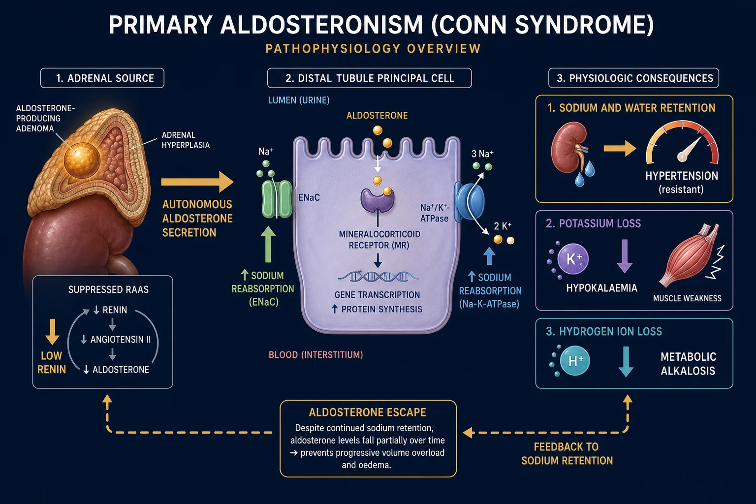
In primary aldosteronism the zona glomerulosa secretes autonomously, so the normal negative-feedback loop is broken. Aldosterone drives sodium and water retention, the expanded extracellular volume raises blood pressure, and the elevated pressure and volume appropriately suppress renin — but, crucially, suppressed renin cannot suppress the tumour or the hyperplastic glands. The loop therefore never closes. This dissociation — high aldosterone in the face of suppressed renin — is the biochemical signature that defines the disease and that the ARR is designed to capture. Three downstream consequences follow and define the phenotype:[1][2]
- Sodium and water retention → hypertension, typically resistant to standard agents because the underlying mineralocorticoid drive is not blocked.
- Distal potassium wasting → hypokalaemia, with muscle weakness, cramps, fatigue, polyuria and nocturia. Chronic hypokalaemia damages the renal concentrating mechanism, producing a nephrogenic diabetes insipidus.
- Hydrogen ion loss → metabolic alkalosis, with raised venous bicarbonate and, on ECG, prominent U waves and QT prolongation when potassium is low. [1]
Two concepts that examiners reward deserve emphasis, because both explain non-intuitive clinical features and are favourite viva corners. Aldosterone escape is the observation that, as blood pressure and extracellular volume climb, pressure natriuresis and atrial natriuretic peptide (ANP) counter the sodium-retaining effect of aldosterone, so the patient reaches a new steady state of mild sodium retention with only modest volume expansion. The practical consequence is that primary aldosteronism does not cause oedema despite mineralocorticoid excess — prominent peripheral oedema in a suspected Conn patient should prompt a search for a secondary (oedematous) cause such as heart failure, renal failure or another cause of hypokalaemia. Non-epithelial (off-target) aldosterone effects explain why the cardiovascular damage of primary aldosteronism exceeds that of essential hypertension at the same blood pressure: aldosterone acts directly on cardiomyocytes, vascular smooth muscle, cardiac fibroblasts and renal mesangial cells to drive inflammation and fibrosis independent of blood pressure — the mechanism behind the excess of left ventricular hypertrophy, atrial fibrillation, stroke, heart failure and chronic kidney disease that defines the disease's cardiovascular burden.[1][6][9]
The molecular genetics of sporadic aldosterone-producing adenomas have reshaped the field: roughly 40 percent of Conn adenomas carry a somatic mutation in KCNJ5 (the G-protein inwardly rectifying potassium channel Kir3.4), producing membrane depolarisation and calcium influx that drives CYP11B2 (aldosterone synthase) expression. Other somatic drivers include CACNA1D (voltage-gated calcium channel), ATP1A1 (Na-K ATPase alpha) and ATP2B3 (plasma-membrane calcium ATPase). These germline and somatic mutations converge on zona glomerulosa cell depolarisation and calcium signalling as the final common pathway of autonomous aldosterone synthesis.[7][8]
Clinical Presentation
The presentation of primary aldosteronism is dominated by hypertension that is usually already established and often labelled "essential" — the diagnosis is made because someone thought to look for it. The blood pressure is characteristically moderate to severe, frequently resistant to three or more agents including a diuretic, and may be the only finding. The notion that primary aldosteronism requires hypokalaemia is outdated: most screen-positive patients are normokalaemic, which is exactly why case detection must be driven by the clinical phenotype (resistant hypertension, early onset, incidentaloma, family history) rather than waiting for a low potassium.[1][2]
The classic symptom cluster appears once hypokalaemia is present and reflects potassium depletion plus the metabolic alkalosis it generates: [1]
- Neuromuscular — muscle weakness (proximal, occasionally profound), cramps, fatigue and, rarely, hypokalaemic periodic paralysis or tetany (from alkalosis lowering ionised calcium). The periodic paralysis variant is over-represented in patients of Asian descent and can be the presenting emergency.
- Renal — polyuria, nocturia and polydipsia from hypokalaemia-induced nephrogenic diabetes insipidus (the distal nephron loses its responsiveness to vasopressin).
- Cardiovascular — symptoms of end-organ damage: palpitations and exertional dyspnoea from left ventricular hypertrophy, paroxysmal atrial fibrillation, and headache (which may also reflect any coexisting sympathetic drive).
- Neurological — the consequences of hypertension and hypokalaemia combine to raise stroke risk disproportionately. [1]
Atypical and easily-missed presentations are deliberately tested. Normokalaemic resistant hypertension is now the commonest presentation and must not be falsely reassuring. Normotensive primary aldosteronism is described but rare, and should prompt consideration of an alternative (especially secondary causes of hypokalaemia). Elderly patients may present with atrial fibrillation, heart failure or a first stroke as the dominant problem, with the underlying aldosterone excess only discovered later. Pregnant patients may have worsening hypertension and hypokalaemia, sometimes misattributed to pre-eclampsia; note that pregnancy normally raises both renin and aldosterone, which complicates the ARR. Children and young adults with primary aldosteronism should always raise the question of a familial form (FH-I to IV) before any operation is planned.[2][8]
Differential Diagnosis
Hypertension with hypokalaemia and metabolic alkalosis is the shared phenotype of several distinct disorders, but the renin-and-aldosterone pair sorts them cleanly. The essential discriminator is whether renin is high or low, and whether aldosterone is high or low. This is a perennial examination topic because a single wrong answer on the renin/aldosterone pattern leads to a dangerous management choice (for example, giving spironolactone to a patient with Liddle syndrome, which is ENaC-driven and requires amiloride).[1]
Primary aldosteronism
low renin, HIGH aldosterone
- Autonomous adrenal aldosterone; suppressed renin
- Hypertension with hypokalaemic alkalosis; often normokalaemic
- Treated by adrenalectomy (unilateral) or MRA (bilateral)
Secondary aldosteronism
HIGH renin, high aldosterone
- Renin-driven: renal artery stenosis, accelerated/malignant hypertension, renin-secreting tumour
- Oedematous states: heart failure, cirrhosis, nephrotic syndrome (oedema distinguishes these from PA)
- Diuretic abuse; the renin AND aldosterone are both high — opposite of PA
Liddle syndrome
low renin, LOW aldosterone
- Gain-of-function mutation of the ENaC beta/gamma subunit (SCNN1B/G)
- Mimics PA biochemically except aldosterone is LOW
- Treat with amiloride or triamterene — NOT spironolactone (the defect is downstream of the receptor)
Apparent mineralocorticoid excess / liquorice
low renin, LOW aldosterone
- 11-beta-hydroxysteroid dehydrogenase type 2 is deficient or inhibited (glycyrrhetinic acid in liquorice/carbenoxolone)
- Cortoid activates the mineralocorticoid receptor
- Treat by stopping liquorice or with amiloride/spironolactone; aldosterone is low
Congenital adrenal hyperplasia
low renin, LOW aldosterone
- 11-beta-hydroxylase or 17-alpha-hydroxylase deficiency
- Hypertension with hypokalaemia, low aldosterone and low cortisol, with androgen features (11-beta) or sexual infantilism (17-alpha)
- Treat with glucocorticoid replacement
Cushing syndrome
variable renin, variable aldosterone
- Cortisol excess overwhelms 11-beta-HSD2 capacity, activating the mineralocorticoid receptor
- Differentiated by clinical stigmata, elevated cortisol and failure of dexamethasone suppression
- Hypokalaemia more common in ectopic ACTH / severe Cushing
The can't-miss mimics in practice are renal artery stenosis (high renin, high aldosterone — and a cause of refractory hypertension that has its own, very different management), Liddle syndrome (low renin, low aldosterone — the single most-tested distractor), and apparent mineralocorticoid excess from chronic liquorice ingestion (a reversible, easily-missed drug/diet cause).[1]
Clinical & Bedside Assessment
There is no single pathognomonic bedside sign for primary aldosteronism, and physical examination is performed principally to gauge the severity and consequences of the hypertension and to exclude alternative diagnoses. A focused assessment documents: [1]
- Blood pressure — often severe; measure both arms and look for a postural drop that might suggest a different cause. Establish whether it is truly resistant (uncontrolled on three agents including a diuretic, or controlled only on four).
- Cardiovascular — examine for left ventricular hypertrophy (displaced, heaving apex; loud aortic component), a fourth heart sound, signs of heart failure, and an irregularly irregular pulse of atrial fibrillation — end-organ consequences that should heighten suspicion.
- Abdomen — there is nothing to feel in primary aldosteronism itself; a palpable mass or bruit redirects toward a pheochromocytoma, adrenal carcinoma (mass over 4 cm, irregular) or renal artery stenosis (a flank or abdominal bruit).
- Oedema — the absence of peripheral oedema is expected (aldosterone escape); its presence argues for a secondary, oedematous cause and against uncomplicated PA.
- Neurological and fundoscopic — hypertensive retinopathy and any focal deficit from prior stroke document target-organ damage and raise the stakes for diagnosis.
- Stigmata of Cushing (central obesity, striae, bruising, proximal myopathy) and neurofibromata/cafe-au-lait patches (pointing to a pheochromocytoma) must be excluded because they are alternative causes of an adrenal incidentaloma with hypertension.[2]
The bedside assessment also includes a careful drug history, because interfering medications are the commonest reason for a false ARR (see Investigations) and for a paradoxically low potassium (diuretics), and a family history of early-onset hypertension, stroke under 40 or sudden cardiac death, which mandates testing for familial hyperaldosteronism.[2][8]
Investigations
Investigation is the heart of the topic and follows the staged algorithm: screen → confirm → localise → subtype. Each stage has named thresholds, and examiners test the whole ladder. The Endocrine Society's most recent clinical practice guideline (2025) updates but does not replace the underlying four-step logic of the 2016 guideline.[2][3]
Who to screen — the case-detection criteria
The Endocrine Society case-detection criteria — screen with the ARR if ANY apply
Resistant hypertension
Blood pressure uncontrolled on three agents including a diuretic, or controlled on four or more agents
Spontaneous or diuretic-induced hypokalaemia
Any hypertensive patient with K below 3.5 mmol/L spontaneously, or provoked by a routine diuretic
Adrenal incidentaloma
Any hypertensive patient with an adrenal mass discovered incidentally
Early-onset hypertension
Hypertension onset under 40, or early-onset stroke under 40
Family history of early-onset HTN or stroke
A first-degree relative with hypertension onset or stroke under 40 — also screen the index case for familial hyperaldosteronism
Hypertension discovered before age 40
Regardless of family history — screen for familial forms before any surgery
Step 1 — Screen with the aldosterone-to-renin ratio (ARR)
The ARR is the screening test of choice. A screen-positive result is defined by both an elevated ratio and a concurrently elevated absolute aldosterone, because a high ratio can arise from a very low renin with a normal aldosterone and is then non-diagnostic. Because the cut-off depends on assay, units and posture, no single universal value applies, but the commonly used teaching thresholds are an ARR over 30 (when aldosterone is measured in ng per dL and renin in ng per mL per hour) with a concurrent aldosterone above 15 ng per dL — equivalently, in SI units, an aldosterone above about 330 to 415 pmol per L with a suppressed renin. A screen that is borderline or ambiguous should be repeated, ideally under standardised conditions (morning, ambulatory, seated, potassium replete), before proceeding. A suppressed renin with a frankly elevated aldosterone is screen-positive; a suppressed renin with a normal aldosterone is non-diagnostic and should be repeated or interpreted with caution.[2][3]
ARR — the screening thresholds (assay-dependent; the two parts must BOTH be abnormal)
Step 2 — Confirm autonomy with a suppression test
A positive screen is never sufficient to operate on; autonomy must be proven by demonstrating that aldosterone fails to suppress under salt or volume loading. Four established confirmatory tests exist, all with explicit cut-offs; the choice is institutional.[2][3]
The four confirmatory tests — protocol, threshold, interpretation
Step 3 — Localise and lateralise (CT then AVS)
Once autonomy is confirmed, computed tomography of the adrenal glands is the first localising test. A discrete unilateral low-density lipid-rich mass under 4 cm with a normal contralateral gland supports a Conn adenoma; bilateral thickening or multiple small nodules suggests bilateral hyperplasia. But CT is fundamentally insufficient for the lateralisation decision: a unilateral nodule may be a non-functioning incidentaloma coexisting with bilateral hyperplasia (leading to wrong-sided surgery), and bilateral nodularity may hide a single functioning adenoma. Adrenal venous sampling (AVS) is therefore the gold standard for proving unilateral disease before surgery.[1][4]
The SPARTACUS trial randomised patients to treatment assignment by CT alone versus by AVS, and although it found no overall blood-pressure benefit of AVS-guided management at one year, AVS remains the standard because CT-only assignment leads to a substantial rate of inappropriate adrenalectomy (operating on non-functioning nodules) and missed surgically curable cases — and contemporary outcome definitions and long-term follow-up favour AVS-guided decisions.[4][5]
AVS catheterises the left adrenal vein (which drains to the left renal vein) and the right adrenal vein (which drains directly to the inferior vena cava), sampling aldosterone and cortisol from each and from the inferior vena cava. Cortisol is measured alongside aldosterone to confirm successful cannulation (the adrenal-to-IVC cortisol ratio should be at least 2 to 3 without cosyntropin, or over 3 with cosyntropin). The lateralisation index (LI) is the aldosterone-to-cortisol ratio on the dominant side divided by that on the non-dominant side: [1]
Step 4 — Subtype (familial) testing
Before any operation in a young patient (under 40) or anyone with a family history of early-onset hypertension or stroke, test for familial hyperaldosteronism type I (glucocorticoid-remediable aldosteronism) with the dexamethasone suppression test and confirm with genetic testing for the CYP11B1/CYP11B2 chimeric gene (long-range PCR), because FH-I is treated medically with glucocorticoid, not surgically. FH-III (KCNJ5 germline) should be suspected when bilateral macronodular hyperplasia is severe and childhood-onset. Genotyping for KCNJ5, CACNA1H, CACNA1D and the chimeric gene is increasingly part of the subtype workup in selected patients.[7][8]
Baseline and supporting investigations
Every confirmed case should also have electrolytes, renal function, ECG and echocardiogram to document and grade target-organ damage, and urine albumin-to-creatinine ratio because albuminuria predicts cardiovascular and renal outcome. An aldosterone and renin measured under standardised conditions plus cortisol and a dexamethasone suppression test are needed when an adrenal incidentaloma is present to exclude a co-secreting cortisol-producing adenoma.[1]
Management — Resuscitation
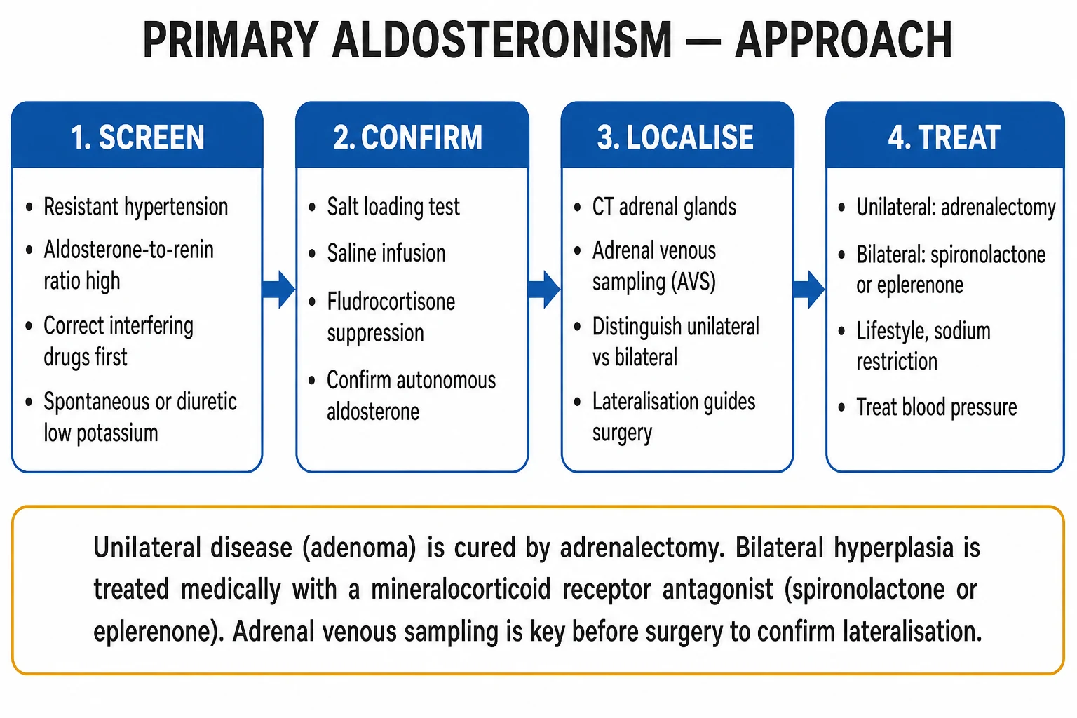
Primary aldosteronism is rarely an immediate resuscitation problem, but two acute presentations demand time-critical management before the diagnostic ladder is even begun: severe hypokalaemia with cardiac arrhythmia or paralysis, and hypertensive emergency (encephalopathy, acute pulmonary oedema, aortic dissection, eclampsia).[2]
Resuscitation bundle for the acute hypokalaemic or hypertensive emergency in primary aldosteronism
Replete potassium urgently
For K below 3.0 mmol/L with arrhythmia or paralysis: intravenous potassium chloride 10 to 20 mmol per hour via a central line with continuous cardiac monitoring, plus oral repletion; recheck every 2 to 4 hours and aim to keep K above 4.0 mmol/L
Block the mineralocorticoid receptor
Start a mineralocorticoid receptor antagonist — spironolactone 25 to 100 mg orally daily — to halt ongoing potassium and hydrogen loss; this also begins to lower blood pressure
Control the blood pressure
For hypertensive emergency, lower mean arterial pressure by no more than 25 percent in the first hour with an intravenous agent (labetalol, nicardipine or nitroprusside), then transition to oral control; avoid diuretics that worsen hypokalaemia
Correct magnesium
Replete magnesium (cofactor for potassium retention) — hypomagnesaemia perpetuates refractory hypokalaemia
Then enter the diagnostic ladder
Once stabilised, proceed to screen, confirm, localise and subtype as above
In hypokalaemic periodic paralysis, the paralysis resolves as potassium is repleted, but the patient must be monitored for rebound hyperkalaemia because total-body potassium deficit is variable. Never give potassium-sparing diuretics and ACE inhibitors together without close monitoring in the acute phase.[2]
Management — Definitive & Stepwise
Definitive management is decided by subtype and follows directly from the lateralisation result. The aim is to normalise aldosterone excess, control blood pressure, correct hypokalaemia and reduce cardiovascular and renal target-organ damage.[1][3]
Unilateral disease — surgical cure
For unilateral aldosterone-producing adenoma or unilateral adrenal hyperplasia proven by AVS, the treatment of choice is unilateral laparoscopic adrenalectomy — curative of the biochemical defect and of the hypertension in a substantial proportion. Pre-operatively, a mineralocorticoid receptor antagonist (spironolactone 25 to 100 mg daily) is given for four to six weeks to correct hypokalaemia, control blood pressure and predict post-operative blood-pressure response (a fall in blood pressure on spironolactone predicts surgical cure). Patients should be counselled on the risk of post-adrenalectomy hypoaldosteronism (transient mineralocorticoid deficiency from chronic suppression of the contralateral zona glomerulosa), which can require short-term fludrocortisone, and on the possibility of persistent hypertension if essential hypertension coexists.[1][5]
Adrenalectomy cures hypertension in about 30 to 60 percent of unilateral cases and improves it in most of the remainder; biochemical cure (normalisation of aldosterone and renin off all mineralocorticoid-blocking therapy) is achieved in over 90 percent. Predictors of clinical cure include younger age, shorter duration of hypertension, absence of a family history of essential hypertension, fewer antihypertensive agents pre-operatively and a larger adenoma. The international PASO consensus (2017) defines five outcome categories — complete, partial and absent clinical success, and complete and partial biochemical success — using standardised blood-pressure and biochemical criteria, and is the reference framework for surgical outcome reporting.[5]
Surgical outcome after adrenalectomy for unilateral primary aldosteronism
Bilateral disease — lifelong medical therapy
For bilateral idiopathic adrenal hyperplasia (or a patient unfit or unwilling for surgery), the mainstay is lifelong mineralocorticoid receptor antagonism. The first-line agent is spironolactone 25 to 100 mg orally daily (titrated to a maximum around 400 mg if needed), which competitively blocks the mineralocorticoid receptor. Its principal drawback is dose-dependent anti-androgen and progestogenic side-effects — gynaecomastia, breast tenderness, erectile dysfunction and menstrual irregularity — that limit its tolerability, particularly in men and premenopausal women. Eplerenone 50 to 100 mg orally daily is a selective mineralocorticoid receptor antagonist (it spares the androgen and progesterone receptors) and is preferred when spironolactone is not tolerated; it is somewhat less potent and more expensive, and dose-equivalence is roughly half. Amiloride 5 to 20 mg orally daily (or triamterene) blocks the epithelial sodium channel directly and is an alternative when both receptor antagonists are contraindicated or poorly tolerated, as in Liddle syndrome. Concomitant sodium restriction, weight loss and additional antihypertensives (ACE inhibitor, angiotensin-receptor blocker or non-dihydropyridine calcium-channel blocker) are usually required for blood-pressure control.[1][2][9]
Familial hyperaldosteronism type I — glucocorticoid
FH-I is uniquely managed with low-dose glucocorticoid: dexamethasone 0.5 to 1 mg orally at night (or an equivalent dose of prednisolone) suppresses ACTH-driven aldosterone synthase and corrects both the biochemical defect and the hypertension. The lowest effective dose is used to avoid iatrogenic Cushing's, and children are dosed by weight and monitored for growth. If blood pressure remains uncontrolled despite biochemical cure, a mineralocorticoid receptor antagonist is added. Surgery has no role in FH-I.[2][8]
General measures and the avoidance of harm
Whatever the subtype, all patients benefit from dietary sodium restriction (which reduces potassium loss and lowers blood pressure), avoidance of potassium-wasting diuretics (thiazides and loops worsen hypokalaemia unless covered by a mineralocorticoid antagonist), weight reduction, smoking cessation and treatment of any dyslipidaemia or diabetes. A common pitfall is adding a thiazide to "control the blood pressure" without recognising that it will unmask profound hypokalaemia in an undiagnosed Conn patient.[1]
Mineralocorticoid receptor antagonists
Specific Subtypes & Scenarios
Each subtype carries an implication for management and is worth a paragraph in its own right. [1]
Aldosterone-producing adenoma (Conn syndrome) is the prototypical surgically curable form — typically a single, unilateral, small (under 2 to 3 cm), lipid-rich adenoma of the zona glomerulosa, often in a younger woman with more severe biochemistry than bilateral disease. The somatic KCNJ5 mutation is the commonest driver. The key action is to prove lateralisation by AVS (or accept a CT adenoma in the under-35 group with marked hypokalaemia) and offer laparoscopic adrenalectomy, which is curative in the majority.[1][7]
Bilateral idiopathic adrenal hyperplasia is the commonest subtype overall and is managed medically for life. Some patients with bilateral macronodular hyperplasia are found to harbour a germline KCNJ5 mutation (FH-III), in whom bilateral adrenalectomy is occasionally considered for severe, refractory disease but is generally avoided because of the commitment to lifelong glucocorticoid and mineralocorticoid replacement.[7][8]
Familial hyperaldosteronism type I (glucocorticoid-remediable aldosteronism) is caused by a chimeric gene from an unequal crossover fusing the 5 prime ACTH-responsive promotor of CYP11B1 with the 3 prime coding region of CYP11B2, placing aldosterone synthase under ACTH control. It is autosomal dominant, presents in childhood, features severe hypertension with early stroke, produces characteristic hybrid steroids (18-oxocortisol, 18-hydroxycortisol), and is uniquely cured by low-dose glucocorticoid (dexamethasone 0.5 to 1 mg nocte). Confirm genetically with the CYP11B chimeric gene test.[2][8]
Familial hyperaldosteronism type II is familial, non-glucocorticoid-remediable, often linked to germline KCNJ5, and behaves like sporadic disease (adenoma or hyperplasia) — managed surgically or medically by subtype. FH-III is a severe childhood form from germline KCNJ5 mutations, usually bilateral macronodular hyperplasia, often needing bilateral adrenalectomy. FH-IV is from germline CACNA1H and is managed medically or surgically depending on laterality. The unifying principle: test ALL young patients (under 40) or those with a family history for FH-I before surgery, because giving glucocorticoid to FH-I avoids an unnecessary operation.[7][8]
Aldosterone-producing adrenocortical carcinoma is rare but should be suspected when the adrenal mass is large (over 4 cm), irregular, with imaging heterogeneity and rapid growth, often with mixed steroid excess (cortisol, androgens). Management is open surgical resection (not laparoscopic) as for any adrenal malignancy, with oncological staging.[1]
Complications & Pitfalls
The complications of primary aldosteronism arise from the disease itself and from its treatment, and examiners test both.[1][6][9]
Disease-related complications are dominated by cardiovascular and renal target-organ damage that is disproportionate to the blood pressure — the central reason that finding and treating primary aldosteronism matters more than simply adding another antihypertensive. The excess is substantial: compared with age-, sex- and blood-pressure-matched essential hypertensives, patients with primary aldosteronism have roughly a four-fold higher rate of stroke, six-fold higher atrial fibrillation, and increased heart failure, left ventricular hypertrophy, myocardial infarction and chronic kidney disease. The mechanism is the non-epithelial, pro-fibrotic effect of aldosterone on the heart, vasculature and kidney independent of blood pressure. Severe hypokalaemia causes cardiac arrhythmia, hypokalaemic periodic paralysis, rhabdomyolysis, nephrogenic diabetes insipidus and impaired glucose tolerance. Chronic kidney disease accrues from long-standing hypertension and direct aldosterone-mediated renal injury.[6][9]
Treatment-related complications include the spironolactone side-effects of gynaecomastia, breast tenderness, erectile dysfunction, menstrual irregularity and hyperkalaemia (switch to eplerenone); surgical and anaesthetic risks of adrenalectomy (bleeding, infection, conversion to open); post-adrenalectomy hypoaldosteronism (transient from chronic suppression of the contralateral gland, occasionally needing short-term fludrocortisone); and persistent hypertension after curative adrenalectomy when essential hypertension coexists. Hyperkalaemia is the commonest complication of an MRA, particularly in the elderly and those with chronic kidney disease, and mandates routine monitoring of potassium and renal function after each dose change.[1]
[1]Prognosis & Disposition
Prognosis depends almost entirely on subtype, the duration of hypertension before diagnosis, and the degree of target-organ damage already accrued. For unilateral disease, adrenalectomy cures hypertension in 30 to 60 percent and improves it in most of the rest; biochemical cure exceeds 90 percent, and targeted treatment reduces cardiovascular and renal morbidity more than escalating standard antihypertensives because it addresses the cause. Quality-of-life data show that adrenalectomy improves health-related quality of life more than continued medical therapy in unilateral disease, which is an argument for pursuing lateralisation rather than defaulting all patients to lifelong medication. For bilateral disease, lifelong mineralocorticoid receptor antagonism normalises potassium and reduces cardiovascular risk, although complete cure of hypertension is uncommon and additional agents are usually required. Familial hyperaldosteronism type I responds well to low-dose glucocorticoid and is the one subtype in which surgery has no role.[1][5][10]
Untreated, cardiovascular mortality exceeds that of age- and blood-pressure-matched essential hypertension, driven by the pro-fibrotic, non-epithelial effects of aldosterone; early diagnosis and subtype-directed treatment substantially reduce this excess. The disposition is therefore specialist endocrinology for the diagnostic ladder (ARR, suppression testing, AVS), specialist endocrine or urological surgery for unilateral adrenalectomy, and long-term endocrine and primary-care follow-up for blood-pressure control, potassium monitoring, and (in medically treated patients) surveillance for MRA side-effects and hyperkalaemia. After adrenalectomy, the safety-net is monitoring for hypoaldosteronism (postural hypotension, hyperkalaemia) for the first weeks to months, with a paired aldosterone-to-renin and electrolytes at six to twelve weeks to confirm biochemical cure.[5][10]
Special Populations
Children and young adults (under 40) presenting with primary aldosteronism must be evaluated for a familial form before any operation — the single most-tested point in this group. Test for FH-I (CYP11B chimeric gene) first; if positive, treat with low-dose glucocorticoid and avoid surgery. Consider FH-III (germline KCNJ5) when disease is severe and bilateral in childhood. Children require weight-based dosing of MRAs and glucocorticoids and growth monitoring on long-term glucocorticoid.[8]
Pregnancy complicates the ARR because oestrogen-induced hepatic production of renin substrate raises both renin and aldosterone physiologically; nonetheless, a suppressed renin with elevated aldosterone in a pregnant hypertensive remains diagnostic, and primary aldosteronism in pregnancy is associated with severe, worsening hypertension and hypokalaemia. Spironolactone is avoided (anti-androgen effects, theoretical fetal risk); eplerenone (more selective) or amiloride are preferred for medical control, and unilateral adrenalectomy (ideally in the second trimester) is offered for confirmed unilateral disease. Bilateral disease is managed medically with close potassium and blood-pressure monitoring.[2][3]
The elderly present more often with normokalaemic resistant hypertension, atrial fibrillation and heart failure and have a high background rate of non-functioning adrenal incidentalomas, which makes the lateralisation step (AVS) even more critical to avoid non-curative surgery. Renal function and potassium must be monitored closely on MRAs because of the higher risk of hyperkalaemia and worsening renal function. Black and South Asian populations have a higher background prevalence of low-renin hypertension, which can inflate the ARR; confirmatory testing is essential and the threshold to proceed to AVS should account for the higher incidentaloma rate with age.[1]
Patients with chronic kidney disease are harder to screen (potassium is often already abnormal, renin is variably suppressed, and MRAs carry hyperkalaemia risk); the diagnosis is still worthwhile because targeted treatment reduces cardiovascular risk, but eplerenone and lower-dose spironolactone with frequent potassium monitoring are preferred.[9]
Evidence, Guidelines & Regional Differences
The evidence base rests on three Endocrine Society clinical practice guidelines — the 2016 guideline (Funder et al, PMID 26934393), its 2025 update (Adler et al, PMID 40658480), and a series of consensus statements — together with the randomised SPARTACUS trial (Dekkers 2016) and the outcome-defining PASO consensus (Williams 2017).[2][3][4][5]
The landmark cardiovascular-outcome paper is Milliez 2005 (PMID 15837256), which established that patients with primary aldosteronism have a substantially higher rate of stroke, atrial fibrillation and myocardial infarction than matched essential hypertensives at the same blood pressure — the foundation for the modern view that primary aldosteronism is a cardiovascular-risk disease, not merely a blood-pressure disease. Catena and Sechi's subsequent work confirmed that targeted treatment (adrenalectomy or MRA) reduces, but does not eliminate, this excess target-organ damage, reinforcing the case for early case detection.[6][9]
SPARTACUS — AVS versus CT to assign treatment (Dekkers, 2016)
Multicentre outcome-based randomised diagnostic trial in the Netherlands
Population: 200 patients with confirmed primary aldosteronism, randomised to subtype assignment by CT alone versus by AVS
Key finding
No significant difference in the co-primary outcomes at one year — leading the authors to suggest CT may suffice in selected cases — but contemporary commentary and longer follow-up note higher rates of inappropriate adrenalectomy with CT-only strategies
PASO — outcome consensus after adrenalectomy (Williams, 2017)
International expert Delphi consensus with retrospective application to a multicentre cohort
Population: 446 patients undergoing adrenalectomy for unilateral primary aldosteronism across 15 centres
Key finding
Established the now-universal PASO outcome definitions; complete clinical cure in roughly 30 to 60 percent and complete biochemical cure in over 90 percent; younger age, shorter hypertension duration and fewer pre-operative agents predicted cure
Exam Pearls
The diagnostic and treatment pathway — SCREEN
SCREEN
high aldosterone-to-renin ratio in resistant hypertension, hypokalaemia or incidentaloma; correct interfering drugs first
suppression test (oral sodium, saline, fludrocortisone or captopril) — aldosterone fails to suppress
the defining feature — high aldosterone with LOW renin distinguishes primary from secondary aldosteronism
aldosterone escape (pressure natriuresis and ANP) prevents oedema despite sodium retention
spironolactone first-line for bilateral disease; switch to eplerenone for gynaecomastia or anti-androgen effects
adrenal venous sampling is essential before surgery — unilateral disease is cured by adrenalectomy
Exam application bank (NEET-PG / INICET)
One-line answer
Primary aldosteronism is autonomous aldosterone secretion that is independent of renin (high aldosterone, suppressed renin), causing sodium retention with hypertension, and potassium and hydrogen loss with hypokalaemic metabolic alkalosis. It is the commonest cause of secondary hypertension, affecting 5 to 10 percent of all hypertensives and over 20 percent of those with resistant hypertension, yet is frequently missed because most patients are normokalaemic. Causes are bilateral idiopathic adrenal hyperplasia (commonest), a unilateral aldosterone-producing adenoma (Conn syndrome), unilateral adrenal hyperplasia, and familial hyperaldosteronism types I to IV. Screen at-risk patients with the aldosterone-to-renin ratio (ARR), confirm autonomy with a suppression test, then localise with CT and adrenal venous sampling (AVS). Treat unilateral disease with laparoscopic adrenalectomy (curative
Worked stems (answer without another resource)
Stem 1 — Classic presentation. Map symptoms to mechanism; name the first investigation and first treatment step with dose/route if drug therapy is standard. [1]
Stem 2 — Unstable / complicated. List red flags that force immediate resuscitation, theatre, ICU, antidote, or reperfusion — and what you do in the first 15 minutes. [1]
Stem 3 — Atypical group. Elderly, pregnancy, child, or immunocompromised: how presentation and thresholds change. [1]
Stem 4 — Differential trap. Name the three closest mimics and one discriminator for each. [1]
Stem 5 — Disposition. Who goes home with safety-netting, who is admitted, who needs HDU/ICU/theatre, and what follow-up is mandatory. [1]
Rapid viva checklist
- Definition + classification
- Pathophysiology chain
- Bedside signs / criteria
- Score with exact components (if any)
- Emergency bundle
- Definitive therapy with doses
- Complications of disease and of treatment
- Special populations
- Guideline/trial name if classic
- Three exam traps
Coverage self-check
If you cannot answer any stem above from this page alone, re-read the matching section — the page is intended to be self-sufficient for final-prof and NEET-PG/INICET questions on Primary Aldosteronism (Conn Syndrome).
References
- [1]Reincke M, Bancos I, Mulatero P, et al. Diagnosis and treatment of primary aldosteronism Lancet Diabetes Endocrinol, 2021.PMID 34798068
- [2]Funder JW, Carey RM, Mantero F, et al. The Management of Primary Aldosteronism: Case Detection, Diagnosis, and Treatment: An Endocrine Society Clinical Practice Guideline J Clin Endocrinol Metab, 2016.PMID 26934393
- [3]Adler GK, Stowasser M, Correa RR, Khan N, Kline G Primary Aldosteronism: An Endocrine Society Clinical Practice Guideline J Clin Endocrinol Metab, 2025.PMID 40658480
- [4]Dekkers T, Prejbisz A, Kool LJS, et al. Adrenal vein sampling versus CT scan to determine treatment in primary aldosteronism: an outcome-based randomised diagnostic trial Lancet Diabetes Endocrinol, 2016.PMID 27325147
- [5]Williams TA, Lenders JWM, Mulatero P, et al. Outcomes after adrenalectomy for unilateral primary aldosteronism: an international consensus on outcome measures and analysis of remission rates in an international cohort Lancet Diabetes Endocrinol, 2017.PMID 28576687
- [6]Milliez P, Girerd X, Plouin PF, Blacher J, Safar ME, Mourad JJ Evidence for an increased rate of cardiovascular events in patients with primary aldosteronism J Am Coll Cardiol, 2005.PMID 15837256
- [7]Mulatero P, Monticone S, Rainey WE, Veglio F, Williams TA, Bollag R, et al. Role of KCNJ5 in familial and sporadic primary aldosteronism Nat Rev Endocrinol, 2013.PMID 23229280
- [8]Monticone S, Buffolo F, Tetti M, Veglio F, Pasini B, Mulatero P GENETICS IN ENDOCRINOLOGY: The expanding genetic horizon of primary aldosteronism Eur J Endocrinol, 2018.PMID 29348113
- [9]Catena C, Colussi G, Nadalini E, Chiuch A, Baroselli S, Lapenna R, Sechi LA Treatment of Primary Aldosteronism and Organ Protection Int J Endocrinol, 2015.PMID 26074961
- [10]Velema M, Dekkers T, Hermus A, et al. Quality of Life in Primary Aldosteronism: A Comparative Effectiveness Study of Adrenalectomy and Medical Treatment J Clin Endocrinol Metab, 2018.PMID 29099925