Endocrinology · General Medicine
Prolactinoma
Also known as Prolactinoma · Prolactin-secreting pituitary adenoma · Lactotroph adenoma · Hyperprolactinaemia
Prolactinoma is a benign pituitary lactotroph adenoma that autonomously secretes prolactin — the most common hormonally active pituitary tumour, accounting for around 40 to 45 percent of functioning pituitary adenomas. Hyperprolactinaemia inhibits hypothalamic GnRH, lowering FSH and LH and producing hypogonadotropic hypogonadism: in women oligomenorrhoea or amenorrhoea, galactorrhoea, infertility and low libido; in men reduced libido, erectile dysfunction and infertility. A macroprolactinoma (10 mm or more) adds mass effect — bitemporal hemianopia (optic chiasm compression), hypopituitarism and cranial nerve palsies. Diagnosis rests on a fasting serum prolactin (over 4000 mU/L or 200 ng/mL essentially diagnostic), exclusion of pregnancy, drugs, hypothyroidism and renal failure, a macroprolactin screen, and a pituitary MRI. First-line treatment is medical — a dopamine agonist, cabergoline (preferred over bromocriptine) — which normalises prolactin, shrinks the tumour and restores fertility. Surgery is reserved for resistance, intolerance, apoplexy, CSF leak or chiasmal compression unresponsive to drugs.
On this page & tools
Your progress
Saved locally on this device.
Exam tags
Red flags
Overview & Definition
A prolactinoma is a benign (almost invariably non-malignant) pituitary lactotroph adenoma that autonomously secretes prolactin (PRL), producing sustained hyperprolactinaemia with its endocrine, reproductive and (in larger tumours) neurological consequences. It is the most common hormonally active pituitary tumour, accounting for roughly 40 to 45 percent of all functioning pituitary adenomas and around 25 to 40 percent of all secretory pituitary tumours overall. Prolactinomas are broadly divided by size into a microprolactinoma (under 10 mm in greatest diameter) and a macroprolactinoma (10 mm or more), a split that tracks prognosis, management and the likelihood of mass effect.[1][2]
The clinical skill in prolactinoma is not the surgery — uniquely among pituitary tumours, medical therapy with a dopamine agonist is first-line and usually sufficient — but rather to recognise the reproductive and galactorrhoeic presentation, to measure a fasting serum prolactin, to exclude the common mimics of hyperprolactinaemia (pregnancy, dopamine-antagonist drugs, primary hypothyroidism, chronic kidney disease, macroprolactin), to size and localise the tumour with pituitary MRI, and to counsel women about the rapid return of fertility once a dopamine agonist is started. The examiner rewards the candidate who knows that prolactinoma is the one pituitary tumour treated medically before surgery, who can distinguish a true prolactinoma from the stalk effect of a non-functioning mass, and who can manage a macroprolactinoma through pregnancy.[1]
Classification
Prolactinomas are classified along four axes — size, behaviour, biological activity of the prolactin measured, and familial context — each of which carries management meaning.[1]
By size (the clinically dominant axis). A microprolactinoma is under 10 mm in greatest diameter; it is the commonest form in women of reproductive age, rarely causes mass effect, and responds excellently to medical therapy with an excellent long-term prognosis. A macroprolactinoma is 10 mm or more; it produces higher prolactin elevations, often presents with mass effect (headache, visual field defect, cranial nerve palsies, hypopituitarism), is the predominant form in men, and usually requires long-term or lifelong therapy and surveillance imaging. Giant macroprolactinomas (over 40 mm, or those with very high prolactin over 1000 ng/mL) behave more aggressively and may invade the cavernous sinus or sphenoid sinus.[1][2]
By biological aggressiveness. Most prolactinomas are slow-growing, well-differentiated and dopamine-responsive. A small subset behave as aggressive or invasive prolactinomas — rapid growth, cavernous sinus or sphenoid invasion, resistance to standard dopamine-agonist doses — and require dose escalation, surgery, radiotherapy and, in the rare malignant form (pituitary carcinoma with craniospinal or distant metastasis), temozolomide chemotherapy.[5]
By prolactin species measured (a critical diagnostic distinction). Circulating prolactin exists as monomeric ("little") prolactin (23.5 kDa, the biologically active form), as "big" prolactin, and as macroprolactin — a bio-inactive complex of monomeric prolactin bound to IgG that is detected by immunometric assays but does not exert a biological effect. Macroprolactinaemia produces an artefactually high serum prolactin in a patient with few or no symptoms, and is excluded by polyethylene glycol (PEG) precipitation before any tumour is sought or treated.[1]
By familial context. Most prolactinomas are sporadic. Familial causes include MEN1 (MEN1 gene — parathyroid, pancreatic neuroendocrine and pituitary tumours, with prolactinoma the commonest pituitary phenotype), MEN4 (CDKN1B), familial isolated pituitary adenoma (FIPA) syndrome (germline AIP mutation — often macroadenomas in young men), Carney complex (PRKAR1A) and, very rarely, McCune-Albright syndrome (mosaic GNAS). A prolactinoma in a young patient or with a family history of endocrine tumours should prompt genetic consideration.[2]
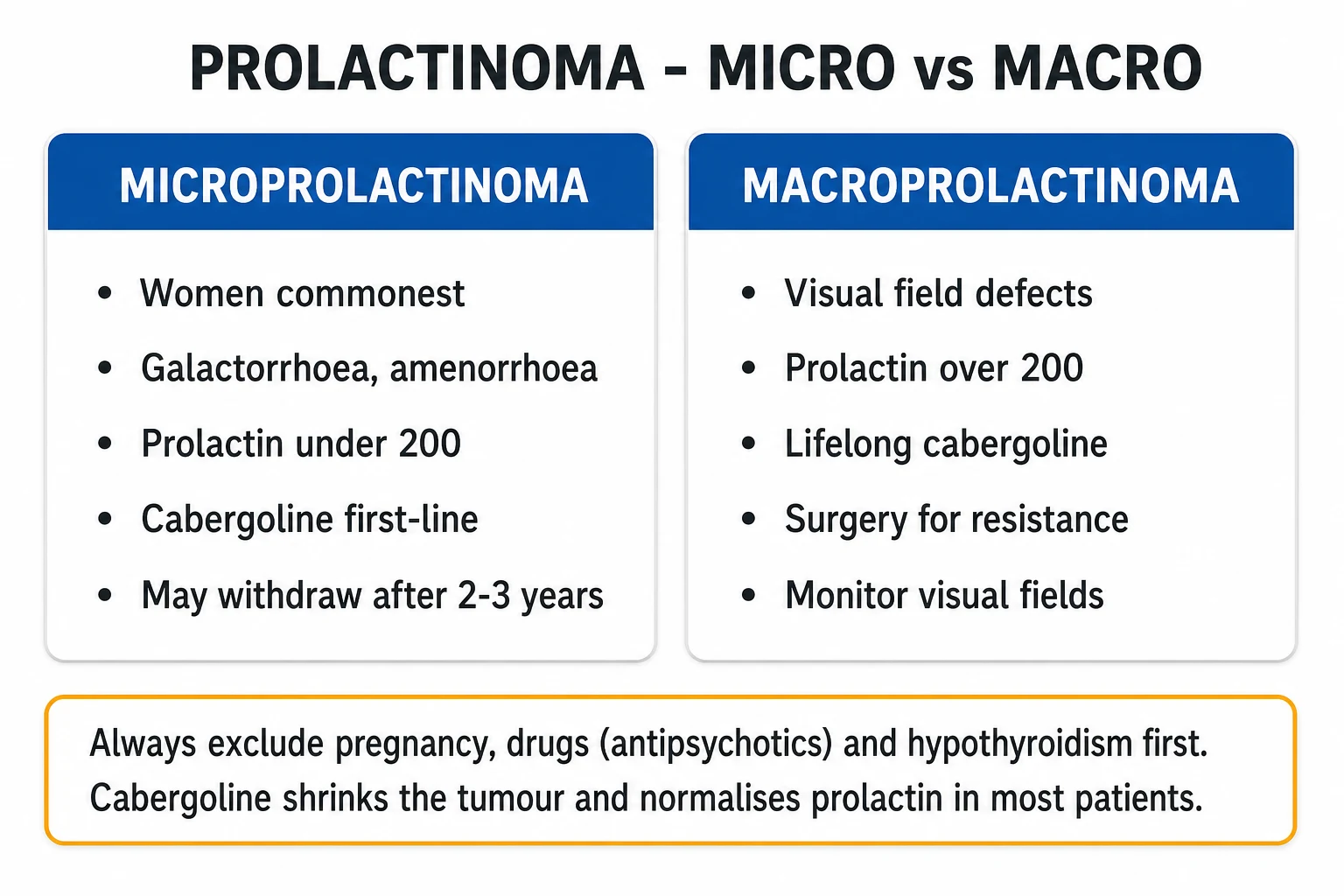
Microprolactinoma
under 10 mm
- Commonest form in reproductive-age women
- Prolactin moderately raised (variable)
- Mass effect uncommon
- Excellent response to dopamine agonist
- May remit; therapy sometimes withdrawn
- Pregnancy: stop DA, low risk of enlargement
Macroprolactinoma
10 mm or more
- Higher prolactin (often over 200 ng/mL)
- Mass effect: headache, hemianopia, CN palsies
- Hypopituitarism from compression
- Predominant form in men; presents later
- Long-term or lifelong therapy
- Pregnancy: continue DA, check visual fields each trimester
Aggressive / malignant
rare
- Rapid growth, cavernous sinus invasion
- Resistance to standard DA doses
- May need surgery + radiotherapy
- Temozolomide for carcinoma or refractory
- Monitor closely for regrowth
Macroprolactinaemia
bio-inactive complex
- Prolactin-IgG complex (big prolactin)
- High assay, few or no symptoms
- Excluded by PEG precipitation
- Not a tumour; do not image or treat
- May coexist with true prolactinoma
Epidemiology & Risk Factors
Prolactinoma is the commonest functioning pituitary adenoma. The clinically significant prevalence is around 6 to 10 per 100 000 population, with an annual incidence of 2 to 3 per 100 000. Autopsy and high-resolution MRI series show that subclinical pituitary microadenomas are common incidental findings (in roughly 10 percent or more of unselected scans), the great majority of which never declare themselves clinically.[1][2]
The sex distribution is sharply skewed by tumour size. Microprolactinomas show a strong female predominance of roughly 10 to 1 — women of reproductive age present early with the disruptive menstrual and fertility phenotype (amenorrhoea, galactorrhoea, infertility), so small tumours are caught. Macroprolactinomas have a more equal sex distribution (around 1 to 1), because the male phenotype — slowly progressive loss of libido and erectile dysfunction — is under-reported and under-investigated, so men present late with larger tumours, mass effect and visual loss. The mean age at diagnosis in women is the late 20s to mid-30s; in men it is the 40s to 50s, reflecting the diagnostic delay rather than a difference in tumour biology.[2][8]
Risk factors are largely genetic. Beyond the familial syndromes (MEN1, MEN4, FIPA / AIP, Carney complex, McCune-Albright), most sporadic prolactinomas carry no single environmental precipitant. Oestrogen exposure is a recognised but debated co-factor: pregnancy and exogenous oestrogen produce lactotroph hyperplasia, and very rare prolactinomas enlarge or first declare during pregnancy. Long-standing primary hypothyroidism (with elevated TRH stimulating lactotrophs) and chronic dopamine antagonist therapy can produce sustained hyperprolactinaemia but do not, in themselves, generate a lactotroph adenoma.[1]
Prolactinoma — key numbers
Pathophysiology
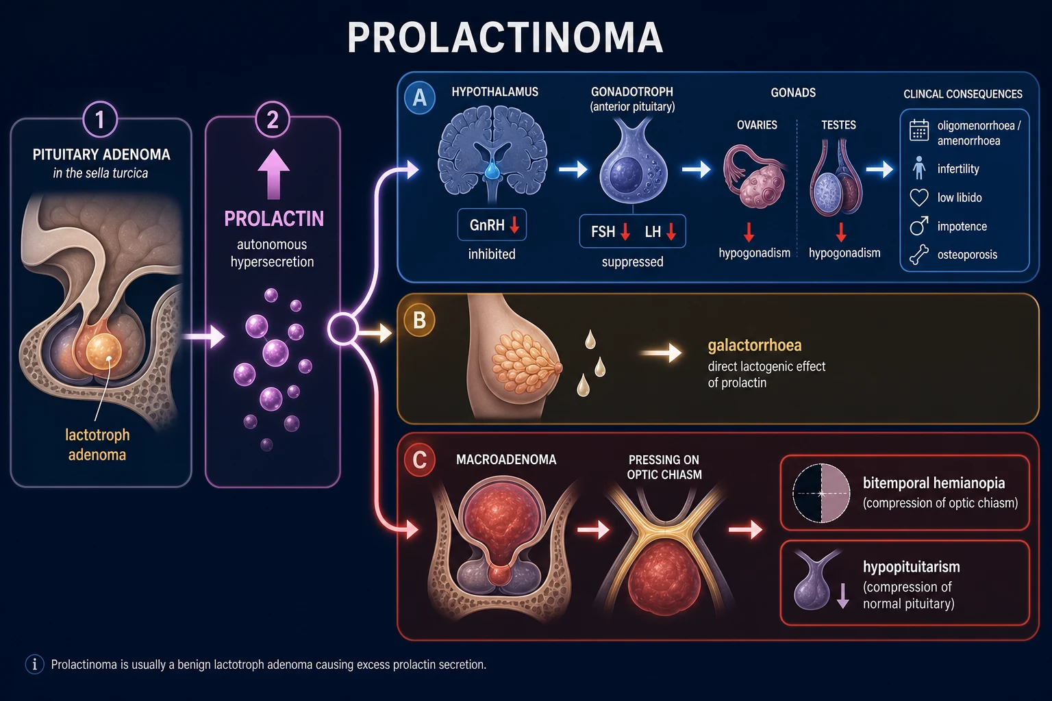
A prolactinoma is a monoclonal lactotroph adenoma — it arises from a single transformed lactotroph progenitor rather than from diffuse hyperplasia — whose cells escape the tonic dopamine inhibition that normally restrains prolactin secretion, and secrete prolactin autonomously. Understanding what the tumour overrides, what prolactin does once it is over-produced, and what happens as the tumour enlarges, explains every clinical feature.[1][2]
Normal physiology — what the tumour overrides. In health, the hypothalamus tonically inhibits the anterior pituitary lactotrophs via dopamine released into the hypophyseal portal system by tuberoinfundibular dopaminergic neurons. Dopamine acts at the D2 receptor on the lactotroph to suppress both prolactin synthesis (gene transcription) and release. Unlike the other anterior pituitary hormones, prolactin is under predominant inhibitory (dopaminergic) control — section of the pituitary stalk, with loss of dopamine delivery, causes prolactin to rise. TRH (thyrotropin-releasing hormone) and oestrogen stimulate prolactin secretion; suckling, stress, sleep and exercise all raise it transiently through hypothalamic pathways. The autonomous lactotroph adenoma ignores the dopaminergic brake; this is precisely why a D2 agonist (cabergoline, bromocriptine) is the rational first-line therapy.[1]
The endocrine consequences — hypogonadotropic hypogonadism. Sustained hyperprolactinaemia inhibits pulsatile hypothalamic GnRH secretion. The downstream effect is reduced pituitary FSH and LH, producing hypogonadotropic hypogonadism: in women, oligomenorrhoea or amenorrhoea, anovulation, infertility, reduced libido, vaginal dryness and dyspareunia; in men, reduced libido, erectile dysfunction, oligospermia and infertility, sometimes with gynaecomastia (prolactin suppresses testosterone but, less directly, also raises the oestrogen-to-testosterone ratio). Over years the chronic sex-steroid deficiency causes osteopenia or osteoporosis with increased fracture risk — a complication often under-recognised but reversible with treatment.[7]
The direct lactogenic effect — galactorrhoea. Prolactin acts directly on breast epithelium to drive milk synthesis and secretion. Galactorrhoea (the spontaneous or expressible milky nipple discharge) is therefore a cardinal feature, but its presence is not obligatory — many women with marked hyperprolactinaemia have no galactorrhoea, and men rarely do, because the breast requires a priming oestrogen milieu to mount a lactogenic response.[1]
Mass effect of a macroprolactinoma. As the tumour enlarges it compresses (in order of clinical importance): the optic chiasm superiorly (the classic bitemporal hemianopia, often beginning in the superior temporal quadrants because the inferior chiasmal fibres are compressed first from below), the surrounding normal pituitary (anterior pituitary failure in the order gonadotrophs, then thyrotrophs, then corticotrophs, then somatotrophs — hypopituitarism), the cavernous sinuses laterally (cranial nerves III, IV, V1, V2 and VI — ophthalmoplegia, facial sensory change), and, rarely, the hypothalamus (disturbance of appetite, temperature, consciousness) or the sphenoid sinus (CSF rhinorrhoea, with the attendant meningitis risk).[1]
The stalk effect — the great mimic. Any large non-functioning sellar mass — a non-functioning pituitary macroadenoma, craniopharyngioma, Rathke cleft cyst, or metastasis — can compress the pituitary stalk and interrupt tonic dopamine delivery to the normal lactotrophs. Without the dopamine brake, normal lactotrophs over-secrete prolactin, but only mildly to moderately — typically under 2000 mU/L (around 100 ng/mL), and almost always under 4000 mU/L. This is the "stalk effect" (also called the pituitary stalk section effect). The cardinal teaching is: in a true prolactinoma, the prolactin level parallels tumour size — a 3 cm tumour is expected to have a prolactin of many thousands of mU/L. A big tumour with a small prolactin rise is not a prolactinoma; it is a non-functioning mass with a stalk effect, and the treatment is surgery of the mass, not a dopamine agonist. Operating on a true macroprolactinoma on the assumption that it is a non-functioning mass is one of the classic — and dangerous — pitfalls in pituitary medicine, because a true macroprolactinoma will shrink with cabergoline and is highly vascular at surgery.[1][2]
Clinical Presentation
The clinical face of prolactinoma is a syndrome of reproductive and galactorrhoeic disturbance layered, in larger tumours, with mass effect and anterior pituitary failure. The presentation differs sharply between women (who present early with small tumours) and men (who present late with large tumours) — a sex gap that is the single most under-recognised feature of the disease.[1][8]
Women — the reproductive phenotype. The classic triad is galactorrhoea with oligomenorrhoea or amenorrhoea and infertility. The menstrual disturbance reflects hypogonadotropic hypogonadism: cycles become irregular, then cease (secondary amenorrhoea), with anovulation and infertility. Reduced libido and vaginal dryness (from oestrogen deficiency) cause dyspareunia. Galactorrhoea — bilateral, milky, non-bloody, often expressible rather than spontaneous — is the marker that brings many women to attention, though its absence does not exclude the diagnosis. Osteopenia or osteoporosis may declare as a fragility fracture in long-standing untreated disease. Many women present through subfertility services, where routine prolactin testing reveals the diagnosis.[1][7]
Men — the under-recognised phenotype. Men typically present with progressive loss of libido, erectile dysfunction and infertility (oligospermia) that are often attributed to ageing, stress or depression for years before prolactin is measured. Gynaecomastia is less common than in other causes of male hypogonadism (prolactin itself does not directly drive breast tissue growth; it suppresses testosterone, and the relative oestrogen excess is mild). Galactorrhoea is rare in men because the male breast lacks the oestrogen-primed ductal system. Because the sexual symptoms are under-reported and under-investigated, men present late, with macroprolactinomas and mass effect — headache, visual field loss, cranial nerve palsies — at a stage when a comparably symptomatic woman would have been diagnosed years earlier with a microadenoma. Visual field defect at presentation is far more common in men than in women.[8]
Mass effect (macroprolactinoma, either sex). Headache (stretch of the diaphragma sellae or raised intrasellar pressure), bitemporal visual field loss (optic chiasm compression — classically a bitemporal hemianopia beginning superiorly, progressing to a complete bitemporal hemianopia), cranial nerve palsies from cavernous sinus invasion (III — ptosis, ophthalmoplegia; IV, VI — diplopia; V1, V2 — facial sensory change), CSF rhinorrhoea if the tumour erodes through the sellar floor into the sphenoid sinus (with consequent meningitis risk), and signs of anterior pituitary failure (secondary hypothyroidism, secondary adrenal insufficiency, growth hormone deficiency, hypogonadism).[1]
Pituitary apoplexy. A macroprolactinoma may undergo haemorrhagic infarction — pituitary apoplexy — presenting as sudden severe headache, vomiting, rapidly progressive visual loss, ophthalmoplegia and, in severe cases, altered consciousness or meningism. This is an endocrine emergency (see Management — Resuscitation).[1]
Chronic consequences of untreated disease. Persistent hypogonadotropic hypogonadism causes osteoporosis and fragility fractures, dyslipidaemia and increased cardiovascular risk, sexual dysfunction, infertility, and psychological distress (from infertility, galactorrhoea and altered body image). Several of these — particularly the bone and cardiovascular effects — are only partially reversible even after biochemical cure, which is why early diagnosis and treatment matter.[7]
Atypical presentations. A prolactinoma may be an incidental finding on MRI done for an unrelated reason (head trauma, sinus disease) — the so-called incidentaloma with a stalk-effect or co-existent prolactin elevation. Adolescent presentation with delayed puberty or primary amenorrhoea raises the question of a germline AIP mutation (familial isolated pituitary adenoma). A very large tumour with a paradoxically low reported prolactin should raise the high-dose hook effect (see Investigations).[1]
Differential Diagnosis of Hyperprolactinaemia
The differential diagnosis of a raised serum prolactin is wide, because prolactin secretion is exquisitely sensitive to anything that lowers dopaminergic tone or raises TRH/oestrogen, and because the assay itself can be confounded. The clinical skill is to work through the categories in order — physiological, drug, systemic, stalk effect, macroprolactin, then a true prolactinoma — before committing a patient to imaging, a dopamine agonist, or surgery. The Endocrine Society guideline codifies this exclusion.[1]
Physiological causes (exclude first). Pregnancy and lactation are the dominant causes — always do a beta-hCG first in any woman of reproductive age with a raised prolactin. Stress (venepuncture, pain, anxiety), strenuous exercise, sleep (prolactin rises during the night and peaks in the early morning — hence a fasting morning resting sample), sexual intercourse and nipple stimulation all raise prolactin transiently. A single mildly raised sample should be repeated on a fasting, resting, non-stressed morning sample before hyperprolactinaemia is declared pathological.[1]
Drug-induced causes (the great mimic). Any drug that depletes dopamine or blocks the D2 receptor will raise prolactin, typically to under 2000 mU/L but occasionally higher. The classic culprits are antipsychotics (especially risperidone, also haloperidol, phenothiazines, but less so clozapine, quetiapine, aripiprazole — which can lower prolactin), antiemetics and prokinetics (metoclopramide, domperidone, prochlorperazine), antidepressants (SSRIs, TCAs, less commonly MAOIs), opioids, the calcium-channel blocker verapamil, methyldopa, reserpine, omeprazole, cimetidine, ranitidine, and high-dose oestrogens. The key teaching point: a drug cause must be excluded (or confirmed by withdrawal and re-testing where safe) before imaging or treatment — withdrawing an essential antipsychotic may be unsafe, in which case the diagnosis is one of exclusion plus imaging.[1]
Systemic causes. Primary hypothyroidism raises TRH, which stimulates lactotrophs — a commonly forgotten cause of mild-moderate hyperprolactinaemia, easily excluded with a TSH and free T4 and corrected by levothyroxine. Chronic kidney disease reduces prolactin clearance; cirrhosis does likewise. Chest wall lesions — herpes zoster (shingles), thoracotomy, mastectomy, severe burns, chronic nipple stimulation — activate the afferent limb of the suckling reflex and drive prolactin. Polycystic ovary syndrome is associated with mildly raised prolactin in a minority. Hypothalamic or infiltrative disease (sarcoidosis, histiocytosis, lymphocytic hypophysitis) can interrupt dopamine delivery.[1]
Macroprolactinaemia (the assay trap). A bio-inactive complex of monomeric prolactin bound to IgG ("big prolactin" or macroprolactin) is detected by the immunometric assay but produces few or no clinical symptoms — the patient with a "high" prolactin but normal menses, no galactorrhoea and no tumour. Macroprolactinaemia accounts for a substantial minority of incidental hyperprolactinaemia in the laboratory. The exclusion test is polyethylene glycol (PEG) precipitation: the sample is treated with PEG, which precipitates the high-molecular-weight complex; the residual monomeric prolactin in the supernatant gives the true bioactive concentration. Macroprolactin should be excluded before any tumour is sought or treated.[1]
Stalk effect from a sellar mass (the great surgical pitfall). A non-functioning pituitary macroadenoma, craniopharyngioma, Rathke cleft cyst, or metastasis compressing the pituitary stalk interrupts dopamine delivery, producing mild-moderate prolactin elevation (typically under 2000 mU/L, almost always under 4000 mU/L). This is not a prolactinoma — the treatment is surgery of the mass, not a dopamine agonist. The cardinal teaching: a big tumour with a small prolactin rise is a stalk effect, not a prolactinoma.[1]
Physiological
common, exclude first
- Pregnancy and lactation (beta-hCG first)
- Stress, exercise, sleep, sexual intercourse
- Nipple stimulation and chest wall lesions
- Resolve on repeat fasting resting sample
Drug-induced
dopamine blockade
- Antipsychotics — risperidone worst; haloperidol, phenothiazines
- Metoclopramide, domperidone, prochlorperazine
- SSRIs, TCAs, opioids
- Verapamil, methyldopa, reserpine, omeprazole, oestrogens
- Usually under 2000 mU/L; withdraw + re-test
Systemic
secondary causes
- Primary hypothyroidism (TRH stimulates prolactin)
- Chronic kidney disease (reduced clearance)
- Cirrhosis; PCOS (mildly raised)
- Chest wall — zoster, trauma, burns (suckling reflex)
- Infiltrative — sarcoidosis, histiocytosis, lymphocytic hypophysitis
Macroprolactin
bio-inactive complex
- Prolactin-IgG complex (big prolactin)
- High assay, few or no symptoms
- Exclude by PEG precipitation
- Not a tumour — do not image or treat
Stalk effect
non-functioning mass
- Large non-functioning pituitary adenoma compressing stalk
- Prolactin only mildly raised (usually under 2000 mU/L)
- Big tumour, small prolactin rise — treat the mass
- DA may lower prolactin but does not shrink the mass
True prolactinoma
lactotroph adenoma
- Prolactin parallels tumour size
- Over 4000 mU/L (200 ng/mL) essentially diagnostic
- DA shrinks the tumour — first-line therapy
- Macroprolactinoma often over 10000 mU/L
Clinical & Bedside Assessment
The bedside assessment is where the diagnosis is first suspected, and it rests on three moves: (1) take a meticulous reproductive, drug and symptom history, (2) examine the visual fields, breasts and cranial nerves, and (3) look for evidence of hypopituitarism and of the bone and metabolic consequences of chronic disease.[1]
History. In a woman of reproductive age with oligomenorrhoea, amenorrhoea, infertility or galactorrhoea, ask systematically: menstrual pattern (onset of oligomenorrhoea or amenorrhoea, previous regularity), galactorrhoea (spontaneous vs expressible, unilateral vs bilateral, milky vs bloody — bloody discharge points to breast pathology, not prolactin), fertility (time to conception, partner fertility), libido and sexual function, headache and visual symptoms (especially bumping into objects, indistinct peripheral vision), drug history (psychiatric medication, antiemetics, verapamil, methyldopa — and over-the-counter antiemetics), thyroid symptoms, renal or hepatic disease, chest wall lesions, and family history of endocrine tumours (parathyroid, pancreatic, pituitary — the MEN1 / FIPA clue). In a man, the same drug and mass-effect history, plus direct questioning about libido and erectile dysfunction (which men rarely volunteer).[1][8]
Examination. Inspect for galactorrhoea (compress the breast in all four quadrants; expressible milky discharge is confirmatory) and gynaecomastia (true glandular tissue, not fatty pseudogynaecomastia). Examine the visual fields by confrontation — a bitemporal hemianopia is detected by comparing the patient's field against your own across a table, finger-by-finger in each quadrant; any field defect mandates urgent MRI and formal perimetry. Examine the cranial nerves (III, IV, V1, V2, VI for cavernous sinus involvement), look for signs of hypothyroidism, hypocortisolism (postural drop, pigmentation), hypogonadism (testicular atrophy, loss of secondary sex hair), and osteoporotic deformity (kyphosis, loss of height). Fundoscopy may reveal optic atrophy in long-standing chiasmal compression.[1]
Bone density. A DEXA scan is part of the baseline workup in long-standing disease, because hypogonadal osteoporosis is common, often silent, and at least partially reversible with treatment.[7]
Investigations
The investigation of suspected prolactinoma is a four-step algorithm: (1) confirm genuine persistent hyperprolactinaemia and exclude the mimics, (2) screen for macroprolactin, (3) image the sella, (4) test the other pituitary axes and assess mass-effect consequences. Examiners expect the exact thresholds and the order of operations.[1]
Step 1 — Serum prolactin: the single best test
A single fasting morning serum prolactin, taken with the patient rested, non-stressed, and not recently exercised or slept, is the single best test. A persistently elevated level (on at least two samples if the rise is mild) confirms true hyperprolactinaemia. The thresholds that guide interpretation:[1]
- Under 1000 mU/L (about 50 ng/mL): usually physiological, drug-related, systemic, or macroprolactin — repeat and exclude causes before any imaging.
- 1000 to 4000 mU/L (about 50 to 200 ng/mL): indeterminate — could be a microprolactinoma, a stalk effect, a drug cause, or macroprolactin; image and exclude mimics.
- Over 4000 mU/L (about 200 ng/mL): essentially diagnostic of a macroprolactinoma, and the level parallels tumour size — a 3 cm tumour is expected to have a prolactin of many thousands of mU/L.
- Over 9400 mU/L (about 470 ng/mL): diagnostic of a macroprolactinoma; very high levels (over 10000 mU/L, over 500 ng/mL) imply a large macroprolactinoma.[1]
The cardinal rule: the prolactin level parallels tumour size in a true prolactinoma. A big tumour with a small prolactin rise is a stalk effect, not a prolactinoma — and operating on a true macroprolactinoma on that assumption is a serious surgical error.[1]
Step 2 — Exclude the mimics
Pregnancy — beta-hCG first, always, in any woman of reproductive age. Hypothyroidism — TSH and free T4 (primary hypothyroidism raises TRH and prolactin; replacement corrects both). Renal disease — U&E and creatinine (chronic kidney disease reduces prolactin clearance). Liver disease — LFTs. A careful drug history. Macroprolactin screen by polyethylene glycol (PEG) precipitation — if the post-PEG recovery is low (typically under 40 percent of total, with the monomeric prolactin in the normal range), the high assay is due to macroprolactin and the patient has no functional hyperprolactinaemia. Macroprolactin should be excluded before any tumour is sought or treated.[1]
Step 3 — Localise and size the tumour: pituitary MRI
A pituitary MRI with gadolinium is the imaging gold standard. It localises the adenoma, measures its greatest diameter (the micro/macro split), defines its relationship to the optic chiasm (contact, displacement, compression), assesses cavernous sinus invasion (the Knosp grade, grades 0 to 4 — grade 3 and 4 indicate cavernous sinus invasion and predict incomplete surgical resection), and looks for sphenoid sinus extension (CSF rhinorrhoea risk). If MRI is contraindicated, a contrast pituitary CT is a poor substitute. A microadenoma under 10 mm with a moderate prolactin elevation in the right clinical context confirms a microprolactinoma; a macroadenoma with a high prolactin (over 4000 mU/L) confirms a macroprolactinoma.[1]
Step 4 — Pituitary axis testing and visual fields
For any macroprolactinoma (and selectively for microprolactinomas), assess the other anterior pituitary axes for hypopituitarism: 9 am cortisol (or short Synacthen test), free T4 and TSH, LH and FSH with oestradiol (women) or testosterone (men), IGF-1, and a prolactin (already done). Visual field testing — Humphrey perimetry — is mandatory for any macroadenoma, any chiasmal contact on MRI, or any visual symptom; the classic defect is a bitemporal hemianopia, often beginning in the superior temporal quadrants. DEXA for bone density in long-standing disease.[1][7]
Dynamic tests — rarely needed
TRH stimulation, metoclopramide challenge, and nomifensine suppression tests were once used to discriminate prolactinoma from other causes; they are now rarely needed in routine practice because the combination of a markedly elevated prolactin, exclusion of mimics, and a pituitary MRI is usually diagnostic. They may have a role in the small indeterminate group with mild-moderate elevation and a normal MRI.[1]
The high-dose hook effect — the trap to remember
In very large macroprolactinomas with extremely high prolactin, the immunometric (sandwich) assay can be overwhelmed by antigen excess, producing a falsely low reported prolactin — the so-called high-dose hook effect (or prozone effect). The clue is a large tumour with a paradoxically modest prolactin: the prolactin looks too small for the tumour. The fix is to repeat the assay on a diluted sample (typically 1 to 100 or 1 to 1000), which restores the antibody-to-antigen ratio and unmasks the true (very high) prolactin. Missing the hook effect leads to the catastrophic error of operating on a macroprolactinoma as if it were a non-functioning mass.[1]
Interpreting serum prolactin (mU/L; multiply by ~0.047 for ng/mL)
Management — Resuscitation
Prolactinoma is overwhelmingly a chronic, elective condition; the diagnostic and treatment pathway is not a resuscitation. The one true emergency is pituitary apoplexy.[1]
Pituitary apoplexy presents with sudden severe headache, vomiting, rapidly progressive visual loss, ophthalmoplegia (cranial nerve III palsy) and, in severe cases, altered consciousness or meningism. The immediate management is:[1]
- Intravenous hydrocortisone 100 to 200 mg stat, then 50 to 100 mg every 6 to 8 hours — because acute cortisol deficiency (corticotroph destruction) is the immediate threat to life. Do not wait for a cortisol result before giving steroids.
- Intravenous fluids and haemodynamic support.
- Urgent pituitary MRI to confirm apoplexy and define the haemorrhage.
- Urgent transsphenoidal surgical decompression if there is deteriorating vision, ophthalmoplegia or depressed consciousness; conservative management with close monitoring is acceptable in clinically stable patients without a visual deficit.
- Endocrine and ophthalmology review.[1]
A patient who presents in acute adrenal crisis from hypopituitarism (hypotension, hyponatraemia, hypoglycaemia, confusion) needs IV hydrocortisone, fluids and IV glucose before any prolactinoma-specific therapy; the prolactinoma itself is treated once the patient is stabilised. A patient with CSF rhinorrhoea from a macroprolactinoma eroding the sphenoid floor needs urgent neurosurgical review (risk of ascending meningitis) — a dopamine agonist will shrink the tumour but may worsen the leak as the tumour shrinks away from the defect.[1]
Management — Definitive & Stepwise
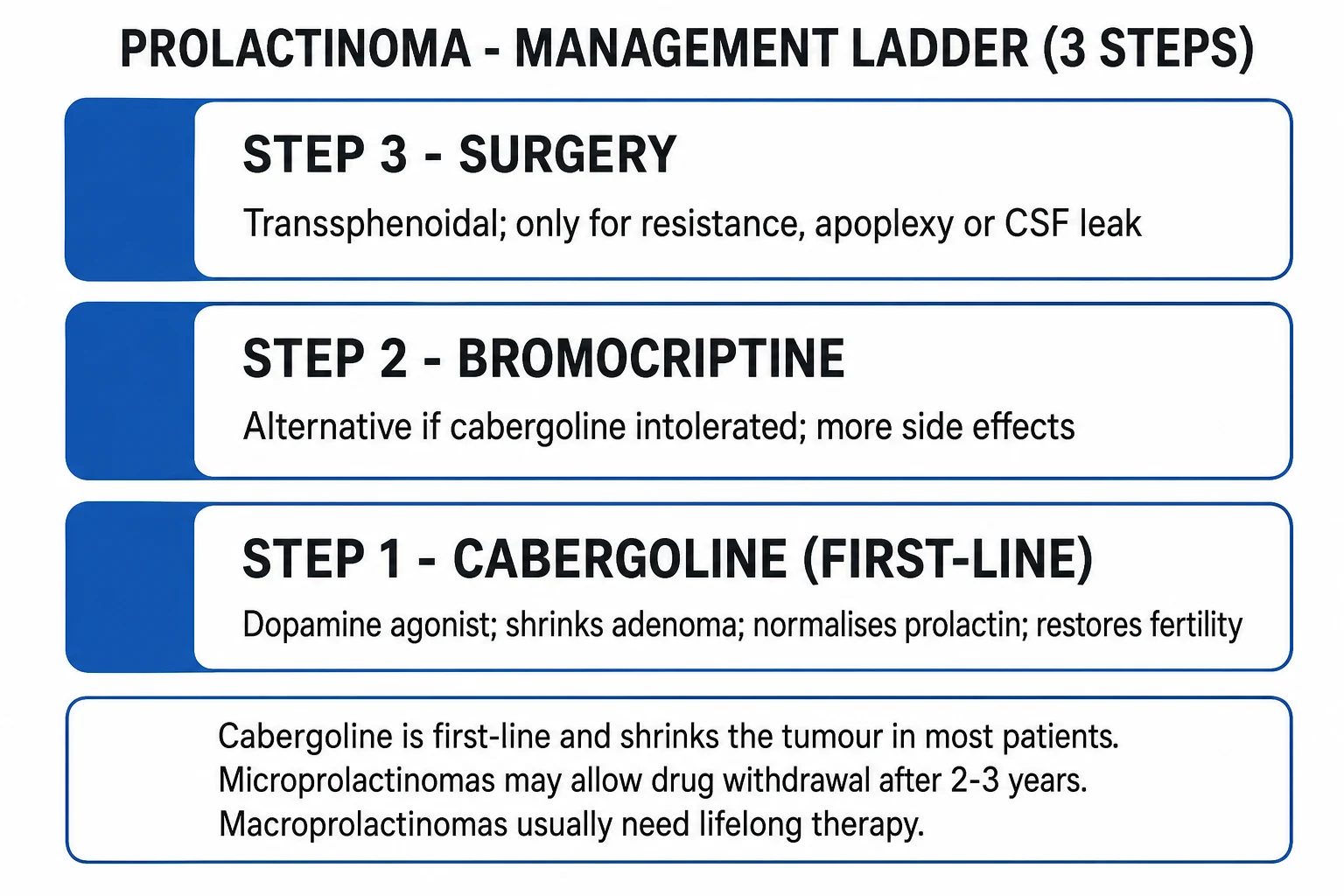
The treatment of prolactinoma is built around four modalities — dopamine agonists (first-line), transsphenoidal surgery (selective), radiotherapy (rare), and temozolomide (rare, for aggressive/malignant disease) — layered onto treatment of the cause (drug withdrawal, hypothyroid replacement), replacement of deficient pituitary axes, and lifelong surveillance. The aim is biochemical normalisation of prolactin, restoration of gonadal function and fertility, tumour shrinkage with relief of mass effect, and preservation of pituitary function.[1][2]
1. Dopamine agonists — first-line, for both micro- and macroprolactinoma
A dopamine D2 receptor agonist is first-line therapy for virtually all prolactinomas, micro and macro, with or without mass effect — this sets prolactinoma apart from every other pituitary tumour, where surgery is usually first. The D2 agonist restores the dopaminergic brake that the tumour has escaped, suppressing prolactin synthesis and secretion, shrinking the tumour (in 60 to 90 percent of macroprolactinomas), relieving mass effect and visual field defects often within weeks, restoring gonadal function and fertility, and stopping galactorrhoea. Even a macroprolactinoma with a complete bitemporal hemianopia is treated medically first, with visual fields monitored closely — the tumour usually shrinks away from the chiasm.[1][2]
Cabergoline is the preferred first-line agent because it is more efficacious at normalising prolactin and shrinking tumour, longer-acting (twice-weekly dosing), and better tolerated than bromocriptine. The starting dose is 0.25 mg once or twice weekly, titrated by prolactin response to a typical maintenance dose of 0.5 to 1 mg once or twice weekly (most patients are controlled on a total weekly dose of 0.5 to 2 mg; standard prolactinoma doses do not exceed around 2 mg per week).[1][2]
Bromocriptine (2.5 mg once daily, titrated by prolactin response up to 2.5 to 15 mg daily in divided doses with food) is the alternative, with the largest body of pregnancy safety data — its use in prolactinoma predates cabergoline by two decades. It is shorter-acting and less well tolerated (more nausea, postural hypotension, nasal congestion), but is preferred in some centres for women seeking pregnancy, particularly in the first trimester, where the safety database is largest. Quinagolide (75 micrograms daily, titrated to 75 to 150 micrograms) is a non-ergot D2 agonist available in some markets.[1]
How to start a dopamine agonist safely. Begin at a low dose at night with food to minimise nausea and postural hypotension (cabergoline 0.25 mg once weekly at night; bromocriptine 1.25 mg at night), and titrate upward at 2 to 4 week intervals guided by prolactin. Warn the patient about nausea, dizziness, postural hypotension, nasal congestion, headache and fatigue (mostly dose-related and transient), and — particularly with bromocriptine at first dose — about the rare hypotensive collapse. Warn sexually active women that ovulation and fertility may return within weeks — advise contraception if pregnancy is not desired.[1]
Expected outcomes. Cabergoline normalises prolactin in roughly 80 to 90 percent of microprolactinomas and 70 to 80 percent of macroprolactinomas, and shrinks the tumour in 60 to 90 percent of macroprolactinomas — often visibly on MRI within 6 to 12 weeks, with continued shrinkage over months. Visual field defects improve in the great majority of macroadenomas with chiasmal compression. Gonadal function and fertility are restored in most. Galactorrhoea stops within weeks.[2]
2. Transsphenoidal surgery — selective second-line (or first-line in selected patients)
Endoscopic transsphenoidal surgery by an experienced pituitary neurosurgeon is reserved for:[3]
- Dopamine-agonist resistance (prolactin fails to normalise and tumour fails to shrink on maximal tolerated doses).
- Dopamine-agonist intolerance (intractable nausea, postural hypotension, psychiatric effects, impulse control disorder).
- Pituitary apoplexy with visual compromise.
- CSF rhinorrhoea (the leak may worsen as the tumour shrinks on a dopamine agonist, so surgical repair is preferred).
- Optic chiasm compression unresponsive to medical therapy (rare — most respond within weeks).
- Patient choice after balanced counselling.
- Pregnancy in a woman with a macroprolactinoma that is enlarging despite dopamine agonist (surgery in the second trimester is an option).
- Atypical/aggressive tumour with cystic component that may not shrink on a dopamine agonist.[3]
Surgical remission rates are around 60 to 90 percent for microprolactinomas but lower (around 30 to 50 percent) for macroprolactinomas, especially those with cavernous sinus invasion. A 2020 systematic review and meta-analysis by Zamanipoor and colleagues** reopened the debate about surgery as a viable first-line alternative** in selected microprolactinomas and small macroprolactinomas, with comparable long-term remission and lower relapse than after dopamine-agonist withdrawal — but medical therapy remains the international standard first-line, with surgery reserved for the indications above. Complications — in expert hands — include hypopituitarism, diabetes insipidus, CSF leak, meningitis and cranial nerve injury.[3]
3. Radiotherapy / stereotactic radiosurgery — third-line
Stereotactic radiosurgery (gamma knife) or fractionated stereotactic radiotherapy is reserved for refractory or residual disease after medical and surgical treatment, or for patients who cannot tolerate or adhere to long-term medical therapy. Drawbacks include a slow onset of biochemical effect (months to years), the risk of hypopituitarism (the commonest late complication, mandating lifelong axis surveillance), and small long-term risks of cerebrovascular disease and second tumours. Radiotherapy does not replace a dopamine agonist in the immediate control of a macroprolactinoma with mass effect.[1][2]
4. Temozolomide — for aggressive and malignant prolactinoma
The rare aggressive or invasive prolactinoma resistant to dopamine agonist, surgery and radiotherapy, and pituitary carcinoma (with craniospinal or distant metastasis), may respond to temozolomide — an oral alkylating agent given as 150 to 200 mg per square metre daily for 5 days of every 28-day cycle, assessed over 3 to 6 cycles. Hagen and colleagues reported tumour shrinkage and prolactin fall in resistant macroadenomas and a pituitary carcinoma. MGMT promoter methylation in the tumour predicts response. This is a specialist decision in a pituitary tumour centre.[5]
5. Treat the cause and replace deficient axes
Withdraw offending drugs where clinically safe; an antipsychotic cannot always be stopped, in which case the hyperprolactinaemia is managed expectantly (with imaging if symptoms or prolactin warrant) or with a dopamine agonist in liaison with the treating psychiatrist. Replace levothyroxine for primary hypothyroidism. Replace cortisol (hydrocortisone 15 to 25 mg daily in divided doses; or prednisolone 3 to 5 mg daily) for ACTH deficiency — always before thyroid replacement, to avoid precipitating an adrenal crisis. Give oestrogen (women) or testosterone (men) replacement if gonadal function does not recover on a dopamine agonist, after contraception/pregnancy considerations are addressed.[1][7]
Withdrawal of dopamine agonist
In microprolactinomas with normalised prolactin and a shrunken or absent tumour on MRI after 2 to 5 years of therapy, a structured withdrawal can be attempted — prolactin and MRI are monitored, and roughly 30 to 40 percent of microprolactinomas remain in remission. Macroprolactinomas almost always relapse on withdrawal and need long-term or lifelong therapy. Withdrawal should be supervised by an endocrinologist.[1][6]
Exam application bank (NEET-PG / INICET)
One-line answer
Prolactinoma is a benign pituitary lactotroph adenoma that autonomously secretes prolactin — the most common hormonally active pituitary tumour, accounting for around 40 to 45 percent of functioning pituitary adenomas. Hyperprolactinaemia inhibits hypothalamic GnRH, lowering FSH and LH and producing hypogonadotropic hypogonadism: in women oligomenorrhoea or amenorrhoea, galactorrhoea, infertility and low libido; in men reduced libido, erectile dysfunction and infertility. A macroprolactinoma (10 mm or more) adds mass effect — bitemporal hemianopia (optic chiasm compression), hypopituitarism and cranial nerve palsies. Diagnosis rests on a fasting serum prolactin (over 4000 mU/L or 200 ng/mL essentially diagnostic), exclusion of pregnancy, drugs, hypothyroidism and renal failure, a macroprolactin screen, and a pituitary MRI. First-line treatment is medical — a dopamine agonist, cabergoline
Worked stems (answer without another resource)
Stem 1 — Classic presentation. Map symptoms to mechanism; name the first investigation and first treatment step with dose/route if drug therapy is standard. [1]
Stem 2 — Unstable / complicated. List red flags that force immediate resuscitation, theatre, ICU, antidote, or reperfusion — and what you do in the first 15 minutes. [1]
Stem 3 — Atypical group. Elderly, pregnancy, child, or immunocompromised: how presentation and thresholds change. [1]
Stem 4 — Differential trap. Name the three closest mimics and one discriminator for each. [1]
Stem 5 — Disposition. Who goes home with safety-netting, who is admitted, who needs HDU/ICU/theatre, and what follow-up is mandatory. [1]
Rapid viva checklist
- Definition + classification
- Pathophysiology chain
- Bedside signs / criteria
- Score with exact components (if any)
- Emergency bundle
- Definitive therapy with doses
- Complications of disease and of treatment
- Special populations
- Guideline/trial name if classic
- Three exam traps
Coverage self-check
If you cannot answer any stem above from this page alone, re-read the matching section — the page is intended to be self-sufficient for final-prof and NEET-PG/INICET questions on Prolactinoma.
Prolactinoma surveillance timeline
Specific Subtypes & Scenarios
Microprolactinoma. The classic patient is a young woman with oligomenorrhoea or amenorrhoea, galactorrhoea and infertility. A prolactin of 1000 to 5000 mU/L with a microadenoma under 10 mm on MRI confirms the diagnosis. Cabergoline is first-line, with excellent response; after 2 to 5 years of remission, a supervised withdrawal succeeds in roughly a third. Mass effect is rare. Pregnancy management is conservative (see Special Populations).[1]
Macroprolactinoma. Higher prolactin (often over 4000 mU/L, frequently over 10000 mU/L), with mass effect (headache, bitemporal hemianopia, cranial nerve palsies, hypopituitarism, occasionally CSF rhinorrhoea or apoplexy). Cabergoline is still first-line, even with visual loss — most macroadenomas shrink within weeks and visual fields improve. Cavernous sinus invasion (Knosp grade 3 to 4) predicts incomplete surgical resection if surgery is later needed. Long-term or lifelong therapy is the rule; relapse on withdrawal is the norm.[2]
Aggressive, invasive and malignant prolactinoma. A small subset behave aggressively — rapid growth, cavernous sinus or sphenoid invasion, resistance to standard dopamine-agonist doses. Management is dose escalation of cabergoline (under specialist supervision), surgical debulking, stereotactic radiotherapy, and, for true resistance or pituitary carcinoma (the rarest form, with craniospinal or distant metastasis), temozolomide (150 to 200 mg per square metre daily for 5 days of a 28-day cycle). MGMT promoter methylation predicts response. Pegvisomant and pasireotide have no role in pure prolactinoma.[5]
Idiopathic hyperprolactinaemia. Persistent mild-moderate prolactin elevation with no tumour on MRI and no identifiable cause. Treat with a dopamine agonist if symptomatic; some evolve into a visible microadenoma on later imaging.[1]
Macroprolactinaemia. High assay prolactin with no symptoms and no tumour; excluded by PEG precipitation. No treatment, no imaging beyond the exclusion; reassure. May coexist with a true prolactinoma, so do not dismiss a symptomatic patient solely on the basis of a "macroprolactin" result.[1]
Stalk effect from a non-functioning macroadenoma. Mild-moderate prolactin elevation (typically under 2000 mU/L) from a large non-functioning mass compressing the stalk. Treat the mass with surgery — a dopamine agonist will lower the prolactin but will not shrink the non-functioning tumour, and so will not relieve the mass effect.[1]
Complications & Pitfalls
The complications of prolactinoma arise from the disease itself (chronic hypogonadism, mass effect, apoplexy) and from its treatment (dopamine-agonist adverse effects, surgical and radiotherapy complications). Several are silent and only partially reversible, which is why early diagnosis and treatment matter.[1][7]
Disease-related — chronic hypogonadism. Persistent hypogonadotropic hypogonadism causes osteoporosis and fragility fractures (often silent until a fracture; partially reversible with treatment, but established bone loss may not fully recover), dyslipidaemia and increased cardiovascular risk, sexual dysfunction (loss of libido, erectile dysfunction, anorgasmia), infertility, and psychological distress from galactorrhoea, infertility and altered body image. These are not trivial — they materially affect quality of life and (for the bone and cardiovascular effects) longevity.[7]
Disease-related — mass effect. Bitemporal hemianopia (may become permanent if long-standing), cranial nerve palsies (III, IV, V1, V2, VI from cavernous sinus invasion), hypopituitarism (gonadotrophs first, then thyrotrophs, then corticotrophs, then somatotrophs), CSF rhinorrhoea (with meningitis risk), pituitary apoplexy (the emergency), and, rarely, hypothalamic disturbance.[1]
Dopamine-agonist adverse effects. Common and dose-related: nausea, vomiting, abdominal cramps, postural hypotension, nasal congestion, headache, dizziness, fatigue, insomnia. Less common: vivid dreams, anxiety, depression, hallucinations, impulse control disorder (pathological gambling, hypersexuality, compulsive shopping — particularly with cabergoline; warn patients and ask directly at review), CSF rhinorrhoea (as a shrunken macroprolactinoma pulls away from a pre-existing sphenoid floor defect), pericardial, pleural or retroperitoneal fibrosis and cardiac valve regurgitation (at high cumulative doses, as in Parkinson's disease — much less of a concern at standard prolactinoma doses, but a baseline echocardiogram is recommended for patients on long-term or higher-dose cabergoline).[4]
Surgical complications. Hypopituitarism, diabetes insipidus (usually transient), CSF leak, meningitis, cranial nerve injury, nasal complications. Expert pituitary surgeons have low complication rates.[3]
Radiotherapy complications. Hypopituitarism (the commonest late effect, requiring lifelong axis surveillance), cerebrovascular disease and second brain tumours (small long-term risks), optic neuropathy (avoided by keeping the dose to the chiasm low).[1]
Diagnostic pitfalls. (1) Missing the hook effect — a very large macroprolactinoma with a falsely low prolactin; always dilute the sample if the tumour looks much bigger than the prolactin implies. (2) Treating a stalk-effect mass with a dopamine agonist instead of surgery — the prolactin falls, the tumour does not, and the mass effect worsens. (3) Imaging and treating macroprolactinaemia — high assay, no symptoms, no tumour; exclude with PEG. (4) Forgetting a drug cause — risperidone, metoclopramide, verapamil, methyldopa. (5) Forgetting pregnancy — beta-hCG first, always. (6) Forgetting primary hypothyroidism — check TSH.[1]
Cabergoline in prolactinoma — what to monitor
Prognosis & Disposition
With cabergoline, prolactin normalises and the tumour shrinks in the great majority of patients, fertility is restored, mass effect is relieved, and quality of life returns to normal. Microprolactinomas have an excellent prognosis; some undergo spontaneous remission, and after years of stable remission a supervised withdrawal succeeds in around a third. Macroprolactinomas usually require long-term or lifelong dopamine-agonist therapy and surveillance imaging, but the prognosis for vision and fertility is excellent if the tumour shrinks as expected. Untreated or refractory disease causes persistent infertility, osteoporosis with fragility fractures, and progressive visual loss — much of it preventable with early treatment.[1][6]
Disposition is long-term endocrine follow-up in a multidisciplinary pituitary service (endocrinology, with neurosurgery, ophthalmology, reproductive medicine and (rarely) radiation oncology as needed). The surveillance schedule is prolactin every 3 to 6 months, MRI annually for macroadenomas until stable (every 1 to 2 years for microadenomas), Humphrey perimetry for any macroadenoma, annual pituitary axis testing, and DEXA at baseline.[1]
Special Populations
Pregnancy — the central scenario. Oestrogen-driven lactotroph hyperplasia means any prolactinoma may enlarge during pregnancy, and the risk is size-dependent. Microprolactinoma: risk of symptomatic tumour enlargement is low — under 5 percent. Stop the dopamine agonist once pregnancy is confirmed; monitor clinically (headache, visual symptoms), with MRI only if symptoms develop and visual fields only if symptomatic. Macroprolactinoma: risk of symptomatic enlargement is substantial — over 20 percent. Two strategies: (a) continue bromocriptine or cabergoline through pregnancy, with visual fields each trimester and MRI if symptoms or field defect develop; or (b) stop the dopamine agonist and monitor closely, restarting if there is any growth or symptomatic enlargement. Bromocriptine has the largest pregnancy safety database and is the agent many centres prefer pre-conception and in the first trimester; cabergoline has growing safety data and is increasingly used. If a macroprolactinoma enlarges despite a dopamine agonist and threatens vision, transsphenoidal surgery in the second trimester is an option. Breastfeeding is generally safe after delivery; the dopamine agonist can be resumed post-partum if needed, and is held during breastfeeding if the tumour is stable.[1]
Fertility restoration — the warning to give. Cabergoline restores ovulation rapidly — warn sexually active women that fertility may return within weeks of starting therapy, and advise contraception if pregnancy is not desired. For a woman seeking pregnancy, cabergoline (or bromocriptine) is continued until conception, then stopped (micro) or continued (macro) as above. Mechanical barrier contraception is preferred over the combined oral contraceptive (which can raise prolactin in some, and obscures the return of menses as a marker of disease control).[1]
Men. Present late, with macroprolactinomas and mass effect. Cabergoline normalises prolactin, raises testosterone, restores libido and erectile function, and improves fertility; testosterone replacement is reserved for persistent hypogonadism after prolactin normalisation. Bone density should be checked and osteoporosis treated. Persistent hypogonadism despite normalised prolactin raises the question of a co-existent primary testicular disorder or irreversible gonadotroph damage from long-standing mass effect.[8]
Adolescents. A prolactinoma in an adolescent (especially a macroadenoma in a young man) raises the question of a germline AIP mutation (familial isolated pituitary adenoma — FIPA), and genetic testing is advised. Cabergoline is first-line; surgery for resistance or apoplexy. The implications for fertility and lifelong bone health are addressed proactively.[2]
Elderly. May present with visual loss, headache or hypopituitarism from a long-standing macroprolactinoma; the galactorrhoea and menstrual features are absent (post-menopausal) or attributed to ageing. Cabergoline remains first-line; comorbidities (cardiac valve disease, postural hypotension) influence drug choice.[1]
Renal or hepatic disease. Cabergoline and bromocriptine are hepatically metabolised and need dose caution in hepatic impairment; in significant renal disease, prolactin is elevated by reduced clearance and the threshold for imaging is higher. Domperidone (no longer available in some markets for cardiac reasons) and metoclopramide are commoner drug causes in renal patients.[1]
Evidence, Guidelines & Regional Differences
The 2011 Endocrine Society clinical practice guideline on hyperprolactinaemia (Melmed, Casanueva and colleagues) remains the practice-defining document: it codified the diagnostic exclusion of pregnancy, drugs, hypothyroidism, renal failure and macroprolactin, established dopamine agonists (cabergoline first-line) as the cornerstone of therapy, set the threshold for essentially diagnostic prolactin, and laid out the pregnancy management algorithm by tumour size. It remains the international standard reference.[1] The Gillam 2006 Endocrine Reviews review (prolactinoma treatment advances) underpins much of the cabergoline-versus-bromocriptine efficacy and tolerability evidence.[2] The Zamanipoor 2020 JCEM meta-analysis reopened the debate on surgery as a viable first-line alternative in selected patients, with comparable long-term remission and lower relapse than after dopamine-agonist withdrawal — but medical therapy remains the international standard.[3] The Valassi 2010 JCEM clinical review quantified the cardiac valve risk of dopamine agonists, which is dose-dependent (high in Parkinson's-disease doses, low at standard prolactinoma doses) and informs the baseline echocardiogram recommendation for long-term or higher-dose cabergoline.[4] The Hagen 2009 EJE paper established temozolomide for resistant/malignant prolactinoma.[5] Recent reviews by Lee (2024), Uygur (2024) on bone comorbidity and Rudman (2025) on male hypogonadism consolidate the tailored-therapy, bone-protection and male-presentation agendas.[6][7][8]
Some European centres (notably the Dutch group behind the Zamanipoor meta-analysis) favour surgery as a first-line alternative for selected micro- and small macroprolactinomas, particularly where lifelong dopamine-agonist therapy and surveillance are burdensome. In resource-variable settings (including much of India), cabergoline (cheap, oral, highly effective) remains the workhorse first-line, with the Endocrine Society algorithm followed; bromocriptine is still used where cabergoline is unaffordable or unavailable. There is no ICMR prolactinoma-specific guideline. Temozolomide is reserved for the rare aggressive/malignant case in specialist centres.[1][3]
Where the evidence is weak. The exact threshold for "essentially diagnostic" (200 vs 250 ng/mL) is consensus-dependent; the role of routine surgery in microprolactinoma is debated (Zamanipoor and colleagues make a strong case, but international practice remains medical); the timing and necessity of echocardiographic screening at standard prolactinoma doses of cabergoline is unresolved (the valve risk is low but not zero); the place of temozolomide in non-malignant aggressive prolactinoma is being defined; and the optimal duration of therapy before a withdrawal attempt in microprolactinoma is individualised.[3][6]
Exam Pearls
Prolactinoma is one of the most patterned topics in endocrinology — the cabergoline-first message, the female-to-male presentation gap, the stalk-effect versus true prolactinoma distinction, and the pregnancy algorithm recur across NEET-PG, INICET, USMLE and PLAB.[1]
The definitional one-liner: a benign lactotroph pituitary adenoma that autonomously secretes prolactin (the commonest functioning pituitary tumour). Prolactin inhibits GnRH → low FSH/LH → hypogonadotropic hypogonadism (amenorrhoea, galactorrhoea, infertility, low libido, erectile dysfunction) plus a direct lactogenic effect (galactorrhoea); a macroprolactinoma compresses the optic chiasm (bitemporal hemianopia) and normal pituitary (hypopituitarism).[1]
The diagnostic quartet: (1) fasting serum prolactin (over 4000 mU/L / 200 ng/mL essentially diagnostic; level parallels tumour size); (2) exclude pregnancy (beta-hCG), drugs, hypothyroidism, renal failure, macroprolactin; (3) pituitary MRI to size and localise; (4) Humphrey perimetry + anterior pituitary axes for any macroadenoma.[1]
The management ladder is fixed and examinable: cabergoline first-line (0.25 to 1 mg once or twice weekly), bromocriptine as alternative (most pregnancy safety data), surgery for resistance, intolerance, apoplexy, CSF leak or chiasmal compression, radiotherapy third-line, temozolomide for aggressive/malignant.[2]
Key contrasts the exam rewards: cabergoline is first-line for prolactinoma (medical) versus surgery first-line for acromegaly, Cushing's disease and non-functioning macroadenoma. Prolactin parallels tumour size in a true prolactinoma versus the stalk effect (big tumour, small prolactin, treat the mass). Macroprolactinaemia = high assay, no symptoms, excluded by PEG, not a tumour. Hook effect = very large tumour with falsely low prolactin; dilute the sample. Men present late with macroadenomas; women present early with microadenomas. Pregnancy: stop DA for micro, continue for macro with visual fields each trimester.[1][8]
The single most quotable fact: prolactinoma is the one pituitary tumour treated medically first — cabergoline normalises prolactin, shrinks the tumour and restores fertility, even for a macroprolactinoma with visual loss.[2]
Quick self-test — cover and answer
Q1. A 28-year-old woman presents with secondary amenorrhoea, galactorrhoea and infertility. What is the first test, and why? — Always a beta-hCG (pregnancy) first; then serum prolactin. Q2. Her prolactin is 4500 mU/L (about 225 ng/mL). What does that level mean? — Essentially diagnostic of a macroprolactinoma; image the pituitary. Q3. MRI shows a 12 mm pituitary adenoma with chiasmal contact and a bitemporal hemianopia. What is the first-line treatment? — Cabergoline — medical therapy first, even with visual loss; most macroprolactinomas shrink within weeks. Q4. A 45-year-old man has a 4 cm pituitary mass, bitemporal hemianopia and a prolactin of 1800 mU/L (about 90 ng/mL). Is this a prolactinoma? — No — this is a stalk effect from a non-functioning macroadenoma. Big tumour, small prolactin. Treat the mass with surgery, not a dopamine agonist. Q5. A 35-year-old man has a 3 cm macroprolactinoma with a reported prolactin of 1500 mU/L. What should you do? — Suspect the high-dose hook effect; repeat the assay on a diluted sample. Q6. She conceives on cabergoline. How does management differ for a micro- versus a macroprolactinoma? — Micro: stop DA once pregnant, low risk of enlargement (under 5 percent). Macro: continue DA and check visual fields each trimester (over 20 percent risk of symptomatic enlargement). Q7. Long-standing untreated prolactinoma in a woman — what silent complication must you screen for? — Osteoporosis (DEXA) — partially reversible with treatment.
Causes of hyperprolactinaemia — PROLACTIN
PROLACTIN
always do a beta-hCG first in a woman of reproductive age
reduced prolactin clearance; also cirrhosis
antipsychotics, metoclopramide, domperidone, SSRIs, opioids, verapamil, methyldopa
the true prolactinoma — prolactin parallels tumour size
hypothyroidism raises TRH which stimulates prolactin
shingles, trauma, nipple stimulation via the suckling reflex
non-functioning sellar mass compressing the stalk interrupts dopamine
sarcoidosis, histiocytosis, lymphocytic hypophysitis
bio-inactive prolactin-IgG complex — high assay, few symptoms; exclude with PEG
Cabergoline side effects — CABERGOLINE
CABERGOLINE
at high cumulative doses (Parkinson's); baseline echo for long-term use
first-dose syncope — start at night with food
impulse control disorder — gambling, hypersexuality; ask directly
dose-related; titrate slowly with food
rare; chest imaging if symptomatic
the desired effect — within weeks
warn about fertility returning; advise contraception
testosterone and oestradiol recover as prolactin falls
pathological gambling, hypersexuality — cabergoline > bromocriptine
common and usually transient
high doses; reduce or switch
References
- [1]Melmed S, Casanueva FF, Hoffman AR, Kleinberg DL, Montori VM, Schlechte JA, Wass JA. Diagnosis and treatment of hyperprolactinemia: an Endocrine Society clinical practice guideline J Clin Endocrinol Metab, 2011.PMID 21296991
- [2]Gillam MP, Molitch ME, Lombardi G, Colao A. Advances in the treatment of prolactinomas Endocr Rev, 2006.PMID 16705142
- [3]Zamanipoor Najafabadi AH, Zandbergen IM, de Vries F, Broersen LHA, van den Akker-van Marle ME, Pereira AM, et al. Surgery as a Viable Alternative First-Line Treatment for Prolactinoma Patients. A Systematic Review and Meta-Analysis J Clin Endocrinol Metab, 2020.PMID 31665485
- [4]Valassi E, Klibanski A, Biller BM. Clinical Review#: Potential cardiac valve effects of dopamine agonists in hyperprolactinemia J Clin Endocrinol Metab, 2010.PMID 20130078
- [5]Hagen C, Schroeder HD, Hansen S, Andersen M. Temozolomide treatment of a pituitary carcinoma and two pituitary macroadenomas resistant to conventional therapy Eur J Endocrinol, 2009.PMID 19654234
- [6]Lee MH, Hong JW, Kim K, Ku CR. Elucidating Clinical Queries for Tailored Therapy in Patients with Prolactinoma Endocrinol Metab (Seoul), 2024.PMID 39397514
- [7]Uygur MM, Menotti S, Santoro S, Giustina A. Modern approach to bone comorbidity in prolactinoma Pituitary, 2024.PMID 39541075
- [8]Rudman Y, Shimon I. Hypogonadism in men with prolactinoma: Diagnosis, treatment, and management of persistent hypogonadism Vitam Horm, 2025.PMID 40812946