Endocrinology · General Medicine
Thyroid Nodules & Thyroid Cancer
Also known as Thyroid nodule · Thyroid cancer · Papillary thyroid cancer · Differentiated thyroid cancer
Thyroid nodules are very common but mostly benign — palpable in around 5 percent of adults and seen on ultrasound in 30 to 50 percent — yet only 5 to 10 percent are malignant. The clinical task is to identify that minority through ultrasound risk-stratification (ACR TI-RADS) and fine-needle aspiration cytology (Bethesda System), guided by clinical risk (neck radiation, family history, rapid growth, hoarseness, fixed nodule, lymphadenopathy). The four main thyroid cancers are papillary (commonest, around 80 percent; excellent prognosis; BRAF V600E, RET-PTC, psammoma bodies, orphan Annie-eye nuclei), follicular (10 percent; vascular and capsular invasion; haematogenous spread to bone and lung; RAS mutation), medullary (5 percent; parafollicular C cells; calcitonin; MEN-2; RET proto-oncogene) and anaplastic (1 to 2 percent; undifferentiated; elderly; survival measured in months). Differentiated cancer is treated with thyroidectomy plus or minus radioactive iodine-131 ablation and TSH suppression; medullary needs surgery plus RET genetic testing (no RAI benefit); anaplastic is largely palliative. Always check serum TSH first in any thyroid nodule.
On this page & tools
Your progress
Saved locally on this device.
Exam tags
Red flags
Overview & Definition
A thyroid nodule is a discrete lesion within the thyroid gland, radiologically distinct from the surrounding parenchyma. Nodules are ubiquitous: palpable in about 5 percent of adults and 30 to 50 percent on high-resolution ultrasound, rising to over half of older people at autopsy. Despite their abundance, only 5 to 10 percent harbour malignancy. The entire diagnostic discipline of thyroid nodule medicine is therefore one of selective triage — ultrasound-risk-stratify which nodules warrant fine-needle aspiration, and treat precisely according to the cancer type found. Reassuring the benign majority while unmasking the malignant minority is the heart of the topic.[1][2]
Thyroid cancer arises from one of two cell lineages, and that single embryological fact governs behaviour, marker and treatment. Follicular epithelial cells give rise to the differentiated cancers — papillary and follicular — and to their undifferentiated end-point, anaplastic carcinoma. Parafollicular C cells (neural-crest-derived) give rise to medullary carcinoma. Differentiated cancers produce thyroglobulin, retain the sodium-iodide symporter and so take up radioactive iodine (RAI), and respond to TSH suppression with levothyroxine. Medullary cancer produces calcitonin and carcinoembryonic antigen (CEA), does not concentrate iodine, and is driven by RET proto-oncogene mutations — molecular distinctions that translate directly into the management algorithm.[1]
Classification
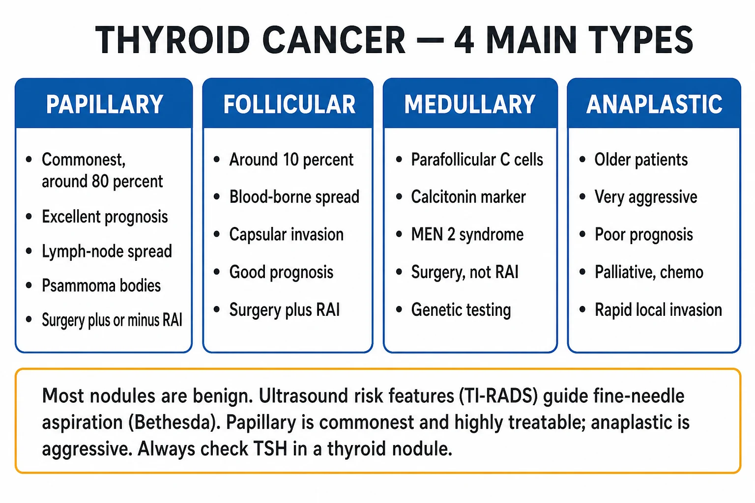
Thyroid nodules themselves are classified as benign (colloid nodule, simple or haemorrhagic cyst, benign follicular adenoma, nodular goitre, focal thyroiditis) or malignant, with a clinically critical indeterminate zone that cytology cannot resolve on its own. Malignant nodules divide along cell of origin:[1]
Papillary (PTC)
- Commonest — 80 to 85 percent of all thyroid cancers
- Women, peak 25 to 65 years; excellent prognosis
- Lymphatic spread to cervical nodes (levels III to VI)
- Orphan Annie-eye (ground-glass) nuclei, grooves, pseudoinclusions, psammoma bodies
- Driver: BRAF V600E (about 45 percent), RET-PTC rearrangement (about 15 percent)
- Takes up RAI; treated with thyroidectomy plus or minus RAI and TSH suppression
Follicular (FTC)
- About 10 percent; commoner in iodine-deficient regions
- Haematogenous spread to bone and lung; node-sparing early
- Defined by CAPSULAR and VASCULAR invasion — cannot be diagnosed on FNA alone
- Driver: RAS mutations, PAX8-PPAR-gamma fusion
- Hürthle-cell (oncocytic) variant is more aggressive and less RAI-avid
- Total thyroidectomy plus RAI; surveillance with thyroglobulin
Medullary (MTC)
- About 2 to 5 percent; parafollicular C-cell (neural-crest) origin
- Secretes CALCITONIN and CEA; amyloid stroma
- RET proto-oncogene mutation — familial in 25 percent (MEN-2A, MEN-2B)
- Does NOT take up RAI and does NOT produce thyroglobulin
- Total thyroidectomy plus central and lateral neck dissection
- Exclude PHAEOCHROMOCYTOMA before surgery (MEN-2); vandetanib or cabozantinib if advanced
Anaplastic (ATC)
- 1 to 2 percent but causes most thyroid-cancer deaths
- Older patients (over 60); rapidly enlarging hard painful mass
- Undifferentiated pleomorphic spindle and giant cells
- TP53, TERT promoter and beta-catenin mutations; dedifferentiation of prior PTC/FTC
- Stage IV by definition; median survival 4 to 6 months
- Largely palliative — multimodal if resectable; dabrafenib plus trametinib if BRAF-mutant
Thyroid nodules and cancer — the numbers that frame the topic
Epidemiology & Risk Factors
Thyroid nodules are among the commonest of all clinical findings. Palpable prevalence is 5 to 10 percent in adults, but ultrasound-detected prevalence reaches 30 to 50 percent in adults and over 60 percent in older cohorts, with autopsy series detecting nodules in roughly half of all glands. Only 5 to 10 percent of clinically significant nodules are malignant, so the pre-test probability is firmly benign — the discipline lies in identifying the minority.[1][2]
Thyroid cancer is the commonest endocrine malignancy. Its age-adjusted incidence has risen sharply over the last three decades, almost entirely driven by increased detection of small papillary carcinomas through wider use of neck ultrasound, carotid Doppler, CT and MRI — a phenomenon of over-diagnosis rather than a true epidemic. Mortality, in contrast, has been essentially flat, confirming that most newly detected cancers are indolent. Women are affected two to four times more often than men; differentiated cancer peaks between 25 and 65 years, while anaplastic cancer typically presents in those over 60.[1]
Risk factors
Established risk factor
- CHILDHOOD neck radiation (atomic survivors, therapeutic mantle radiotherapy for Hodgkin lymphoma, repeated childhood X-rays) — strongest risk, latency 10 to 40 years
- Female sex (oestrogen, reproductive factors)
- Family history of thyroid cancer (FNMTC syndrome)
- Iodine deficiency — increases follicular carcinoma risk
- Iodine excess — associated with papillary carcinoma
- Obesity, metabolic syndrome and insulin resistance
- MEN-2 syndrome and germline RET mutation — for medullary cancer
Protective / lower-risk
- Adequate dietary iodine intake lowers follicular and anaplastic rates
- Autonomous hyperfunctioning (hot) nodule on scintigraphy — very low malignancy risk
- Pure cyst — almost always benign
- Spongiform nodule — less than 3 percent malignant
- TR1/TR2 on ACR TI-RADS — no FNA required
Iodine deficiency deserves a special note: endemic goitre regions show a shift toward follicular and anaplastic carcinoma, while iodine-sufficient (or iodine-supplemented) populations show a predominance of papillary carcinoma. Population iodine supplementation has been shown to shift the histological mix towards papillary and away from the more aggressive follicular and anaplastic subtypes.[2]
Pathophysiology
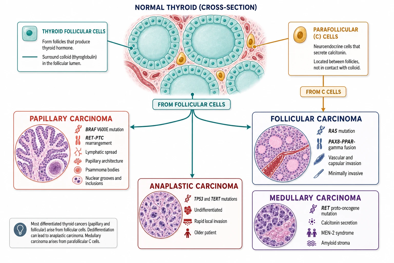
Most thyroid nodules are benign: colloid nodules (the commonest), simple or haemorrhagic cysts, benign follicular adenomas, multinodular goitre, and focal thyroiditis. Malignant transformation proceeds along distinct, well-characterised molecular pathways that have become therapeutically actionable.[1]
Papillary carcinoma (PTC) is driven by activation of the MAPK signalling pathway, most often by BRAF V600E (present in about 45 percent) and RET-PTC rearrangements (about 15 percent, classically after radiation), with RAS mutations in a smaller fraction. The MAPK cascade drives the distinctive nuclear morphology — optically clear ground-glass (orphan Annie-eye) nuclei, nuclear grooves and intranuclear pseudoinclusions — and the laminated calcifications called psammoma bodies. PTC spreads lymphatically to cervical nodes (most often central level VI, then lateral levels III to IV) and only uncommonly haematogenously to lung and bone.[1]
Follicular carcinoma (FTC) arises through a different molecular pathway: RAS mutations and the PAX8-PPAR-gamma fusion. Its histological hallmark is capsular and vascular (capsular-space) invasion — invasion through the tumour capsule and into blood vessels — which is why it cannot be diagnosed reliably on fine-needle aspiration (FNA samples cells, not architecture). FTC spreads haematogenously to bone and lung, with characteristically sparing of regional lymph nodes early on. The Hürthle-cell (oncocytic) variant has more abundant mitochondria-rich cytoplasm, is more aggressive and less avid for radioactive iodine.[1]
Medullary carcinoma (MTC) arises from parafollicular C cells (neural-crest-derived, calcitonin-secreting). Its defining molecular event is a gain-of-function mutation in the RET proto-oncogene: germline in the familial forms (MEN-2A, MEN-2B and familial medullary thyroid carcinoma — together about 25 percent of all MTC), and somatic in roughly half of sporadic cases. The RET M918T mutation (exon 16) confers the most aggressive phenotype and is the hallmark of MEN-2B. C cells produce calcitonin (a sensitive tumour marker) and CEA (a marker of tumour burden and dedifferentiation), and the tumour stroma typically contains amyloid (congophilic on staining). MTC does not produce thyroglobulin and does not concentrate iodine.[1]
Anaplastic carcinoma (ATC) is undifferentiated, comprising pleomorphic spindle, giant and squamoid cells. It is thought usually to arise by dedifferentiation of a pre-existing papillary or follicular cancer, accumulating TP53, TERT promoter and beta-catenin (CTNNB1) mutations that override cell-cycle control. BRAF V600E is often co-present (from the antecedent PTC). ATC is among the most aggressive of all human cancers — rapidly invading trachea, oesophagus, great vessels and overlying skin, with a doubling time often measured in weeks.[1]
Clinical Presentation
The classic presentation is a painless neck lump found by the patient, a partner, or a clinician on routine examination, or detected incidentally on carotid Doppler, neck CT, MRI or PET performed for another reason. Most patients are clinically and biochemically euthyroid — thyroid function tests are done to exclude a hyperfunctioning (hot) nodule rather than to diagnose thyroid status per se.[1]
Examination focuses on features that raise malignancy probability. The high-yield clinical risk features are:[1]
- History of childhood neck irradiation, family history of thyroid cancer or MEN-2, age under 20 or over 70, male sex (cancer is less common in men but more often aggressive when present).
- Growth — rapid enlargement of a previously stable nodule is ominous (and a sentinel clue to anaplastic transformation).
- Consistency — a firm or fixed nodule adherent to trachea or strap muscles.
- Voice — hoarseness, a sign of recurrent laryngeal nerve involvement (one of the strongest single predictors of malignancy).
- Nodes — palpable cervical lymphadenopathy, especially levels II to VI.
- Compression — dysphagia, dyspnoea or stridor from a large or rapidly growing mass. [1]
Atypical presentations are deliberately examined. Medullary carcinoma may present with the calcitonin syndrome (flushing, secretory diarrhoea) or as part of MEN-2 — screen for phaeochromocytoma (hypertensive crises), primary hyperparathyroidism (hypercalcaemia) and the MEN-2B marfanoid phenotype with mucosal neuromas. Anaplastic carcinoma presents dramatically as a rapidly enlarging, hard, painful neck mass in an older patient with systemic deterioration, compressive symptoms and sometimes skin invasion. Thyroid lymphoma arises almost exclusively in long-standing Hashimoto thyroiditis and presents as rapid enlargement of a pre-existing goitre in an older woman. Metastases to the thyroid (kidney, breast, lung, melanoma) may mimic a primary nodule.[1]
Differential Diagnosis
The differential of a thyroid nodule is broad, and the role of imaging and FNA is to triage among them:[1]
Benign — common
- Colloid (adenomatous) nodule — commonest of all; mixed solid-cystic, isoechoic, no suspicious features
- Simple or haemorrhagic cyst — anechoic with comet-tail artefacts; benign
- Benign follicular adenoma — homogeneous, well-circumscribed; cannot exclude FTC on FNA
- Multinodular goitre — multiple nodules of varying echogenicity; iodine deficiency or autoimmune
- Focal thyroiditis (Hashimoto, subacute/De Quervain) — tender or painless; elevated antibodies
Malignant
- Papillary carcinoma — solid, hypoechoic, taller-than-wide, microcalcifications, irregular margins
- Follicular carcinoma — looks deceptively benign on ultrasound; vascular and capsular invasion on histology
- Medullary carcinoma — hypoechoic, calcified; calcitonin elevated
- Anaplastic carcinoma — large, invasive, necrotic, heterogeneous; fixed mass in elderly
- Poorly differentiated thyroid cancer — intermediate between differentiated and anaplastic
- Thyroid lymphoma — rapid growth in long-standing Hashimoto; homogeneous hypoechoic
Non-thyroid neck mass
- Reactive or metastatic cervical lymph node
- Thyroglossal duct cyst (midline, moves with swallowing or tongue protrusion)
- Branchial cleft cyst (anterolateral neck)
- Sebaceous or dermoid cyst, lipoma
- Parathyroid adenoma or cyst (posterior to thyroid)
- Normal structure mistaken for a nodule (omohyoid, prominent isthmus)
The clinically distinguishing question is always: does this nodule carry enough risk to warrant FNA? The next two sections — ultrasound TI-RADS and Bethesda cytology — answer that. [1]
Clinical & Bedside Assessment
A focused thyroid consultation takes five minutes and decides the entire work-up. Take a structured history: age and sex, radiation exposure (childhood neck radiotherapy, repeated childhood X-rays, atomic-survivor exposure), family history of thyroid cancer or MEN-2, tempo of nodule growth, and compressive or voice symptoms. Ask specifically about flushing, diarrhoea and hypertension (MEN-2 / medullary) and hypercalcaemia symptoms (parathyroid).[1]
Examine the patient from the front. Inspect the neck for asymmetry and a palpable mass, then palpate the thyroid from behind while the patient swallows a sip of water (thyroid moves up with swallowing, distinguishing it from other neck masses). Document the nodule's size, consistency (soft, firm, hard), surface (smooth, irregular), mobility (mobile, fixed), tenderness, and relation to the trachea. Then systematically palpate the cervical lymph node chains (levels I to VI), particularly the central (level VI) and jugular (levels III to IV) stations. Assess for tracheal deviation and compressive signs (stridor). Check vocal cord function — clinically through voice quality, and by indirect or flexible laryngoscopy in any patient with hoarseness or before any thyroid surgery (a paralysed cord predicts malignant nerve involvement and changes the surgical plan). Measure blood pressure (phaeochromocytoma screen in suspected MEN-2) and look for hyperparathyroidism (hypercalcaemia) and the marfanoid habitus and mucosal neuromas of MEN-2B.[1]
Investigations
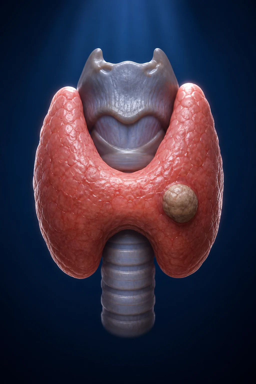
The investigation of a thyroid nodule follows a strict, evidence-based sequence — TSH first, ultrasound second, fine-needle aspiration third, ancillary tests fourth.[1][2]
Step 1 — Serum TSH
Serum TSH is the first test in every thyroid nodule. A normal or raised TSH prompts ultrasound risk-stratification. A suppressed (subnormal) TSH with a raised free T4 (and free T3) suggests an autonomous (hot) nodule — which carries a much lower malignancy risk — and the next step is radionuclide scintigraphy (technetium-99m pertechnetate or iodine-123) to confirm a hyperfunctioning (tracer-avid) nodule, which generally does not need FNA. Within the normal range, a TSH in the upper half has itself been shown to slightly increase the probability of malignancy.[1]
Step 2 — High-resolution thyroid ultrasound with ACR TI-RADS
High-resolution ultrasound is the single most useful imaging test in thyroid nodule evaluation. The American College of Radiology Thyroid Imaging Reporting and Data System (ACR TI-RADS) assigns points across five feature categories and converts the sum into a risk level (TR1 to TR5) that, combined with nodule size, decides whether to FNA, follow up or ignore.[3]
The FNA / follow-up thresholds by TI-RADS and size: TR1 and TR2 — no FNA. TR3 — FNA if at least 2.5 cm, follow-up if at least 1.5 cm. TR4 — FNA if at least 1.5 cm, follow-up if at least 1 cm. TR5 — FNA if at least 1 cm, follow-up if at least 0.5 cm. Smaller TR3 to TR5 nodules may still be biopsied if there are high-risk clinical features, suspicious nodes, or an extra-thyroidal extension. The pattern of cervical lymph nodes (round, cystic, lost hilum, punctate echogenic foci, calcifications) is itself a major indication for FNA, even when the nodule is small.[3]
Step 3 — Fine-needle aspiration cytology (Bethesda System)
Fine-needle aspiration under ultrasound guidance is the cornerstone of nodule triage. Cytology is reported using The Bethesda System for Reporting Thyroid Cytopathology (TBSRTC), which assigns every thyroid FNA to one of six categories, each carrying an implicit malignancy risk and a linked management recommendation.[4]
Bethesda I — Nondiagnostic
- Inadequate cellular material or non-diagnostic cyst fluid
- Implied malignancy risk 5 to 10 percent
- Repeat ultrasound-guided FNA (typically a different site or under better visualisation)
Bethesda II — Benign
- Benign follicular nodule, colloid, cyst contents
- Risk of malignancy 0 to 3 percent
- Clinical and ultrasound surveillance; no surgery unless symptomatic
Bethesda III — AUS / FLUS
- Atypia of undetermined significance or follicular lesion of undetermined significance
- Risk 10 to 30 percent
- Repeat FNA, molecular testing, or diagnostic lobectomy
Bethesda IV — FN / SFN
- Follicular neoplasm or suspicious for follicular neoplasm (includes Hürthle-cell)
- Risk 25 to 40 percent
- Diagnostic lobectomy; molecular testing may help avoid surgery
Bethesda V — Suspicious
- Suspicious for malignancy (often papillary)
- Risk 50 to 75 percent
- Near-total or total thyroidectomy, or lobectomy if low-risk
Bethesda VI — Malignant
- Malignant — papillary, medullary, anaplastic, lymphoma or metastatic
- Risk 97 to 99 percent
- Definitive surgery as appropriate to the cancer type
The 2017 Bethesda revision updated the implied malignancy risks upward, partly because of the reclassification of non-invasive follicular thyroid neoplasm with papillary-like nuclear features (NIFTP) — a tumour previously called encapsulated follicular variant papillary carcinoma that is now regarded as very-low-risk, non-malignant and managed by lobectomy alone. This single reclassification reduced apparent cancer rates across Bethesda III, IV and V without changing outcomes.[4]
For the troublesome indeterminate zone (Bethesda III and IV), molecular testing refines the risk and can spare surgery: gene-expression classifiers (Afirma GSC) and next-generation sequencing panels (ThyroSeq, ThyGeNEXT / ThyraMIR) stratify nodules into low-risk (observe) and high-risk (operate) groups, reducing diagnostic lobectomies by around half.[1]
Step 4 — Ancillary tests
- Serum calcitonin — order if medullary cancer is suspected (family history, cytology suggesting MTC, or for some guidelines as a baseline screen). A basal calcitonin over 100 pg/mL (or a stimulated rise after pentagastrin) is highly suggestive; provocative testing with calcium or pentagastrin can be used for borderline values. CEA is a useful tumour-burden marker in MTC and is also raised in anaplastic cancer.[1]
- Cross-sectional CT or MRI of neck and chest — for large nodules, suspected local invasion, retrosternal extension, bulky nodal disease, or anaplastic cancer to map tumour extent and plan surgery. CT with contrast is generally avoided in patients who may receive radioactive iodine, because iodinated contrast loads stable iodine and blocks RAI uptake for weeks; MRI is the preferred alternative in that setting.[1]
- Vocal-cord assessment — flexible laryngoscopy before any thyroid surgery if hoarse.
- Baseline thyroglobulin — measured before definitive treatment of differentiated cancer (it cannot distinguish benign from malignant, but serves as a non-secretion reference after thyroidectomy and RAI).
- RET genetic testing and family screening — for any patient with medullary cancer and first-degree relatives; positive carriers proceed to prophylactic thyroidectomy timed to the mutation risk class.[1]
Management — Resuscitation and Peri-operative Preparation
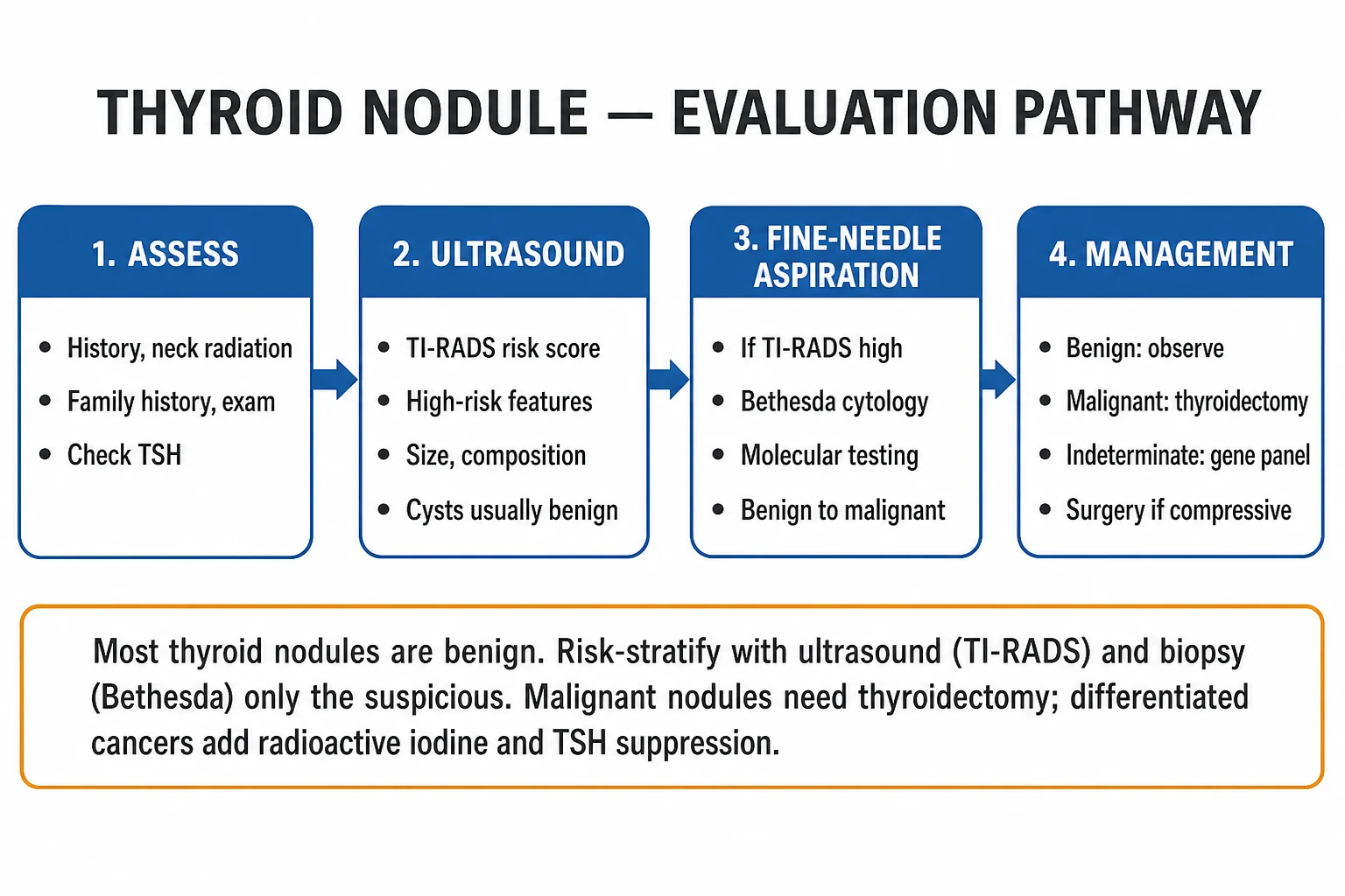
Most thyroid nodule and cancer work-ups are outpatient. Acute resuscitation is rarely required except for three situations:[1]
- Airway compromise from a large or anaplastic mass — stridor, respiratory distress, or rapid tracheal compression requires urgent airway assessment (awake fibreoptic intubation may be needed), multidisciplinary oncology and palliative input, and may demand emergency tracheostomy or debulking radiotherapy.
- Neck haematoma after thyroid surgery — a surgical emergency: the wound must be opened at the bedside to release the clot and relieve airway compression, then the patient returned to theatre for haemostasis.
- Bilateral recurrent laryngeal nerve injury — post-extubation stridor may demand re-intubation and sometimes tracheostomy. [1]
Peri-operative preparation is high-yield exam material: (a) document vocal-cord function by laryngoscopy before surgery (a pre-existing palsy predicts malignant nerve involvement); (b) check serum calcium, phosphate, vitamin D and parathyroid hormone (PTH) pre-operatively to anticipate and recognise post-operative hypoparathyroidism; (c) render the patient biochemically euthyroid before elective surgery (a hyperthyroid patient is at risk of thyroid storm); (d) for medullary cancer, exclude phaeochromocytoma first (24-hour urinary metanephrines or plasma free metanephrines) to avoid a lethal intra-operative hypertensive crisis; (e) group and save and discuss the risk of tracheostomy, permanent hypocalcaemia and voice change in consent.[1]
Management — Definitive and Stepwise
Definitive management is dictated by the cancer type, the stage, and the recurrence risk of the individual patient. The framework below is built on the 2015 American Thyroid Association guidelines.[1]
Surgery — the central curative modality
Surgical extent ranges from diagnostic lobectomy to total thyroidectomy with neck dissection, chosen on histology and risk.[1]
Diagnostic lobectomy
- For Bethesda III / IV indeterminate nodules, low-risk single-lobe lesions, or small unifocal intrathyroidal cancers
- Removes the ipsilateral lobe and isthmus
- Avoids bilateral complications; preserves contralateral lobe and normal parathyroids
- Converted to completion thyroidectomy if histology reveals high-risk cancer
Total thyroidectomy
- Standard for differentiated cancers over 4 cm, multifocal or bilateral disease, extra-thyroidal extension, nodal metastases, or when RAI is planned
- Enables accurate thyroglobulin surveillance and RAI ablation
- Risks: bilateral recurrent laryngeal nerve injury, permanent hypoparathyroidism
- Required for almost all medullary and anaplastic cancers
Prophylactic central neck dissection
- Removal of level VI (prelaryngeal, pretracheal, paratracheal) nodes
- Considered for T3 or higher, or known lateral-node disease
- Improves staging and may reduce locoregional recurrence in high-volume centres
- Higher transient hypocalcaemia; reserved for intermediate-high risk
Therapeutic lateral neck dissection
- Levels II to V for biopsy-proven lateral nodal metastasis (usually papillary or medullary)
- Selective therapeutic, not prophylactic, in papillary cancer
- Comprehensive (not berry-picking) — removes all fibro-fatty lymphatic tissue in the field
- Mandatory in medullary cancer given high nodal involvement rate
Radioactive iodine (131-I) ablation
Radioactive iodine-131 ablation is given after total thyroidectomy to destroy residual thyroid tissue and micrometastases, enable sensitive thyroglobulin surveillance, and reduce recurrence in intermediate- and high-risk differentiated cancer. Patients are prepared either by thyroid hormone withdrawal (to raise endogenous TSH) or by recombinant human TSH (rhTSH, Thyrogen) injections, the latter avoiding iatrogenic hypothyroidism. Activity is stratified by recurrence risk — low-activity ablation (around 30 mCi / 1.1 GBq) for low-to-intermediate risk, and higher-activity therapy (100 to 150 mCi) for high-risk disease with nodal metastases, extra-thyroidal extension or distant metastases. RAI has NO role in medullary or anaplastic cancer, because those cells do not concentrate iodine.[1]
TSH suppression with levothyroxine
After total thyroidectomy, all patients require lifelong levothyroxine replacement — typically 1.6 micrograms per kilogram per day as a starting adult dose, adjusted to a risk-adapted TSH target. The 2015 ATA guidance stratifies suppression intensity:[1]
- High-risk cancer — TSH below 0.1 mU/L indefinitely, balancing against bone and cardiac toxicity.
- Intermediate-risk — TSH 0.1 to 0.5 mU/L for several years, then relaxed.
- Low-risk — TSH in the low-normal reference range (0.5 to 2.0 mU/L); aggressive suppression is unnecessary and harmful. [1]
Excess suppression carries real harms: osteoporosis (especially post-menopausal women) and atrial fibrillation (especially older patients) — a compelling reason to relax suppression once recurrence risk has fallen.[1]
Risk-adapted approach and follow-up
The 2015 ATA guidelines formalised dynamic risk stratification: an initial staging estimate (TNM-8 and the ATA response-to-therapy categories) is updated at each follow-up by thyroglobulin trends, neck ultrasound and, when needed, cross-sectional imaging or diagnostic iodine scans. Excellent responders (undetectable stimulated thyroglobulin, normal ultrasound) move to lighter surveillance; biochemical incomplete (rising thyroglobulin) or structural incomplete (imaging-positive disease) responders escalate to further local therapy or systemic treatment.[1]
Targeted and systemic therapy
For RAI-refractory differentiated cancer, multitargeted tyrosine-kinase inhibitors have transformed outcomes. Lenvatinib (24 mg daily) and sorafenib (400 mg twice daily) both improve progression-free survival; lenvatinib produced a striking 18.3 versus 3.6 month median PFS in the SELECT trial.[5]
SELECT — lenvatinib for RAI-refractory differentiated thyroid cancer
Schlumberger M et al. N Engl J Med 2015
Phase III RCT, 392 patients with progressive iodine-131-refractory DTC: lenvatinib 24 mg/day vs placebo (2:1)
Key finding
Median progression-free survival 18.3 months (lenvatinib) vs 3.6 months (placebo); hazard ratio 0.21; response rate 64.8 percent vs 1.5 percent
Practice change
Lenvatinib is a first-line standard for progressive RAI-refractory differentiated thyroid cancer
For medullary cancer, RET and VEGFR-directed TKIs are the systemic mainstay. Vandetanib (300 mg daily) and cabozantinib (140 mg daily) both prolong progression-free survival; cabozantinib's effect is independent of RET mutation status.[6][7]
ZETA — vandetanib for advanced medullary thyroid cancer
Wells SA Jr et al. J Clin Oncol 2012
Phase III RCT, 331 patients with advanced MTC: vandetanib 300 mg/day vs placebo (2:1)
Key finding
Prolonged progression-free survival (hazard ratio 0.46); objective response rate higher with vandetanib
Practice change
First systemic therapy approved for advanced medullary thyroid cancer; note QTc prolongation
EXAM — cabozantinib for progressive medullary thyroid cancer
Elisei R et al. J Clin Oncol 2013
Phase III RCT, 330 patients with progressive metastatic MTC: cabozantinib 140 mg/day vs placebo (2:1)
Key finding
Median progression-free survival 11.2 months (cabozantinib) vs 4.0 months (placebo); hazard ratio 0.28; response rate 28 percent
Practice change
Cabozantinib standard for progressive metastatic MTC, irrespective of RET mutation status
For anaplastic cancer with a BRAF V600E mutation (about half of cases), the combination of dabrafenib plus trametinib (BRAF and MEK inhibition) has produced remarkable responses in a disease that was until recently uniformly fatal within weeks. For RET-mutant medullary cancer, the highly selective RET inhibitors selpercatinib and pralsetinib are emerging as first-line targeted therapy.[2]
Specific Subtypes and Scenarios
Papillary thyroid carcinoma (PTC)
PTC accounts for 80 to 85 percent of thyroid cancers, peaks between 25 and 65, is two to three times more common in women, and is the cancer most strongly linked to childhood neck radiation. Histological variants include the classic type, the follicular variant (now partly reclassified as NIFTP when encapsulated and non-invasive), the aggressive tall-cell, columnar-cell and hobnail variants, and the diffuse sclerosing variant (more aggressive, lymphotropic). Standard treatment of clinically evident PTC is total thyroidectomy plus or minus prophylactic central neck dissection, followed by selective RAI ablation and risk-adapted TSH suppression. Active surveillance of low-risk papillary microcarcinoma (T1a, no invasion, no nodes), pioneered by Japanese and Memorial Sloan Kettering cohorts, is now an accepted alternative to immediate surgery, with very low 10-year progression rates.[1][2]
Follicular thyroid carcinoma (FTC)
FTC accounts for 5 to 10 percent of thyroid cancers, is commoner in iodine-deficient regions, presents slightly later than PTC, and spreads haematogenously to bone and lung. It is defined by capsular and vascular invasion — and so cannot be diagnosed on FNA cytology alone (Bethesda IV triggers diagnostic lobectomy to inspect architecture). It is divided into minimally invasive (excellent prognosis) and widely invasive (worse prognosis). The Hürthle-cell (oncocytic) variant behaves more aggressively, is less RAI-avid and is managed with total thyroidectomy plus RAI and lifelong thyroglobulin surveillance.[1]
Medullary thyroid carcinoma (MTC)
MTC accounts for 2 to 5 percent of thyroid cancers but a disproportionate share of thyroid-cancer deaths, because it is biologically more aggressive and does not respond to RAI or TSH suppression. About 25 percent are hereditary (MEN-2A, MEN-2B, familial MTC) and are caused by germline RET proto-oncogene mutations; the remaining sporadic cases carry somatic RET mutations in roughly half. Every patient with a new diagnosis of MTC must undergo RET genetic testing and family screening, because identifying an asymptomatic RET carrier allows prophylactic thyroidectomy — in infancy for MEN-2B (the most aggressive), in early childhood for MEN-2A and FMTC.[1]
Surgery is the only curative therapy: total thyroidectomy plus routine central (level VI) and ipsilateral lateral (levels II to V) neck dissection, with contralateral lateral dissection if nodal disease is present. Before surgery, exclude phaeochromocytoma (24-hour urinary fractionated metanephrines or plasma free metanephrines) — operating on an unrecognised phaeo in MEN-2 precipitates a lethal hypertensive crisis. Surveillance is with serial calcitonin and CEA (doubling times are strong prognostic markers — calcitonin doubling time under six months is ominous). For progressive metastatic disease, vandetanib, cabozantinib, or the selective selpercatinib (for RET-mutant disease) prolong progression-free survival.[6][7]
Anaplastic thyroid carcinoma (ATC)
ATC accounts for only 1 to 2 percent of thyroid cancers but is responsible for a third to a half of all thyroid-cancer deaths. It arises in older patients (over 60), presents as a rapidly enlarging, hard, painful, fixed neck mass, often with stridor, dysphagia, hoarseness and systemic decline. Histology shows undifferentiated pleomorphic spindle and giant cells; TP53, TERT promoter and beta-catenin mutations are characteristic, and BRAF V600E is often co-present (indicating dedifferentiation from a prior papillary cancer). All ATC are stage IV at diagnosis. Treatment is multimodal where resectable — surgery (rarely curative, often debulking), external-beam radiotherapy and chemotherapy (taxanes such as paclitaxel or docetaxel, sometimes combined with carboplatin). For the BRAF V600E-mutant subgroup, dabrafenib plus trametinib has produced meaningful responses and improved survival. Despite all this, median survival is 4 to 6 months and 5-year survival is under 5 percent; the priority is airway, symptom control and early palliative care.[1][2]
AJCC TNM-8 staging — a major change
The 8th edition (2017, effective 2018) of the AJCC / UICC TNM staging made three changes that materially improve staging accuracy for differentiated thyroid cancer. First, the prognostic age cut-off was moved from 45 to 55 years (reflecting data that age confers a much smaller risk gradient below 55). Second, minimal extra-thyroidal extension was removed from the T3 definition, leaving only tumour size over 4 cm (T3a) or gross strap-muscle invasion (T3b). Third, N1 disease no longer automatically stages an older patient to stage I, since nodal metastasis in well-differentiated PTC confers a much smaller survival penalty than was historically assumed.[1]
Differentiated (PTC / FTC) — age under 55
- Stage I — any T, any N, M0
- Stage II — any T, any N, M1 (distant metastasis is the only upstage)
- Reflects excellent prognosis — even node-positive disease is stage I if no distant spread
Differentiated — age 55 or over
- Stage I — T1 or T2 (up to 4 cm), N0 or NX, M0
- Stage II — T3 (over 4 cm or minimal strap-muscle invasion) with N0/NX M0, OR T1 to T3 with N1 M0
- Stage III — T4a (subcutaneous, larynx, trachea, oesophagus, RLN), any N, M0
- Stage IV — T4b (prevertebral fascia, mediastinal vessels, carotid encasement) OR any M1
Medullary (all ages)
- Stage I — T1 (up to 2 cm), N0, M0
- Stage II — T2 or T3, N0, M0
- Stage III — T1 to T3, N1a (central nodes), M0
- Stage IVA — T4a any N M0, OR T1 to T3 with N1b (lateral nodes) M0
- Stage IVB — T4b any N M0; Stage IVC — any M1
Anaplastic — all stage IV
- Stage IVA — T1 to T3, N0 or NX, M0 (intrathyroidal; potentially resectable)
- Stage IVB — T1 to T3 with N1, OR T4a any N, M0
- Stage IVC — T4b any N M0, OR any T any N M1 (the commonest presentation)
- Reflects uniformly dismal prognosis — age and T category no longer improve the stage
Complications and Pitfalls
Disease-related complications
- Local invasion — recurrent laryngeal nerve (hoarseness), trachea (stridor, haemoptysis), oesophagus (dysphagia), especially in anaplastic and advanced medullary disease.
- Nodal and distant metastasis — papillary to cervical nodes and lung; follicular to bone and lung (haematogenous); medullary to nodes, liver and bone; anaplastic widely metastatic.
- Calcitonin syndrome — flushing, secretory diarrhoea in advanced medullary cancer. [1]
Surgical complications
Surgery for thyroid cancer is safe in high-volume hands but carries three classic complications every student must know.[1]
- Recurrent laryngeal nerve (RLN) injury — unilateral causes hoarseness; bilateral causes stridor and may require tracheostomy. Transient in 5 to 10 percent, permanent in 1 to 2 percent with intra-operative nerve monitoring in expert hands.
- Hypoparathyroidism — transient hypocalcaemia in up to 25 percent after total thyroidectomy with central dissection; permanent in 1 to 4 percent. Managed with oral calcium and calcitriol; severe cases need intravenous calcium gluconate. Always check calcium and PTH post-operatively.
- Neck haematoma — rare (under 1 percent) but life-threatening; open the wound at the bedside to release the clot and decompress the airway, then return to theatre.
- Superior laryngeal nerve injury — loss of high-pitch voice and aspiration of liquids.
- Chyle leak, seroma, infection, hypertrophic scar — less common. [1]
Treatment-related complications
- Radioactive iodine — sialadenitis and dry mouth, transient neck pain, nausea, small secondary malignancy risk (notably salivary and leukaemia), and pregnancy / breastfeeding restrictions (avoid pregnancy for 6 to 12 months).
- TSH suppression — osteoporosis (post-menopausal women) and atrial fibrillation (older patients); justify only in high-risk disease.
- TKI therapy — hypertension, diarrhoea, fatigue, hand-foot syndrome, QTc prolongation (vandetanib), proteinuria and bleeding risk. [1]
Classic pitfalls
- Operating on a medullary cancer patient with an undiagnosed phaeochromocytoma — provokes a fatal intra-operative hypertensive crisis. Always exclude phaeo first.
- Diagnosing follicular carcinoma on FNA — impossible; it requires histology of capsular/vascular invasion. Bethesda IV is a diagnostic lobectomy indication.
- Misreading a thyroglobulin level after RAI — must be interpreted with anti-thyroglobulin antibody status (antibodies cause false-low readings) and only after thyroidectomy and ablation.
- Giving RAI for medullary or anaplastic cancer — useless; those cells do not concentrate iodine.
- Treating iodinated-contrast CT as routine in differentiated cancer — iodine loading blocks subsequent RAI for weeks; use MRI if cross-sectional imaging is needed before ablation.
- Over-suppressing TSH in a low-risk older patient — precipitates atrial fibrillation and osteoporosis without benefit. [1]
Prognosis and Disposition
Cancer type
- Papillary — 5-year survival over 95 percent overall; over 98 percent for localised disease
- Follicular — 5-year survival around 85 percent; worse with vascular invasion or distant metastasis
- Medullary — 5-year survival around 75 to 85 percent overall; stage-dependent; calcitonin doubling-time under 6 months is ominous
- Anaplastic — median survival 4 to 6 months; 5-year survival under 5 percent; almost uniformly fatal
Determinants
- AGE (under 55 favourable in differentiated cancer), tumour SIZE, extra-thyroidal extension, nodal status and distant metastasis (TNM-8)
- Histological variant — tall-cell, columnar-cell, hobnail PTC and widely-invasive FTC and Hürthle-cell carry worse prognosis
- Molecular profile — BRAF plus TERT co-mutation predicts aggressive behaviour in PTC; RET M918T predicts aggressive MTC
- Response to initial therapy — excellent, biochemically incomplete, structurally incomplete (dynamic risk stratification)
Differentiated thyroid cancer is among the most curable of all solid tumours. Even patients with cervical nodal metastasis have excellent survival — a fact reflected in the AJCC-8 staging change that keeps node-positive disease stage I in patients under 55. Follicular cancer is slightly worse, especially with vascular invasion or distant metastasis. Medullary prognosis is stage- and calcitonin-doubling-time-dependent; MEN-2B and the RET M918T mutation carry the worst outcomes. Anaplastic carcinoma is almost uniformly fatal within months, though the small BRAF-mutant subgroup now benefits dramatically from dabrafenib-trametinib.[1][2]
Disposition is risk-adapted. Most differentiated cancer patients are managed as outpatients with lifelong structured surveillance: six-monthly thyroglobulin and anti-thyroglobulin antibodies, periodic neck ultrasound, and diagnostic whole-body iodine scans when biochemistry or imaging is discordant. Medullary cancer surveillance centres on calcitonin and CEA doubling times, with ultrasound and cross-sectional imaging for rising markers. Anaplastic cancer demands rapid inpatient multidisciplinary management and early palliative care. Benign nodules are observed safely — small low-risk papillary microcarcinomas may even be actively surveilled with excellent long-term outcomes.[1][2]
Special Populations
- Pregnancy — fine-needle aspiration is safe in pregnancy and is indicated for the same ultrasound features as in non-pregnant patients. If a differentiated cancer is found in the first or early second trimester, surgery can usually be deferred to the postpartum unless there are aggressive features (rapid growth, extra-thyroidal extension, nodes); cancers discovered later in pregnancy are almost always observed through delivery. Radioactive iodine is absolutely contraindicated in pregnancy and breastfeeding.[1]
- Children and adolescents — thyroid nodules are less common in children but carry a higher malignancy risk (around 20 to 25 percent). Paediatric papillary cancer is more often disseminated at presentation (larger primaries, more nodal and pulmonary metastasis) but remains highly curable. Children exposed to Chernobyl fallout demonstrated the strong radiation-PTC link. RAI doses and surveillance are weight-adapted.
- Known MEN-2 / RET carriers — prophylactic thyroidectomy is performed before medullary cancer develops, timed by the ATA risk class of the RET mutation: infancy for MEN-2B (highest-risk codons, e.g. M918T), before age 5 years for high-risk MEN-2A, and individualised for lower-risk mutations. Calcitonin surveillance begins early; phaeochromocytoma screening is lifelong.[1]
- The elderly patient with a rapidly growing neck mass — assume anaplastic until proven otherwise; secure the airway, image, biopsy urgently, and involve the multidisciplinary team and palliative care early.
- Patients on amiodarone — the iodine load can precipitate both hyper- and hypothyroidism and cause nodular change; check thyroid function before and during amiodarone.
Evidence, Guidelines and Regional Differences
The 2015 American Thyroid Association (ATA) guidelines for adult patients with thyroid nodules and differentiated thyroid cancer are the most widely used international framework, defining the TSH-first work-up, the role of ultrasound risk stratification, the surgical extent, the indications for radioactive iodine, the risk-adapted TSH suppression targets, and the dynamic risk-stratification approach to follow-up. They are endorsed or adapted by most national societies.[1]
Key controversies
- Over-diagnosis and over-treatment — the steep rise in thyroid cancer incidence from increased imaging has led to concerns about overtreatment of indolent papillary microcarcinoma. Active surveillance of low-risk T1a tumours, molecular testing to reduce diagnostic lobectomy, and the NIFTP reclassification are all responses.
- Extent of surgery — total thyroidectomy versus lobectomy for low-risk unilateral disease remains debated; lobectomy avoids bilateral complications but complicates RAI and thyroglobulin surveillance.
- RAI in low-risk disease — increasingly avoided in patients at very low recurrence risk, to spare toxicity.
- TSH suppression intensity — relaxed for low-risk disease to avoid bone and cardiac toxicity.
- Prophylactic central neck dissection — its routine use in clinically node-negative papillary cancer varies between centres; the ATA recommends it be considered, not mandated.[1]
Exam Pearls and High-Yield Minutiae
High-risk features of a thyroid nodule — the RADIUS mnemonic
RADIUS
childhood neck irradiation — the strongest risk factor for papillary thyroid cancer
age under 20 or over 70; palpable cervical lymphadenopathy
hoarseness (recurrent laryngeal nerve) or compressive symptoms
family history of thyroid cancer or MEN-2 (RET mutation)
solid, hypoechoic, taller-than-wide, irregular margins, microcalcifications (high TI-RADS)
rapid growth of a firm, fixed nodule; male sex raises malignancy risk
Cancer types by behaviour — the PBMA mnemonic
PBMA
commonest 80 percent; best prognosis; lymphatic; psammoma bodies; BRAF V600E
10 percent; capsular and vascular invasion; bone and lung metastasis; RAS; needs iodine
5 percent; C cells; calcitonin; MEN-2; RET; NO RAI; exclude phaeo first
1 to 2 percent; undifferentiated; elderly; survival in months; stage IV by definition
Quick self-test — what is the first test in a thyroid nodule, and why?
Serum TSH. A normal or raised TSH prompts ultrasound (TI-RADS) and selective FNA; a suppressed TSH points to an autonomous (hot) nodule with a low malignancy risk, which is then assessed by radionuclide scintigraphy. Skipping TSH means you cannot interpret the rest of the work-up.[1]
Quick self-test — why can follicular carcinoma NOT be diagnosed on FNA?
Because FTC is defined by capsular and vascular invasion, which are architectural features — FNA samples cells, not the relationship of the tumour to its capsule and vessels. Bethesda IV (follicular neoplasm) therefore triggers diagnostic lobectomy for histological assessment. (Papillary, in contrast, is diagnosable cytologically by its nuclear features.)[1][4]
Quick self-test — what MUST you exclude before operating on a medullary thyroid cancer?
A phaeochromocytoma (MEN-2). Operating on an unrecognised phaeo precipitates a fatal intra-operative hypertensive crisis. Screen with 24-hour urinary fractionated metanephrines or plasma free metanephrines before any surgery; if positive, the phaeochromocytoma is resected first (usually after alpha-blockade).[1][6]
Exam application bank (NEET-PG / INICET)
One-line answer
Thyroid nodules are very common but mostly benign — palpable in around 5 percent of adults and seen on ultrasound in 30 to 50 percent — yet only 5 to 10 percent are malignant. The clinical task is to identify that minority through ultrasound risk-stratification (ACR TI-RADS) and fine-needle aspiration cytology (Bethesda System), guided by clinical risk (neck radiation, family history, rapid growth, hoarseness, fixed nodule, lymphadenopathy). The four main thyroid cancers are papillary (commonest, around 80 percent; excellent prognosis; BRAF V600E, RET-PTC, psammoma bodies, orphan Annie-eye nuclei), follicular (10 percent; vascular and capsular invasion; haematogenous spread to bone and lung; RAS mutation), medullary (5 percent; parafollicular C cells; calcitonin; MEN-2; RET proto-oncogene) and anaplastic (1 to 2 percent; undifferentiated; elderly; survival measured in months). Differenti
Worked stems (answer without another resource)
Stem 1 — Classic presentation. Map symptoms to mechanism; name the first investigation and first treatment step with dose/route if drug therapy is standard. [1]
Stem 2 — Unstable / complicated. List red flags that force immediate resuscitation, theatre, ICU, antidote, or reperfusion — and what you do in the first 15 minutes. [1]
Stem 3 — Atypical group. Elderly, pregnancy, child, or immunocompromised: how presentation and thresholds change. [1]
Stem 4 — Differential trap. Name the three closest mimics and one discriminator for each. [1]
Stem 5 — Disposition. Who goes home with safety-netting, who is admitted, who needs HDU/ICU/theatre, and what follow-up is mandatory. [1]
Rapid viva checklist
- Definition + classification
- Pathophysiology chain
- Bedside signs / criteria
- Score with exact components (if any)
- Emergency bundle
- Definitive therapy with doses
- Complications of disease and of treatment
- Special populations
- Guideline/trial name if classic
- Three exam traps
Coverage self-check
If you cannot answer any stem above from this page alone, re-read the matching section — the page is intended to be self-sufficient for final-prof and NEET-PG/INICET questions on Thyroid Nodules & Thyroid Cancer.
[1]References
- [1]Haugen BR, Alexander EK, Bible KC, et al. 2015 American Thyroid Association Management Guidelines for Adult Patients with Thyroid Nodules and Differentiated Thyroid Cancer: The American Thyroid Association Guidelines Task Force on Thyroid Nodules and Differentiated Thyroid Cancer Thyroid, 2016.PMID 26462967
- [2]Grani G, Sponziello M, Filetti S, Durante C. Thyroid nodules: diagnosis and management Nat Rev Endocrinol, 2024.PMID 39152228
- [3]Tessler FN, Middleton WD, Grant EG, et al. ACR Thyroid Imaging, Reporting and Data System (TI-RADS): White Paper of the ACR TI-RADS Committee J Am Coll Radiol, 2017.PMID 28372962
- [4]Cibas ES, Ali SZ. The 2017 Bethesda System for Reporting Thyroid Cytopathology Thyroid, 2017.PMID 29091573
- [5]Schlumberger M, Tahara M, Wirth LJ, et al. Lenvatinib versus placebo in radioiodine-refractory thyroid cancer N Engl J Med, 2015.PMID 25671254
- [6]Wells SA Jr, Robinson BG, Gagel RF, et al. Vandetanib in patients with locally advanced or metastatic medullary thyroid cancer: a randomized, double-blind phase III trial J Clin Oncol, 2012.PMID 22025146
- [7]Elisei R, Schlumberger MJ, Muller SP, et al. Cabozantinib in progressive medullary thyroid cancer J Clin Oncol, 2013.PMID 24002501