Endocrinology · General Medicine
Vitamin D Deficiency (Rickets & Osteomalacia)
Also known as Vitamin D deficiency · Rickets · Osteomalacia · Vitamin D insufficiency · Calcipenic rickets
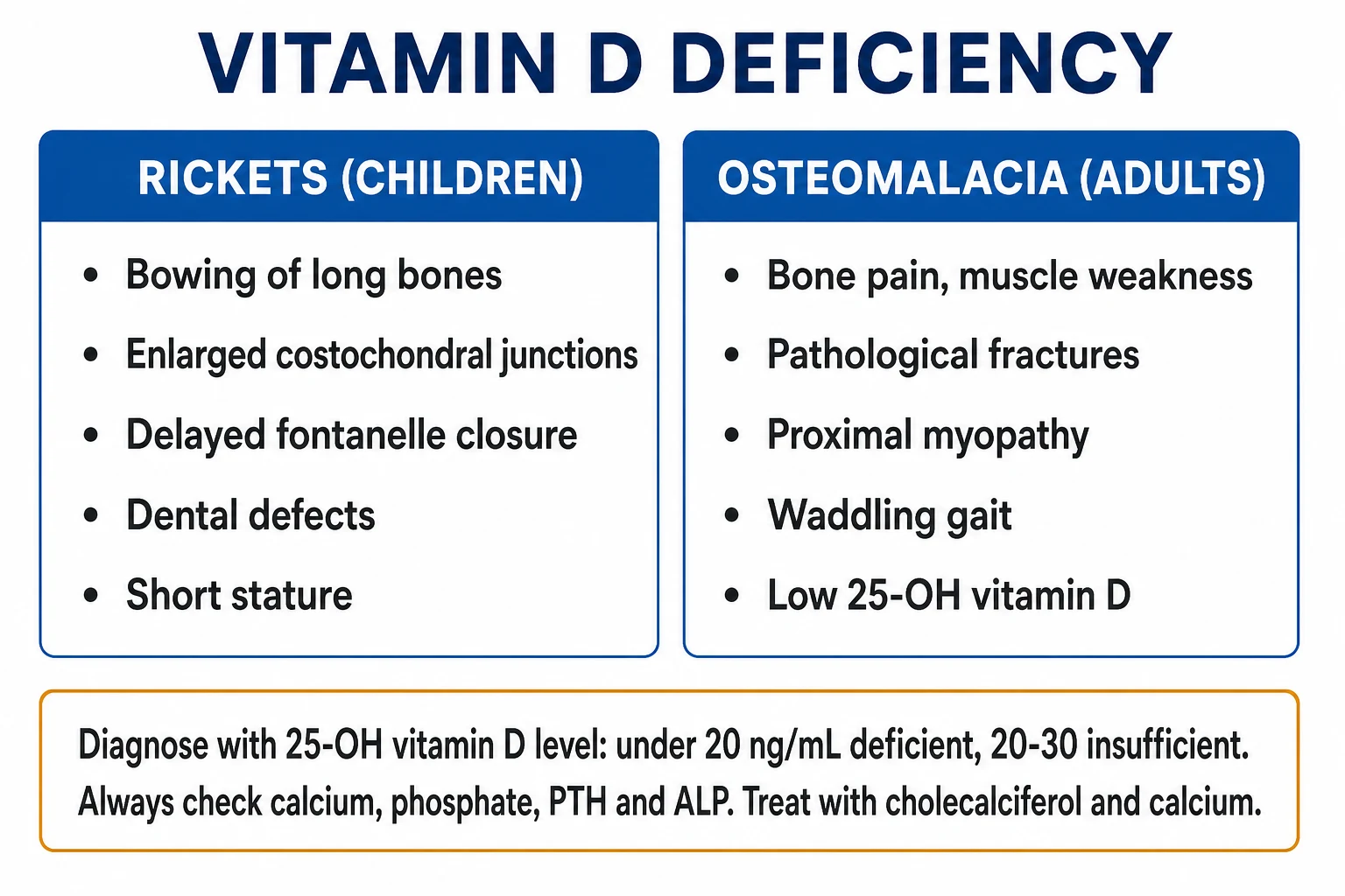
Vitamin D deficiency is the commonest nutritional deficiency worldwide, caused by inadequate UVB skin synthesis, dark skin, malabsorption, CKD, liver disease, obesity and enzyme-inducing drugs. In the growing skeleton it causes nutritional rickets — craniotabes, rachitic rosary, Harrison sulcus, widened wrists, genu varum, delayed fontanelle closure and dental defects. In the mature skeleton it causes osteomalacia — diffuse bone pain, proximal muscle weakness, a waddling gait and Looser zones (pseudofractures). The diagnostic test is 25-hydroxyvitamin D (calcidiol): under 30 nmol/L (12 ng/mL) deficient, 30 to 50 nmol/L insufficient, 50 nmol/L or above sufficient, with the classical biochemical tetrad of low or normal calcium, low phosphate, high PTH (secondary hyperparathyroidism) and raised alkaline phosphatase. Treatment is colecalciferol (vitamin D3) — a loading dose of 300,000 IU over 6 to 8 weeks (e.g. 20,000 IU weekly for 7 weeks) then maintenance 800 to 2000 IU daily — with adequate calcium; CKD and malabsorption require active vitamin D (calcitriol 0.25 to 1 mcg daily).
On this page & tools
Your progress
Saved locally on this device.
Exam tags
Red flags
Overview & Definition
Vitamin D is a prohormone rather than a vitamin in the strict sense, because the body can synthesise it entirely in the skin under ultraviolet-B (UVB) light. It occupies the centre of a tightly regulated calcium–phosphate–parathyroid hormone (PTH)–bone axis whose purpose is to maintain extracellular calcium and phosphate concentrations within narrow limits and to permit normal mineralisation of newly formed osteoid and growth-plate cartilage. When the supply of vitamin D — whether cutaneous or dietary — is inadequate, gut calcium and phosphate absorption fall, the serum calcium is defended at the cost of secondary hyperparathyroidism, and unmineralised osteoid accumulates. In the growing skeleton this manifests as nutritional rickets; in the mature skeleton as osteomalacia.[1][2]
Vitamin D deficiency is best understood as a continuum of severity rather than a binary state, because the biochemical and clinical features emerge in sequence as the vitamin D store depletes. The first detectable abnormality is a fall in 25-hydroxyvitamin D; next comes a rise in PTH (secondary hyperparathyroidism) with renal phosphate wasting; only later do alkaline phosphatase rise, radiographic changes appear and symptoms develop. Recognising the early, biochemical stage — before irreversible skeletal deformity — is the clinical art. [1]
The clinical stakes are real but largely preventable and reversible. Severe infantile rickets can present with hypocalcaemic seizures, laryngeal stridor and dilated cardiomyopathy, and nutritional rickets remains endemic in South Asia, the Middle East and parts of Africa despite being entirely preventable by a cheap, safe daily supplement. In adults, undiagnosed osteomalacia is a common and treatable cause of bone pain, proximal weakness and falls, frequently mistaken for polymyalgia rheumatica, fibromyalgia, inflammatory myopathy or metastatic bone disease. [1]
Classification
Vitamin D status is classified by the serum 25-hydroxyvitamin D concentration, the storage metabolite. Thresholds differ slightly between guidelines, but the UK Scientific Advisory Committee on Nutrition (SACN 2016) and NICE conventions are used here, with the Endocrine Society (US) equivalents in parentheses. To convert nmol/L to ng/mL, divide by 2.496.[1][2]
SUFFICIENT (over 50 nmol/L)
- 25-OH-D of 50 nmol/L or above (about 20 ng/mL or above)
- Normal calcium homeostasis and bone mineralisation
- UK SACN target for population musculoskeletal health
- Endocrine Society uses 75 nmol/L (30 ng/mL) as the 'sufficient' floor — a more stringent threshold
INSUFFICIENT (30 to 50 nmol/L)
- 25-OH-D between 30 and 50 nmol/L (12 to 20 ng/mL)
- PTH often begins to rise; subtle increase in bone turnover
- Usually asymptomatic but represents 'at-risk' status
- Correct with maintenance-dose vitamin D and lifestyle advice
DEFICIENT (under 30 nmol/L)
- 25-OH-D below 30 nmol/L (under 12 ng/mL)
- Secondary hyperparathyroidism established; osteoid mineralisation impaired
- Symptomatic rickets or osteomalacia at sustained low levels
- Requires loading-dose replacement then maintenance
SEVERE (under 12.5 nmol/L)
- 25-OH-D under 12.5 nmol/L (under 5 ng/mL)
- High risk of symptomatic hypocalcaemia and overt bone disease
- Seizures, cardiomyopathy and Looser zones may be present
- Treat aggressively and investigate for malabsorption
A second classification axis — central to paediatric rickets — separates calcipenic rickets (vitamin D and/or calcium deficiency, with low/normal calcium, low phosphate, high PTH) from phosphopenic rickets (renal phosphate wasting — X-linked hypophosphataemia from PHEX, tumour-induced osteomalacia from FGF23 — with low phosphate, normal calcium, normal or low PTH). The distinction is fundamental because the treatments diverge: calcipenic rickets responds to vitamin D and calcium, whereas phosphopenic rickets requires oral phosphate and active vitamin D and is worsened by calcium/vitamin D alone.[1]
Epidemiology & Risk Factors
Vitamin D deficiency is the commonest nutritional deficiency worldwide, with an estimated 1 billion people affected and around 50 per cent of the global population classified as insufficient. Subclinical deficiency is so prevalent in temperate latitudes — particularly through winter and spring — that it is best regarded as the default state in several high-risk groups unless actively supplemented.[1][2]
The high-risk groups the examiner expects you to name are predictable because they cluster around the two sources of vitamin D — the skin and the gut — and the three metabolic steps that activate it. In the skin, melanin competes for UVB photons, so people with dark South Asian, African, African-Caribbean or Middle Eastern skin need substantially more sun exposure (often 3 to 5 times longer) to synthesise the same amount of vitamin D3 as a fair-skinned person at the same latitude. Cultural and religious covering, housebound or institutionalised status, high latitude (above 35 degrees), winter season, air pollution, sunscreen (SPF 15 cuts synthesis by roughly 95 per cent), and window glass (which blocks UVB) all reduce cutaneous synthesis. [1]
Vitamin D deficiency — the numbers examiners ask
In the gut, malabsorption is the dominant mechanism — coeliac disease, inflammatory bowel disease, post-bariatric surgery (Roux-en-Y, sleeve gastrectomy), cystic fibrosis, chronic pancreatitis, short bowel syndrome and cholestatic liver disease all impair the fat-soluble absorption of vitamin D. Obesity sequesters vitamin D in adipose tissue, lowering the bioavailable pool and roughly doubling the maintenance requirement; nephrotic syndrome wastes vitamin D-binding protein (DBP) in the urine. Exclusive breastfeeding is a frequent infant cause, because breast milk contains only about 25 IU/L of vitamin D — far below the infant requirement — so all exclusively or partially breastfed infants need a supplement from birth. Prematurity compounds this because the fetal vitamin D store is laid down in the third trimester. [1]
The metabolic causes target the liver (impaired 25-hydroxylation in cirrhosis), the kidney (impaired 1-alpha-hydroxylation in chronic kidney disease — the single most important adult cause), and drug-induced catabolism (enzyme inducers — phenytoin, carbamazepine, phenobarbitone, rifampicin, isoniazid, some antiretrovirals such as efavirenz, and high-dose glucocorticoids — which up-regulate CYP24A1 and accelerate vitamin D degradation). Age itself is a risk: skin 7-dehydrocholesterol content falls by more than half between ages 20 and 70, and elderly, housebound populations combine reduced synthesis, reduced intake and reduced sun exposure. [1]
Pathophysiology
Vitamin D metabolism is a three-organ cascade — skin, liver, kidney — whose product, calcitriol (1,25-dihydroxyvitamin D), acts through a nuclear receptor on the gut, bone, kidney and parathyroid gland. Understanding each step is the key to understanding every cause, every laboratory abnormality and every treatment choice.[1][2]
[1]Calcitriol acts by binding the vitamin D receptor (VDR), a nuclear receptor that heterodimerises with the retinoid X receptor (RXR) and binds vitamin D response elements in target genes. In the intestine it transcriptionally up-regulates TRPV6 (apical calcium channel), calbindin-D9k (cytosolic calcium shuttle) and the Ca-ATPase (PMCA1b) — together doubling-to-tripling calcium absorption, from about 10 to 15 per cent at baseline to 30 to 40 per cent when calcitriol is abundant. Phosphate absorption rises in parallel through NaPi-IIb up-regulation. In bone, calcitriol permits — and is permissive for — normal osteoid mineralisation, and modulates osteoblast RANKL and osteoprotegerin (OPG) expression. In the kidney, calcitriol augments distal calcium reabsorption through TRPV5 and calbindin-D28k. In the parathyroid gland, calcitriol suppresses PTH gene transcription and parathyroid cell proliferation. [1]
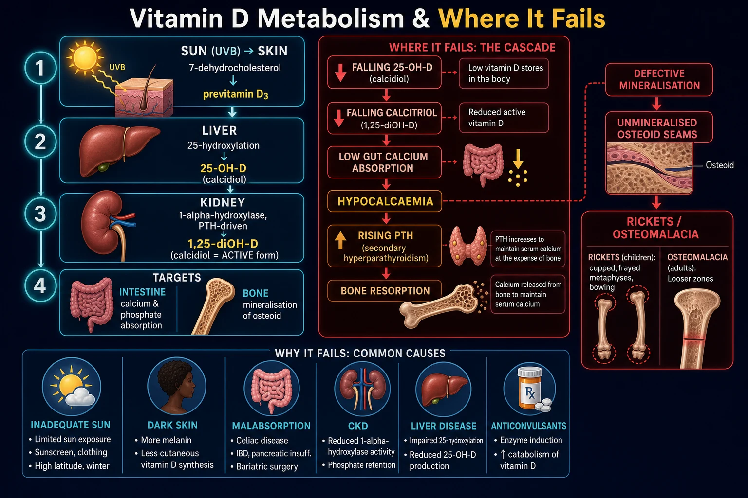
The failure cascade follows directly. When calcitriol falls, gut calcium absorption drops to a level too low to defend serum calcium. The parathyroid CaSR senses the falling ionised calcium within seconds and PTH rises — the appropriate secondary hyperparathyroidism of vitamin D deficiency. PTH mobilises calcium from bone (driving osteoclast-mediated resorption and a rise in alkaline phosphatase from coupled osteoblast activity) and wastes phosphate in the urine by down-regulating NaPi-2a in the proximal tubule — which is why phosphate falls even though bone resorption releases it. The serum calcium is partially defended by PTH and so reads low or low-normal, never profoundly low until stores are exhausted. Meanwhile osteoid continues to be laid down by osteoblasts but is not mineralised, widening the osteoid seam and producing the histological signature of osteomalacia. In the growing child the same defect strikes the hypertrophic zone of the growth plate, where unmineralised cartilage accumulates, the metaphysis widens, and the softened bone bows under weight — the radiographic and clinical picture of rickets. [1]
Calcitriol also has extra-skeletal actions — immunomodulation (macrophage-expressed CYP27B1 locally activates vitamin D to drive cathelicidin and beta-defensin antimicrobial peptides), cell differentiation and anti-proliferation, renin suppression, and modulation of insulin secretion. These observations underpinned two decades of epidemiological associations linking low 25-OH-D to cardiovascular disease, cancer, diabetes and infections — associations that, as the Evidence section shows, have not been confirmed by large randomised trials of supplementation.[6][7]
Causes
The causes of vitamin D deficiency map onto the synthesis chain described above and are best memorised as a single mnemonic. The trick to scoring well is to name the mechanism (synthesis failure, intake failure, malabsorption, impaired 25-hydroxylation, impaired 1-alpha-hydroxylation, increased catabolism, sequestration) as well as the cause.[1][2]
Causes of vitamin D deficiency — SUNLAMP
SUNLAMP
housebound, institutionalised, high latitude (above 35 degrees), winter, veiled or culturally covered clothing
dark skin (melanin), sunscreen, window glass, air pollution; age-related fall in skin 7-dehydrocholesterol
exclusive breastfeeding (breast milk only about 25 IU/L), vegan diet, malnutrition, prematurity
cirrhosis and cholestasis impair 25-hydroxylation (CYP2R1) and bile-salt-dependent absorption
coeliac, IBD, post-bariatric, cystic fibrosis, pancreatic insufficiency, short bowel
enzyme inducers up-regulate CYP24A1 — phenytoin, carbamazepine, phenobarbitone, rifampicin, efavirenz, glucocorticoids
CKD (loss of 1-alpha-hydroxylase), obesity (adipose sequestration), nephrotic syndrome (urinary DBP loss)
The rare genetic causes deserve a focused mention because they explain the refractory rickets stem. Vitamin-D-dependent rickets type 1 (VDDR-I) is autosomal recessive loss of CYP27B1 — the kidney cannot 1-alpha-hydroxylate, so calcitriol is low and the picture resembles severe nutritional deficiency despite adequate intake; it responds dramatically to physiological doses of calcitriol. Vitamin-D-dependent rickets type 2 (VDDR-II, hereditary vitamin-D-resistant rickets) is autosomal recessive loss-of-function of the VDR — calcitriol is high but ineffective; patients have alopecia (a key discriminating sign) and require very high doses of calcium and calcitriol. These must be distinguished from X-linked hypophosphataemic rickets (PHEX mutation), a phosphopenic rickets driven by FGF23 excess, in which calcium and PTH are normal and phosphate is low — the treatment is oral phosphate plus calcitriol, not vitamin D alone.[1]
Clinical Presentation
The clinical face of vitamin D deficiency depends on age (growing vs mature skeleton) and on severity. Many adults are entirely asymptomatic and the diagnosis is made on a screening 25-OH-D; the symptomatic extremes are rickets in children and osteomalacia in adults, with symptomatic hypocalcaemia as the acute emergency that crosses both. [1]
Rickets — the growing skeleton
Rickets is the failure of mineralisation at the growth plate (physis). The clinical features cluster into skeletal, dental, neuromuscular and acute hypocalcaemic groups.[1]
The head shows craniotabes (soft, ping-pong-ball skull, best felt over the occiput and parietal bones, in infants under 1 year), frontal bossing, delayed fontanelle closure (anterior fontanelle still open beyond 18 months), and delayed dentition with enamel hypoplasia and early caries. The chest has the rachitic rosary — palpable, beaded, anterior costochondral junctions that may be visible and tender — and a Harrison sulcus, a horizontal groove along the line of diaphragmatic insertion produced by the pull of the diaphragm on a softened rib cage. The limbs are the most examined: widening of the wrists (distal radius and ulna) and ankles, genu varum (bow legs) or genu valgum (knock knees), anterior bowing of the tibia, coxa vara, double malleoli, greenstick fractures, and reluctance to walk or a delayed walking milestone. The spine may show kyphoscoliosis and the pelvis flattens (forward projection of the promontory), which in adolescent girls can obstruct future vaginal delivery. Short stature (stunting) is common and is a key population-level marker of endemic nutritional rickets. [1]
The acute hypocalcaemic presentation is the emergency to remember. Exclusively breastfed infants of vitamin-D-deficient mothers, typically aged 3 to 18 months, can present with generalised tonic-clonic seizures, laryngeal stridor, carpopedal spasm, apnoea or cardiac failure — and rarely dilated cardiomyopathy and sudden death. An infant presenting with an afebrile seizure, particularly of South Asian or African origin and exclusively breastfed, must have calcium, magnesium, 25-OH-D, PTH and ALP checked before discharge; missing severe infantile rickets can be fatal. [1]
Osteomalacia — the mature skeleton
Osteomalacia is the failure of mineralisation of trabecular and cortical bone. Because the growth plates are fused, there is no metaphyseal cupping; instead, the patient complains of diffuse, dull, aching bone pain — typically in the lower back, pelvis, ribs and lower legs — that is worse on activity and reproduced by bone palpation (especially the tibial shafts and ribs).[2]
The signature feature is a proximal muscle weakness (a true myopathy of vitamin D deficiency, not a primary muscle disease). Patients describe difficulty rising from a low chair or the floor, climbing stairs, combing their hair or lifting objects overhead, and adopt a characteristic waddling (Trendelenburg-like) gait. The weakness is proximal and symmetrical, easily mistaken for polymyalgia rheumatica, inflammatory myositis, statin myopathy or thyroid myopathy — but the creatine kinase is normal (muscle is structurally intact; the defect is calcium-dependent muscle function) and the 25-OH-D is low. [1]
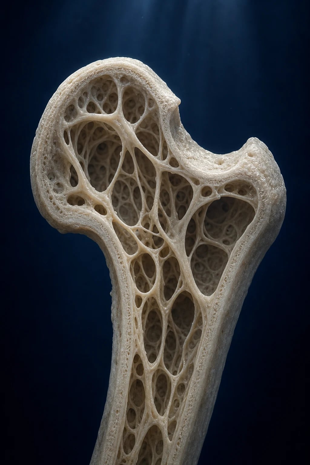
Pathological and insufficiency fractures occur, particularly at sites of Looser zones (see Investigations). Vertebral involvement causes loss of height and kyphosis. The disease is frequently mistaken for osteoporosis on DEXA — both lower bone mineral density — but osteomalacia has a low or normal calcium with high PTH that osteoporosis does not, and the treatment is different. [1]
Atypical and occult presentations
Examiners test the corners. The elderly, housebound patient may present with falls, proximal weakness, low-trauma fracture or unexplained bone pain rather than a textbook osteomalacia picture. The post-bariatric surgery patient (especially after Roux-en-Y or duodenal switch) develops deficiency months to years after surgery and may also have calcium, iron, B12 and thiamine deficiency. The institutionalised or chronically ill patient on long-term anticonvulsants has a drug-induced component layered on top of sun deprivation. The pregnant or lactating woman with deep, pelvic, weight-bearing pain may have transient osteoporosis of pregnancy or osteomalacia. The dark-skinned immigrant to a temperate climate often presents in the first spring after migration with biochemically overt deficiency. Cardiomyopathy in an exclusively breastfed infant is an under-recognised and occasionally fatal presentation. [1]
Differential Diagnosis
The differential is built by asking two questions: (1) is the bone disease rickets/osteomalacia at all? and (2) if it is, is it calcipenic or phosphopenic? The laboratory panel — especially calcium, phosphate, PTH and ALP — answers both.[1]
Calcipenic rickets/osteomalacia (LOW/normal Ca, LOW PO4, HIGH PTH, HIGH ALP)
- Vitamin D deficiency (sun, diet, dark skin, malabsorption) — the prototype
- Calcium deficiency alone (rare, low-calcium diet with adequate vitamin D)
- Vitamin-D-dependent rickets type 1 (CYP27B1 loss — low calcitriol)
- Vitamin-D-dependent rickets type 2 (VDR loss — HIGH calcitriol, alopecia)
- CKD with low calcitriol (also hyperphosphataemic, CKD-MBD)
- Anticonvulsant/rifampicin/efavirenz-induced increased catabolism
Phosphopenic rickets (LOW PO4, normal Ca, normal/low PTH)
- X-linked hypophosphataemic rickets (PHEX, FGF23 excess) — the prototype
- Autosomal dominant/recessive hypophosphataemic rickets (FGF23, DMP1, ENPP1)
- Tumour-induced osteomalacia (FGF23-secreting mesenchymal tumour — adult)
- Hereditary hypophosphataemic rickets with hypercalciuria (SLC34A3)
- Fanconi syndrome, proximal RTA, ifosfamide — renal phosphate wasting
- Does NOT respond to vitamin D alone — needs oral phosphate + calcitriol
Mimics NOT to confuse with rickets/osteomalacia
- Hypophosphatasia — LOW alkaline phosphatase (opposite of rickets); do NOT give bisphosphonate
- Osteogenesis imperfecta — blue sclera, hearing loss, family history, normal biochemistry
- Primary hyperparathyroidism — HIGH calcium (not low), HIGH PTH
- Metastatic bone disease / multiple myeloma — older adult, lytic lesions, anaemia, high ESR
- Polymyalgia rheumatica / inflammatory myositis — proximal pain but normal 25-OH-D, raised inflammatory markers / CK
- Scurvy (vitamin C deficiency) — bleeding gums, perifollicular haemorrhage
- Sickle cell disease — painful crises, normal biochemistry
Three pitfalls deserve emphasis. Hypophosphatasia (loss-of-function ALPL) presents with rachitic-like bone changes but a LOW alkaline phosphatase — the opposite of nutritional rickets — and is worsened by bisphosphonates; genetic testing and ALP are essential before treating "rickets" with a low ALP. Primary hyperparathyroidism shares high PTH and high ALP with vitamin D deficiency, but has HIGH calcium (not low) and is often discovered incidentally. Metastatic bone disease and multiple myeloma in older adults produce bone pain, fractures and lytic lesions that can superficially resemble osteomalacia; an immunoglobulin/serum-free light-chain screen, prostate-specific antigen and a myeloma screen are appropriate in the atypical older adult. [1]
Clinical & Bedside Assessment
The bedside assessment has three goals: identify the skeletal and muscular features, screen for the underlying cause, and detect the acute hypocalcaemic emergency. Begin with the tempo of presentation (acute seizure vs chronic bone pain) and the risk-factor history (sunlight, diet, dark skin, malabsorption symptoms — diarrhoea, weight loss, steatorrhoea — bariatric surgery, medications, CKD, family history). [1]
In the child with suspected rickets the examination is systematic. Head: feel for craniotabes (occipital/parietal softening), inspect for frontal bossing, check fontanelle size (delayed closure beyond 18 months) and the teeth (enamel hypoplasia, delayed eruption). Chest: palpate the costochondral junctions for the rachitic rosary and inspect for Harrison sulcus. Abdomen: hepatosplenomegaly may suggest malabsorption. Limbs: measure and palpate the wrists, ankles, knees for widening; observe genu varum/valgum with the child standing (intermalleolar/intercondylar distance); watch the gait. Growth: plot height and weight (stunting is a key marker). Neurology: tone, power, deep tendon reflexes (hypotonia is common). [1]
In the adult with suspected osteomalacia the examination focuses on the gait (waddling), the proximal muscle power (rising from a chair, stair-climbing, shoulder abduction), bone tenderness (rib and tibial shaft pressure reproduces the pain), the spine (kyphosis, vertebral tenderness) and a search for fractures (rib, pubic ramus, femoral neck). Look for skin and dietary clues (sun-avoidance, cultural covering), an abdominal scar (prior bariatric or bowel surgery), signs of chronic liver or kidney disease, and a drug history for enzyme inducers. Neuromuscular irritability signs (Chvostek, Trousseau) are positive only if hypocalcaemic; their presence indicates severe deficiency. [1]
In the acutely hypocalcaemic infant (seizure, stridor, apnoea, cardiac failure) the assessment is resuscitative: airway, breathing, circulation, ECG (prolonged QTc), IV access, bedside glucose and a stat calcium before treatment. [1]
Investigations
The diagnosis of vitamin D deficiency rests on a short panel — a single 25-hydroxyvitamin D level with calcium, phosphate, PTH and alkaline phosphatase — supported by radiographs in symptomatic bone disease. The trick is to interpret the panel together, because each abnormality points to a different point in the cascade.[1][2]
The blood panel — and what each result means
The single best marker of vitamin D status is 25-hydroxyvitamin D (calcidiol). It is the storage form, has a half-life of 2 to 3 weeks, reflects total-body input from both skin and diet, and the hepatic 25-hydroxylation step is substrate-driven (little regulated), so its level mirrors supply rather than activation. Do NOT measure calcitriol (1,25-(OH)2-D) to diagnose deficiency: it can be normal or even elevated in severe deficiency because secondary hyperparathyroidism drives renal 1-alpha-hydroxylase, masking the deficiency. Calcitriol measurement is reserved for CKD, hypercalcaemia of granulomatous disease (sarcoid, TB, lymphoma), suspected VDDR-II, and hypercalcaemia of malignancy.[2]
The classical biochemical tetrad of established vitamin D deficiency is: low or normal calcium (defended by PTH until late), low phosphate (PTH-driven phosphaturia), high PTH (appropriate secondary hyperparathyroidism) and high alkaline phosphatase (osteoblast activity coupled to PTH-driven resorption). The pattern distinguishes deficiency from primary hyperparathyroidism (high calcium, high PTH), CKD-MBD (low calcium, high phosphate, high PTH) and osteoporosis (normal calcium, phosphate, PTH and ALP). [1]
The classical biochemistry tetrad — read it as a pattern
Always also send a renal profile (eGFR — CKD is the commonest metabolic cause), liver function (cirrhosis impairs 25-hydroxylation), albumin (correct the calcium), magnesium (hypomagnesaemia causes refractory hypocalcaemia), and a FBC (anaemia may suggest malabsorption). In suspected malabsorption add coeliac serology (anti-tissue transglutaminase IgA plus total IgA), and consider faecal calprotectin or endoscopy. In the refractory case, send 1,25-(OH)2-D (high in VDDR-II, low in VDDR-I) and genetic testing (PHEX, CYP27B1, VDR, ALPL). [1]
Radiographs and bone imaging
Wrist and knee radiographs in the child show the pathognomonic cupped, frayed and widened metaphyses — the unmineralised hypertrophic cartilage of the growth plate bulges and splays under weight. The distal radial and ulnar metaphyses are the most reliable site. Other features include generalised osteopenia, bowing of long bones, greenstick fractures, delayed epiphyseal appearance, skull craniotabes and the rachitic rosary on chest X-ray. [1]
Pelvic and long-bone radiographs in the adult show Looser zones (pseudofractures) — symmetrical, narrow, transverse, radiolucent bands perpendicular to and through the cortex, without displacement, classically at the medial femoral neck, pubic rami, ribs, lateral scapula and clavicle. Extensive Looser zones are sometimes called Milkman syndrome. Other findings include osteopenia, codfish (biconcave) vertebrae and a triradiate pelvis (inlet deformity from softened bones). [1]
DEXA shows low bone mineral density (low T-score in adults, low Z-score in children and premenopausal women) and may be indistinguishable from osteoporosis — but the biochemistry distinguishes the two. Bone turnover markers (CTX, P1NP) are typically elevated. Tetracycline-labelled transiliac bone biopsy is the gold standard — demonstrating increased osteoid width and prolonged mineralisation lag time — but is rarely needed in clinical practice; it is reserved for atypical or refractory cases, suspected aluminium-related bone disease in dialysis, or before labelling chronic kidney disease bone disease. [1]
Self-test — A 28-year-old vegan woman with Crohn's disease has diffuse bone pain, waddling gait and difficulty climbing stairs. 25-OH-D 14 nmol/L, calcium 2.05 mmol/L, phosphate 0.6 mmol/L, PTH 12 pmol/L, ALP 320 U/L. What is the pattern and the next imaging test?
The biochemical pattern is the classical tetrad of calcipenic osteomalacia — low 25-OH-D, low/normal calcium, low phosphate (PTH phosphaturia), high PTH (secondary hyperparathyroidism), high ALP (osteoblast activity). The cause is Crohn's-related malabsorption layered on a vegan diet. The next imaging test is a pelvic and proximal femoral radiograph to look for Looser zones (pseudofractures) — symmetrical transverse radiolucent lines perpendicular to the cortex at the medial femoral neck, pubic rami and ribs. Treat with high-dose colecalciferol (with absorption-failure dosing — typically 2 to 3 times standard), adequate calcium, treat the Crohn's, and consider calcitriol if Colecalciferol cannot sustain levels.
Management — Resuscitation
Most vitamin D deficiency is not an emergency and is managed electively. The resuscitative scenarios are symptomatic hypocalcaemia (seizure, tetany, laryngeal stridor, prolonged QT, cardiomyopathy) and severe infantile rickets with hypocalcaemia.[1][2]
[1]The cardiomyopathy of severe infantile rickets is a dilated cardiomyopathy from chronic hypocalcaemia, occasionally the presenting feature, and is reversible with calcium and vitamin D over weeks; do not assume a viral aetiology in an at-risk infant without checking calcium and 25-OH-D. [1]
Management — Definitive & Stepwise
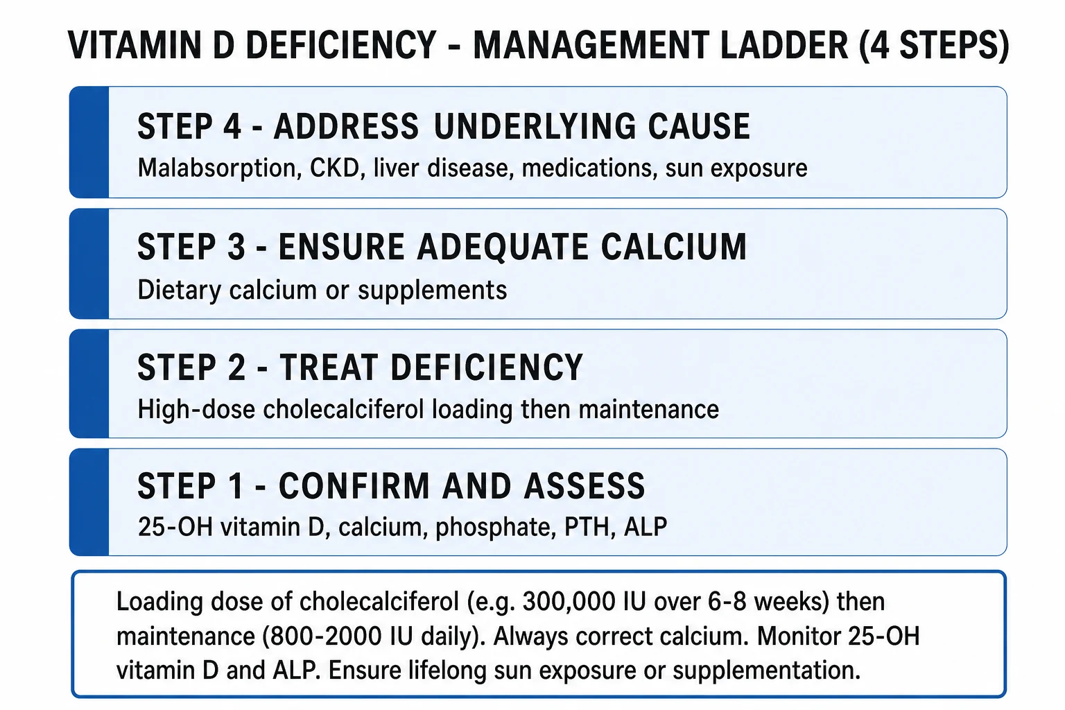
Once hypocalcaemia is excluded or treated, the definitive management has four pillars: (1) replace the vitamin D store with a loading dose then maintenance, (2) ensure adequate calcium, (3) use active vitamin D when 1-alpha-hydroxylation is impaired (CKD, severe liver disease, VDDR-I/II), and (4) treat the underlying cause.[1][2]
Step 1 — Replace the vitamin D store
The principle is a loading dose to replete the store, followed by a maintenance dose to sustain it. Colecalciferol (vitamin D3) is preferred over ergocalciferol (vitamin D2) because it raises 25-OH-D more reliably and for longer. Both are inactive prohormones that require hepatic 25-hydroxylation and (for full activity) renal 1-alpha-hydroxylation, so they are inappropriate as sole therapy when 1-alpha-hydroxylation is impaired.[2]
Standard loading (UK)
- Colecalciferol (vitamin D3) 20,000 IU weekly for 7 weeks (total 140,000 IU), OR 25,000 IU monthly for 6 months
- Alternative: 300,000 IU intramuscularly as a single stat dose (rarely needed; useful in non-adherence or severe malabsorption)
- Endocrine Society (US): 50,000 IU weekly for 8 weeks (total 400,000 IU)
- Munns global consensus (children): 2000 IU/day for 3 months, OR 50,000 IU weekly for 6 weeks
- All routes achieve a similar end-store; the principle is the same — repletion then maintenance
Maintenance
- Colecalciferol 800 to 2000 IU daily for adults (UK NICE up to 1000 IU/day for maintenance; Endocrine Society 1500 to 2000 IU/day)
- Infants and children under 1 year: 400 IU/day (UK) to 400 to 1000 IU/day (Munns global)
- Children 1 to 18 years: 600 to 1000 IU/day
- Pregnancy and lactation: 400 to 600 IU/day minimum; replete if deficient
- Obesity, malabsorption, anticonvulsants: 2 to 3 times the standard maintenance dose
Active vitamin D (when 1-alpha-hydroxylase is absent)
- Calcitriol (1,25-(OH)2-D) 0.25 to 1 mcg daily — for CKD, severe liver disease, VDDR-I, hypoparathyroidism, post-parathyroidectomy
- Alfacalcidol (1-alpha-hydroxycholecalciferol) 0.25 to 1 mcg daily — liver converts to calcitriol; widely used in CKD
- Acts in 1 to 2 days (vs weeks for colecalciferol) — preferred for acute hypocalcaemia or rapid effect
- Risk: hypercalcaemia and hypercalciuria — monitor calcium closely; do NOT give native colecalciferol alone in CKD
Calcium
- Adult elemental calcium 1000 to 1200 mg/day (dietary preferred — dairy, fortified foods; supplement the gap)
- Child elemental calcium 500 to 1000 mg/day; infant 200 to 500 mg/day
- Calcium carbonate (40 per cent elemental) — take with food; calcium citrate (21 per cent) — for PPI users or achlorhydria
- Without adequate calcium, vitamin D alone does NOT heal rickets — calcium is co-essential
Step 2 — Treat the underlying cause
Cure the deficiency and it recurs unless the cause is addressed. Coeliac disease needs a gluten-free diet and a malabsorption work-up. Inflammatory bowel disease needs disease control. Post-bariatric patients need lifelong higher-dose supplementation and micronutrient surveillance. Cholestatic liver disease may benefit from a water-miscible vitamin D preparation. CKD needs calcitriol and a CKD-MBD pathway (phosphate binders, vitamin D analogues, calcimimetics). Enzyme-inducing drugs should be reviewed and the dose of vitamin D increased where they cannot be stopped. Sun exposure advice (15 minutes of midday sun on face and forearms, May to September, lighter-skinned; longer for darker skin, without sunscreen during that window) and dietary advice (oily fish — salmon, mackerel, sardines; eggs; fortified foods; cod-liver oil) complete the package. [1]
Step 3 — Monitor
Recheck 25-OH-D at 3 to 4 months after starting loading (the store takes this long to plateau), then annually in high-risk groups. Track calcium, phosphate, ALP and PTH to confirm biochemical healing — ALP falls and PTH normalises as rickets heals, often before radiographic resolution. DEXA in adults with low bone density. In children, repeat wrist/knee radiographs at 3 months to document metaphyseal healing, and monitor growth. Calcitriol therapy in CKD requires close calcium monitoring (every 1 to 2 weeks during titration) to avoid hypercalcaemia. [1]
Specific Subtypes & Scenarios
Nutritional rickets in exclusively breastfed infants is the global prototype. Breast milk contains only about 25 IU/L of vitamin D, so any exclusively or partially breastfed infant is at risk unless supplemented. The Munns global consensus recommends 400 IU/day from birth, with a higher treatment dose (2000 IU/day for 3 months) for established deficiency. Maternal supplementation (at least 600 to 1000 IU/day, higher if deficient) raises the breast-milk concentration and protects the infant.[1]
Vitamin D deficiency in CKD is fundamentally a problem of lost 1-alpha-hydroxylase. As eGFR falls below about 60 mL/min, calcitriol production declines, phosphate retention rises, and secondary hyperparathyroidism (CKD-MBD) develops. Native colecalciferol repletes the store but cannot correct the calcitriol deficit, so active vitamin D analogues (calcitriol or paricalcitol), phosphate binders, and — for refractory secondary hyperparathyroidism — calcimimetics (cinacalcet, etelcalcetide) are added. Denosumab in dialysis patients can precipitate profound, prolonged hypocalcaemia (a hungry-bone-like state) and requires pre-dose calcium, vitamin D and phosphate optimisation. [1]
Malabsorption (coeliac, post-bariatric, cystic fibrosis, short bowel) combines impaired absorption with steatorrhoea-driven fat-soluble vitamin loss. Standard doses fail; 2 to 3 times the maintenance dose (or a water-miscible preparation) is needed, with periodic monitoring. The cause should be treated (gluten-free diet for coeliac; disease control for Crohn's) to restore absorptive capacity.[2]
Obesity sequesters vitamin D in adipose tissue, lowering the bioavailable pool. The Endocrine Society recommends 2 to 3 times the standard maintenance dose in obesity (and after bariatric surgery), reflecting the larger distribution volume. The deficiency is functional rather than absolute — weight loss raises 25-OH-D — but supplementation is needed in the interim. [1]
Pregnancy and lactation increase vitamin D requirement. NICE (UK) recommends 400 IU/day for all pregnant and breastfeeding women; the Endocrine Society suggests 1500 to 2000 IU/day is safe. Severe deficiency in pregnancy is associated with neonatal hypocalcaemia, neonatal rickets and (controversially) pre-eclampsia — repletion is recommended at booking. [1]
Drug-induced deficiency (enzyme inducers — phenytoin, carbamazepine, phenobarbitone, rifampicin, efavirenz, glucocorticoids) accelerates catabolism via CYP24A1. Increase the maintenance dose 2 to 3 fold and monitor; review the drug history in any unexplained deficiency. [1]
Vitamin-D-dependent rickets type 1 and 2 are rare autosomal recessive disorders presenting in infancy with severe, refractory rickets. VDDR-I (CYP27B1 loss) has low calcitriol and responds to physiological calcitriol (0.25 to 1 mcg/day). VDDR-II (VDR loss-of-function) has high calcitriol, alopecia, and requires very high-dose calcium and calcitriol; some respond only to IV calcium infusions in childhood.[1]
Complications & Pitfalls
The acute complications of severe deficiency are symptomatic hypocalcaemia (seizures, tetany, laryngeal stridor, prolonged QT, torsades de pointes, hypotension), dilated cardiomyopathy in infants, and pathological fractures through Looser zones or softened metaphyses. These are the indications for urgent IV calcium and high-dose vitamin D.[1]
The chronic complications of untreated disease are permanent skeletal deformity (bowing, kyphoscoliosis, triradiate pelvis — the latter obstructing future vaginal delivery), short stature, proximal myopathy with falls and fracture, secondary hyperparathyroidism driving further bone resorption, dental caries and enamel defects, and (in CKD) progression to renal osteodystrophy with vascular calcification. [1]
The pitfalls of treatment are several and examinable. Treating without calcium fails to heal rickets — vitamin D permits but does not substitute for calcium. Giving native colecalciferol alone in CKD is futile because 1-alpha-hydroxylation is impaired — use calcitriol or alfacalcidol. Using annual mega-dose bolus (500,000 IU) in the elderly paradoxically increases falls and fractures (Sanders 2010, JAMA) — the proposed mechanism is a transient surge in bone resorption after the bolus; daily or weekly dosing is safer.[3] Over-treatment produces hypercalcaemia (nausea, vomiting, polyuria, constipation, confusion, nephrocalcinosis) and hyperphosphataemia — rare with native vitamin D at standard doses but a real risk of calcitriol in CKD. The hungry-bone phenomenon during early treatment of severe rickets/osteomalacia can transiently worsen hypocalcaemia as the skeleton avidly re-mineralises; monitor closely and titrate calcium. Missing the diagnosis of hypophosphatasia (low ALP) and treating with bisphosphonates can be catastrophic.
Prognosis & Disposition
Vitamin D deficiency responds rapidly and completely to replacement. Biochemical markers (ALP, PTH) begin to normalise within weeks; radiographic healing of rickets is visible at 3 months; bone pain and muscle weakness improve within 4 to 8 weeks; and 25-OH-D reaches its plateau at 3 to 4 months after loading. Residual skeletal deformity (bowing, kyphosis, short stature) may persist in children treated late but usually remodels over years if growth continues; severe pelvic deformity in adolescent girls may require caesarean delivery.[1][2]
Disposition depends on context. The asymptomatic adult with screening-detected deficiency is managed entirely in primary care with oral colecalciferol. The symptomatic adult or child with bone disease is managed in outpatient endocrinology or paediatrics with radiographs and a 3-month review. The acutely hypocalcaemic infant (seizure, stridor, cardiomyopathy) is admitted for IV calcium and high-dose vitamin D, monitored on a paediatric ward with cardiac telemetry. CKD-related deficiency is co-managed with nephrology through a CKD-MBD pathway. Failure to respond to adequate doses mandates a search for malabsorption, ongoing drug effect, non-adherence, or a phosphopenic/genetic rickets — the dose is right but the diagnosis is wrong. [1]
Long-term maintenance is lifelong in high-risk groups (dark skin, elderly, malabsorption, CKD, post-bariatric, anticonvulsants) because the underlying cause rarely resolves; relapse is the rule without continued supplementation. [1]
Special Populations
Infants and children — universal supplementation from birth is the cornerstone of prevention. The Munns global consensus and UK NICE both recommend 400 IU/day from birth (regardless of feeding mode), with higher treatment doses for established deficiency. Premature infants need supplementation from birth regardless of feeding because the fetal store is laid down in the third trimester.[1]
Pregnant and breastfeeding women — 400 IU/day minimum (UK NICE); the Endocrine Society tolerates up to 1500 to 2000 IU/day. Severe maternal deficiency causes neonatal hypocalcaemia, congenital rickets and (debated) pre-eclampsia; repletion at the booking visit is standard. [1]
The elderly — combine 800 to 2000 IU/day with calcium 1000 to 1200 mg/day and falls-prevention advice. Skin 7-dehydrocholesterol content halves between ages 20 and 70, and housebound or institutionalised elderly are uniformly deficient unless supplemented. [1]
CKD — needs calcitriol (or paricalcitol, alfacalcidol) because 1-alpha-hydroxylation is impaired; native colecalciferol alone is insufficient. Co-manage through a CKD-MBD pathway with phosphate binders and calcimimetics as needed. [1]
Dark skin, veiled or culturally covered patients, and immigrants to high latitudes — higher maintenance doses (1000 to 2000 IU/day), routine screening, and culturally appropriate sun-exposure advice. The whole family is often deficient and should be screened and treated together. [1]
Obesity and post-bariatric patients — 2 to 3 times the standard maintenance dose to overcome adipose sequestration and malabsorption; lifelong micronutrient surveillance after bariatric surgery. [1]
Anticonvulsant / rifampicin / antiretroviral therapy — increase maintenance dose 2 to 3 fold and monitor; review the drug regimen where possible. [1]
Evidence, Guidelines & Regional Differences
The evidence base for vitamin D has been transformed in the last decade by a wave of large randomised controlled trials that tested whether supplementation reduces non-skeletal outcomes (cancer, cardiovascular disease, diabetes, falls, infections, mortality). The consistent answer has been no — vitamin D supplementation is a bone-health intervention, not a panacea. The regional guidelines have diverged on thresholds and maintenance doses, and these deltas are examinable.[2][6][7]
VITAL — Vitamin D and Omega-3 Trial
New England Journal of Medicine, 2019
2x2 factorial RCT of 25,871 US adults (men 50+, women 55+) given vitamin D3 2000 IU/day and/or omega-3 1 g/day vs placebo for a median 5.3 years.
Key finding
Vitamin D3 supplementation did NOT reduce the incidence of invasive cancer or major cardiovascular events, nor all-cause mortality, in a generally replete population.
Practice change
Established that routine high-dose vitamin D supplementation does NOT prevent cancer or cardiovascular disease in community-dwelling adults with baseline sufficient levels. Vitamin D is a bone-health intervention, not a panacea.
ViDA — Vitamin D Assessment Study
JAMA Cardiology, 2017
RCT of 5108 New Zealand adults given monthly bolus vitamin D3 100,000 IU vs placebo for a median 3.3 years.
Key finding
Monthly high-dose bolus did NOT reduce cardiovascular events, and post-hoc analysis raised concerns about increased falls in the elderly with the bolus regimen.
Practice change
Reinforced that large intermittent bolus dosing is inferior to daily/weekly maintenance; bolus may transiently increase falls and fractures in the elderly.
Trivedi et al — four-monthly cholecalciferol fracture trial
BMJ, 2003
RCT of 2686 community-dwelling UK adults (65-85) given oral cholecalciferol 100,000 IU every 4 months (300,000 IU/year) vs placebo for 5 years.
Key finding
Reduced first hip, wrist, forearm or spine fracture by 22 per cent (relative risk 0.78) and showed a non-significant trend to lower all-cause mortality.
Practice change
Supported intermittent moderate-dose vitamin D (300,000 IU/year as 4-monthly boluses) for community fracture prevention in the elderly — but distinguished from annual 500,000 IU mega-bolus, which is harmful.
Bischoff-Ferrari et al — fall-prevention meta-analysis
BMJ, 2009
Meta-analysis of 8 RCTs (n = 2426) of supplemental (700 to 1000 IU/day) or active vitamin D for fall prevention in older adults.
Key finding
Vitamin D 700 to 1000 IU/day reduced falls by 19 per cent (relative risk 0.81); doses below 700 IU/day were ineffective. Benefit was seen with supplemental but not high-dose active forms alone.
Practice change
Established the 700 to 1000 IU/day minimum for fall prevention and supported routine supplementation in falls-prone elderly.
Autier et al — non-skeletal outcomes systematic review
Lancet Diabetes & Endocrinology, 2017
Systematic review of meta-analyses and large RCTs of vitamin D supplementation for cardiovascular disease, cancer, diabetes, infections and mortality.
Key finding
No consistent benefit of vitamin D supplementation for any non-skeletal outcome. Epidemiological associations of low 25-OH-D with disease reflect reverse causation (illness lowers vitamin D), not causality.
Practice change
Reframed vitamin D as a bone-health intervention; cautioned against routine broad screening and high-dose supplementation for non-skeletal indications.
- Universal infant supplementation 400 IU/day from birth regardless of feeding mode.
- Diagnosis of rickets: 25-OH-D under 30 nmol/L with biochemical/radiographic features.
- Treatment of rickets: 2000 IU/day for 3 months (under 1 year) or 3000 IU/day (over 1 year), OR 50,000 IU weekly for 6 weeks, plus calcium 500 mg/day (under 1 year) or 1000 mg/day (older).
- Adequate maternal vitamin D (600 IU/day minimum, more if deficient) protects exclusively breastfed infants.
The central controversy is whether low 25-OH-D is a cause or a consequence of poor health. Observational associations are abundant and striking — low 25-OH-D with cardiovascular disease, cancer, infections, autoimmunity, mortality — but Mendelian randomisation and the large RCTs (VITAL, ViDA) suggest most associations are reverse causation: illness, inflammation, obesity and immobility lower 25-OH-D rather than the reverse. The current evidence-based position is: vitamin D supplementation prevents and treats rickets, osteomalacia and (with calcium) secondary hyperparathyroidism, and modestly reduces falls in the elderly; it does not prevent cancer, cardiovascular disease, diabetes or infections in the generally replete population.[6][7]
Prevention
Prevention is simple, cheap and effective, and is the public-health centrepiece of the global response to nutritional rickets.[1]
Supplementation is the most reliable strategy. The Munns global consensus and UK NICE PH56 converge on universal supplementation of all infants from birth (400 IU/day), with targeted higher-dose supplementation for pregnant and breastfeeding women, the elderly, those with limited sun exposure, and people with dark skin. Adherence is the limiting factor — rates of infant supplementation in high-risk populations are often below 30 per cent. [1]
Fortification of staple foods is the population-level strategy. Mandatory fortification of milk and dairy (USA, Canada), margarine (some countries), and increasingly flour or cooking oil (Finland, India pilot programmes) has demonstrably raised population 25-OH-D levels and reduced the prevalence of rickets. The UK has historically relied on voluntary fortification only, which delivers an inconsistent dose. [1]
Sun exposure advice is culturally calibrated: 15 minutes of midday sun on exposed face and forearms, May to September, without sunscreen (lighter skin), with longer exposure for darker skin, while avoiding erythema and skin-cancer risk. Sun exposure between November and March at latitudes above about 35 degrees is ineffective because the UVB flux is too low; in those regions, supplementation is essential through winter. [1]
Maternal supplementation during pregnancy and lactation raises breast-milk vitamin D and protects the exclusively breastfed infant — the highest-yield preventive intervention of all. [1]
Monitoring
Recheck 25-OH-D at 3 to 4 months after starting loading therapy (the store takes this long to plateau). Confirm the level has reached 50 nmol/L or above; if not, investigate adherence, malabsorption, ongoing drug effect or a wrong diagnosis. Once stable, monitor annually in high-risk groups (CKD, malabsorption, post-bariatric, anticonvulsants, dark skin, elderly). Track calcium, phosphate, ALP and PTH to confirm biochemical healing — ALP and PTH normalise as rickets heals, often before radiographic resolution.[1][2]
In children, repeat wrist and knee radiographs at 3 months to document metaphyseal healing, and monitor growth (height velocity accelerates as rickets heals). In adults on calcitriol or alfacalcidol (CKD, hypoparathyroidism), check calcium every 1 to 2 weeks during dose titration to avoid hypercalcaemia and hypercalciuria, then every 3 to 6 months once stable. In CKD, follow the full CKD-MBD panel (calcium, phosphate, PTH, ALP, 25-OH-D) on a defined schedule. [1]
Red Flags
Exam application bank (NEET-PG / INICET)
One-line answer
Vitamin D deficiency is the commonest nutritional deficiency worldwide, caused by inadequate UVB skin synthesis, dark skin, malabsorption, CKD, liver disease, obesity and enzyme-inducing drugs. In the growing skeleton it causes nutritional rickets — craniotabes, rachitic rosary, Harrison sulcus, widened wrists, genu varum, delayed fontanelle closure and dental defects. In the mature skeleton it causes osteomalacia — diffuse bone pain, proximal muscle weakness, a waddling gait and Looser zones (pseudofractures). The diagnostic test is 25-hydroxyvitamin D (calcidiol): under 30 nmol/L (12 ng/mL) deficient, 30 to 50 nmol/L insufficient, 50 nmol/L or above sufficient, with the classical biochemical tetrad of low or normal calcium, low phosphate, high PTH (secondary hyperparathyroidism) and raised alkaline phosphatase. Treatment is colecalciferol (vitamin D3) — a loading dose of 300,000 IU ove [1]
Worked stems (answer without another resource)
Stem 1 — Classic presentation. Map symptoms to mechanism; name the first investigation and first treatment step with dose/route if drug therapy is standard. [1]
Stem 2 — Unstable / complicated. List red flags that force immediate resuscitation, theatre, ICU, antidote, or reperfusion — and what you do in the first 15 minutes. [1]
Stem 3 — Atypical group. Elderly, pregnancy, child, or immunocompromised: how presentation and thresholds change. [1]
Stem 4 — Differential trap. Name the three closest mimics and one discriminator for each. [1]
Stem 5 — Disposition. Who goes home with safety-netting, who is admitted, who needs HDU/ICU/theatre, and what follow-up is mandatory. [1]
Rapid viva checklist
- Definition + classification
- Pathophysiology chain
- Bedside signs / criteria
- Score with exact components (if any)
- Emergency bundle
- Definitive therapy with doses
- Complications of disease and of treatment
- Special populations
- Guideline/trial name if classic
- Three exam traps
Coverage self-check
If you cannot answer any stem above from this page alone, re-read the matching section — the page is intended to be self-sufficient for final-prof and NEET-PG/INICET questions on Vitamin D Deficiency (Rickets & Osteomalacia).
Exam Pearls
References
- [1]Munns CF, Shaw N, Kiely M, et al. Global Consensus Recommendations on Prevention and Management of Nutritional Rickets J Clin Endocrinol Metab, 2016.PMID 26745253
- [2]Holick MF, Binkley NC, Bischoff-Ferrari HA, et al. Evaluation, treatment, and prevention of vitamin D deficiency: an Endocrine Society clinical practice guideline J Clin Endocrinol Metab, 2011.PMID 21646368
- [3]Bischoff-Ferrari HA, Dawson-Hughes B, Staehelin HB, et al. Fall prevention with supplemental and active forms of vitamin D: a meta-analysis of randomised controlled trials BMJ, 2009.PMID 19797342
- [4]Trivedi DP, Doll R, Khaw KT. Effect of four monthly oral vitamin D3 (cholecalciferol) supplementation on fractures and mortality in men and women living in the community: randomised double blind controlled trial BMJ, 2003.PMID 12609940
- [5]Scragg R, Stewart AW, Waayer D, et al. Effect of Monthly High-Dose Vitamin D Supplementation on Cardiovascular Disease in the Vitamin D Assessment Study : A Randomized Clinical Trial JAMA Cardiol, 2017.PMID 28384800
- [6]Manson JE, Cook NR, Lee IM, et al. Vitamin D Supplements and Prevention of Cancer and Cardiovascular Disease N Engl J Med, 2019.PMID 30415629
- [7]Autier P, Mullie P, Macchi A, et al. Effect of vitamin D supplementation on non-skeletal disorders: a systematic review of meta-analyses and randomised trials Lancet Diabetes Endocrinol, 2017.PMID 29102433