Gastroenterology · General Medicine
Acute & Chronic Diarrhoea
Also known as Diarrhoea · Acute gastroenteritis · Chronic diarrhoea · Infective diarrhoea · Traveller's diarrhoea · Antibiotic-associated diarrhoea
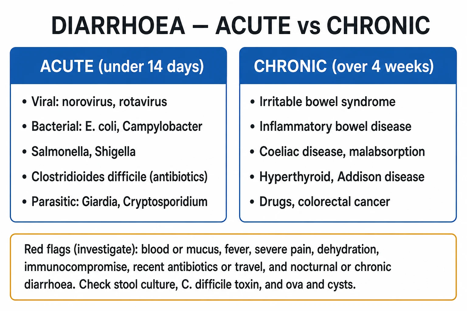
Diarrhoea is three or more loose or watery stools in 24 hours (or more frequent than is normal for the individual). Acute diarrhoea (under 14 days) is usually infective and self-limiting — viral (norovirus, rotavirus), bacterial (enterotoxigenic E. coli, Campylobacter, Salmonella, Shigella), Clostridioides difficile (after antibiotics) or parasitic (Giardia, Cryptosporidium, Entamoeba) — and the central clinical task is to separate the self-limiting viral illness (reassure, rehydrate) from disease that needs testing or specific treatment: invasive bacterial infection, C. difficile, or an underlying chronic cause. Chronic diarrhoea (over 4 weeks) has a wide differential including irritable bowel syndrome (commonest), inflammatory bowel disease, coeliac disease, malabsorption, hyperthyroidism, drugs and colorectal cancer. Management is rehydration first — low-osmolarity oral rehydration salts exploit the intact SGLT1 sodium-glucose co-transporter — then stool testing and cause-specific treatment, with antibiotics reserved for severe, bloody, febrile, immunocompromised or high-risk cases and contraindicated in Shiga-toxin E. coli (haemolytic uraemic syndrome risk). C. difficile is managed by stopping the offending antibiotic and giving fidaxomicin (preferred) or metronidazole/vancomycin.
On this page & tools
Your progress
Saved locally on this device.
Exam tags
Red flags

Overview & Definition
Diarrhoea is among the commonest clinical presentations worldwide and one of the leading causes of outpatient attendance and, in children, of death. The WHO definition is three or more loose or watery stools in 24 hours, or stool that takes the shape of its container — or, more pragmatically, an increase in stool frequency or fluidity that is abnormal for the individual. Dysentery is diarrhoea with visible blood and mucus, usually reflecting invasive colonic infection. [1]
The central clinical task is triage by duration and red flags: separate the self-limiting acute viral illness (reassure, rehydrate) from disease that needs investigation or specific treatment — invasive bacterial infection, C. difficile, Shiga-toxin E. coli with haemolytic uraemic syndrome (HUS), or a chronic cause. The duration of symptoms (acute under 14 days; persistent 14 to 29 days; chronic over 4 weeks) and the red flags (blood/mucus, fever, severe pain, dehydration, immunocompromise, recent antibiotics or travel, nocturnal or weight-losing stools) are the key tools.[1][2]
Classification
Diarrhoea is classified along three independent axes: duration, mechanism, and inflammatory vs non-inflammatory stool pattern. All three are examinable and each changes the management. [1]
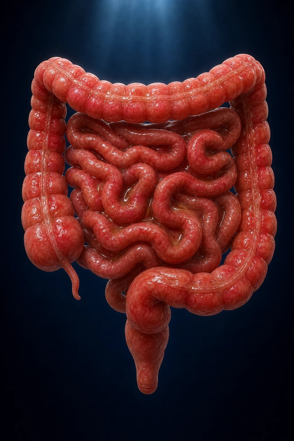
By duration
- Acute diarrhoea — under 14 days. Usually infective (viral, bacterial, parasitic) or a foodborne toxin, and most often self-limiting within a few days.
- Persistent diarrhoea — 14 to 29 days. An intermediate group; think of persistent infection (Giardia, C. difficile, Cystoisospora in immunocompromise), post-infectious irritable bowel syndrome, or the early presentation of an evolving chronic cause.
- Chronic diarrhoea — over 4 weeks (some authorities over 30 days). Demands a structured workup; the commonest cause overall is irritable bowel syndrome with diarrhoea (IBS-D), but organic causes — IBD, coeliac disease, malabsorption, hyperthyroidism, drugs, colorectal cancer — must be excluded by red flags and targeted testing.[2]
By mechanism (the pathophysiology axis — see Pathophysiology)
Secretory
- Active chloride secretion (CFTR) drives water into the lumen
- Toxins: cholera, ETEC heat-labile/LT and heat-stable/ST, VIPoma
- Persists on fasting; large volume; low stool osmotic gap
- No red cells/leukocytes
Osmotic
- Unabsorbed solute retains water (lactose, sorbitol, Mg2+, PEG, malabsorbed fat)
- Stops on fasting (the diagnostic clue)
- Stool osmotic gap over 50 mOsm/kg
- Causes: lactase deficiency, laxatives, coeliac, pancreatic insufficiency
Inflammatory/exudative
- Mucosal invasion/destruction -> blood, mucus, pus, protein leak
- Invasive bacteria (Shigella, Salmonella, Campylobacter, EIEC, EHEC) and cytotoxins (C. difficile TcdA/TcdB)
- Faecal leukocytes, lactoferrin, calprotectin raised
- Fever, tenesmus, lower-abdominal pain
Motility / dysbiosis
- Rapid transit (hyperthyroid, IBS-D, diabetic autonomic neuropathy, carcinoid, post-vagotomy)
- Slow transit/stasis -> SIBO (scleroderma, blind-loop, strictures)
- Bile-acid diarrhoea (ileal disease/resection, post-cholecystectomy)
- Mixed secretory + osmotic features
By stool pattern (inflammatory vs non-inflammatory)
Non-inflammatory (watery)
- Small-bowel, enterotoxin-mediated
- Large-volume, watery, no blood
- Pathogens: rotavirus, norovirus, ETEC, cholera, Giardia, Cryptosporidium
- Low fever, little systemic upset
Inflammatory (dysenteric)
- Colonic, invasive or cytotoxic
- Small-volume, frequent, blood and mucus, tenesmus
- Pathogens: Shigella, Campylobacter, Salmonella, EIEC, EHEC, Entamoeba, C. difficile
- Fever, marked systemic upset, faecal leukocytes
Enteric fever
- Typhoid/paratyphoid (Salmonella Typhi/Paratyphi)
- Constipation early, then 'pea-soup' diarrhoea; rose spots, relative bradycardia, splenomegaly
- Diagnose on blood/marrow culture in week 1, stool/urine culture later; Widal is supportive
Bristol Stool Scale (reproduce verbatim)
The Bristol Stool Chart grades stool form into seven types, used both for diagnosis and to monitor response: [1]
- Type 1 — separate hard lumps, like nuts (severe constipation)
- Type 2 — sausage-shaped but lumpy (mild constipation)
- Type 3 — sausage with cracks on the surface (optimal)
- Type 4 — smooth, soft sausage or snake (optimal)
- Type 5 — soft blobs with clear edges (lacking fibre)
- Type 6 — mushy, ragged, fluffy (mild diarrhoea)
- Type 7 — watery, no solid pieces (severe diarrhoea) [1]
Diarrhoea = Bristol types 5, 6 and especially 7. [1]
Epidemiology & Risk Factors
Diarrhoeal disease is one of the commonest reasons people seek medical care and, in children under 5, remains among the leading infectious causes of death worldwide (rotavirus and other pathogens causing several hundred thousand paediatric deaths annually, predominantly in low- and middle-income countries). In adults in industrialised settings it is a major cause of work loss, and its prevalence rises with foodborne outbreaks and international travel. [1]
Acute & chronic diarrhoea — the headline numbers
Causes by age
- Children — rotavirus (the dominant cause before vaccination, now much reduced where the vaccine is in the programme), norovirus, enteric adenovirus, enterotoxigenic E. coli, Salmonella, Shigella, Campylobacter, and Cryptosporidium.
- Adults — norovirus (the commonest overall), Campylobacter jejuni, Salmonella, Shigella, enterotoxigenic E. coli (travellers), and Clostridioides difficile (healthcare and antibiotic exposure).[1]
Host risk factors for infectious diarrhoea
- Reduced gastric acidity — proton-pump inhibitors and antacids, achlorhydria, post-gastrectomy/vagotomy, atrophic gastritis, biguanides; all reduce the gastric acid barrier to ingested organisms.
- Immunocompromise — HIV/AIDS (CD4 under 200 — Cryptosporidium, Microsporidium, Cystoisospora, CMV colitis), chemotherapy, transplant and immunosuppressants, hypogammaglobulinaemia, and the immunosuppressive drugs used in IBD.
- Extremes of age — infants and the elderly dehydrate faster and tolerate infection less well.
- Malnutrition — impairs gut immunity and mucosal repair, and worsens outcome.
- Comorbidity — IBD, haemoglobinopathies (Salmonella bacteraemia in sickle cell disease), aortic aneurysm (Salmonella endovascular infection), malignancy. [1]
Incubation periods (high-yield table — reproduce verbatim)
The incubation period of a foodborne illness is the single most discriminating historical clue: [1]
Drugs that cause diarrhoea (a long list — examine for these in chronic and antibiotic-associated diarrhoea)
Metformin (very common, dose-dependent), colchicine, proton-pump inhibitors, antibiotics (macrolides, penicillins, cephalosporins — both osmotic and C. difficile), SSRIs, orlistat (steatorrhoea), magnesium-containing antacids and laxatives, sorbitol (in sugar-free foods and liquid medicines), digoxin (toxicity), NSAIDs (enteropathy), mycophenolate, olmesartan (sprue-like enteropathy), gold, methotrexate, 6-mercaptopurine/azathioprine, and immune checkpoint inhibitors (immune-mediated colitis).[2]
Pathophysiology
Diarrhoea results whenever intestinal secretion exceeds absorption, when unabsorbed solute retains fluid in the lumen, when mucosal inflammation leaks fluid and blood, or when altered motility or dysbiosis impairs normal absorption. Understanding the four mechanisms — and how each therapy interrupts them — is exactly what the examiner probes at viva depth. [1]
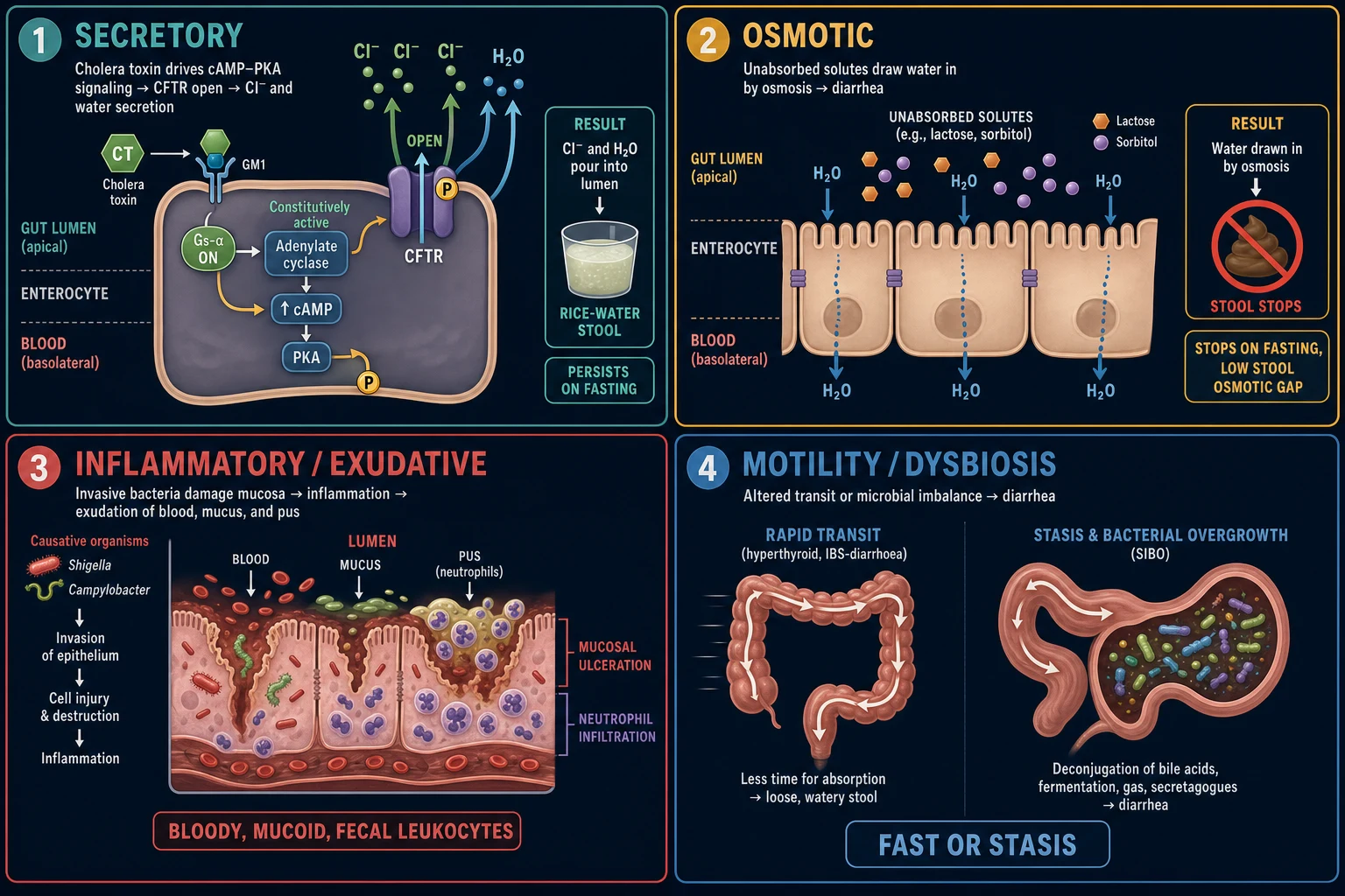
1. Secretory diarrhoea
The classic example is cholera. Vibrio cholerae produces cholera toxin, whose A-subunit catalyses the ADP-ribosylation of the Gs-alpha subunit of the stimulatory G-protein, locking adenylate cyclase permanently ON. The resulting sustained rise in intracellular cAMP activates protein kinase A (PKA), which phosphorylates and opens the CFTR chloride channel at the apical (luminal) membrane of the enterocyte. Chloride — followed by bicarbonate and, via paracellular flux, sodium and water — pours into the gut lumen. The gut simply cannot reabsorb the volume, producing the profuse, painless rice-water stool of cholera (up to a litre per hour).[1]
The crucial point: the enterocyte itself is structurally intact — only its signalling is hijacked. The SGLT1 sodium-glucose co-transporter remains functional, so oral sodium and glucose are still absorbed, dragging water with them. This is the molecular rationale for oral rehydration salts, which exploit the intact transporter to deliver water even when the toxin is over-secreting it. ETEC heat-labile toxin (LT) uses an identical mechanism; ETEC heat-stable toxin (ST) binds guanylyl cyclase C, raising cGMP, which similarly opens CFTR. Secretory diarrhoea is large-volume, persists on fasting, has a low stool osmotic gap (under 50 mOsm/kg), and shows no blood or leukocytes. [1]
2. Osmotic diarrhoea
When solute that cannot be absorbed accumulates in the lumen, it retains water by osmosis and the stool remains liquid. The classic causes are lactose (in lactase deficiency, including the temporary lactase loss after viral enteritis), sorbitol and xylitol (sugar-free foods and liquid medicines), magnesium (antacids, laxatives), polyethylene glycol, and malabsorbed carbohydrate or fat in coeliac disease or pancreatic insufficiency. The defining features: it stops on fasting (the solute is no longer ingested) and the stool osmotic gap is over 50 mOsm/kg. The stool is generally less voluminous than secretory diarrhoea and is accompanied by bloating, flatus and abdominal cramps. [1]
3. Inflammatory / exudative diarrhoea
Here the mucosa is invaded or destroyed, and the stool contains blood, mucus, pus and protein. Two sub-mechanisms operate: [1]
- Direct invasion — Shigella, Campylobacter jejuni, Salmonella, enteroinvasive E. coli (EIEC) and Entamoeba histolytica invade the colonic epithelium, triggering neutrophil influx, ulceration and an exudative colitis. The stool is small-volume, bloody and mucoid, with faecal leukocytes, lactoferrin and calprotectin all raised.
- Cytotoxins — Clostridioides difficile produces toxin A (TcdA, enterotoxic) and toxin B (TcdB, cytotoxic), which glucosylate and inactivate Rho-family GTPases, causing actin depolymerisation, cytoskeletal collapse and enterocyte death. The resulting fibrin-rich neutrophilic exudate overlying necrotic mucosa forms the pseudomembrane of pseudomembranous colitis.[4]
Shiga-toxin E. coli (STEC/EHEC, serotype O157:H7 and others) is a special, dangerous case. Shiga toxin binds the globotriaosylceramide (Gb3) receptor on colonic and renal endothelium; the A-subunit is internalised and cleaves a specific adenine residue from the 28S rRNA, inhibiting protein synthesis. The resulting endothelial injury and microvascular thrombosis produce the haemolytic uraemic syndrome (HUS) — microangiopathic haemolytic anaemia, thrombocytopenia and acute kidney injury. This is why antibiotics are avoided in suspected/confirmed EHEC — they can amplify toxin release and increase the risk of HUS.[5]
4. Altered motility and dysbiosis
- Rapid transit — in hyperthyroidism, IBS-D, diabetic autonomic neuropathy, post-vagotomy, carcinoid syndrome and some bile-acid diarrhoea, the gut moves contents through too quickly for adequate absorption, producing watery stool; nocturnal diarrhoea in particular points to an organic (non-IBS) cause.
- Slow transit / stasis — in systemic sclerosis (scleroderma), diabetic enteropathy, blind-loop syndrome, strictures and fistulae, small intestinal bacterial overgrowth (SIBO) develops; the bacteria deconjugate bile salts (causing fat malabsorption and steatorrhoea) and damage the mucosa directly.
- Bile-acid diarrhoea — ileal disease or resection, or post-cholecystectomy, spills bile acids into the colon, where they stimulate chloride secretion and irritate the mucosa (the BAAT/FXR pathway). [1]
Clinical Presentation
Typical acute infective diarrhoea
Abrupt onset of loose or watery stools with nausea, abdominal cramps, urgency, bloating, low-grade fever and sometimes vomiting, resolving within 1 to 5 days. Most cases are viral and self-limiting. Specific pathogens leave characteristic fingerprints (see below). [1]
Inflammatory / dysenteric diarrhoea
Frequent small-volume stools that are bloody and mucoid, with tenesmus, lower abdominal pain, fever and systemic upset. Causes: Shigella (often the most severe dysentery), Campylobacter, Salmonella, EIEC, EHEC (O157:H7), and Entamoeba histolytica (amoebic dysentery, classically "anchovy-paste" blood-stained mucus). [1]
Quantifying dehydration (the WHO/IMCI framework)
The single most important bedside assessment is the degree of dehydration, because it determines the route and rate of fluid replacement. The WHO/IMCI framework grades dehydration into three: [1]
- No dehydration — well, alert, drinks normally, normal perfusion, moist mucosae, brisk skin pinch.
- Some dehydration — two or more of: restless or irritable; sunken eyes; drinks eagerly or is thirsty; skin pinch goes back slowly. (WHO Plan B: ORS 75 mL/kg over 4 hours.)
- Severe dehydration — two or more of: lethargic or unconscious; sunken eyes; unable to drink or drinks poorly; skin pinch goes back very slowly. (WHO Plan C: IV Ringer's lactate 100 mL/kg in divided boluses.)[1]
In adults, look for postural blood pressure drop, tachycardia, oliguria, dry axillae, cool peripheries and altered consciousness as markers of severe volume depletion. [1]
Organism-specific clinical fingerprints (high-yield)
Pathogens by stool character — 'Watery vs Bloody'
WETBLOOD
Cholera, ETEC, rotavirus, norovirus, Giardia, Cryptosporidium — secretory/osmotic, small-bowel, no blood
Bloody diarrhoea after undercooked beef; risk of HUS — avoid antibiotics and antimotility
S. aureus and B. cereus emetic — vomiting within hours, brief illness
Shigella, Campylobacter, Salmonella, EIEC, Entamoeba — invasive colitis, tenesmus, faecal leukocytes
Avoid antimotility in bloody, febrile or C. difficile diarrhoea — toxic megacolon risk
Clostridioides difficile — test GDH + toxin, isolate, soap-and-water hand hygiene
Norovirus (closed communities, cruise ships, care homes) — vomiting prominent, short incubation
Metformin, colchicine, PPI, olmesartan, mycophenolate, checkpoint inhibitors — take a full drug history
Other classical clues: profuse painless rice-water stool (cholera); hamburger/E. coli O157:H7 with bloody stool and falling platelets (EHEC, HUS risk); antibiotic or healthcare exposure (C. difficile); camping/stream water with foul-smelling greasy floating stools and bloating (Giardia); right-lower-quadrant pain and pseudoappendicitis (Yersinia); reactive arthritis weeks later (Campylobacter, Salmonella, Shigella, Yersinia, Chlamydia); 'pea-soup' diarrhoea with rose spots, relative bradycardia and splenomegaly (typhoid). [1]
Atypical presentations (the exam traps)
- Elderly — may present with confusion, falls, anorexia or functional decline rather than classic symptoms; dehydrate rapidly; and mesenteric ischaemia, ischaemic colitis and diverticulitis must be excluded as surgical mimics. Consider faecal impaction with overflow in the bed-bound.
- Immunocompromised — wider organism differential (Cryptosporidium, Microsporidium, Cystoisospora, CMV colitis), severe and prolonged course, lower threshold to investigate and treat; neutropenic enterocolitis (typhlitis) in chemotherapy.
- Diabetic — autonomic neuropathy causes chronic diarrhoea, and SIBO is common; exclude mesenteric ischaemia in vascular disease.
- Pregnant — Listeria monocytogenes (bacteraemia, miscarriage risk), Salmonella (bacteraemia), and hepatitis E (fulminant in pregnancy) are of particular concern; dehydration threatens the fetus, so rehydrate early.[3]
Differential Diagnosis
The differential is built around the duration (acute vs chronic) and the mechanism (watery non-inflammatory vs bloody inflammatory). The key differentials to hold in mind, ranked by how commonly they are tested and how often they are missed:
- Viral gastroenteritis (norovirus, rotavirus) — the commonest acute cause; watery, self-limiting, no blood.
- Invasive bacterial diarrhoea (Campylobacter, Salmonella, Shigella, EIEC, EHEC) — bloody, febrile, faecal leukocytes.
- Clostridioides difficile — antibiotic/healthcare-associated; test GDH + toxin.
- Parasitic infection (Giardia, Cryptosporidium, Entamoeba) — travel/water exposure, foul-smelling greasy stool, persistent.
- Irritable bowel syndrome (IBS-D) — the commonest chronic cause; Rome IV, no red flags, no nocturnal stools.
- Inflammatory bowel disease (Crohn's, ulcerative colitis, microscopic colitis) — chronic bloody or watery diarrhoea with weight loss and raised calprotectin.
- Coeliac disease — chronic diarrhoea, weight loss, iron deficiency; tTG IgA positive.
- Endocrine and drug causes — hyperthyroidism, Addison's disease, metformin, PPI, olmesartan, checkpoint inhibitors.
- Surgical mimics not to miss — mesenteric ischaemia, appendicitis, diverticulitis, ischaemic colitis, ectopic pregnancy/ovarian torsion (women), colorectal cancer. [1]
Differential of acute diarrhoea
Viral gastroenteritis (norovirus, rotavirus) is the commonest. Then bacterial food poisoning, traveller's diarrhoea (ETEC), C. difficile, food intolerance (lactose), and drug side-effect. The surgical mimics that must not be missed are appendicitis (diarrhoea can precede the classical shift of pain to the right iliac fossa), mesenteric ischaemia (elderly, vascular disease, pain out of proportion, metabolic acidosis), partial small-bowel obstruction, diverticulitis, and — in women — ectopic pregnancy and ovarian torsion. [1]
Differential of chronic diarrhoea (ranked by frequency/examinability)
Irritable bowel syndrome with diarrhoea (IBS-D) is the commonest overall, then inflammatory bowel disease (Crohn's, ulcerative colitis), coeliac disease, microscopic colitis (collagenous/lymphocytic — think in older women on NSAIDs/PPIs), hyperthyroidism, drugs, small intestinal bacterial overgrowth, bile-acid malabsorption, chronic pancreatitis/pancreatic insufficiency (and pancreatic cancer), lactose intolerance, colorectal cancer, neuroendocrine tumours (carcinoid, VIPoma, gastrinoma/Zollinger-Ellison), HIV/AIDS-related, and factitious/laxative abuse.[2]
Distinguishing functional (IBS-D) from organic disease
The Rome IV criteria define IBS: recurrent abdominal pain at least 1 day per week over the last 3 months, associated with two or more of — related to defecation; associated with a change in stool frequency; associated with a change in stool form. Red flags that exclude IBS and mandate investigation: weight loss, nocturnal diarrhoea (rousing from sleep to defecate), visible blood, anaemia, family history of IBD/coeliac/colorectal cancer, new onset over 50, fever, and abnormal screening tests. Nocturnal diarrhoea is the single most useful discriminator — IBS by definition does not wake the patient from sleep to defecate.[2]
Distinguishing the inflammatory colitides
| Feature | Crohn's | Ulcerative colitis | Microscopic colitis | Ischaemic colitis | Infective colitis |
|---|---|---|---|---|---|
| Site | Anywhere mouth-to-anus, skip lesions, transmural | Colon only, continuous from rectum, mucosa only | Colon, macroscopically normal | Watershed (splenic flexure, sigmoid) | Variable |
| Stool | Loose, may be bloody; steatorrhoea if small-bowel | Bloody, mucoid, frequent | Watery (non-bloody) | Bloody, with pain | Bloody or watery |
| Histology | Non-caseating granulomas, transmural | Crypt abscesses, mucosal | Subepithelial collagen band / lymphocytes | Ischaemic necrosis | Acute inflammation, organisms |
Clinical & Bedside Assessment
Focused history
- Stool character — onset, frequency, volume, Bristol type, blood/mucus/pus, colour, floating or greasy (steatorrhoea), and nocturnal occurrence.
- Associated symptoms — vomiting, fever, abdominal pain (site), tenesmus, rash, arthropathy (reactive arthritis).
- Epidemiology — food (and the incubation period clue), travel, animal/water contacts, sick contacts, daycare, healthcare exposure, recent antibiotics/PPI, occupation (food-handler, healthcare worker).
- Drugs — antibiotics, PPI, metformin, colchicine, laxatives, NSAIDs, olmesartan, immunosuppressants, checkpoint inhibitors.
- Comorbidity and immune status — HIV, transplant, chemotherapy, IBD, thyroid disease, diabetes, vascular disease. [1]
Examination — dehydration first
Assess capillary refill, pulse, blood pressure (with a postural drop), JVP, mucous membranes, skin turgor, sunken eyes (and fontanelle in infants), and conscious level, and quantify dehydration by the WHO grade above. In adults, look specifically for a postural drop, oliguria and cool peripheries. [1]
Abdominal examination
Note distension, tenderness (diffuse vs localised — right iliac fossa in Yersinia/Crohn's/pseudoappendicitis; left iliac fossa in diverticulitis and ischaemic colitis), guarding or rigidity (peritonism — perforation, ischaemia), masses, and bowel sounds (absent in ileus/peritonism; tinkling/high-pitched in obstruction). Toxic megacolon presents with a distended, tender, tympanic abdomen with systemic upset — a surgical emergency. [1]
Rectal and systemic examination
A digital rectal examination may reveal an anal fissure (a pointer to Crohn's), a mass, faecal impaction with overflow in the elderly, or frank blood/melena. Look for the extraintestinal manifestations of IBD (erythema nodosum, pyoderma gangrenosum, uveitis, aphthous ulcers, large-joint arthritis) and the eye and tremor signs of hyperthyroidism. [1]
Investigations
The cardinal principle: most acute, uncomplicated, watery diarrhoea needs no testing — it is a clinical diagnosis of viral gastroenteritis. Test when there is a reason to test.[1][3]
When to test the stool (IDSA 2017)
Send stool when there is blood or mucus; severe, prolonged or bloody illness; fever; immunocompromise; recent antibiotics or healthcare exposure (suspect C. difficile); recent travel; daycare attendance or food-handling; or a suspected outbreak. Do not test formed stool, and do not send "test of cure" routinely after C. difficile. [1]
Stool tests for acute infectious diarrhoea
- Stool culture — Salmonella, Shigella, Campylobacter, and E. coli O157 (on sorbitol-MacConkey or by Shiga-toxin PCR).
- Ova, cysts and parasites — Giardia, Cryptosporidium, Entamoeba; needs three samples on alternate days and a concentration method; antigen or PCR is more sensitive for Giardia and Cryptosporidium.
- Clostridioides difficile — a two-step or algorithmic approach: GDH (glutamate dehydrogenase) plus toxin A/B EIA, with NAAT (PCR) as a reflex arbiter for discordant results; or NAAT plus toxin. Never test formed stool; a positive NAAT without toxin indicates a carrier, not active disease.
- Multiplex molecular stool panels — rapid, sensitive panels for viruses, bacteria and parasites; use where available, with reflex culture for public-health typing.
- Faecal inflammatory markers — faecal leukocytes, lactoferrin and calprotectin mark inflammation and support an invasive/inflammatory cause.[3]
Blood tests in the unwell patient
FBC (haemoconcentration; leucocytosis; eosinophilia in parasitic infection), U&E and creatinine (acute kidney injury, hypokalaemia, hyponatraemia), venous bicarbonate and lactate (acidosis — a marker of severe illness or mesenteric ischaemia), CRP, glucose, blood cultures (fever, suspected typhoid, sepsis), and group and crossmatch if major bleeding. Where HUS is suspected (after bloody diarrhoea, especially in children), send FBC with blood film, LDH, haptoglobin, reticulocytes and U&E — the triad is microangiopathic haemolytic anaemia (schistocytes), thrombocytopenia and AKI.[5]
Reproduced: WHO/IMCI dehydration grading (verbatim)
No dehydration
- Not enough signs to classify as some or severe dehydration
- Plan A: ORS at home, continue feeding, advise when to return
Some dehydration
- Two or more of: restless/irritable, sunken eyes, drinks eagerly/thirsty, skin pinch goes back slowly
- Plan B: ORS 75 mL/kg over 4 hours in a supervised setting, then reassess
Severe dehydration
- Two or more of: lethargy/unconscious, sunken eyes, unable to drink or drinks poorly, skin pinch goes back very slowly
- Plan C: IV Ringer's lactate 100 mL/kg — 30 mL/kg in 30 min (under 1 y: over 30 min) then 70 mL/kg over 2.5 h; reassess; switch to ORS when able
Reproduced: the Vesikari severity score (children)
The Vesikari score quantifies the severity of acute gastroenteritis in children from three days of symptoms: it sums frequency of diarrhoea and vomiting, duration, maximum temperature, and the treatment given (rehydration and hospitalisation). A score of 0 to 5 is mild, 6 to 9 moderate, 11 or more severe — used to compare severity in trials and rotavirus vaccine studies. [1]
The structured workup of chronic diarrhoea
- First-line bloods and screening — FBC, CRP, coeliac serology (tissue transglutaminase IgA + a total IgA to exclude deficiency), thyroid function, ferritin/iron studies, vitamin B12 and folate.
- Faecal calprotectin — under 50 micrograms per gram in a patient not on NSAIDs effectively rules out inflammatory bowel disease and supports a functional diagnosis.
- Stool — ova/cysts/parasites (three samples) and Giardia antigen/PCR; consider C. difficile if relevant exposure.
- Stool osmotic gap — calculated as 290 minus 2 x (stool Na + stool K); over 50 suggests osmotic, under 50 secretory diarrhoea. A stool osmolality below 290 suggests factitious diarrhoea (added water).
- Stool elastase — low in pancreatic exocrine insufficiency (chronic pancreatitis, cystic fibrosis, pancreatic cancer).
- Bile-acid malabsorption — SeHCAT (75-selenium-homotaurocholic acid) retention scan (under 15 percent at 7 days is positive); 7alpha-hydroxy-4-cholesten-3-one (C4) is a blood marker of bile-acid synthesis.
- Breath tests — lactose hydrogen breath test (lactose intolerance); glucose hydrogen breath test for SIBO.
- Endoscopy — colonoscopy with ileoscopy and biopsies (IBD, microscopic colitis, ischaemic colitis, CMV); OGD with duodenal biopsies (coeliac — Marsh grading).
- Cancer triage — faecal immunochemical test (FIT) and colonoscopy in the over-50s or anyone with red flags; CT colonography if colonoscopy is contraindicated.[2]
Management — Resuscitation
The first step in any dehydrating diarrhoea is to assess the airway, breathing and circulation, quantify the dehydration, and rehydrate. Fluid loss — and its electrolyte consequences — kills; the infecting organism rarely does, in an otherwise well adult. [1]
Oral rehydration — the cornerstone
Low-osmolarity ORS is first-line wherever the gut works (i.e. the patient can drink and is not vomiting relentlessly). The WHO low-osmolarity formulation contains sodium 75 mmol/L, glucose 75 mmol/L, potassium 20 mmol/L, citrate 10 mmol/L, chloride 65 mmol/L, osmolarity about 245 mOsm/L. Its genius is the SGLT1 sodium-glucose co-transporter: even when a toxin is driving secretion (cholera, ETEC), the enterocyte is structurally intact and SGLT1 still actively co-transports sodium with glucose, dragging water with it — so oral rehydration is as effective as intravenous in most cases, and far safer and cheaper.[1]
Volume targets (WHO): [1]
- Plan A (no dehydration) — ORS at home after each loose stool (~10 mL/kg), continue feeding.
- Plan B (some dehydration) — ORS 75 mL/kg over 4 hours under supervision, then reassess; switch to Plan A or C as needed.
- Plan C (severe dehydration) — IV Ringer's lactate (or 0.9% saline) 100 mL/kg, given as 30 mL/kg in the first 30 minutes (over 30 minutes if under 1 year) then 70 mL/kg over 2.5 hours; reassess and switch to ORS as soon as the patient can drink.[1]
Intravenous fluid resuscitation in the adult
For the adult with severe dehydration, hypovolaemic shock or AKI, gain IV access, take bloods, and give 0.9% sodium chloride or a balanced crystalloid (Ringer's lactate or Hartmann's) in 10 to 20 mL/kg boluses titrated to perfusion, heart rate and blood pressure. Then continue maintenance fluid with potassium replacement guided by serial U&E (correct hypokalaemia cautiously — gut losses of potassium are large). Correct metabolic acidosis with fluid resuscitation rather than bicarbonate unless severe. [1]
Recognising and managing the deteriorating patient
- Hypovolaemic shock — large-bore IV access, 500 mL crystalloid boluses, crossmatch, blood cultures, and broad-spectrum antibiotics if septic.
- Suspected HUS (EHEC, falling platelets, AKI, schistocytes) — urgent FBC, film, U&E; supportive fluid and blood-pressure support; no antibiotics, no antimotility; refer to nephrology; consider eculizumab in severe complement-mediated HUS.[5]
- Suspected mesenteric ischaemia / peritonism — keep nil by mouth, IV fluids, analgesia, urgent surgical review, and CT angiogram.
- Toxic megacolon (C. difficile, CMV, severe colitis) — nil by mouth, IV fluids and antibiotics, urgent surgical review, and daily abdominal X-ray to monitor colonic diameter.
Public-health and isolation measures
Isolate suspected or confirmed C. difficile (contact precautions; soap-and-water hand hygiene — alcohol gel does not kill spores; sporicidal environmental cleaning). Notify the relevant organisms (in the UK, food poisoning, cholera and dysentery are notifiable; in India, cholera and typhoid are notifiable under the Integrated Disease Surveillance Programme). Exclude food-handlers, healthcare workers and children in daycare until 48 hours after the first normal stool — longer for specific organisms where negative cultures are required.[3][4]
Management — Definitive & Stepwise
Most acute diarrhoea is supportive
For most acute, uncomplicated, watery diarrhoea — a clinical diagnosis of viral gastroenteritis — management is supportive only: low-osmolarity ORS, continue normal diet and feeding (do not starve — early refeeding shortens the illness), and no routine antibiotics or antimotility. The evidence supports early refeeding, and breast-feeding should continue in infants.[1]
When to give antibiotics (ACG 2016 / IDSA 2017)
Empirical antibiotics are reserved for severe, bloody, febrile or high-risk acute diarrhoea: [1]
- Moderate-to-severe traveller's diarrhoea — azithromycin 500 mg daily for 3 days (or 1000 mg as a single dose) is preferred, especially in regions of high fluoroquinolone resistance (Campylobacter in South/Southeast Asia and India); rifaximin 200 mg three times daily for 3 days for non-invasive watery diarrhoea; fluoroquinolones (e.g. ciprofloxacin 500 mg twice daily for 1 to 3 days) where resistance is low — avoid in pregnancy and children.
- Invasive dysentery (bloody, febrile) — azithromycin is first-line, covering Shigella, Campylobacter and Salmonella.
- Immunocompromise, fever with systemic illness, severe or prolonged illness — empirical therapy after stool sampling.[1][3]
Specific antimicrobial therapy
| Organism | First-line agent and dose | Notes |
|---|---|---|
| Shigella | Azithromycin 500 mg daily x 3 d, or ciprofloxacin if sensitive | Severe dysentery; resistance common |
| Campylobacter | Azithromycin 500 mg daily x 3 d | Usually self-limiting; treat if severe |
| Salmonella (severe) | Ciprofloxacin or azithromycin | Withhold in mild — antibiotics prolong carriage |
| Cholera | Doxycycline 300 mg single dose (or azithromycin/ciprofloxacin) | Always with aggressive ORS — the lifesaver |
| ETEC (traveller's) | Azithromycin, rifaximin or fluoroquinolone | Rifaximin only for non-invasive watery |
| Giardia | Metronidazole 400 mg TDS x 5 to 7 d; or tinidazole 2 g single; or nitazoxanide | Confirm with antigen/PCR |
| Entamoeba histolytica | Metronidazole 750 mg TDS x 10 d then a luminal agent (diloxanide furoate or paromomycin) | Treat the cysts to prevent relapse |
| Cyclospora / Cystoisospora | Co-trimoxazole 960 mg BD x 7 to 10 d | Common in immunocompromise |
| Typhoid | Ceftriaxone then azithromycin or fluoroquinolone | Treat carriers; surgery for gallbladder carriage |
| EHEC (O157:H7) | NO antibiotics — supportive; avoid antimotility | Antibiotics may increase HUS risk |
C. difficile management (SHEA/IDSA 2021 — reproduce the ladder)
Initial episode — non-severe
- Fidaxomicin 200 mg twice daily x 10 days (preferred)
- OR metronidazole 400 mg three times daily x 10 days if fidaxomicin/vancomycin unavailable
- Stop the precipitating antibiotic and review PPI
Initial episode — severe
- Severe = WBC over 15 x 10^9/L OR creatinine over 1.5 mg/dL (133 micromol/L) relative to baseline
- Fidaxomicin 200 mg BD x 10 d, OR vancomycin 125 mg QDS x 10 d
- Monitor for fulminant progression
Fulminant colitis
- Hypotension, ileus, or toxic megacolon
- Vancomycin 500 mg four times daily oral/NG PLUS IV metronidazole 500 mg three times daily
- Urgent surgical review for colectomy
Recurrence
- First recurrence: fidaxomicin, or vancomycin tapered/pulsed
- Multiple recurrences: vancomycin taper/pulse, fidaxomicin, or faecal microbiota transplant
- Bezlotoxumab (anti-toxin B monoclonal) for high-risk recurrence prevention
Severity thresholds: non-severe (WBC under 15 and creatinine under 1.5 mg/dL); severe (WBC over 15 or creatinine over 1.5 mg/dL); fulminant (hypotension, ileus, megacolon — surgical emergency). Always stop the offending antibiotic and deprescribe the PPI; contact precautions and soap-and-water hand hygiene (alcohol gel does not kill spores); sporicidal environmental cleaning.[4]
Antimotility and antisecretory agents — use and cautions
Loperamide (4 mg loading dose, then 2 mg per loose stool, maximum 16 mg per day) is safe and effective for non-bloody, afebrile watery diarrhoea in adults, and is a useful adjunct with antibiotics in traveller's diarrhoea. Avoid it in bloody or febrile diarrhoea, in young children (ileus, toxic megacolon), and in C. difficile (risk of toxic megacolon). Bismuth subsalicylate has both antisecretory and modest antimicrobial action and avoids the antimotility risks. Racecadotril (an enkephalinase inhibitor that is antisecretory rather than antimotility) is used in children in many countries. Zinc 20 mg daily for 10 to 14 days in children under 5 is a WHO recommendation — it shortens the current episode and reduces future episodes.[1]
Management of chronic diarrhoea by cause
- IBS-D — low-FODMAP diet, loperamide, peppermint oil, low-dose tricyclic (amitriptyline), rifaximin for IBS-D, eluxadoline; alosetron in restricted use.[2]
- Microscopic colitis — budesonide 9 mg daily, taper; stop NSAIDs and PPIs.
- Bile-acid diarrhoea — colesevelam (start 1.25 g, titrate) or cholestyramine.
- SIBO — a rotating cycle of antibiotics (rifaximin, metronidazole, doxycycline, norfloxacin).
- Pancreatic insufficiency — pancreatic enzyme replacement (PERT) — Creon 25,000 to 50,000 units with meals, plus a PPI.
- Coeliac disease — strict lifelong gluten-free diet.
- IBD — 5-ASA, steroids, biologics (see the IBD topic).
- Hyperthyroidism — carbimazole (see the topic).
- Neuroendocrine tumours (carcinoid, VIPoma, gastrinoma) — octreotide, then resect.
When to admit and when to discharge
Admit for severe dehydration, AKI, sepsis, bloody diarrhoea with systemic upset, immunocompromise, suspected HUS, toxic megacolon, suspected mesenteric ischaemia, inability to tolerate oral fluids, or social or safety concerns / diagnostic uncertainty. Manage as an outpatient when the patient is mildly unwell, well-perfused, has no red flags, can rehydrate orally, and has reliable follow-up. [1]
Specific Subtypes & Scenarios
Traveller's diarrhoea
Defined as three or more loose stools in 24 hours plus another symptom (cramps, nausea, vomiting, fever, tenesmus) within 14 days of return from a high-risk area. The commonest cause is enterotoxigenic E. coli (ETEC), then Campylobacter, Shigella, Salmonella and parasites (Giardia, Entamoeba, Cyclospora). Most cases are self-limiting within a few days; management is ORS plus a short course of standby antibiotics (azithromycin or rifaximin) for moderate-to-severe illness, and antimotility (loperamide) as an adjunct in non-bloody cases. Prevention — food and water hygiene ("boil it, cook it, peel it, or forget it"), bismuth, and (where available) vaccination.[1]
Clostridioides difficile infection (end to end)
Risk factors — antibiotic exposure (clindamycin, fluoroquinolones, cephalosporins, broad-spectrum penicillins), age over 65, healthcare exposure, PPI use, chemotherapy, and immunocompromise. Spectrum — from mild watery diarrhoea through pseudomembranous colitis to fulminant colitis with toxic megacolon, perforation and death. Diagnosis — a two-step GDH + toxin EIA (with NAAT reflex), or NAAT plus toxin; sigmoidoscopy showing pseudomembranes if toxin is negative and suspicion is high. Management — see the ladder above (fidaxomicin first-line; vancomycin for severe; vancomycin 500 mg + IV metronidazole for fulminant, with surgical review for colectomy). Recurrence is 15 to 30 percent after a first episode; manage with vancomycin taper/pulse, fidaxomicin, or faecal microbiota transplant for multiple recurrences. Infection control — contact precautions, soap-and-water hand hygiene, sporicidal cleaning.[4]
Acute gastroenteritis in children (see the dedicated topic)
Rotavirus (now much reduced by vaccination) and norovirus dominate, with a high dehydration risk. Use the WHO/IMCI assessment and ORS first-line (Plans A/B/C); zinc 20 mg daily for 10 to 14 days; avoid antimotility and unnecessary antibiotics; and give the rotavirus vaccine (India: ROTAVAC, in the Universal Immunisation Programme). Counsel parents on the red-flag return signs (blood, severe pain, dehydration, drowsiness, reduced urine).[1]
Food poisoning by preformed toxin
Staphylococcus aureus (creams, meats — onset 2 to 6 hours, prominent vomiting, recovery within a day); Bacillus cereus emetic (fried rice, 1 to 6 hours, vomiting) and diarrhoeal (8 to 16 hours, cramps and watery diarrhoea); Clostridium perfringens (reheated meat, 8 to 16 hours, watery diarrhoea). Botulism is a distinct toxin-mediated illness — descending flaccid paralysis (cranial nerves first, then symmetrical), with or without diarrhoea, treated with botulinum antitoxin and supportive care (including ventilation). [1]
Inflammatory and organic mimics not to miss
Ischaemic colitis (elderly, vascular disease, pain out of proportion, left-sided "thumb-printing" on X-ray); acute mesenteric ischaemia (severe pain, metabolic acidosis, risk of infarction — a surgical emergency); diverticulitis; appendicitis (diarrhoea can precede the classical shift of pain); and colorectal cancer presenting as altered bowel habit with blood in the older patient. [1]
Infectious diarrhoea in the immunocompromised
In HIV/AIDS with CD4 under 200 — Cryptosporidium, Microsporidium, Cystoisospora, CMV colitis; in transplant/neutropenic patients — neutropenic enterocolitis (typhlitis); in chemotherapy — C. difficile. There is a wider organism differential, a lower threshold to investigate and admit, the need to isolate, and empirical therapy guided by the CD4 count or level of immunosuppression, with early involvement of infectious diseases and microbiology.[3]
Complications & Pitfalls
Complications of acute infectious diarrhoea
Dehydration and hypovolaemic shock, acute kidney injury (pre-renal, and intrinsic with HUS), electrolyte disturbance (hypokalaemia, hyponatraemia, hypomagnesaemia, metabolic acidosis), reactive arthritis and Reiter's syndrome (HLA-B27, after Campylobacter, Salmonella, Shigella, Yersinia, Chlamydia), Guillain-Barre syndrome (Campylobacter jejuni, AMAN/AMSAN), haemolytic uraemic syndrome (EHEC Shiga toxin), toxic megacolon and perforation (C. difficile, CMV, severe colitis, IBD flare), bacteraemia and metastatic infection (Salmonella — osteomyelitis in sickle cell disease, mycotic aneurysm), post-infectious irritable bowel syndrome (about 10 percent after a bacterial gastroenteritis), and temporary lactose intolerance (lactase loss after viral enteritis).[3][5]
Post-infectious associations (high-yield pairing)
Post-infectious sequelae — 'after the bug, what?'
GREH
After Campylobacter jejuni — AMAN/AMSAN, ascending paralysis
After Shigella, Campylobacter, Salmonella, Yersinia, Chlamydia; HLA-B27; triad of urethritis, conjunctivitis/uveitis, arthritis
After Yersinia (classically), Salmonella, Campylobacter
After Shiga-toxin E. coli (O157:H7) — schistocytes, thrombocytopenia, AKI
Diagnostic pitfalls
Missing C. difficile in any antibiotic-associated or nosocomial diarrhoea; missing HUS in a child or adult with bloody diarrhoea after EHEC; over-treating Salmonella (antibiotics prolong carriage) or EHEC (antibiotics raise HUS risk); mistaking mesenteric ischaemia or peritonism for 'gastroenteritis' in the elderly; attributing an IBD flare to infection without stool testing (especially C. difficile, which can coexist and worsen IBD); failing to test for coeliac in chronic diarrhoea; chalking chronic nocturnal or weight-losing diarrhoea up to IBS; and not checking thyroid function in chronic diarrhoea. [1]
Therapeutic pitfalls
Giving loperamide in inflammatory/bloody diarrhoea, in young children, or in C. difficile (toxic megacolon); using fluoroquinolones where resistance is high (Campylobacter in South/Southeast Asia) or in pregnancy/children; unnecessary antibiotics in self-limiting viral illness; failing to stop the offending antibiotic in C. difficile; relying on alcohol hand-rub for C. difficile spores; and under-resuscitating severe dehydration. [1]
Prognosis & Disposition
Acute viral gastroenteritis resolves within 1 to 5 days with an excellent prognosis once rehydrated. Bacterial causes are mostly self-limiting in 5 to 7 days (Salmonella carriage may persist for weeks). Cholera can kill over 50 percent of untreated severe cases — but under 1 percent with proper ORS/IV rehydration. C. difficile recurs in 15 to 30 percent and carries a 5 to 10 percent mortality in the elderly and severe. HUS has a case-fatality under 5 percent, though some residual chronic kidney disease remains. [1]
Chronic diarrhoea by cause: IBS is benign (chronic relapsing-remitting, no excess mortality); coeliac excellent on a gluten-free diet (small increased lymphoma risk if non-adherent); IBD variable; microscopic colitis benign and relapsing; pancreatic cancer and neuroendocrine tumours cause-specific; colorectal cancer stage-dependent.[2]
Disposition: outpatient for mild, self-limiting illness with reliable follow-up; admit for moderate-to-severe dehydration, AKI, sepsis, bloody diarrhoea with systemic upset, immunocompromise, suspected HUS, toxic megacolon, suspected mesenteric ischaemia, or inability to tolerate oral fluids; ICU for shock, severe sepsis, multi-organ failure, or megacolon with perforation risk. Discharge when rehydrated and haemodynamically stable, tolerating oral fluids, with recovering renal function, no red flags, and clear safety-net advice (return if blood, severe pain, dehydration, fever, or worsening). [1]
Special Populations
Children (weight-based)
WHO ORS Plans A/B/C; 20 mL/kg boluses in shock; zinc 20 mg daily for 10 to 14 days (under 5 years); avoid loperamide; azithromycin 10 mg/kg daily for 3 days for severe dysentery; treat dehydration first. Give the rotavirus vaccine (India: ROTAVAC in the Universal Immunisation Programme) and follow the Integrated Management of Neonatal and Childhood Illness (IMNCI) framework.[1]
Pregnancy
Organisms of greater concern: Listeria monocytogenes (bacteraemia, miscarriage), Salmonella (bacteraemia), hepatitis E (fulminant in pregnancy). Safe antimicrobials — metronidazole, penicillins, cephalosporins, azithromycin. Avoid — fluoroquinolones, tetracyclines, doxycycline. Prioritise hydration to protect the fetus; avoid loperamide in the third trimester except where essential.[3]
Elderly
Anticipate atypical presentation (confusion, falls, anorexia), high risk of dehydration, AKI and electrolyte disturbance; exclude mesenteric ischaemia, diverticulitis, ischaemic colitis, C. difficile and colorectal cancer; drug-induced and antibiotic-associated diarrhoea are common; use lower fluid bolus volumes in heart failure; and consider faecal impaction with overflow.[2]
Immunocompromised
A wider organism differential (Cryptosporidium, Microsporidium, Cystoisospora, CMV colitis, neutropenic enterocolitis/typhlitis), lower threshold to investigate and admit, isolate, and empiric therapy guided by the CD4 count or level of immunosuppression, with early involvement of infectious diseases and microbiology.[3]
Anticoagulated, renal and cardiac patients
Correct dehydration promptly to avoid AKI and intravascular stasis; hold or adjust DOACs and warfarin if dehydrated or bleeding; avoid NSAIDs; ensure electrolyte stability (hypokalaemia precipitates digoxin toxicity); and adjust antimicrobial doses for renal function (e.g. reduce the fluoroquinolone dose, dose-adjust vancomycin in CKD, avoid/reduce nitazoxanide in severe renal impairment). [1]
Evidence, Guidelines & Regional Differences
Key controversies
Empirical antibiotics in traveller's diarrhoea — resistance, microbiome disruption, and C. difficile risk; probiotics (Lactobacillus rhamnosus GG, Saccharomyces boulardii) modestly shorten viral diarrhoea but carry risk in the immunocompromised; bismuth vs antimotility; bezlotoxumab (monoclonal anti-toxin B) for high-risk C. difficile recurrence prevention; faecal microbiota transplant formulations (capsule vs enema); and whether asymptomatic C. difficile carriers should be isolated. [1]
Exam Pearls
- Incubation period is the single best historical clue — under 6 hours (S. aureus, B. cereus emetic, vomiting-dominant); 8 to 16 hours (C. perfringens, B. cereus diarrhoeal); 1 to 3 days (norovirus, rotavirus, Salmonella, Shigella, Campylobacter, E. coli); over a week (Giardia, Cryptosporidium, amoebae, Yersinia, typhoid).
- The 'avoid antibiotics' organisms — Salmonella (prolongs carriage), EHEC/Shiga-toxin E. coli (raises HUS risk), C. difficile (worsens if unrecognised); the 'treat' organisms — severe Shigella, Campylobacter, cholera, Giardia, Entamoeba, typhoid, C. difficile.[1][3]
- Three killer mimics of 'gastroenteritis' not to miss — mesenteric ischaemia (elderly, pain out of proportion, metabolic acidosis), appendicitis / ectopic / ovarian torsion (especially in women), and ischaemic or toxic megacolon / peritonism.[2]
- The 'watery vs bloody' split — watery non-inflammatory (cholera, ETEC, rotavirus, norovirus, Giardia) vs bloody inflammatory (Shigella, Campylobacter, Salmonella, EIEC, EHEC, Entamoeba, C. difficile).
- Post-infectious associations — Campylobacter -> Guillain-Barre; Shigella/Campylobacter/Salmonella/Chlamydia -> reactive arthritis/Reiter; Yersinia -> erythema nodosum and pseudoappendicitis; EHEC -> HUS; any severe gastroenteritis -> post-infectious IBS.[5]
- C. difficile dose mnemonic — "fidaxomicin 200 mg BD x 10 d (preferred); metronidazole 400 mg TDS x 10 d; vancomycin 125 mg QDS oral (500 mg QDS if fulminant)". Alcohol hand-rub does NOT kill C. difficile spores — use soap and water.[4]
- ORS in one line — "the SGLT1 sodium-glucose co-transporter exploits the intact enterocyte (even in secretory diarrhoea) to absorb water and electrolytes — the rationale for low-osmolarity ORS over IV fluids wherever the gut works."[1]
Exam application bank (NEET-PG / INICET)
One-line answer
Diarrhoea is three or more loose or watery stools in 24 hours (or more frequent than is normal for the individual). Acute diarrhoea (under 14 days) is usually infective and self-limiting — viral (norovirus, rotavirus), bacterial (enterotoxigenic E. coli, Campylobacter, Salmonella, Shigella), Clostridioides difficile (after antibiotics) or parasitic (Giardia, Cryptosporidium, Entamoeba) — and the central clinical task is to separate the self-limiting viral illness (reassure, rehydrate) from disease that needs testing or specific treatment: invasive bacterial infection, C. difficile, or an underlying chronic cause. Chronic diarrhoea (over 4 weeks) has a wide differential including irritable bowel syndrome (commonest), inflammatory bowel disease, coeliac disease, malabsorption, hyperthyroidism, drugs and colorectal cancer. Management is rehydration first — low-osmolarity oral rehydration sa
Worked stems (answer without another resource)
Stem 1 — Classic presentation. Map symptoms to mechanism; name the first investigation and first treatment step with dose/route if drug therapy is standard. [1]
Stem 2 — Unstable / complicated. List red flags that force immediate resuscitation, theatre, ICU, antidote, or reperfusion — and what you do in the first 15 minutes. [1]
Stem 3 — Atypical group. Elderly, pregnancy, child, or immunocompromised: how presentation and thresholds change. [1]
Stem 4 — Differential trap. Name the three closest mimics and one discriminator for each. [1]
Stem 5 — Disposition. Who goes home with safety-netting, who is admitted, who needs HDU/ICU/theatre, and what follow-up is mandatory. [1]
Rapid viva checklist
- Definition + classification
- Pathophysiology chain
- Bedside signs / criteria
- Score with exact components (if any)
- Emergency bundle
- Definitive therapy with doses
- Complications of disease and of treatment
- Special populations
- Guideline/trial name if classic
- Three exam traps
Coverage self-check
If you cannot answer any stem above from this page alone, re-read the matching section — the page is intended to be self-sufficient for final-prof and NEET-PG/INICET questions on Acute & Chronic Diarrhoea.
References
- [1]Riddle MS, DuPont HL, Connor BA, et al. ACG Clinical Guideline: Diagnosis, Treatment, and Prevention of Acute Diarrheal Infections in Adults Am J Gastroenterol, 2016.PMID 27068718
- [2]Schiller LR. Evaluation of chronic diarrhea and irritable bowel syndrome with diarrhea in adults in the era of precision medicine Am J Gastroenterol, 2018.PMID 29713027
- [3]Shane AL, Mody RR, Crump JA, et al. 2017 Infectious Diseases Society of America Clinical Practice Guidelines for the Diagnosis and Management of Infectious Diarrhea Clin Infect Dis, 2017.PMID 29194529
- [4]Johnson S, Lavallee EE, Bohnen LM, et al. Clinical Practice Guideline by the Infectious Diseases Society of America (IDSA) and Society for Healthcare Epidemiology of America (SHEA): 2021 Focused Update Guidelines on Management of Clostridioides difficile Infection in Adults Clin Infect Dis, 2021.PMID 34492699
- [5]Tarr PI, Gordon CA, Chandler WL. Shiga-toxin-producing Escherichia coli and haemolytic uraemic syndrome Lancet, 2005.PMID 15781103