Gastroenterology · General Medicine
Acute Liver Failure
Also known as Acute liver failure · ALF · Fulminant hepatic failure · Acute hepatic failure · Fulminant hepatic failure (FHF)
Acute liver failure (ALF) is severe acute liver injury with coagulopathy (INR at least 1.5) and any degree of hepatic encephalopathy, developing within 26 weeks in a patient without pre-existing cirrhosis (Wilson's disease and reactivation of chronic hepatitis B are the accepted exceptions). The commonest cause in the developed world is paracetamol (acetaminophen) toxicity; in the developing world viral hepatitis (HAV, HBV, HEV) predominates. Other causes are idiosyncratic drug-induced liver injury (anti-TB, antiepileptics), autoimmune hepatitis, ischaemic/shock liver, Budd-Chiari, Wilson's disease, mushroom (Amanita phalloides) poisoning and pregnancy-related syndromes (HELLP, acute fatty liver of pregnancy). ALF is a critical medical emergency: death comes not from the liver itself but from cerebral oedema with raised intracranial pressure, sepsis and multi-organ failure. Management is in intensive care — identify and treat the cause (give N-acetylcysteine early, for paracetamol AND non-paracetamol ALF), support failing organs, control intracranial pressure, and make an early referral for emergency liver transplantation guided by the King's College Criteria.
On this page & tools
Your progress
Saved locally on this device.
Practise this topic
Exam tags
Red flags

Overview & Definition
Acute liver failure (ALF) is a rare, life-threatening syndrome of rapid loss of hepatocyte function. The defining triad, used worldwide, is severe acute liver injury, coagulopathy (INR at least 1.5) and any degree of hepatic encephalopathy, all developing within 26 weeks in a patient without pre-existing cirrhosis.[1][3]
The terminology has evolved, and examiners test the lineage. The original 1970 description by Trey and Davidson coined "fulminant hepatic failure" for encephalopathy appearing within eight weeks of the first symptom in a patient with no prior liver disease. Bernuau and colleagues in Paris later proposed a split into fulminant (encephalopathy within two weeks of jaundice) and subfulminant (between two weeks and three months). The modern AASLD/Paris consensus collapses these into "acute liver failure", timed from onset of symptoms to encephalopathy, up to 26 weeks, and divides it by tempo into hyperacute, acute and subacute. The reason all three definitions converge on the same practical point is that encephalopathy is the non-negotiable feature — a raised INR with a fully alert patient is severe acute hepatitis, not ALF, and behaves clinically like a different disease.[1][3]
Two conditions are the accepted exceptions to the "no chronic disease" rule, because they can present as ALF yet sit on a chronic substrate: Wilson's disease and reactivation of chronic hepatitis B. Recognising them matters because their treatment and transplant listing differ from the rest — Wilson's ALF uses its own prognostic score (the Wilson index), not the King's College Criteria.[1]
The clinical skill in ALF is not the diagnosis (that follows from the triad and the history) but three things done in parallel and under time pressure: (1) identifying and treating the cause (antidotes change the outcome — NAC, silibinin, aciclovir, delivery, chelation); (2) supporting the failing brain, circulation and kidney while the liver regenerates; and (3) deciding early whether the patient needs an emergency liver transplant, using the King's College Criteria.[2][6] Because cerebral oedema and multi-organ failure evolve over hours, early ICU care and prompt transplant-centre referral are decisive.
Classification
ALF is classified two ways — by time (O'Grady's syndromic classification) and by cause. Both carry prognostic weight. [1]
By the interval from jaundice to encephalopathy (O'Grady, 1993):[4]
- Hyperacute — encephalopathy within 7 days of jaundice. Typical of paracetamol and ischaemic injury. It carries the highest risk of cerebral oedema and severe coagulopathy, yet paradoxically the best transplant-free survival: if the patient survives the acute insult, hepatocyte regeneration restores function.
- Acute — encephalopathy at 8 to 28 days. Typical of many viral and drug-induced cases.
- Subacute — encephalopathy at 29 days to 26 weeks. Typical of indeterminate, idiosyncratic drug and some Wilson's cases; prominent jaundice and ascites, and a poor prognosis without transplant. [1]
Hyperacute (under 7 days)
- Typical cause: paracetamol, ischaemic 'shock liver'
- Highest risk of cerebral oedema and severe coagulopathy
- Paradoxically the BEST transplant-free survival (hepatocyte regeneration)
- Onset of encephalopathy can precede deep jaundice
Acute (8 to 28 days)
- Typical cause: viral hepatitis (HAV, HBV, HEV), many drug reactions
- Intermediate prognosis
- Jaundice usually well established before encephalopathy
Subacute (29 days to 26 weeks)
- Typical cause: indeterminate, idiosyncratic DILI, Wilson's, some Budd-Chiari
- Prominent jaundice and ascites
- WORST transplant-free survival; usually needs transplant
- Least cerebral oedema of the three
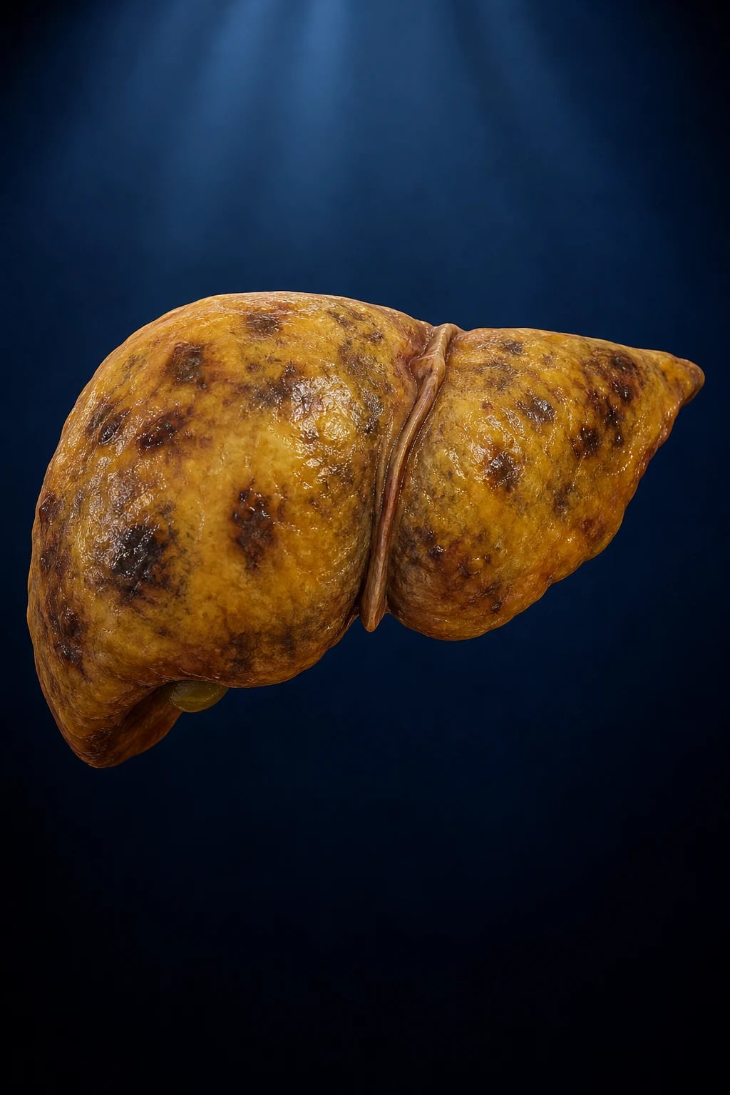
Epidemiology & Risk Factors
ALF is rare (incidence roughly 1 to 8 per million per year in the developed world; an estimated 2000 cases per year in the United States and around 400 per year in the UK) but dramatic: it strikes previously well, often young, people and carries a high short-term mortality. The aetiological mix is strongly regional:[1][6]
- Developed world (US, UK, Europe, Australasia): paracetamol (acetaminophen) is the single commonest cause (around 40 to 50 percent of cases in the UK), followed by idiosyncratic drug-induced liver injury and viral hepatitis; a substantial minority (around 10 to 20 percent) remain indeterminate.
- Developing world (India, South-East Asia, Africa): viral hepatitis predominates — hepatitis A, B and especially E (HEV is notorious for causing ALF and a high case-fatality in pregnancy, where mortality can approach 20 to 25 percent). Paracetamol overdose is far less common. [1]
Aetiology by category (the complete list):[1][3]
| Category | Specific causes and the exam clue |
|---|---|
| Drugs — dose-related | Paracetamol (top cause, developed world); mechanism is NAPQI / glutathione depletion |
| Drugs — idiosyncratic (DILI) | Isoniazid / anti-TB (rifampicin, pyrazinamide), valproate, halothane, phenytoin, carbamazepine, nitrofurantoin, amiodarone, statins, NSAIDs, sulphonamides, herbal / AYUSH preparations |
| Viral | HAV, HBV, HEV (HEV worst in pregnancy), HSV (immunosuppressed, pregnant — often missed), EBV, CMV, adenovirus, yellow fever, dengue |
| Vascular | Ischaemic hepatitis ("shock liver" after arrest/severe hypotension/heart failure), Budd-Chiari syndrome (hepatic vein thrombosis), portal vein thrombosis |
| Autoimmune | Autoimmune hepatitis (type I/II) |
| Metabolic | Wilson's disease (young), alpha-1 antitrypsin, galactosaemia, tyrosinaemia (children) |
| Toxins | Amanita phalloides mushroom; industrial carbon tetrachloride; herbal toxins |
| Pregnancy-related | Acute fatty liver of pregnancy (AFLP), HELLP syndrome (third trimester) |
| Malignancy / infiltration | Lymphoma, leukaemia, metastatic infiltration, melanoma |
| Miscellaneous | Heat stroke, Reye's syndrome (children, aspirin), veno-occlusive disease/sinusoidal obstruction syndrome |
| Indeterminate | No cause found after full work-up; poor prognosis without transplant |
Risk factors for severe paracetamol hepatotoxicity (the exam-tested modifiers that deplete glutathione or induce CYP2E1, lowering the toxic threshold): chronic alcohol misuse, malnutrition, fasting/anorexia, chronic liver disease, AIDS, and concomitant enzyme-inducing drugs (rifampicin, phenytoin, carbamazepine, isoniazid). These patients develop toxicity at doses nearer the therapeutic range — the rationale for treating any of them with NAC at lower thresholds.[1]
Causes of acute liver failure — PAVED-IM
PAVED-IM
the top cause in the developed world (also other drugs)
autoimmune hepatitis (ANA, ASMA, anti-LKM1, high IgG)
ischaemic / shock liver and Budd-Chiari syndrome
HAV, HBV, HEV (worst in pregnancy), HSV in the immunosuppressed
isoniazid, valproate, halothane, phenytoin, anti-TB, AYUSH/herbal
no cause found — poor prognosis without transplant
Amanita phalloides; infiltrative lymphoma/leukaemia
Plus two special-scenario causes to add deliberately: Wilson's disease (young + Coombs-negative haemolysis + Kayser-Fleischer rings) and pregnancy (AFLP/HELLP) (third trimester + hypoglycaemia + high ammonia). Both are common exam stems. [1]
Pathophysiology
The liver normally performs three jobs that ALF destroys: synthesis (clotting factors, albumin, glucose), detoxification (ammonia, drugs), and excretion (bilirubin). The mechanism cascade below explains every clinical feature.[1][3]
1. Massive hepatocyte necrosis. Whatever the cause — paracetamol's NAPQI, a virus, ischaemia, immune attack — the dominant lesion is loss of functioning hepatocytes, classically centrilobular (zone 3) in paracetamol and ischaemic injury because zone 3 is farthest from the oxygenated portal triad and richest in CYP2E1. The wholesale loss of functional hepatocyte mass is the engine of every downstream event: loss of synthesis, loss of detoxification, and loss of the liver's role in immune clearance. [1]
2. Loss of synthetic function leads to the laboratory and bleeding phenotype:
- Coagulopathy — the liver stops making the vitamin-K-dependent and non-dependent clotting factors (II, V, VII, IX, X, fibrinogen); factor VII has the shortest half-life so the INR/PT rises first. The INR is therefore the best single marker of hepatic synthetic function and of prognosis, and the cornerstone of the King's College Criteria.[2]
- Hypoglycaemia — impaired gluconeogenesis plus hyperinsulinaemia from reduced insulin clearance; dangerous because it worsens encephalopathy and mimics cerebral oedema. Check glucose hourly.
- Hypoalbuminaemia — contributes to oedema and reduced drug binding.
The "rebalanced haemostasis" concept. Although the INR is markedly prolonged, ALF patients bleed far less than the number suggests. This is because the liver also synthesises the natural anticoagulants (protein C, protein S, antithrombin) and clotting factors in parallel, and both fall together. The system is therefore "rebalanced" rather than auto-anticoagulated, and the INR overstates bleeding risk — which is why thromboelastography (TEG/ROTEM) is preferred over the INR for assessing functional bleeding risk, and why prophylactic FFP is not given.[1]
3. Loss of detoxification leads to the neurological phenotype — the single most important mechanism to understand, because it is how patients die. Ammonia and other neurotoxins are no longer cleared, cross a blood-brain barrier made leaky by circulating cytokines, and enter astrocytes where glutamine synthetase converts ammonia to glutamine. The accumulating glutamine is osmotically active, drawing in water — astrocyte swelling (cytotoxic oedema). Two further mechanisms amplify this: loss of cerebral autoregulation (blood flow passively follows arterial pressure, so hypotension directly causes cerebral hypoperfusion and hypertension risks hyperaemia), and a synergistic neurotoxicity between ammonia and lactate. The cascade progresses to cerebral oedema, raised intracranial pressure (ICP) and ultimately brainstem herniation, the leading cause of death in ALF.[3]
4. The systemic inflammatory / haemodynamic phenotype. Like sepsis, ALF produces a vasoplegic, hyperdynamic circulation: low systemic vascular resistance, high cardiac output, hypotension, microcirculatory shunting and tissue hypoxia. Nitric oxide and pro-inflammatory cytokines drive this. The degree of the systemic inflammatory response syndrome (SIRS) independently predicts encephalopathy progression and death. The result is acute kidney injury (ATN and/or functional hepatorenal physiology), lactic acidosis and progression to multi-organ failure.[1]
5. Immune dysfunction. The acutely failing liver cannot clear gut-derived endotoxin and bacteria from the portal blood, and Kupffer-cell function collapses. Patients with ALF are therefore functionally immunocompromised — a compensatory anti-inflammatory response syndrome (CARS) follows the initial SIRS, leaving the patient vulnerable to bacterial and fungal sepsis, the second leading cause of death. Fever and leucocytosis are often absent, so surveillance cultures are mandatory.[3]
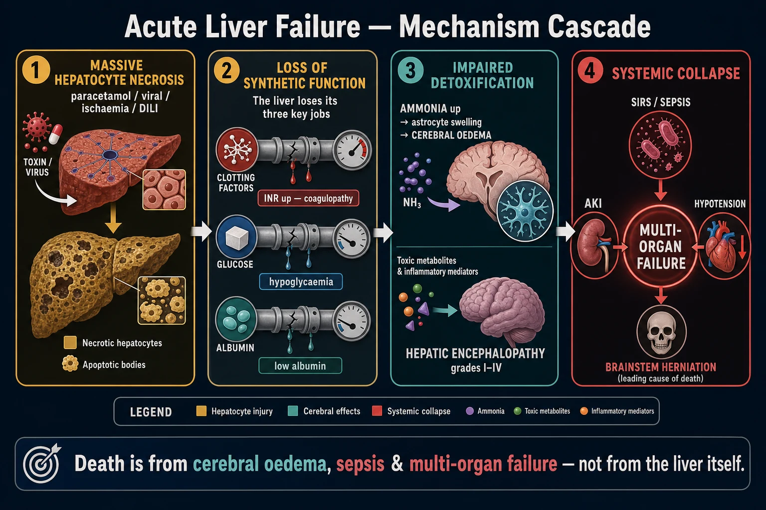
Acute liver failure — the numbers that decide management
Paracetamol toxicity — the molecular mechanism (examinable in detail). Paracetamol is mostly glucuronidated and sulphated; a small fraction (5 to 10 percent) is oxidised by CYP2E1 to the reactive intermediate NAPQI (N-acetyl-p-benzoquinone imine). At therapeutic doses NAPQI is immediately detoxified by conjugation with glutathione. After overdose, glutathione is depleted, NAPQI binds covalently to hepatocyte proteins (forming adducts), mitochondrial injury releases apoptosis-inducing factors, and zone-3 (centrilobular) necrosis follows. This is why N-acetylcysteine (NAC) works: it is a glutathione precursor (a cysteine donor) that repletes stores and detoxifies NAPQI. The threshold for toxicity is lowered by anything that depletes glutathione (alcohol, fasting, malnutrition) or induces CYP2E1 (rifampicin, isoniazid, phenytoin).[1][5]
Clinical Presentation
The presentation reflects speed of onset and the underlying cause, but the common features are: [1]
- Encephalopathy — the cardinal feature. West Haven grading: I mild confusion, euphoria/anxiety, asterixis (liver flap), sleep reversal; II lethargy, disorientation, marked asterixis; III somnolent but rousable, severe confusion, aggressive; IV coma (unresponsive to pain). Cerebral oedema becomes common from grade III to IV.[3]
- Jaundice — usually present, though in hyperacute (paracetamol) jaundice can be mild or lag behind encephalopathy.
- Coagulopathy — bleeding (gum, nasal, GI, puncture sites), bruising; the INR rises early.
- Right upper quadrant discomfort / tender hepatomegaly, nausea, vomiting, anorexia.
- Ascites — especially in subacute disease.
- Hypoglycaemia, metabolic acidosis, hypokalaemia, hypophosphataemia, infection (fever may be absent).
West Haven hepatic encephalopathy grade (and what each grade triggers)
Somnolent but rousable
Somnolent but rousable, severe confusion, aggression; cerebral oedema risk rising — start ICP-directed care and prepare to intubate
Asterixis (liver flap): with the arms outstretched and wrists hyperextended, a flapping, irregular flexion-extension at the wrist/metacarpophalangeal joints — a sign of grade I to II encephalopathy (metabolic brain dysfunction). It is lost as the patient progresses to deeper coma. [1]
Cause-specific clues (high-yield stems):[1]
- Paracetamol — deliberate overdose (often hours to a day before presentation), the four phases of paracetamol toxicity (0 to 24 h nausea/pallor; 24 to 72 h RUQ pain with rising INR/AST; 72 to 96 h peak hepatotoxicity with renal failure; over 5 d recovery or progression to ALF).
- Wilson's ALF — young patient, Coombs-negative intravascular haemolysis (low haemoglobin, high bilirubin), Kayser-Fleischer rings, low urate, low ceruloplasmin; ALP much lower than expected for the bilirubin.
- Amanita phalloides — mushroom foraging; a biphasic course: GI phase (nausea, vomiting, diarrhoea) 6 to 24 h after ingestion, then an apparent recovery, then hepatorenal failure at 2 to 4 days.
- Ischaemic hepatitis — context of cardiopulmonary arrest, severe shock, heart failure; very high AST/ALT (often over 10 000) that falls rapidly once perfusion is restored.
- Pregnancy (AFLP/HELLP) — third trimester, malaise, nausea, epigastric pain, hypoglycaemia, high ammonia, thrombocytopenia, haemolysis.
- HSV hepatitis — immunosuppressed or pregnant; often anicteric (jaundice may be absent); high mortality — treat empirically with aciclovir while awaiting PCR. [1]
The four phases of paracetamol toxicity
Atypical presentations examiners test deliberately. The elderly patient may have a blunted confusional state attributed to infection when it is early encephalopathy. The diabetic patient with paracetamol toxicity can collapse first from hypoglycaemia. The pregnant patient in the third trimester with AFLP presents with vomiting and abdominal pain that can masquerade as HELLP or even gastritis. The immunocompromised patient with HSV hepatitis is often anicteric with a flu-like prodrome, and the diagnosis is missed until the AST exceeds 5000. The alcoholic patient with malnutrition develops paracetamol toxicity at doses closer to therapeutic than to a true overdose — the so-called "therapeutic misadventure".[1]
[1]Differential Diagnosis
An acutely jaundiced, encephalopathic patient is not always ALF. Distinguish:[1][3]
- Acute-on-chronic liver failure / decompensated cirrhosis — the key exclusion. Look for stigmata of chronic disease, low platelets, splenomegaly, imaging/elastography showing a shrunken nodular liver, and previous LFTs. These patients do NOT meet ALF criteria (pre-existing cirrhosis) and are NOT listed under the same transplant pathway.
- Severe sepsis with cholestatic jaundice and septic encephalopathy — sepsis causes jaundice and confusion but, in the absence of shock liver, does not cause INR over 1.5 with encephalopathy. Look for the source, cultures, lactate.
- Ischaemic hepatitis (shock liver) — profound hypotension/cardiogenic shock; AST/ALT often over 1000 to 10 000 U/L with a rapid fall on reperfusion; the INR rises but encephalopathy is usually from the underlying insult rather than the liver.
- Intracranial event / metabolic encephalopathy — a stroke, subdural, uraemia, drug intoxication, Wernicke's or hypoglycaemia can mimic ALF encephalopathy; the liver function and INR will be near-normal (unless coexistent liver disease).
- Severe acute viral hepatitis without encephalopathy — by definition not ALF until encephalopathy appears.
- Acute biliary obstruction with cholangitis — fever, RUQ pain, jaundice (Charcot's triad); AST/ALT and INR usually only mildly abnormal; ultrasound/MRCP shows dilated ducts. [1]
Patterns that help distinguish the cause of a very high AST/ALT (over 1000 U/L):[1]
| Pattern | Most likely cause |
|---|---|
| AST/ALT over 1000 to 10 000 U/L, rapid rise and fall, context of hypotension | Ischaemic hepatitis |
| AST/ALT over 10 000 U/L after known overdose | Paracetamol toxicity |
| AST/ALT 400 to 2000 U/L, prodromal illness, viral risk | Acute viral hepatitis |
| AST/ALT modestly raised with very high bilirubin, young, haemolysis | Wilson's ALF |
| Anicteric transaminitis in the immunosuppressed/pregnant | HSV hepatitis |
Clinical & Bedside Assessment
ABCDE first. The priority in grade III to IV encephalopathy is airway protection and intracranial-pressure control — these patients need early intubation and mechanical ventilation.[3]
Focused assessment:
- Conscious level and encephalopathy grade — orientation (time, place, person), simple arithmetic, day/date; asterixis; then assign the West Haven grade I to IV.
- Cerebral oedema signs — the preterminal picture is Cushing's triad: hypertension + bradycardia + irregular (Cheyne-Stokes) respiration, with pupillary changes, decerebrate posturing and loss of brainstem reflexes as herniation progresses.
- Stigmata of chronic liver disease — palmar erythema, spider naevi, caput, gynaecomastia, Dupuytren's — to look for (but not rule out) underlying cirrhosis. Their absence does not exclude chronic disease.
- Kayser-Fleischer rings (slit-lamp) — Wilson's.
- Haemodynamic monitoring — hypotension, tachycardia; assess perfusion (capillary refill, lactate, urine output), not pressure alone. [1]
Collateral history (the single most useful bedside act): timing and dose of any paracetamol or drug ingestion (single vs staggered, co-ingestants), mushroom foraging, viral risk (travel, contacts, IV drug use, sexual exposure), pregnancy status, and available previous LFTs / imaging.[1]
Investigations
ALF investigations serve two purposes at once — prognostic monitoring (what is the liver doing, is it failing further, does the patient meet transplant criteria?) and aetiological diagnosis (what is the cause, is there a treatable antidote?). Send both panels on admission and repeat the prognostic markers (INR, lactate, glucose, pH, creatinine, phosphate) serially.[1][6]
First-line (every ALF patient): [1]
- Coagulation — INR/PT (the prognostic and transplant-trigger marker), APTT, fibrinogen.
- Biochemistry — LFTs (AST, ALT, bilirubin, ALP, GGT, albumin), glucose (hourly), lactate, ammonia (arterial preferred), arterial blood gas (arterial pH), U&E, creatinine, phosphate, magnesium, calcium, CRP.
- Haematology — full blood count, blood film, group and save.
- Microbiology — blood, urine and (if ascites) peritoneal cultures; surveillance cultures on admission and then daily.
- Pregnancy test in any woman of childbearing potential.
- Imaging — abdominal ultrasound with Doppler (hepatic/portal vein patency — Budd-Chiari, portal vein thrombosis; liver texture; biliary dilatation); CT/MRI brain for cerebral oedema, herniation or an alternative intracranial cause. Transient elastography to detect occult cirrhosis. [1]
Diagnostic work-up to identify the cause:[1]
- Paracetamol level (and salicylate, ethanol, toxicology screen).
- Viral serology — HAV IgM, HBsAg, anti-HBc IgM, anti-HCV, anti-HEV IgM/HEV RNA; HSV/EBV/CMV PCR in the immunosuppressed or pregnant.
- Autoimmune markers — ANA, anti-smooth muscle antibody (ASMA), anti-LKM1, immunoglobulins.
- Wilson's — serum ceruloplasmin, 24-hour urinary copper, slit-lamp for Kayser-Fleischer rings; the ALP-to-bilirubin ratio below 2 is characteristic.
- Pregnancy — AFLP/HELLP panel (platelets, haemolysis markers, glucose, ammonia, urate).
- Liver biopsy — rarely needed and hazardous (coagulopathy); considered for autoimmune, malignancy, or indeterminate disease, usually via the transjugular route. [1]
Serial lactate kinetics — the dynamic prognostic marker. A single lactate is informative, but the trajectory is more powerful. Bernal and colleagues (Lancet 2002) showed that arterial lactate measured early and again after fluid resuscitation outperformed the static King's College Criteria in paracetamol ALF: a lactate above 3.5 mmol/L early (before fluids) or above 3.0 mmol/L after 12 hours of resuscitation predicts poor outcome (death or transplant) with high sensitivity. Measure arterial lactate on admission, at 4 hours and again at 12 hours. A falling lactate, a falling INR and improving encephalopathy together signal hepatic regeneration and a favourable course; a static or rising lactate mandates transplant referral.[7][9]
Phosphate. Persistent hypophosphataemia in paracetamol ALF reflects hepatic regeneration (regenerating hepatocytes consume phosphate) and is paradoxically a favourable sign; it should be replaced to keep serum phosphate above 0.6 mmol/L. Hyperphosphataemia or adrenal failure are adverse.[1]
King's College Criteria — reproduced verbatim
The King's College Criteria (O'Grady, 1989) are the standard tool to decide emergency liver transplantation in ALF, and must be reproduced exactly. They were derived from the King's College Hospital cohort and remain the global default because they use bedside-available variables (pH, INR, creatinine, encephalopathy grade, age, cause, interval).[2]
For paracetamol-induced ALF — transplant if EITHER:
- Arterial pH below 7.3 after adequate fluid resuscitation (the single most powerful predictor), OR
- ALL THREE of: INR over 6.5 (the original 1989 paper expressed this as prothrombin time over 100 seconds), serum creatinine over 300 micromol/L (3.4 mg/dL), and grade III to IV encephalopathy. [1]
For non-paracetamol ALF — transplant if EITHER:
- INR over 6.5 alone, OR
- ANY THREE of: age under 10 or over 40; an unfavourable cause (non-A non-B hepatitis, drug-induced/halothane); interval from jaundice to encephalopathy over 7 days; INR over 3.5; bilirubin over 300 micromol/L (17.5 mg/dL). [1]
Paracetamol ALF
- Arterial pH below 7.3 after fluids, OR
- ALL three: INR over 6.5, creatinine over 300 micromol/L, grade III to IV encephalopathy
- Additive: arterial lactate over 3.5 mmol/L early (or over 3.0 after fluids)
- Hyperacute onset; best transplant-free survival if survives
Non-paracetamol ALF
- INR over 6.5 alone, OR
- ANY three of: age under 10 or over 40; non-A non-B / drug / halothane cause; jaundice-to-encephalopathy over 7 days; INR over 3.5; bilirubin over 300 micromol/L
- Includes DILI, autoimmune, viral (non-HAV/HBV), indeterminate
- Subacute / indeterminate patterns do worst
The lactate modifier and other prognostic models. Because King's criteria prioritise specificity over sensitivity (they miss some who will die), the arterial lactate modifier (Bernal, 2002) is added in paracetamol ALF: an early lactate over 3.5 mmol/L, or a post-resuscitation lactate over 3.0 mmol/L, independently predicts poor outcome.[7] In France the Clichy criteria (from HBV-related ALF) trigger transplant for factor V below 20 percent of normal in patients under 30 (with encephalopathy), or below 30 percent in those over 30. The dynamic model of Bernal (2016) tracks the trajectory of lactate, INR and encephalopathy over time and is more accurate than a single static assessment.[3]
Additional prognostic markers:[1][3]
- Arterial ammonia — over 100 to 150 mcg/dL predicts cerebral oedema and raised ICP; a falling ammonia suggests improvement.
- Factor V — low factor V (under 20 percent) is a poor prognostic marker (used in the Clichy criteria, France).
- Phosphate — persistent hypophosphataemia paradoxically favours regeneration (see above).
- Cause, age, interval jaundice-to-encephalopathy, and number of organ failures all carry independent prognostic weight. [1]
Management — Resuscitation
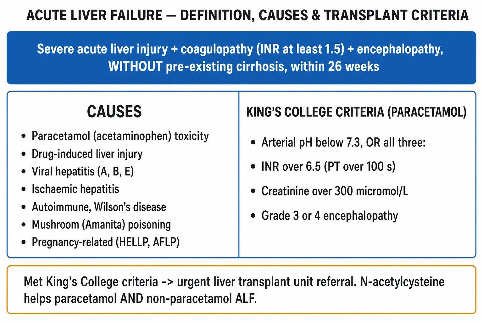
ALF is managed in intensive care, with early discussion and transfer to a liver transplant centre — do not wait for every King's criterion to be met before calling. The resuscitation priorities, done in parallel, are:[1][3][6]
ABCDE and organ support.
- Airway/Breathing — intubate and ventilate for grade III to IV encephalopathy (airway protection + ICP control); oxygen to maintain normoxia; target normocapnia (PaCO2 around 4.5 to 5.0 kPa; avoid hypercapnia which raises ICP).
- Circulation — two large-bore cannulae; arterial line; resuscitate with albumin (and balanced crystalloid); use noradrenaline (norepinephrine) as the first-line vasopressor for vasoplegic hypotension, with vasopressin as an adjunct; avoid over-resuscitation (worsens cerebral oedema).
- Disability — check and correct glucose (give 10 percent or 50 percent dextrose); maintain glucose over 7 mmol/L; assess encephalopathy grade.
- Exposure — full examination, source control, surveillance cultures. [1]
Time-critical antidotes (give EARLY and EMPIRICALLY).
- N-acetylcysteine (NAC) — give to ALL patients in whom paracetamol cannot be confidently excluded, and continue in paracetamol-induced ALF regardless of timing. NAC is safe, inexpensive and, crucially, the randomised trial by Lee et al (2009) showed IV NAC improves transplant-free survival in non-paracetamol ALF too.[5]
- Targeted antidotes once the cause is clear — see Definitive management.
Sepsis bundle. Patients with ALF are functionally immunocompromised and at high risk of infection. Send surveillance cultures, and start broad-spectrum prophylactic antibiotics plus an antifungal per local protocol; treat any infection promptly.[3]
Renal replacement therapy. Indicated for the standard AKI criteria (acidosis, fluid overload, hyperkalaemia, uraemia) and to help lower ammonia and control cerebral oedema. Prefer continuous modes (CVVHDF) over intermittent haemodialysis in cerebral oedema, to avoid rapid solute shifts that worsen ICP.[3]
Management — Definitive & Stepwise
Once resuscitated, management follows the five priorities, with cause-specific therapy layered on.[1]
The five priorities of acute liver failure management
ICU admission + identify and treat the cause
Stop all hepatotoxins; give N-acetylcysteine empirically; administer the specific antidote once the cause is clear (table below)
Encephalopathy and cerebral oedema
Head of bed 30 degrees midline; hypertonic saline to Na 145 to 155 mmol/L; mannitol 0.5 g/kg for rising ICP; continuous RRT; induced hypothermia 33 to 34 C refractory; consider ICP monitoring in grade III to IV
Coagulopathy and metabolic
Keep glucose over 7 mmol/L; vitamin K 10 mg IV; give FFP, cryoprecipitate or platelets ONLY if bleeding or before a procedure — never prophylactically; correct electrolytes and phosphate
Circulation, infection and renal
Noradrenaline first-line, albumin, vasopressin adjunct; surveillance cultures plus broad-spectrum antimicrobials and antifungal; continuous renal replacement therapy
Early transplant referral
Apply the King's College Criteria; refer the moment criteria are met, not when multi-organ failure develops; super-urgent national listing where available
2. Encephalopathy / cerebral oedema — the goal is to keep cerebral perfusion pressure above 60 mmHg and ICP below 20 mmHg.
- Head of bed elevated 30 degrees, midline (optimises venous drainage).
- Maintain the five norms: normoxia, normocapnia, normotension, normoglycaemia, normothermia. Avoid hyponatraemia (it worsens cerebral oedema).
- Hypertonic saline (30 percent bolus, e.g. 20 to 30 mL of 30 percent NaCl, or a 5 percent infusion) to a target sodium of 145 to 155 mmol/L — the cornerstone of ICP control in ALF.
- Mannitol 0.5 g/kg IV bolus (20 percent) for rising ICP, repeated if serum osmolality is under 320 mOsm/L and urine output is adequate; use continuous RRT to clear the mannitol and control ammonia.
- Continuous renal replacement therapy to control ammonia, fluid and acid-base.
- Rescue therapies for refractory ICP: induced hypothermia (33 to 34 degrees C), indomethacin (reduces cerebral blood flow), barbiturates (thiopentone) to suppress cerebral metabolism.
- ICP monitoring — considered in selected centres for grade III to IV patients, weighing the bleeding risk (which is lower than the INR suggests) against the benefit of an ICP-targeted approach.[3]
3. Coagulopathy / hypoglycaemia / metabolic.
- Correct glucose and keep it over 7 mmol/L (10 percent dextrose infusion at 50 to 100 mL/h).
- Vitamin K 10 mg IV slow (in case of vitamin-K deficiency from cholestasis, antibiotics or poor intake).
- Give fresh frozen plasma, cryoprecipitate, platelets — only if bleeding or before a procedure (e.g. line insertion). Do NOT correct the INR prophylactically — it is the key prognostic marker and transplant trigger, and routine FFP erases it. Thromboelastography (TEG/ROTEM) better reflects functional bleeding risk than the INR.[1]
- Stress-ulcer prophylaxis (PPI) and DVT prophylaxis (LMWH once bleeding risk acceptable).
- Replace phosphate to keep it above 0.6 mmol/L, correct potassium and magnesium.
4. Circulation / infection / renal. As above — noradrenaline, albumin, surveillance cultures plus antimicrobials, continuous renal replacement therapy. Enteral nutrition should be started early (the gut is functional); avoid overfeeding protein unless encephalopathy worsens. [1]
5. Early transplant referral. Apply the King's College Criteria; refer the moment criteria are met, not when multi-organ failure develops. The transplant assessment evaluates aetiology, grade of encephalopathy, comorbidity, age, psychosocial suitability and contraindications. Where available, super-urgent national listing (UK) activates within hours.[2]
Specific antidotes and cause-directed therapy
| Cause | Therapy (agent, dose, rationale) |
|---|---|
| Paracetamol | N-acetylcysteine — 150 mg/kg IV over 1 h, then 50 mg/kg over 4 h, then 100 mg/kg over 16 h (total 300 mg/kg over 21 h); continue until INR improving and paracetamol level undetectable. Glutathione precursor; detoxifies NAPQI.[5] |
| Amanita phalloides | Silibinin (silymarin) IV + benzylpenicillin high-dose (1 g/kg/day); add NAC; supportive care; early transplant referral. |
| HSV hepatitis | Aciclovir IV (10 mg/kg every 8 h) — start empirically in the immunosuppressed/pregnant if HSV suspected. |
| Autoimmune hepatitis | Prednisolone 40 to 60 mg/day (or methylprednisolone IV); biopsy if feasible; transplant if no response. |
| Wilson's disease | Chelation (penicillamine) as a bridge only; urgent transplant (King's does NOT apply — use the Wilson index). |
| Hepatitis B | Nucleos(t)ide analogues (entecavir/tenofovir); post-exposure prophylaxis (HBIG + vaccine) for contacts. |
| Budd-Chiari | Anticoagulation, TIPS, transplant if fulminant. |
| HELLP / AFLP (pregnancy) | Urgent delivery (often the cure); supportive ICU care; coagulation correction for delivery; transplant if no recovery. |
| Ischaemic hepatitis | Treat the underlying cardiac/haemodynamic cause; supportive. |
N-acetylcysteine — the regimen, reproduced
N-acetylcysteine (NAC) — intravenous, for acute liver failure
Glutathione precursor; antidote for paracetamol ALF (and improves transplant-free survival in non-paracetamol ALF)
Dose
150 mg/kg over 1 h, then 50 mg/kg over 4 h, then 100 mg/kg over 16 h (total 300 mg/kg over 21 h)
The standard IV NAC regimen for ALF is 150 mg/kg over 1 hour (loading), then 50 mg/kg over 4 hours, then 100 mg/kg over 16 hours — a total of 300 mg/kg over 21 hours.[5] In paracetamol ALF, continue until the INR is improving and the paracetamol level is undetectable. Adverse effects (nausea, flushing, bronchospasm, urticarial rash — histamine-mediated) are managed by slowing or pausing the infusion, antihistamines, and rarely switching to oral NAC. Give NAC early and empirically whenever the diagnosis of ALF is entertained — late NAC still helps.
High-volume plasma exchange (HELP)
The HELP trial (Larsen et al, J Hepatol 2016) randomised ALF patients to high-volume plasma exchange (8 to 12 litres of plasma exchange over three sessions) plus standard medical therapy. It improved transplant-free survival — partly by removing inflammatory mediators and improving haemodynamics — and is a useful bridge to transplant or to spontaneous recovery, though it is not a substitute for transplant in those who meet King's criteria.[8]
Specific Subtypes & Scenarios
- Paracetamol ALF — the prototypical cause; mechanism (NAPQI / glutathione / zone-3 necrosis), the Rumack-Matthew nomogram (a treatment line on a plot of paracetamol level against time since ingestion; give NAC if the level is above the line, within 24 h of a single ingestion), the four phases, and the King's College paracetamol criteria. Staggered overdoses are riskier than a single ingestion — the nomogram cannot be used, so give NAC on history alone.[1]
- Drug-induced liver injury (DILI) — idiosyncratic. Classic exam offenders: isoniazid (add pyridoxine; consider in anti-TB therapy with baseline and monthly LFT monitoring), valproate, halothane (modern anaesthesia has largely retired it; "halothane hepatitis"), phenytoin, nitrofurantoin, amiodarone, statins, carbamazepine, sulphonamides, and herbal/AYUSH preparations. Stop the offending drug; consider steroids for autoimmune or hypersensitivity (DRESS) overlap; supportive care; transplant if progressive.[1]
- Viral ALF. HAV/HBV/HEV dominate in the developing world (HEV worst in pregnancy). HSV hepatitis is easily missed — often anicteric, high mortality, treat empirically with aciclovir in the immunosuppressed or pregnant. Prevention: HAV/HBV vaccination; post-exposure HBIG + vaccine for HBV contacts.[1]
- Wilson's ALF. The classic stem — young, Coombs-negative haemolysis, Kayser-Fleischer rings, ALP-to-bilirubin ratio below 2, low ceruloplasmin, high urinary copper, low urate. King's College Criteria do NOT apply — use the Wilson index and refer for urgent transplant; chelation is only a bridge.[1]
- Pregnancy-related ALF (AFLP/HELLP). Third trimester; hypoglycaemia, high ammonia, coagulopathy, thrombocytopenia, haemolysis. Definitive treatment is urgent delivery; supportive ICU care; transplant if liver failure does not recover postpartum.[1]
- Amanita phalloides. Biphasic (GI then hepatorenal); silibinin IV plus high-dose benzylpenicillin plus NAC; early transplant referral.[1]
- Ischaemic hepatitis (shock liver). Profound hypotension/cardiac failure/arrest; very high AST/ALT with rapid fall on reperfusion; manage the underlying cardiac/haemodynamic cause. The "shock" transaminitis typically resolves in 3 to 7 days once perfusion is restored.
- Budd-Chiari syndrome. Hepatic vein thrombosis; painful hepatomegaly, ascites; anticoagulation, TIPS, transplant if fulminant.
- Indeterminate ALF. No cause after full work-up; poor prognosis; transplant pathway.
Complications & Pitfalls
Cerebral oedema / raised ICP
- Leading cause of death (up to 80 percent of grade IV without intervention)
- Astrocyte glutamine accumulation drives cytotoxic oedema
- Cushing triad (hypertension, bradycardia, irregular respiration) = preterminal
- Treat: head up 30 degrees, hypertonic saline to Na 145 to 155, mannitol 0.5 g/kg, hypothermia 33 to 34 C
Sepsis
- Second leading cause of death
- Functionally immunocompromised — bacterial and fungal; fever often absent
- Mandatory surveillance cultures; prophylactic antibiotics plus antifungal
- Worsens encephalopathy and precipitates multi-organ failure
Renal failure
- ATN and/or functional hepatorenal physiology
- Contributes to fluid overload, acidosis, ammonia clearance failure
- Prefer continuous RRT (CVVHDF) over intermittent haemodialysis
Coagulopathy / metabolic
- Bleeding paradoxically uncommon (rebalanced haemostasis)
- Hypoglycaemia worsens encephalopathy — check hourly
- Lactic/metabolic acidosis, hypokalaemia, hypophosphataemia, hyponatraemia
The full complication set:[1][3]
- Cerebral oedema with raised ICP and brainstem herniation — the leading cause of death.
- Infection / sepsis — bacterial (chest, catheter, urinary, spontaneous bacteraemia) and fungal; often clinically silent (no fever); worsens encephalopathy and multi-organ failure.
- Renal failure — ATN and/or functional hepatorenal physiology; contributes to fluid overload and acidosis.
- Coagulopathy / bleeding — GI, intracranial, puncture-site; balanced by a paradoxically low rate of spontaneous major bleeding (rebalanced haemostasis).
- Metabolic — hypoglycaemia, lactic and metabolic acidosis, electrolyte disturbance (hypokalaemia, hypophosphataemia, hyponatraemia — worsens cerebral oedema).
- Cardiorespiratory — arrhythmias, ARDS, aspiration (reduced conscious level).
- Pancreatitis — particularly after paracetamol overdose.
- Multi-organ failure — the final common pathway. [1]
Classic management pitfalls (exam favourites):[1][6]
- Not giving NAC early/empirically — NAC is safe, inexpensive, and works in paracetamol AND non-paracetamol ALF.
- Prophylactically correcting the INR with FFP — this erases the key prognostic and transplant marker; correct only if bleeding or before procedures.
- Late transplant referral — refer the moment King's College Criteria are met.
- Missing treatable causes — HSV, autoimmune, Wilson's, Budd-Chiari, pregnancy.
- Fluid overload / hypotension / hypoxia / hypoglycaemia / hyponatraemia — each directly worsens cerebral oedema and outcome.
- Delayed intubation in grade III to IV encephalopathy — airway and ICP control require it.
- Using intermittent haemodialysis in cerebral oedema — rapid solute shifts raise ICP; use continuous RRT. [1]
Prognosis & Disposition
Prognosis depends on cause, age and speed of onset.[1][3]
- Best outcomes: paracetamol and hepatitis A — transplant-free survival 50 to 70 percent in specialist centres (hyperacute regeneration).
- Worst outcomes without transplant: indeterminate, idiosyncratic DILI, Wilson's and subacute ALF.
- Overall survival with modern ICU care and transplantation now exceeds 70 percent; post-transplant survival is over 80 percent at one year. Without transplant, mortality ranges from 30 to over 50 percent depending on cause and grade, and approaches 80 to 90 percent in grade IV coma with cerebral oedema. [1]
Prognostic markers reproduced: the King's College Criteria, arterial pH, arterial lactate (admission, 4 h and 12 h), phosphate, encephalopathy grade, cause, age, interval jaundice-to-encephalopathy, and the number of organ failures. The dynamic paracetamol model (Bernal, 2016) — tracking the trajectory of lactate, INR and encephalopathy — outperforms static thresholds.[3][9]
Disposition: ALL ALF patients are managed in intensive care; the principle is to discuss with, and transfer early to, a liver transplant centre — do not wait for every King's criterion to be met before referring. Living-donor transplantation may be needed where deceased donors are scarce. [1]
Prevention
Prevention is cause-specific and is heavily examined because it is where public health meets the bedside.[1]
- Paracetamol pack-size restrictions. The UK introduced legislation in 1998 limiting paracetamol pack sizes sold without prescription to 16 tablets (32 in pharmacies). This measurably reduced both fatal and non-fatal paracetamol overdoses and the number of liver units admissions — a public-health success cited in the prophylaxis question.
- Hepatitis B vaccination. Universal infant HBV vaccination (and catch-up programmes) and post-exposure prophylaxis with HBIG plus vaccine after needle-stick, sexual or vertical exposure prevents HBV-related ALF.
- Hepatitis A and E prevention. HAV vaccination for travellers and high-risk groups; safe water, safe food and sanitation are the foundation of HEV prevention (no widely available HEV vaccine in most countries, though a licensed vaccine exists in China).
- Drug-monitoring programmes. Baseline and regular LFT monitoring on anti-tubercular therapy (isoniazid, rifampicin, pyrazinamide) and on other high-risk idiosyncratic drugs, with prompt cessation at the first sign of hepatotoxicity.
- Antidote availability. Timely access to NAC in emergency departments, and to silibinin/benzylpenicillin in mushroom-endemic regions.
- Reye's syndrome avoidance. Avoid aspirin in children under 16 with viral illness. [1]
Special Populations
- Pregnancy (AFLP/HELLP). Diagnose early, deliver (often the cure), correct coagulation for delivery/anaesthesia, supportive ICU care, transplant if liver failure persists postpartum.[1]
- Children. Different aetiological spectrum (metabolic — tyrosinaemia, galactosaemia; viral; drug); weight-based NAC dosing (same mg/kg); lower threshold for transplant; paediatric transplant criteria (Paediatric End-stage Liver Disease, PELD).
- The elderly. Poorer prognosis, higher paracetamol/DILI risk, comorbidity limits transplant candidacy, atypical/blunted presentation.
- The immunocompromised. Broader differential (HSV, adenovirus, EBV, CMV, drug); empiric aciclovir if HSV suspected; lower threshold for biopsy.
- The anticoagulated patient. Complex coagulopathy; use thromboelastography (TEG/ROTEM) over the INR for functional bleeding risk; reverse anticoagulation only if bleeding or before procedures.
- Wilson's ALF in the young. A true emergency — King's does not apply; urgent transplant referral; bridging chelation.
Evidence, Guidelines & Regional Differences
The evidence base that shapes modern ALF practice rests on three landmark studies.[2][5][8]
O'Grady 1989 — derivation of the King's College Criteria
Gastroenterology 1989
Cohort of patients with fulminant hepatic failure at King's College Hospital; statistical derivation of clinical predictors of mortality.
Key finding
Arterial pH under 7.3 alone, and the triad of INR over 6.5 with creatinine over 300 and grade III to IV encephalopathy (paracetamol), independently predicted mortality and became transplant triggers.
Practice change
Became the global default bedside tool for emergency transplant listing in ALF.
Lee et al 2009 — IV NAC in non-acetaminophen ALF
Gastroenterology 2009
173 adults with non-acetaminophen ALF and early-grade encephalopathy, randomised to IV N-acetylcysteine versus placebo.
Key finding
Significant improvement in transplant-free survival at 3 weeks and 1 year (driven mainly by non-A non-B, drug-induced and autoimmune causes).
Practice change
Established IV NAC for ALL ALF, not only paracetamol — the rationale for empiric NAC.
Larsen et al 2016 — HELP trial, high-volume plasma exchange
J Hepatol 2016
183 ALF patients (all causes), open-label randomised controlled trial of high-volume plasma exchange (8 to 12 L over 3 sessions) plus standard medical therapy versus standard therapy alone.
Key finding
Improved transplant-free survival; reduced vasopressor requirements and lower inflammatory markers.
Practice change
Plasma exchange is a useful bridge to transplant or to spontaneous recovery, not a substitute for transplant in those meeting King's criteria.
Guidelines: the AASLD position paper (Polson and Lee, 2005) and the 2023 AASLD guidance update (US practice); the EASL Clinical Practice Guidelines (European practice, King's College transplant listing); NICE and the UK liver transplant guidelines use King's as the default for super-urgent listing.[6]
Regional differences:[1]
- US — paracetamol leading; AASLD guidance; high transplant access (deceased and living donor).
- UK / Europe — paracetamol leading; King's College Criteria dominant; early specialist-liver-unit referral; super-urgent national transplant listing.
- India / developing world — viral hepatitis (HAV/HBV/HEV) dominant, HEV worst in pregnancy; limited transplant access in many regions; prevention (vaccination, safe water/food) is paramount. [1]
Controversies: routine prophylactic antibiotics/antifungals; the risk to benefit ratio of ICP monitor placement (bleeding risk vs benefit — but the bleeding risk is lower than the INR suggests); high-volume plasma exchange as routine; induced hypothermia and indomethacin for refractory ICP; living-donor versus deceased-donor transplantation; the utility of ammonia targets; the futility of transplant in established severe cerebral oedema with fixed pupils; the role of MARS (molecular adsorbent recirculating system) and other liver-support devices, which have not shown a survival benefit but may bridge to transplant. [1]
Exam Pearls
- The defining triad: acute liver injury + INR at least 1.5 + encephalopathy, within 26 weeks, no cirrhosis (exceptions: Wilson's, HBV reactivation).[1]
- Commonest cause developed world = paracetamol; developing world (India) = viral hepatitis.
- Death is from cerebral oedema, sepsis and multi-organ failure — NOT from the liver itself.
- Give N-acetylcysteine EARLY and EMPIRICALLY — works for paracetamol AND improves non-paracetamol ALF survival (Lee 2009).[5]
- King's College — paracetamol: arterial pH below 7.3, OR all three of INR over 6.5, creatinine over 300 micromol/L, grade III to IV encephalopathy.[2]
- King's College — non-paracetamol: INR over 6.5 alone, OR any three of age under 10/over 40, unfavourable cause, interval over 7 days, INR over 3.5, bilirubin over 300.
- Wilson's ALF: Coombs-negative haemolysis + low ceruloplasmin + Kayser-Fleischer rings + ALP-to-bilirubin ratio below 2 + low urate; King's does NOT apply.
- Pregnancy third trimester ALF = AFLP/HELLP then URGENT DELIVERY is treatment.
- Amanita poisoning: silibinin + high-dose benzylpenicillin + NAC.
- Do NOT prophylactically correct the INR — it is the key prognostic and transplant marker.
- Hyperacute (paracetamol) paradoxically has the best transplant-free survival if the patient survives the insult.
- Target Na 145 to 155 mmol/L with hypertonic saline for cerebral oedema; mannitol 0.5 g/kg for rising ICP.
- Grade III to IV encephalopathy = intubate (airway + ICP), continuous RRT, ICP monitoring in select centres.
- Cushing's triad (hypertension + bradycardia + irregular respiration) = raised ICP, preterminal.
- Serial arterial lactate (admission, 4 h, 12 h) refines prognosis in paracetamol ALF (Bernal 2002).[7]
- UK 1998 paracetamol pack-size legislation reduced overdose deaths — the key prevention fact.
Exam application bank (NEET-PG / INICET)
One-line answer
Acute liver failure (ALF) is severe acute liver injury with coagulopathy (INR at least 1.5) and any degree of hepatic encephalopathy, developing within 26 weeks in a patient without pre-existing cirrhosis (Wilson's disease and reactivation of chronic hepatitis B are the accepted exceptions). The commonest cause in the developed world is paracetamol (acetaminophen) toxicity; in the developing world viral hepatitis (HAV, HBV, HEV) predominates. Other causes are idiosyncratic drug-induced liver injury (anti-TB, antiepileptics), autoimmune hepatitis, ischaemic/shock liver, Budd-Chiari, Wilson's disease, mushroom (Amanita phalloides) poisoning and pregnancy-related syndromes (HELLP, acute fatty liver of pregnancy). ALF is a critical medical emergency: death comes not from the liver itself but from cerebral oedema with raised intracranial pressure, sepsis and multi-organ failure. Management is
Worked stems (answer without another resource)
Stem 1 — Classic presentation. Map symptoms to mechanism; name the first investigation and first treatment step with dose/route if drug therapy is standard. [1]
Stem 2 — Unstable / complicated. List red flags that force immediate resuscitation, theatre, ICU, antidote, or reperfusion — and what you do in the first 15 minutes. [1]
Stem 3 — Atypical group. Elderly, pregnancy, child, or immunocompromised: how presentation and thresholds change. [1]
Stem 4 — Differential trap. Name the three closest mimics and one discriminator for each. [1]
Stem 5 — Disposition. Who goes home with safety-netting, who is admitted, who needs HDU/ICU/theatre, and what follow-up is mandatory. [1]
Rapid viva checklist
- Definition + classification
- Pathophysiology chain
- Bedside signs / criteria
- Score with exact components (if any)
- Emergency bundle
- Definitive therapy with doses
- Complications of disease and of treatment
- Special populations
- Guideline/trial name if classic
- Three exam traps
Coverage self-check
If you cannot answer any stem above from this page alone, re-read the matching section — the page is intended to be self-sufficient for final-prof and NEET-PG/INICET questions on Acute Liver Failure.
References
- [1]Stravitz RT, Lee WM. Acute liver failure Lancet, 2019.PMID 31498101
- [2]O'Grady JG, Gimson AES, O'Brien CJ, Pucknell A, Hughes RD, Williams R. Early indicators of prognosis in fulminant hepatic failure Gastroenterology, 1989.PMID 2490426
- [3]Bernal W, Wendon J. Acute liver failure N Engl J Med, 2013.PMID 24369077
- [4]O'Grady JG, Schalm SW, Williams R. Acute liver failure: redefining the syndromes Lancet, 1993.PMID 8101303
- [5]Lee WM, Hynan LS, Rossaro L, et al. Intravenous N-acetylcysteine improves transplant-free survival in early stage non-acetaminophen acute liver failure Gastroenterology, 2009.PMID 19524577
- [6]Polson J, Lee WM; American Association for the Study of Liver Diseases. AASLD position paper: the management of acute liver failure Hepatology, 2005.PMID 15841455
- [7]Bernal W, Donaldson N, Wyncoll D, Wendon J. Blood lactate as an early predictor of outcome in paracetamol-induced acute liver failure: a cohort study Lancet, 2002.PMID 11867109
- [8]Larsen FS, Schmidt LE, Bernsmeier C, et al. High-volume plasma exchange in patients with acute liver failure: An open randomised controlled trial J Hepatol, 2016.PMID 26325537
- [9]Craig DG, Ford AC, Hayes PC, Simpson KJ. Systematic review: prognostic tests of paracetamol-induced acute liver failure Aliment Pharmacol Ther, 2010.PMID 20180786