Gastroenterology · General Medicine
Acute Pancreatitis
Also known as Acute pancreatitis · Haemorrhagic pancreatitis · Necrotising pancreatitis · Gallstone pancreatitis · Alcoholic pancreatitis
Acute pancreatitis is an acute inflammatory process of the pancreas caused by premature intracellular activation of trypsinogen to trypsin within pancreatic acinar cells, with autodigestion of the gland and a systemic inflammatory response. Diagnosed when 2 of 3 criteria are met: characteristic epigastric pain radiating to the back, serum lipase or amylase over 3 times the upper limit of normal, or characteristic imaging (CT/MRI). The two commonest causes are gallstones (40 to 50%) and alcohol (25 to 35%); remember I GET SMASHED for the full list. Severity is graded by the Revised Atlanta classification into mild (no organ failure, no complications — 80% of cases), moderately severe (transient organ failure under 48 h, or local complications), and severe (persistent organ failure over 48 h — high mortality). Resuscitate with goal-directed moderate lactated Ringer's (WATERFALL trial — aggressive fluids harm), give adequate IV opioid analgesia, and start early enteral feeding within 24 to 48 h (no longer 'nil by mouth'). Routine prophylactic antibiotics do NOT help — give them only for infected necrosis or cholangitis (carbapenem or quinolone plus metronidazole). ERCP within 24 h only for cholangitis or persistent biliary obstruction. Infected necrosis is managed by the step-up approach (drain first, surgery delayed around 4 weeks). Look for Cullen's sign (periumbilical bruising) and Grey-Turner's sign (flank bruising) = severe necrotising or haemorrhagic disease.
On this page & tools
Your progress
Saved locally on this device.
Practise this topic
Exam tags
Red flags
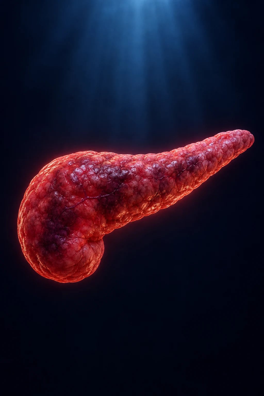
Overview & Definition
Acute pancreatitis is an acute inflammatory disorder of the pancreas in which pancreatic digestive enzymes are activated inside the gland (rather than in the duodenal lumen), causing autodigestion, oedema, fat necrosis, and — in severe disease — parenchymal necrosis, haemorrhage and a systemic inflammatory response that can progress to multi-organ failure.[9][1]
The diagnostic standard since the 2012 Revised Atlanta classification is the two of three rule — acute pancreatitis is diagnosed when any two of the following three are present:[1]
- Characteristic epigastric pain — acute onset of severe, constant epigastric pain, frequently radiating straight through to the back, often relieved by sitting forward.
- Serum lipase or amylase over 3 times the upper limit of normal (lipase is preferred — more specific and persists longer than amylase).
- Characteristic imaging findings on contrast-enhanced CT, MRI or transabdominal ultrasound. [1]
Imaging is not required for diagnosis when both the clinical and biochemical criteria are met. Conversely, a normal amylase or lipase does not exclude pancreatitis — typically in hypertriglyceridaemic pancreatitis (lipaemic serum interferes with the assay) and in late presentations after enzymes have already cleared.[9]
The clinical challenge is not making the diagnosis — it is (a) grading severity early, (b) providing goal-directed rather than aggressive fluids, (c) avoiding the three outdated dogmas (prolonged nil by mouth, routine prophylactic antibiotics, early surgery for necrosis), and (d) excluding the life-threatening mimics (perforated peptic ulcer, mesenteric ischaemia, leaking abdominal aortic aneurysm) that can also raise amylase.[3][9]
Classification
Acute pancreatitis is classified along three independent axes — morphology, severity, and local complications by timing. Together these define management and prognosis. [1]
By morphology (cross-sectional imaging):[1]
- Interstitial oedematous pancreatitis — the commoner pattern — diffuse gland enlargement, peripancreatic fat stranding, no necrosis. Usually mild course.
- Necrotising pancreatitis — non-enhancing pancreatic parenchyma and/or peripancreatic tissue on contrast-enhanced CT (necrosis = lack of enhancement). Affects around 20%; higher complication and mortality rate; risk of infection. [1]
By aetiology (the axis that drives definitive treatment):[3]
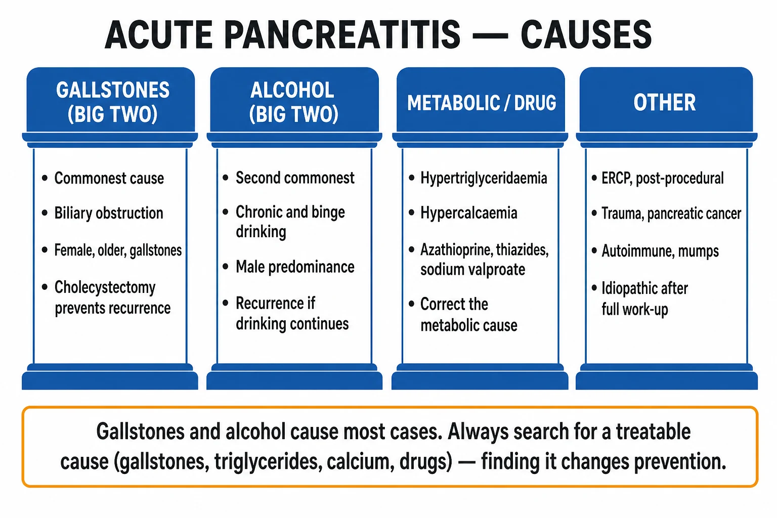
- Gallstone pancreatitis (40 to 50%) — needs cholecystectomy to prevent recurrence.
- Alcoholic pancreatitis (25 to 35%) — needs cessation counselling.
- Hypertriglyceridaemic (serum triglycerides over 11.3 mmol/L / 1000 mg/dL) — insulin ± apheresis.
- Hypercalcaemic, drug-induced, post-ERCP, traumatic, autoimmune, infective, idiopathic — see Epidemiology. [1]
Interstitial oedematous (mild)
- Around 80% of cases
- Diffuse gland enlargement, NO necrosis on contrast CT
- Usually mild course; resolves within a week
- Local complication: acute peripancreatic fluid collection, pseudocyst
Necrotising (severe)
- Around 20% of cases
- Non-enhancing pancreatic/peripancreatic tissue on contrast CT
- Higher complication rate (infection, haemorrhage)
- Local complication: acute necrotic collection, walled-off necrosis
- Mortality 10 to 30%, higher if infected
Epidemiology & Risk Factors
Acute pancreatitis is the commonest gastrointestinal cause of hospital admission in many countries, with an annual incidence of 13 to 45 per 100,000 that is rising worldwide (more gallstone disease in women, more alcohol-related disease in men, rising obesity driving hypertriglyceridaemic pancreatitis). It accounts for over 250,000 admissions per year in the United States. The lifetime risk is around 1 in 50.[9]
The two big causes together account for 70 to 80% of cases:[3][9]
- Gallstones — around 40 to 50%. Small gallstones (under 5 mm) and microlithiasis (biliary sludge) are particularly prone to transiently obstruct the ampulla of Vater and trigger an episode.
- Alcohol — around 25 to 35%. Typically chronic heavy use (over 50 to 80 g/day for 5 or more years); a recognisable acute episode may be the first presentation of underlying chronic pancreatitis. [1]
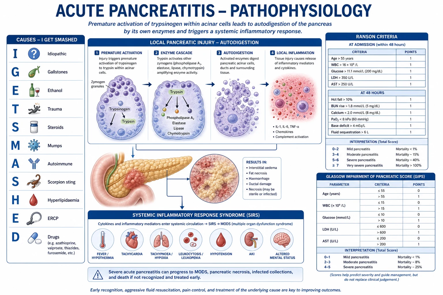
Other causes (examined in the I GET SMASHED mnemonic):[3]
| Mnemonic letter | Cause and notes |
|---|---|
| I | Idiopathic (around 10%) — after thorough exclusion, consider occult microlithiasis, sphincter of Oddi dysfunction, genetic (PRSS1, SPINK1, CFTR) |
| G | Gallstones (and microlithiasis / biliary sludge) — commonest cause |
| E | Ethanol (alcohol) — second commonest |
| T | Trauma (blunt abdominal, post-operative, ERCP 3 to 10%) |
| S | Steroids / Smoking — smoking is now an independent risk factor |
| M | Mumps (and other infections: coxsackie, CMV, Mycoplasma, Salmonella, Legionella, Mycobacterium), Malignancy (pancreatic cancer, ampullary) |
| A | Autoimmune (IgG4-related, type 1 and type 2) |
| S | Scorpion sting (Tityus trinitatis in the Caribbean) |
| H | Hypercalcaemia (hyperparathyroidism), Hypertriglyceridaemia (TG over 11.3 mmol/L / 1000 mg/dL), Hypothermia |
| E | ERCP (3 to 10% risk), Embolic |
| D | Drugs (azathioprine/6-mercaptopurine, didanosine, thiazides, sodium valproate, mesalazine, oestrogens, tetracyclines, sulphonamides, GLP-1 agonists) — a diagnosis of exclusion |
Drug-induced pancreatitis is a diagnosis of exclusion — the drug must be temporally related, other causes excluded, and pancreatitis resolves on withdrawal (and recurs on rechallenge, in classical cases). The highest-risk drugs are azathioprine/6-mercaptopurine, didanosine, thiazides, sodium valproate, mesalazine, oestrogens, tetracyclines and, more recently, the GLP-1 receptor agonists.[3]
Metabolic causes (high-yield exam triggers):[3]
- Hypertriglyceridaemia — serum triglycerides over 11.3 mmol/L (1000 mg/dL), usually in familial hypertriglyceridaemia, poorly controlled diabetes, alcohol, or pregnancy. The amylase may be falsely normal (lipaemic serum interferes with the assay). Look for lipaemic (milky) plasma and eruptive xanthomata. Treat with insulin infusion, apheresis/plasmapheresis, lipid-lowering therapy.
- Hypercalcaemia — usually primary hyperparathyroidism; calcium is thought to activate trypsinogen. Always check serum calcium (corrected) in any unexplained pancreatitis — but remember the serum calcium falls in severe pancreatitis due to saponification of peripancreatic fat. [1]
Iatrogenic and mechanical causes: ERCP (3 to 10% — preventable with rectal NSAIDs and a prophylactic pancreatic stent), abdominal trauma (handlebar injury in children, blunt deceleration injury in adults), post-operative (cardiopulmonary bypass, upper-abdominal surgery), sphincter of Oddi manometry.[9]
Pathophysiology
The central event in acute pancreatitis is the premature intracellular activation of trypsinogen to trypsin within the pancreatic acinar cell, before the digestive enzymes have been secreted into the ductal system and duodenal lumen where activation is normally intended.[9]
The normal protective mechanisms (and how they fail): [1]
- Trypsinogen is normally activated in the duodenal lumen by enterokinase (enteropeptidase) on the brush border — not inside the pancreas.
- Inside the acinar cell, trypsin is kept inactive by the trypsinogen-activation peptide, serine protease inhibitor Kazal type 1 (SPINK1), and chymotrypsin C-mediated degradation (which destroys prematurely activated trypsin).
- Calcium signalling inside the acinar cell regulates normal, basolateral exocytosis. Abnormal (sustained, high-amplitude) calcium signals — from bile acids, alcohol metabolites, or hypercalcaemia — cause trypsinogen to be activated inside the cell and secreted via the basolateral membrane into the interstitium.
- Failure of SPINK1 (genetic) or of autophagy/lysosomal degradation (the cathepsin-B mediated intracellular activation pathway) allows trypsin to persist and activate the other zymogens — proelastase, prophospholipase A2, procarboxypeptidase, kallikreinogen — and the complement and kinin cascades. [1]
Once trypsin activates the zymogen cascade inside the gland, autodigestion follows:[9]
- Elastase digests the elastic lamina of blood vessels → haemorrhage (the basis of haemorrhagic/necrotising pancreatitis).
- Phospholipase A2 digests cell membrane phospholipids → acinar cell necrosis and, in the lungs, surfactant destruction contributing to ARDS.
- Lipase digests peripancreatic fat → fat necrosis. Free fatty acids bind calcium → calcium soap (saponification) formation → hypocalcaemia.
- Kallikrein, bradykinin activation produces vasodilation, increased vascular permeability and third-space loss → hypovolaemia and hypotension.
- Trypsin itself is chemotactic for neutrophils and activates the complement cascade. [1]
The systemic inflammatory cascade: injured acinar cells release damage-associated molecular patterns (DAMPs) and cytokines — IL-6, IL-8, TNF-alpha, IL-1 beta, MCP-1 — which recruit neutrophils and macrophages and produce systemic inflammatory response syndrome (SIRS).[9] SIRS drives:
- Systemic capillary leak with massive third-space loss (retroperitoneum, peritoneum, bowel wall) — produces the intravascular hypovolaemia that drives shock and the aggressive fluid resuscitation that, when excessive, causes fluid overload (the lesson of the WATERFALL trial).
- Acute respiratory distress syndrome (ARDS) — alveolar capillary leak, surfactant destruction.
- Acute kidney injury — hypovolaemia plus inflammation.
- Disseminated intravascular coagulation — cytokine-driven activation.
- Systemic hypotension and distributive shock. [1]
How gallstones cause pancreatitis (the common bile duct — pancreatic duct relationship): most people share a common channel at the ampulla of Vater where the bile duct and pancreatic duct join before entering the duodenum. A gallstone migrating through the ampulla transiently obstructs the pancreatic duct, raising pressure and/or allowing bile to reflux into the pancreatic duct. Either mechanism promotes premature trypsinogen activation. Importantly, persistent biliary obstruction also causes obstructive jaundice and ascending cholangitis — the indications for urgent ERCP.[9]
How alcohol causes pancreatitis: direct acinar cell toxicity (fatty acid ethyl esters), secretory protein (GP2) precipitation into protein plugs that obstruct small ductules, and spasm of the sphincter of Oddi. Chronic exposure produces chronic pancreatitis; an acute flare may superimpose on chronic disease.[9]
Key pathophysiology numbers
Clinical Presentation
The classic presentation is unmistakable: acute onset of severe, constant, deep-seated epigastric pain that radiates straight through to the back, is relieved by sitting forward (the pancreatico-duodenal autonomic plexus sits posteriorly), and is worsened by lying flat, eating, coughing, and deep inspiration. The pain reaches maximum intensity within minutes to a few hours (distinguishing it from the waxing-and-waning colic of biliary or renal origin). Nausea and vomiting are invariable, and anorexia is complete.[9]
General examination findings depend on severity: [1]
- Mild disease: the patient looks unwell but is systemically well — low-grade fever (under 38.5°C), mild tachycardia, mild abdominal tenderness in the epigastrium. Bowel sounds may be normal or slightly reduced.
- Moderate to severe disease: signs of SIRS — fever over 38°C or hypothermia under 36°C, tachycardia over 90, tachypnoea over 20, WBC over 12 or under 4. Hypotension, oliguria, hypoxia, confusion signal developing organ failure.
- Established severe / haemorrhagic disease: the abdomen is distended (ileus, third-space ascites), tender and guarded in the epigastrium, with absent or markedly reduced bowel sounds (paralytic ileus). There may be a palpable epigastric mass (pseudocyst, later). [1]
The two eponymous bruising signs of severe haemorrhagic/necrotising pancreatitis (rare, but a viva favourite and a marker of severe disease with high mortality):[9]
- Grey-Turner's sign — bluish-green or yellowish-brown flank bruising — blood tracking from the retroperitoneum into the lateral abdominal wall along fascial planes.
- Cullen's sign — periumbilical bruising — blood tracking via the falciform ligament to the umbilicus. [1]
(Less commonly examined: Fox's sign — bruising in the upper thigh from retroperitoneal tracking along the inguinal ligament.) [1]
Atypical presentations:[9]
- The elderly: confusion, falls, hypothermia, or non-specific functional decline rather than classical pain. Fever and leucocytosis may be blunted. High index of suspicion; lower threshold to image and to admit.
- Pregnancy (typically 3rd trimester): usually gallstone pancreatitis, sometimes hypertriglyceridaemic (gestational). May be missed because epigastric pain is attributed to hyperemesis, reflux, or pre-eclampsia. Manage as the non-pregnant; ultrasound and MRI are safe; foetal monitoring is essential.
- Diabetic ketoacidosis may precipitate pancreatitis and can itself be its consequence; check ketones and blood gas in any diabetic with pancreatitis.
- Hypertriglyceridaemic pancreatitis: lipaemic (milky) plasma, eruptive xanthomata on the back, buttocks, extensor surfaces; often recurrent; amylase may be falsely normal.
- Post-ERCP: pain, nausea, and rising amylase within 4 to 24 hours of the procedure. [1]
Differential Diagnosis
The differential of acute severe epigastric pain radiating to the back is the differential of an acute abdomen, several of which can themselves raise amylase and mislead you. Distinguish as follows:[9]
- Perforated peptic ulcer — sudden onset "knife-like" epigastric pain that becomes generalised; peritonism (rigid, board-like abdomen, guarding, rebound, absent bowel sounds); free intraperitoneal gas on erect chest X-ray (under the diaphragm) or CT. Amylase can be mildly raised. Peritoneal/upper-GI surgical emergency.
- Acute cholecystitis / biliary colic — right upper quadrant pain (cholangitis adds jaundice, fever, rigors — Charcot's triad; Reynolds' pentad adds hypotension and confusion). Pain is colicky and localised to the RUQ with a positive Murphy's sign; lipase is normal or only mildly raised. Ultrasound shows gallstones, gallbladder wall thickening, sonographic Murphy sign.
- Mesenteric ischaemia / infarction — pain out of proportion to examination ("painful abdomen with nothing to find"), often in an elderly vascular patient with atrial fibrillation or severe atherosclerosis; lactic acidosis, metabolic acidosis, rectal bleeding; pneumatosis intestinalis or bowel-wall thickening/gas in mesenteric vessels on CT angiography. A raised amylase is common and misleading — keep this differential high.
- Leaking or ruptured abdominal aortic aneurysm — tearing epigastric and back pain, syncope, hypotension, a pulsatile epigastric mass in an elderly male smoker. A raised amylase is well described. A fatal miss — always palpate the aorta and consider CT angiography in the older vasculopath.
- Inferior myocardial infarction — epigastric pain, nausea, vomiting, diaphoresis; inferior ST elevation on ECG, raised troponin. An ECG is mandatory in any adult with acute epigastric pain.
- Diabetic ketoacidosis (DKA) — abdominal pain, vomiting, ketotic breath, high anion gap metabolic acidosis, ketones, hyperglycaemia. DKA may also cause pancreatitis and vice versa.
- Hypercalcaemic crisis — confusion, abdominal pain, polyuria/polydipsia, corrected calcium over 3.5 mmol/L, shortened QT interval on ECG.
- Renal colic — colicky loin-to-groin pain with haematuria, normal amylase and lipase.
- Small bowel obstruction — colicky central abdominal pain, distension, absolute constipation, tinkling bowel sounds, step-ladder dilated loops on X-ray.
- Lower lobe pneumonia / pulmonary embolism — pleuritic chest pain referred to the upper abdomen; chest signs and CXR/CT findings. [1]
Rule of thumb: in any older vasculopath with "pancreatitis" and atypical features, specifically look for a leaking AAA or mesenteric ischaemia before attributing the amylase to the pancreas — both can elevate it.[9]
Clinical & Bedside Assessment
Begin with ABCDE, then a focused abdominal examination, then a structured severity assessment. [1]
ABCDE — look for organ failure (the basis of the Revised Atlanta severe category):[1]
- Airway — patent; protect if vomiting and reduced GCS.
- Breathing — respiratory rate, SpO2 on air, work of breathing. Hypoxia (SpO2 under 92% or PaO2 under 8 kPa/60 mmHg) signals respiratory organ failure.
- Circulation — heart rate, blood pressure, capillary refill, peripheral temperature, jugular venous pressure. Hypotension (systolic under 90 mmHg) signals cardiovascular organ failure.
- Disability — GCS / AVPU; confusion or new disorientation is a marker of severe disease and is a Glasgow / BISAP criterion.
- Exposure — fully expose the abdomen. [1]
Focused abdominal examination — look specifically for: [1]
- Tenderness, guarding, rigidity in the epigastrium (mild to marked depending on severity).
- Distension (ileus, third-space fluid).
- Bowel sounds — reduced or absent (paralytic ileus) in severe disease.
- Cullen's sign and Grey-Turner's sign — present in under 3% of cases but specific for severe necrotising/haemorrhagic disease when present.
- Jaundice — suggests biliary obstruction (gallstone in the common bile duct, cholangitis, or compression by pancreatic head oedema/pseudocyst).
- A palpable epigastric mass — late: pseudocyst, walled-off necrosis. [1]
Bedside observations that define severity: urine output (catheterise for hourly output in moderate-severe disease — target over 0.5 mL/kg/h), continuous SpO2 and ECG, hourly respiratory rate and blood pressure, four-hourly temperature. Hourly urine output is the most sensitive bedside perfusion marker. [1]
Assess for SIRS at the bedside (2 or more of): temperature over 38°C or under 36°C, heart rate over 90, respiratory rate over 20, WBC over 12 or under 4 (or over 10% immature bands). SIRS at admission and at 48 hours predicts severe disease and infected necrosis.[9]
I GET SMASHED — causes of acute pancreatitis
I GET SMASHED
around 10%, after exclusion — consider microlithiasis, sphincter of Oddi, genetics (PRSS1, SPINK1, CFTR)
commonest cause (40 to 50%); small stones under 5 mm and microlithiasis highest risk
second commonest (25 to 35%); chronic use; may be first presentation of chronic disease
blunt abdominal, post-operative, post-ERCP (3 to 10%)
smoking is now an independent risk factor; steroids (autoimmune)
mumps, coxsackie, CMV, Mycoplasma; pancreatic or ampullary cancer
IgG4-related type 1 (with other organ involvement) and type 2 (duct-centric); steroid-responsive
Tityus trinitatis (Caribbean) — rare but classic
calcium (hyperparathyroidism); triglycerides over 11.3 mmol/L
3 to 10% risk; prevent with rectal NSAID + pancreatic stent
azathioprine, didanosine, thiazides, valproate, mesalazine, oestrogens, tetracyclines
Investigations
First-line investigations at admission (everyone):[3]
- Serum lipase (preferred — over 3 × ULN; highly specific, persists longer than amylase).
- Serum amylase (over 3 × ULN) — rises within hours, peaks at 24 hours, returns to normal in 3 to 5 days (so a normal amylase does not exclude late-presenting pancreatitis).
- Full blood count — leucocytosis; haematocrit (basis of haemoconcentration-fluid monitoring).
- Urea and electrolytes — urea (BUN), creatinine (renal failure).
- Liver function tests — ALT over 150 IU/L has a 95% positive predictive value for gallstone aetiology; AST, bilirubin (obstruction), albumin (severity).
- Glucose — hyperglycaemia (Ranson criterion).
- Calcium (corrected for albumin) — hypocalcaemia (Ranson/Glasgow criterion).
- Triglycerides — to identify hypertriglyceridaemic cause (falsely normal amylase).
- CRP — at 48 hours, CRP over 150 mg/L predicts pancreatic necrosis.
- Arterial blood gas and lactate — PaO2 (Ranson/Glasgow criterion), base deficit (Ranson), lactate (severity/perfusion).
- Transabdominal ultrasound — first-line imaging: identifies gallstones, common bile duct dilatation, and (limited) peripancreatic fluid. Often limited by bowel gas. [1]
Severity scores — reproduced verbatim (any one may be examined; reproduce the components exactly):[1][4]
Revised Atlanta classification (severity)
- Mild — no organ failure, no local or systemic complications. Around 80%.
- Moderately severe — transient organ failure (resolves under 48 h), OR local complications (necrosis, fluid collections, pseudocyst), OR systemic complications (exacerbation of pre-existing disease).
- Severe — persistent organ failure (over 48 h), defined by a Marshall score of 2 or more in any one of: respiratory (PaO2/FiO2 under 300), renal (creatinine over 170 micromol/L), or cardiovascular (systolic BP under 90 mmHg unresponsive to fluids). Single, multiple, or new-onset worsening all count. [1]
Ranson criteria
At admission (5 criteria): age over 55, WBC over 16 × 10^9/L, glucose over 10 mmol/L (200 mg/dL), AST over 250 IU/L, LDH over 350 IU/L. [1]
During the first 48 hours (6 criteria): haematocrit fall over 10 percentage points, serum calcium under 2 mmol/L (8 mg/dL), PaO2 under 8 kPa (60 mmHg), base deficit over 4 mEq/L, fluid sequestration over 6 L, urea rise over 1 mmol/L (3.6 mg/dL). [1]
Score interpretation: 0 to 2 = mild (mortality under 1%); 3 to 4 = 15% mortality; 5 to 6 = 40% mortality; over 6 = near 100% mortality. Score of 3 or more = severe. [1]
Glasgow (modified Glasgow / Imrie) criteria
Eight criteria (no race-specific admission variables — applicable internationally), assessed within 48 hours of admission: [1]
- Age over 55 years
- WBC over 15 × 10^9/L
- Glucose over 10 mmol/L
- LDH over 600 IU/L (some versions: over 350)
- AST over 200 IU/L
- Urea over 16 mmol/L
- Calcium under 2 mmol/L
- PaO2 under 8 kPa (60 mmHg) [1]
Score of 3 or more = severe. Albumin under 32 g/L is included in some versions (making it 9 criteria). [1]
BISAP score (5 — early, simple, validated)
A simple 5-point score computed within the first 24 hours, derived by Wu et al. 2008 from a large US population-based dataset:[4]
- Blood Urea Nitrogen over 8.6 mmol/L (25 mg/dL)
- Impaired mental status (GCS under 15)
- SIRS (2 or more of temperature, heart rate, respiratory rate, WBC as above)
- Age over 60 years
- Pleural effusion on imaging [1]
Score 0 to 5; score of 3 or more = high mortality (over 5 to 20 times that of low scores). [1]
APACHE-II
A dynamic, physiological score originally for ICU severity; a score of 8 or more defines severe pancreatitis. More accurate but complex and time-consuming; used in research and ICU. [1]
CRP at 48 hours
CRP over 150 mg/L at 48 hours is an independent predictor of pancreatic necrosis.[1]
Cross-sectional imaging — when and why
- Contrast-enhanced CT (CECT) — not needed at admission in obvious cases. Indicated when: (a) diagnosis uncertain, (b) severity assessment at 48 to 72 hours to detect necrosis (the timing matters — necrosis may not be visible on day 1), or (c) suspected complications (infected necrosis, haemorrhage, pseudocyst). The lack of parenchymal enhancement defines necrosis.
- MRI / MRCP — equivalent diagnostic accuracy to CT; preferred in pregnancy and to characterise the biliary tree non-invasively (stones in the CBD).
- EUS (endoscopic ultrasound) — for occult microlithiasis, sludge, and small pancreatic tumours in unexplained recurrent pancreatitis. [1]
Diagnosing infected necrosis: gas within an area of necrosis on CT is highly suggestive; positive Gram stain or culture on image-guided fine-needle aspiration (FNA) is confirmatory. FNA is reserved for clinically deteriorating patients with necrosis in whom infection is suspected but not proven.[5]
Management — Resuscitation
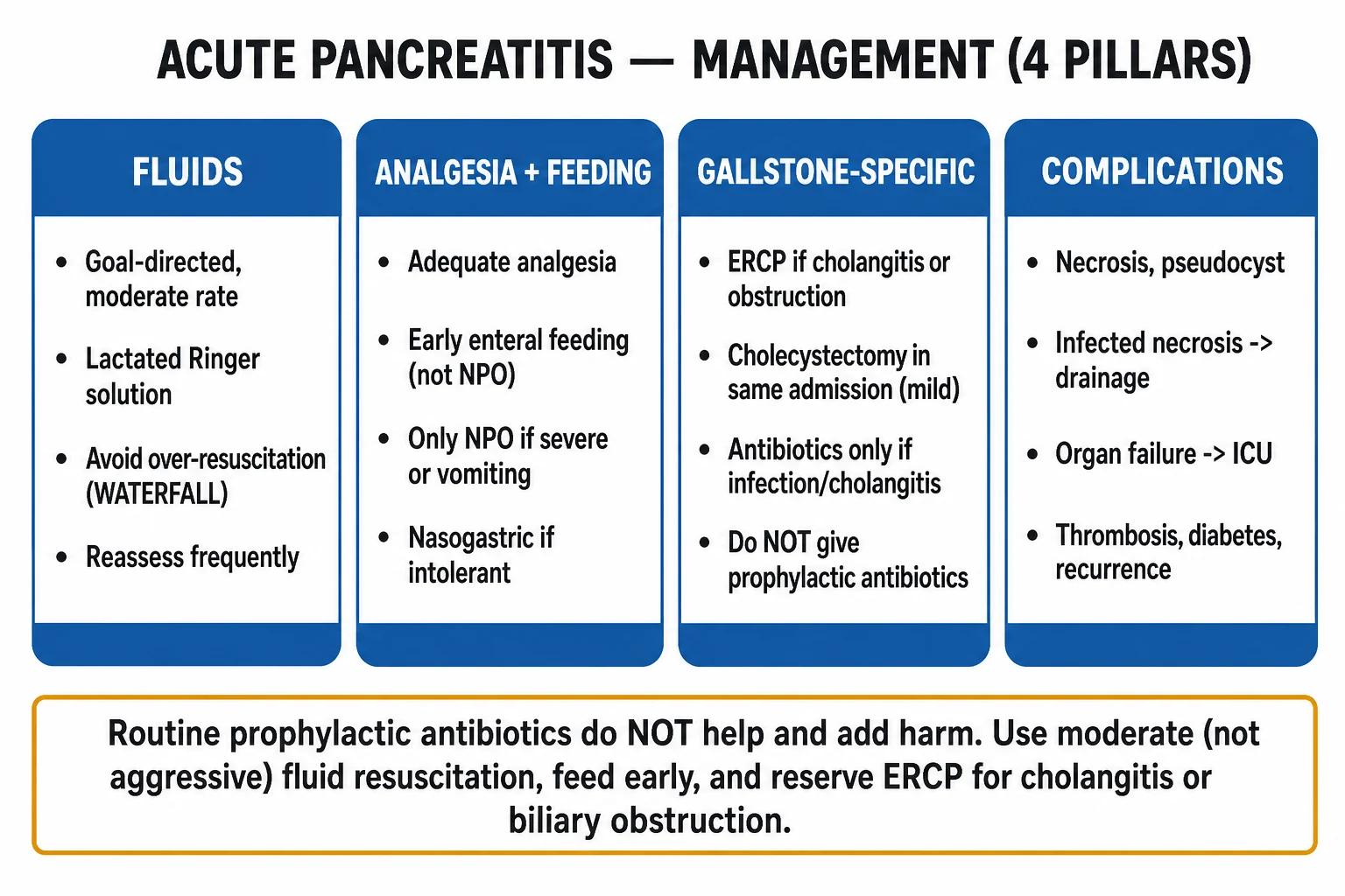
All patients with acute pancreatitis should be admitted to hospital; severe disease warrants ICU or HDU.[3]
ABCDE: high-flow oxygen to target SpO2 94 to 98% (or 88 to 92% if chronic CO2 retainer); two large-bore IV cannulae; urinary catheter for hourly output; continuous SpO2, ECG; reliable IV access before procedures. [1]
The single most important early intervention is goal-directed fluid resuscitation.[2][3]
Fluid strategy (WATERFALL trial-informed, moderate — NOT aggressive): [1]
- Crystalloid of choice: lactated Ringer's (the Wu 2011 RCT showed less SIRS and CRP rise with LR than saline, by avoiding hyperchloraemic acidosis which may worsen trypsin activation).
- Moderate goal-directed rate: an initial 10 to 15 mL/kg/h for the first few hours, reducing to 3 to 5 mL/kg/h by 12 to 24 hours, with frequent reassessment.
- Reassess with goals — a falling haematocrit toward normal, normalising blood urea (BUN), stable or improving lactate, and urine output over 0.5 mL/kg/h.
- Avoid aggressive resuscitation: the WATERFALL trial (de-Madaria et al., NEJM 2022) randomised patients to aggressive (20 mL/kg bolus then 3 mL/kg/h for 72 h) vs moderate LR; aggressive fluids INCREASED fluid overload (6% vs 0.8% — pulmonary oedema, ascites) with NO improvement in outcome.[2]
- Reduce or hold fluids if haematocrit has normalised or signs of overload appear.
Analgesia — adequate analgesia is a quality indicator:[3]
- IV opioid titrated to pain — options include IV morphine 0.05 to 0.1 mg/kg every 4 to 6 hours (or patient-controlled analgesia), IV fentanyl 25 to 50 micrograms every 1 to 2 hours, or IV tramadol 50 to 100 mg every 6 to 8 hours.
- Historically there was concern that morphine causes sphincter of Oddi spasm and worsens pancreatitis — this is not supported by clinical evidence and morphine remains a reasonable first-line opioid. The priority is adequate analgesia.
- Avoid IM injections (interfere with CK and absorb variably). [1]
Monitoring and escalation triggers: [1]
- Hourly urine output (catheter), continuous SpO2 and ECG, frequent vital signs and GCS, daily weights (third-space shifts).
- Escalate to ICU/HDU when organ failure develops or persists (persistent hypoxia, hypotension unresponsive to fluids, oliguria not responding to fluid challenge, confusion) — the definition of severe pancreatitis. [1]
Management — Definitive & Stepwise
Early enteral feeding (within 24 to 48 h) — the standard of care:[3][9]
- Oral feeding as tolerated in mild pancreatitis — start with low-fat solid or liquid diet within 24 to 48 hours; advancing as tolerated.
- In severe disease or gastric intolerance: nasogastric (NG) feeding is as effective as nasojejunal (NJ) in most trials; reserve NJ for those who do not tolerate NG.
- Total parenteral nutrition is reserved for prolonged ileus, fistula, or other failures of enteral feeding — it does NOT improve outcome and increases infection. [1]
Antibiotics — the most important "less is more" lesson:[7]
- Routine prophylactic antibiotics do NOT reduce infected necrosis or mortality and cause harm — selection of resistant organisms, fungal overgrowth, and Clostridioides difficile. Do not give them. This is a meta-analysis-level conclusion (Poropat 2022).[7]
- Give antibiotics ONLY when:
- Infected necrosis — proven by gas in necrosis on CT or positive FNA, OR strongly suspected by clinical deterioration with raised inflammatory markers.
- Acute cholangitis (fever, jaundice, rigors, abdominal pain — Charcot's triad) associated with biliary obstruction.
- An extrapancreatic infection — pneumonia, UTI, bacteraemia.
- Choice for infected necrosis: carbapenem (imipenem-cilastatin 500 mg IV every 6 hours, meropenem 1 g IV every 8 hours) OR quinolone plus metronidazole (ciprofloxacin 400 mg IV every 12 hours plus metronidazole 500 mg IV every 8 hours). These penetrate necrosis well. Continue until clinical and biochemical improvement and resolution/sealing of the collection — typically 4 to 7 days of effective source control.
ERCP — early, but ONLY for specific indications:[8]
- Within 24 hours for acute cholangitis (fever, jaundice, biliary pain — Charcot's triad) and persistent biliary obstruction (jaundice, dilated CBD with stone on imaging, or worsening LFTs).
- NOT routinely in gallstone pancreatitis without obstruction — the Cochrane review (Tse & Yuan 2012) showed no benefit of routine early ERCP in this group, and a trend to harm.[8]
- Perform sphincterotomy and stone extraction; place a biliary stent if clearance is incomplete.
Cholecystectomy — timing depends on severity:[3]
- Mild gallstone pancreatitis: same-admission laparoscopic cholecystectomy (during the index admission, after symptoms settle) — reduces recurrent gallstone-related events from around 20 to 30% to under 5%.
- Severe pancreatitis (necrosis, organ failure): delay cholecystectomy until complete resolution and ideally after a delay to allow inflammation to settle — sometimes staged after recovery from necrosectomy.
- Unfit for surgery: ERCP + sphincterotomy before discharge reduces recurrence (defunctions the sphincter) as a second-line approach. [1]
Hypertriglyceridaemic pancreatitis — cause-specific therapy:[3]
- Insulin infusion (with dextrose to maintain euglycaemia) — activates lipoprotein lipase, clears triglycerides.
- Plasmapheresis / apheresis — for very high TG (over 1000 mg/dL) with severe disease; rapid TG removal.
- Lipid-lowering therapy: fibrate (fenofibrate 145 to 200 mg daily) first-line for hypertriglyceridaemia; omega-3 fatty acids; statin; dietary fat restriction; tight diabetic control. [1]
Autoimmune pancreatitis: prednisolone 30 to 40 mg daily for 4 to 6 weeks, then taper — dramatic radiological and biochemical response in type 1 (IgG4-related).[3]
Step-up approach for infected necrosis (the PANTER and TENSION trials):[5][6]
- Delay intervention ~4 weeks if possible (allows the collection to become walled-off and drainable, reducing bleeding and organ failure).
- Step 1: percutaneous (radiological) or endoscopic (EUS-guided) catheter drainage — controls sepsis in most.
- Step 2: if drainage fails, endoscopic or minimally invasive necrosectomy (PANTER and TENSION: endoscopic step-up superior to open necrosectomy — less organ failure, fewer fistulas, lower diabetes/incisional hernia long-term).[6]
- Open necrosectomy is now reserved for the failures of the step-up approach.
Specific Subtypes & Scenarios
- Gallstone pancreatitis (40 to 50%): the commonest cause, particularly in women over 50. Identify by ALT over 150 IU/L (95% PPV for gallstone aetiology) and ultrasound (gallstones ± CBD dilatation). ERCP within 24 h for cholangitis or persistent obstruction; same-admission laparoscopic cholecystectomy in mild disease (after symptoms settle). Recurrence without cholecystectomy: 20 to 30% within 12 weeks.[3]
- Alcoholic pancreatitis (25 to 35%): typically men in their 30s to 50s with chronic heavy intake. Mechanism is direct acinar toxicity plus protein-plug duct obstruction. Cessation counselling is the definitive therapy — continuing to drink causes recurrence and progression to chronic pancreatitis. Correct thiamine, magnesium, potassium, phosphate; assess for withdrawal (CIWA scale) and give thiamine 100 mg IV/IM daily for 3 to 5 days before any glucose.
- Hypertriglyceridaemic pancreatitis (around 5 to 7%): trigger is serum TG over 11.3 mmol/L (1000 mg/dL); lipaemic serum, eruptive xanthomata, often diabetic or pregnant, often recurrent. Amylase may be falsely normal — diagnose clinically and by imaging. Treat with insulin infusion ± plasmapheresis plus long-term fibrate.[3]
- Severe necrotising pancreatitis: distinguish sterile (most, manage conservatively with fluids, analgesia, nutrition) from infected (gas on CT, positive FNA, or clinical deterioration) necrosis. Infected necrosis is the leading cause of death in severe pancreatitis — give carbapenem and arrange drainage (step-up). Postpone intervention ~4 weeks where possible.
- Post-ERCP pancreatitis (3 to 10%): risk factors include young age, female sex, prior post-ERCP pancreatitis, difficult cannulation, sphincter of Oddi dysfunction, normal bilirubin. Prevent with rectal NSAID — diclofenac 100 mg or indomethacin 100 mg per rectum immediately before or after ERCP — and a prophylactic pancreatic stent in high-risk patients.[3]
- Autoimmune pancreatitis (rare): type 1 is IgG4-related disease (older men, other organ involvement — biliary stricture, retroperitoneal fibrosis, salivary/lacrimal); type 2 is duct-centric without IgG4. Both respond dramatically to steroids (prednisolone 30 to 40 mg daily — diagnostic and therapeutic trial). Imaging: diffuse sausage-shaped pancreas with a capsule-like rim.
Complications & Pitfalls
Local complications (defined by content and timing — Revised Atlanta):[1]
| Collection | Time | Content | Capsule |
|---|---|---|---|
| Acute peripancreatic fluid collection (APFC) | first 4 weeks | fluid, no necrosis | none |
| Pancreatic pseudocyst | after 4 weeks | fluid, no necrosis | encapsulated |
| Acute necrotic collection (ANC) | first 4 weeks | necrosis plus fluid | none |
| Walled-off necrosis (WON) | after 4 weeks | necrosis plus fluid | encapsulated |
A pseudocyst is a fluid collection encapsulated by a non-epithelial fibrous wall, without necrosis, persisting over 4 weeks. Drain (endoscopic or surgical) only if symptomatic, infected, over 6 cm, or persisting over 6 weeks.[1]
Other local complications: [1]
- Infected necrosis — the leading cause of late death. Diagnose by gas in necrosis on CT or positive FNA. Treat with carbapenem + step-up drainage.[5]
- Haemorrhage — from erosion into splenic, gastroduodenal, or pancreatico-duodenal arteries. Diagnose by CT angiography; treat by interventional radiology embolisation.
- Splenic and portal vein thrombosis — from adjacent inflammation; splenic vein thrombosis may cause gastric varices (bleed risk).
- Gastric outlet / duodenal obstruction — compression by a phlegmon or walled-off necrosis.
Systemic complications:[9]
- SIRS and sepsis — from infected necrosis.
- Organ failure — ARDS (surfactant destruction by phospholipase A2), AKI, shock, DIC.
- Metabolic — hypocalcaemia (saponification), hyperglycaemia/diabetes (endocrine loss), hypomagnesaemia.
- Stress ulceration, DVT/PE (prophylactic LMWH once bleeding risk controlled). [1]
Long-term sequelae: exocrine insufficiency (steatorrhoea, weight loss — pancreatic enzyme replacement), endocrine insufficiency (type 3c diabetes), recurrent pancreatitis, chronic pancreatitis. [1]
Classic errors (the viva minefield):[2][3][7]
- Aggressive fluid resuscitation — fluid overload, pulmonary oedema, abdominal compartment syndrome (WATERFALL).
- Prolonged nil by mouth — gut mucosal atrophy, bacterial translocation, infected necrosis.
- Routine prophylactic antibiotics — resistance, fungal infection, C. difficile (Poropat 2022 meta-analysis).
- Early surgery for necrosis — high mortality; use the step-up approach and delay ~4 weeks.
- Routine ERCP without obstruction — causes more ERCP-related pancreatitis/cholangitis.
- Missing the leaking AAA or mesenteric ischaemia because amylase is mildly raised.
- Over-relying on amylase (normal in hypertriglyceridaemia, late presentation).
- Forgetting same-admission cholecystectomy in mild gallstone pancreatitis (recurrence risk 20 to 30%). [1]
Prognosis & Disposition
Overall mortality:[9]
- Mild (around 80% of all pancreatitis): under 1 to 3%.
- Severe (persistent organ failure): 10 to 30%.
- Infected necrosis: 25 to 30% or higher without source control. [1]
Predictors of severity:[3]
- At admission: organ failure, obesity (BMI over 30), older age, comorbidity (cardiac, respiratory, renal, diabetes), confusion, SpO2 under 92%, BISAP of 3 or more.
- At 48 hours: CRP over 150 mg/L, rising Ranson/Glasgow scores, persistent SIRS, ** worsening organ failure**, haematocrit not falling (inadequate resuscitation), rising urea/creatinine. [1]
Disposition:[3]
- Mild — general ward, early oral feeding within 24 to 48 h, same-admission laparoscopic cholecystectomy if gallstone-related, discharge when tolerating diet with analgesia and cause-specific prevention (cholecystectomy, alcohol cessation, triglyceride control).
- Severe — ICU/HDU, multidisciplinary (gastroenterology, surgery, intensive care, interventional radiology, nutrition); prepare for step-up drainage if necrosis becomes infected.
- Necrosis — step-up drainage at around 4 weeks; long-term follow-up for endocrine/exocrine insufficiency. [1]
Recurrence and prevention: remove the cause — cholecystectomy for gallstones; alcohol cessation (refer to alcohol liaison); triglyceride control (fibrate, omega-3, tight diabetic control); drug withdrawal; steroids for autoimmune. Recurrence without cause removal is high (around 20 to 30% within 12 months for untreated gallstone disease).[3]
Special Populations
- Pregnancy — commonest cause is gallstones, with increased risk in the 3rd trimester (gallbladder hypomotility from progesterone) and hyperemesis gravidarum-related biliary stasis; hypertriglyceridaemia also rises. Presentation as the non-pregnant, but may be missed (attributed to hyperemesis, reflux, pre-eclampsia). Safe imaging: ultrasound and MRI/MRCP; avoid CT if possible (radiation). Manage as the non-pregnant — moderate goal-directed LR, early enteral feeding, ERCP for cholangitis (with foetal shielding and obstetric involvement), same-admission or early postpartum cholecystectomy. Maternal mortality is low; foetal loss is increased in severe disease.[3]
- The elderly — atypical/blunted presentation (confusion, falls, hypothermia); higher comorbidity and higher mortality; lower threshold for ICU and for cross-sectional imaging. NSAID analgesia cautious (renal/GI bleeding); DVT prophylaxis.
- The alcoholic — thiamine replacement before glucose (prevent Wernicke); correct magnesium, potassium, phosphate; CIWA-based withdrawal prophylaxis (benzodiazepines); relapse-prevention referral to alcohol liaison; nutritional support. Recurrent pancreatitis is the path to chronic pancreatitis.
- The immunocompromised — broaden the differential to include opportunistic infection (CMV, Mycobacterium avium, invasive fungal) and drug toxicity (didanosine, antiretrovirals). Lower threshold for imaging and for broad microbiological work-up.
- Children — rare; consider trauma (handlebar injury, non-accidental injury), structural (pancreas divisum, choledochal cyst, annular pancreas), systemic (cystic fibrosis, hypercalcaemia), drugs (valproate, corticosteroids), infection (mumps). Manage in a specialist paediatric centre.[3]
Evidence, Guidelines & Regional Differences
International guideline framework:[3]
- IAP/APA 2013 guidelines (Banks et al., Pancreatology) — established the early goal-directed fluid, early enteral feeding, no routine prophylactic antibiotics, ERCP for cholangitis/obstruction, step-up drainage for infected necrosis principles.[1]
- ACG 2024 guidelines (Tenner et al., American Journal of Gastroenterology) — the current US standard; reaffirms the above and stresses the WATERFALL fluid strategy, the avoidance of routine antibiotics, same-admission cholecystectomy, and the step-up approach.[3]
- Revised Atlanta classification (2012) — the international severity framework.[1]
- WATERFALL (de-Madaria et al., NEJM 2022) — aggressive vs moderate LR: aggressive fluids increased fluid overload with no benefit. Foundation of the modern moderate-fluid strategy.[2]
- PANTER (van Santvoort et al., NEJM 2010) — endoscopic step-up (drain then minimally invasive necrosectomy) vs open necrosectomy: step-up reduced the primary composite endpoint of major complications or death.[5]
- TENSION (Onnekink et al., Gastroenterology 2022 long-term follow-up) — endoscopic vs surgical step-up for infected necrotising pancreatitis: endoscopic step-up reduced pro-inflammatory response, organ failure, fistulas, incisional hernias, and new-onset diabetes at long-term follow-up.[6]
- Wu 2011 (Gut) — lactated Ringer's vs saline: LR reduced SIRS and CRP at 24 hours (chloride-sparing effect).[4]
Controversies: [1]
- Continuous vs bolus fluids, exact rate of LR, and role of goal-directed echocardiography in early resuscitation remain debated.
- Antibiotic prophylaxis in severe sterile necrosis: now NOT recommended by most guidelines, but historically debated (early small trials suggested benefit; larger and higher-quality studies and meta-analyses do not).[7]
- Endoscopic vs surgical step-up: TENSION long-term follow-up supports endoscopic step-up, but centre expertise determines the local approach.[6]
Exam Pearls
- Diagnose with 2 of 3: epigastric pain radiating to the back, lipase or amylase over 3 × ULN, characteristic imaging. Imaging is NOT required when both clinical and biochemical criteria are met.[1]
- Lipase is preferred over amylase — more specific, persists longer. Amylase is normal in hypertriglyceridaemia and late presentation.
- Causes mnemonic: I GET SMASHED.
- Two big causes (gallstones 40 to 50%, alcohol 25 to 35%) = 70 to 80% of all cases.
- Cullen's sign (periumbilical) and Grey-Turner's sign (flank) = severe necrotising or haemorrhagic pancreatitis (under 3% of cases but high specificity).
- Severity scores — Ranson (3 or more severe) and Glasgow/Imrie (3 or more severe); APACHE-II (8 or more severe); BISAP (3 or more high mortality); Revised Atlanta (persistent organ failure over 48 h = severe).
- CRP over 150 mg/L at 48 h predicts necrosis.
- Three dogmas overturned: (1) moderate not aggressive fluids (WATERFALL); (2) early enteral feeding not NPO; (3) no routine prophylactic antibiotics.
- No early surgery for necrosis — step-up drainage at ~4 weeks.
- ERCP within 24 h only for cholangitis or persistent obstruction — NOT routine.
- Same-admission cholecystectomy in mild gallstone pancreatitis.
- Infected necrosis → carbapenem (imipenem/meropenem) or quinolone + metronidazole + drainage. Sterile necrosis usually managed conservatively.
- Post-ERCP prophylaxis: rectal indomethacin or diclofenac 100 mg + prophylactic pancreatic stent in high-risk patients.
- Hypertriglyceridaemic pancreatitis: lipaemic serum, eruptive xanthomata, amylase may be normal — treat with insulin ± plasmapheresis.
- Hypocalcaemia = saponification of peripancreatic fat by free fatty acids — not just total-body calcium depletion.
- ALT over 150 IU/L = 95% PPV for gallstone cause.
- Phospholipase A2 digests lung surfactant — basis of ARDS in severe pancreatitis.
Exam application bank (NEET-PG / INICET)
One-line answer
Acute pancreatitis is an acute inflammatory process of the pancreas caused by premature intracellular activation of trypsinogen to trypsin within pancreatic acinar cells, with autodigestion of the gland and a systemic inflammatory response. Diagnosed when 2 of 3 criteria are met: characteristic epigastric pain radiating to the back, serum lipase or amylase over 3 times the upper limit of normal, or characteristic imaging (CT/MRI). The two commonest causes are gallstones (40 to 50%) and alcohol (25 to 35%); remember I GET SMASHED for the full list. Severity is graded by the Revised Atlanta classification into mild (no organ failure, no complications — 80% of cases), moderately severe (transient organ failure under 48 h, or local complications), and severe (persistent organ failure over 48 h — high mortality). Resuscitate with goal-directed moderate lactated Ringer's (WATERFALL trial — agg
Worked stems (answer without another resource)
Stem 1 — Classic presentation. Map symptoms to mechanism; name the first investigation and first treatment step with dose/route if drug therapy is standard. [1]
Stem 2 — Unstable / complicated. List red flags that force immediate resuscitation, theatre, ICU, antidote, or reperfusion — and what you do in the first 15 minutes. [1]
Stem 3 — Atypical group. Elderly, pregnancy, child, or immunocompromised: how presentation and thresholds change. [1]
Stem 4 — Differential trap. Name the three closest mimics and one discriminator for each. [1]
Stem 5 — Disposition. Who goes home with safety-netting, who is admitted, who needs HDU/ICU/theatre, and what follow-up is mandatory. [1]
Rapid viva checklist
- Definition + classification
- Pathophysiology chain
- Bedside signs / criteria
- Score with exact components (if any)
- Emergency bundle
- Definitive therapy with doses
- Complications of disease and of treatment
- Special populations
- Guideline/trial name if classic
- Three exam traps
Coverage self-check
If you cannot answer any stem above from this page alone, re-read the matching section — the page is intended to be self-sufficient for final-prof and NEET-PG/INICET questions on Acute Pancreatitis.
References
- [1]Banks PA, Bollen TL, Dervenis C, et al. Classification of acute pancreatitis--2012: revision of the Atlanta classification and definitions by international consensus Gut, 2013.PMID 23100216
- [2]de-Madaria E, Buxbaum JL, Maisonneuve P, et al. Aggressive or Moderate Fluid Resuscitation in Acute Pancreatitis N Engl J Med, 2022.PMID 36103415
- [3]Tenner S, Vege SS, Sheth SG, et al. American College of Gastroenterology Guidelines: Management of Acute Pancreatitis Am J Gastroenterol, 2024.PMID 38857482
- [4]Wu BU, Johannes RS, Sun X, et al. The early prediction of mortality in acute pancreatitis: a large population-based study Gut, 2008.PMID 18519429
- [5]van Santvoort HC, Besselink MG, Bakker OJ, et al. A step-up approach or open necrosectomy for necrotizing pancreatitis N Engl J Med, 2010.PMID 20410514
- [6]Onnekink AM, Boxhoorn L, Timmerhuis HC, et al. Endoscopic Versus Surgical Step-Up Approach for Infected Necrotizing Pancreatitis (ExTENSION): Long-term Follow-up of a Randomized Trial Gastroenterology, 2022.PMID 35580661
- [7]Poropat G, Gorianec K, Lackovic A, et al. Systematic Review with Trial Sequential Analysis of Prophylactic Antibiotics for Acute Pancreatitis Antibiotics (Basel), 2022.PMID 36139970
- [8]Tse F, Yuan Y. Early routine endoscopic retrograde cholangiopancreatography strategy versus early conservative management strategy in acute gallstone pancreatitis Cochrane Database Syst Rev, 2012.PMID 22592743
- [9]Boxhoorn L, Voermans RP, Bouwense SA, et al. Acute pancreatitis Lancet, 2020.PMID 32891214