Gastroenterology · General Medicine
Alcoholic & MASLD Liver Disease
Also known as Alcohol-related liver disease · Alcoholic hepatitis · Metabolic dysfunction-associated steatotic liver disease · MASLD · NAFLD · Fatty liver disease
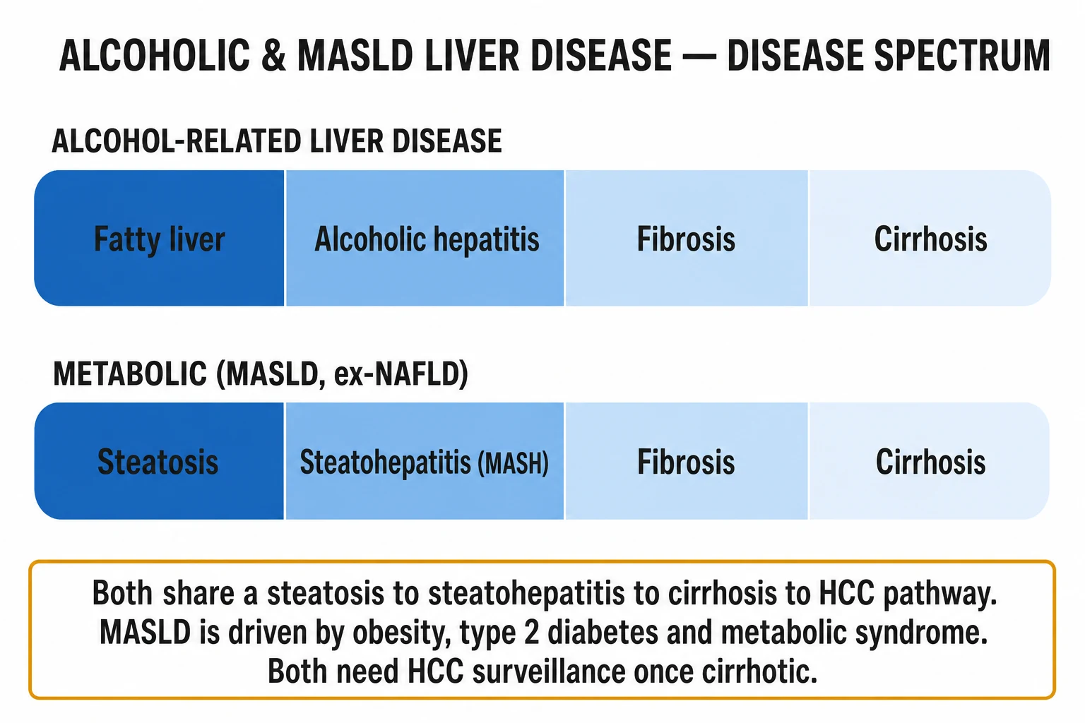
Alcohol-related liver disease and metabolic dysfunction-associated steatotic liver disease (MASLD, formerly NAFLD) are the two leading causes of chronic liver disease worldwide. Both follow the same histological spectrum — steatosis to steatohepatitis to fibrosis to cirrhosis (and hepatocellular carcinoma) — driven by different insults (alcohol versus obesity, type 2 diabetes and metabolic syndrome). Alcoholic hepatitis is the severe inflammatory form: AST greater than ALT (ratio over 2), both under 300, with jaundice and a high MCV, graded by the Maddrey Discriminant Function (32 or more is severe) and MELD. Management centres on abstinence and addiction care for alcohol-related disease (with corticosteroids for severe alcoholic hepatitis, response assessed by the Lille score at day 7), and weight loss of 7 to 10 percent, metabolic control and emerging pharmacotherapy (pioglitazone, GLP-1 agonists, resmetirom) for MASLD. Both warrant 6-monthly hepatocellular carcinoma surveillance once cirrhotic.
On this page & tools
Your progress
Saved locally on this device.
Exam tags
Red flags

Overview & Definition
Alcohol-related liver disease (ALD) and metabolic dysfunction-associated steatotic liver disease (MASLD) together are the commonest chronic liver diseases globally, generating the majority of cirrhosis and a rapidly rising share of hepatocellular carcinoma. They are best taught together because they share a single histological pathway — steatosis (hepatic fat), steatohepatitis (fat plus inflammation and hepatocyte ballooning), fibrosis, cirrhosis — yet arise from two distinct insults: alcohol (ALD) and metabolic dysfunction (MASLD).[1][2]
Definition of MASLD. A 2023 multisociety Delphi consensus renamed NAFLD to MASLD (metabolic dysfunction-associated steatotic liver disease) and NASH to MASH (metabolic dysfunction-associated steatohepatitis). The change is not cosmetic: it anchors the diagnosis to the presence of metabolic dysfunction rather than to the absence of alcohol, removes the stigma of "fatty", harmonises paediatric and adult nomenclature, and creates a new MetALD category for patients in whom alcohol and metabolic disease both contribute substantially (alcohol intake above the MASLD threshold but not high enough to define ALD as the sole cause).[2]
The MASLD diagnosis requires hepatic steatosis (on imaging, histology, or a blood-based score) plus at least one metabolic criterion: overweight/obesity (BMI over 25 kg/m², or over 23 kg/m² in South Asians), type 2 diabetes mellitus, or evidence of metabolic dysregulation (at least two of: increased waist circumference, blood pressure at least 130/85 mmHg or treatment, triglycerides at least 1.7 mmol/L or treatment, HDL cholesterol under 1.0 mmol/L men / 1.3 mmol/L women or treatment, prediabetes or fasting glucose at least 5.6 mmol/L, homeostatic model assessment of insulin resistance at least 2.5, or high-sensitivity CRP over 2 mg/L).[2]
Definition of alcohol-related liver disease. ALD spans alcohol-associated steatosis (asymptomatic, reversible with abstinence), alcoholic steatohepatitis (ASH) — latent inflammation seen on histology — and the clinically overt alcoholic hepatitis (AH) syndrome (rapid jaundice with hepatocellular injury in a heavy drinker), through to alcohol-related fibrosis and cirrhosis.[1]
Definition of alcoholic hepatitis. AH is the clinically recognisable, inflammatory form of ALD presenting with new-onset jaundice in a patient with chronic heavy alcohol use, accompanied by anorexia, tender hepatomegaly, fever and leucocytosis, and biochemically by an AST-to-ALT ratio over 2 (both typically under 300 U/L). It is distinct from the latent steatohepatitis seen only on biopsy.[1]
The upper limit of alcohol that still permits a diagnosis of MASLD is fewer than 20 g/day in women and fewer than 30 g/day in men (around 1.5–2 standard drinks). Above this, ALD is the diagnosis; in the overlap zone, MetALD applies.[2]
Classification
The two diseases are classified in parallel because they share downstream pathology. The cardinal axes are (i) the aetiological driver and (ii) the histological stage. [1]

Alcohol-related (ALD)
- Driver: ethanol and its toxic metabolite acetaldehyde
- Hallmark: AST over ALT (ratio over 2), both under 300 U/L; high GGT and MCV
- Severe form: alcoholic hepatitis, graded by Maddrey DF (32 or more) and MELD
- Cornerstone treatment: complete abstinence; corticosteroids for severe AH; addiction care
MASLD (formerly NAFLD)
- Driver: metabolic dysfunction — obesity, type 2 diabetes, metabolic syndrome
- Hallmark: ALT predominant (AST to ALT under 1), bright liver on ultrasound
- Severe form: MASH with F2 to F4 fibrosis, graded by FIB-4, NFS and FibroScan
- Cornerstone treatment: 7 to 10 percent weight loss; metabolic control; resmetirom, pioglitazone, GLP-1 agonists
MetALD (overlap)
- Both alcohol (over 20 g/day women / over 30 g/day men) and metabolic drivers contribute substantially
- Recognised as a distinct 2023 nomenclature category
- Approach both drivers: harm-reduction of alcohol plus weight loss and metabolic control
The histological staging (shared) is graded by the SAF (Steatosis-Activity-Fibrosis) score or, for MASLD, the NAS (NAFLD Activity Score), with fibrosis staged F0 (none) to F4 (cirrhosis) — the F0–F3 "compensated" and F4 "cirrhotic" distinction governs HCC surveillance and decompensation risk. [1]
Numbers that decide every answer
Epidemiology & Risk Factors
MASLD is the commonest chronic liver disease worldwide. A meta-analysis of 86 studies estimated a global adult prevalence of about 25–30 percent (Younossi 2016); the prevalence is far higher — 56 to over 70 percent — in type 2 diabetes and in obesity, and is rising in parallel with the global metabolic syndrome epidemic.[8] MASLD is now a leading indication for liver transplantation and a leading cause of hepatocellular carcinoma in many countries.
ALD risk depends on the quantity and duration of alcohol, but only a minority of heavy drinkers (roughly 10–20 percent) develop cirrhosis, indicating the importance of host modifiers. Established risk factors:[1]
- Quantity and pattern of alcohol — risk rises above about 30 g/day in women and 50 g/day in men consumed over years; binge drinking adds independent risk.
- Female sex — women develop ALD at lower intake and after shorter exposure, due to lower gastric alcohol dehydrogenase activity and a lower volume of distribution.
- Genetics — PNPLA3 (rs738409, the I148M variant), TM6SF2 (E167K) and HSD17B13 variants are shared susceptibility alleles for both ALD and MASLD; HSD17B13 loss-of-function is protective.
- Malnutrition and sarcopenia — present in over half of patients with severe alcoholic hepatitis, and independently predict mortality.
- Obesity and the metabolic syndrome — act synergistically with alcohol (the basis of the MetALD category); drinkers who are obese have a supra-additive fibrosis risk.
- Coexisting viral hepatitis (HCV, HBV) — accelerates fibrosis; HCV is itself steatogenic (core protein-driven).
- Smoking — independent risk modifier. [1]
MASLD risk factors are metabolic: obesity (especially visceral), type 2 diabetes mellitus, metabolic syndrome, dyslipidaemia, hypothyroidism, polycystic ovary syndrome, obstructive sleep apnoea, and hypopituitarism. The "lean MASLD" phenotype (about 10 percent of all MASLD, commoner in Asian populations) carries a paradoxically higher risk of fibrosis progression per unit hepatic fat, attributed to genetic susceptibility (PNPLA3), sarcopenia and dietary fructose.[2][10]
Pathophysiology
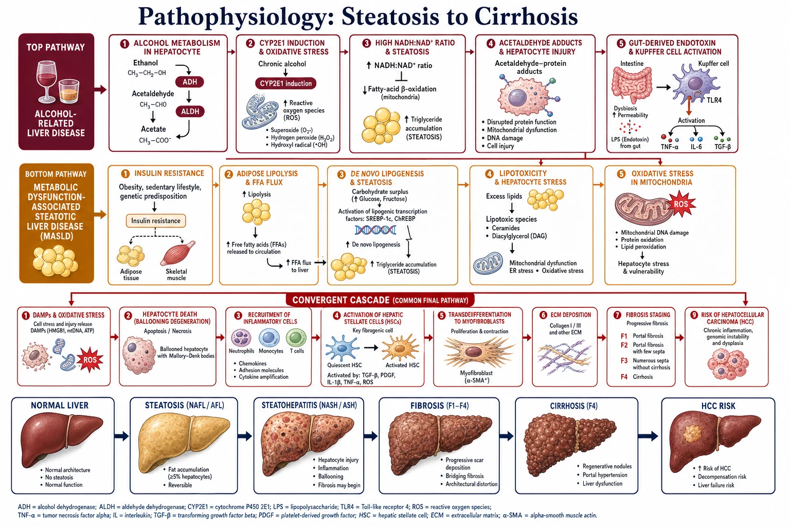
The two diseases converge on a single downstream cascade but enter it by different molecular routes. Understanding both routes is the key to the management logic: remove the driver (alcohol or metabolic overload) early and the disease regresses; let it run and stellate-cell-driven fibrosis becomes irreversible. [1]
Alcohol-related liver disease — the molecular mechanism
- Ethanol metabolism. Alcohol dehydrogenase (ADH) in the cytosol oxidises ethanol to the highly toxic acetaldehyde; mitochondrial aldehyde dehydrogenase (ALDH2) then oxidises acetaldehyde to acetate. Two minor pathways — the microsomal ethanol-oxidising system (CYP2E1) and catalase — become important in chronic drinkers because CYP2E1 is induced, generating large amounts of reactive oxygen species (ROS).[1]
- The redox shift drives steatosis. Both ADH and ALDH2 reduce NAD+ to NADH, so heavy drinking produces a high hepatic NADH:NAD+ ratio. This blocks the beta-oxidation of fatty acids in mitochondria and simultaneously diverts metabolism toward triglyceride synthesis — the biochemical basis of alcoholic steatosis.
- Acetaldehyde and adducts. Acetaldehyde forms adducts with proteins and DNA, impairing their function and generating neoantigens that recruit immune injury. Lipid peroxidation (malondialdehyde, 4-hydroxynonenal) damages membranes.
- The gut–liver axis and inflammation. Alcohol disrupts intestinal tight junctions, increasing translocation of lipopolysaccharide (LPS, endotoxin) to the portal circulation. LPS binds TLR4/CD14 on Kupffer cells, activating NF-kB and releasing TNF-alpha, IL-1, IL-6 and TGF-beta. TNF-alpha induces hepatocyte apoptosis; TGF-beta is the master activator of hepatic stellate cells.
- Stellate cell activation and fibrosis. The hepatic stellate cell (HSC, Ito cell) is a vitamin-A storing pericyte lying in the space of Disse. In health it is quiescent; injured hepatocytes, Kupffer cells and platelets release TGF-beta and PDGF, which transdifferentiate HSCs into alpha-smooth-muscle-actin-positive myofibroblasts that deposit type I and III collagen, tissue inhibitors of metalloproteinases (TIMPs), and contractile elements — the structural basis of cirrhosis.
MASLD — the multiple-hit pathogenesis
The original "two-hit" hypothesis (Day and James) has been superseded by a multiple-parallel-hit model:[10]
- Insulin resistance and lipotoxicity. Peripheral (adipose) insulin resistance drives unregulated lipolysis, increasing free fatty acid (FFA) flux to the liver. In parallel, hyperinsulinaemia activates SREBP-1c and hyperglycaemia activates ChREBP, both driving de novo lipogenesis; VLDL export is overwhelmed, and triglyceride accumulates as steatosis.
- The toxic lipid species. Not triglyceride itself but saturated FFAs (palmitate), ceramides, diacylglycerol and lysophosphatidylcholine are lipotoxic. They trigger endoplasmic reticulum stress, the unfolded protein response, mitochondrial dysfunction with ROS, and hepatocyte apoptosis (the histological correlate of ballooning degeneration).
- Inflammation and cell death. Dying hepatocytes release damage-associated molecular patterns (DAMPs) that activate Kupffer cells and recruit neutrophils and monocytes — the histological signature of steatohepatitis.
- Stellate cell activation and fibrosis — the convergent step. The same TGF-beta and PDGF signals that drive fibrosis in ALD drive it in MASH. Genetic modifiers (PNPLA3, TM6SF2) alter lipid handling and increase susceptibility; HSD17B13 loss-of-function is protective.
- Progression and HCC. Simple steatosis is benign and reversible in most patients; MASH with advanced fibrosis (F3–F4) progresses to cirrhosis in a substantial fraction and carries a markedly elevated risk of hepatocellular carcinoma. Uniquely among chronic liver diseases, MASLD can cause HCC even in the absence of established cirrhosis, which complicates surveillance.[10]
STELLATE — the fibrogenic cell
STELLATE
the perisinusoidal space where HSCs (Ito cells) lie
the master activator of stellate cells (with PDGF)
type I and III collagen laid down by activated myofibroblasts
ROS and lipid aldehydes that injure hepatocytes
gut-derived TLR4 ligand activating Kupffer cells in ALD
marker of transdifferentiated (activated) stellate cells
tissue inhibitors of metalloproteinases that block matrix breakdown
the two upstream drivers that converge on HSC activation
Clinical Presentation
Both diseases are frequently asymptomatic for years and are detected on incidentally abnormal liver function tests, an echogenic ("bright") liver on ultrasound, or hepatomegaly found on examination. The clinical picture depends on the stage. [1]
Early / compensated disease
- MASLD: often discovered incidentally; the patient may describe fatigue and a dull right-upper-quadrant fullness, but most are entirely well. Hepatomegaly is common. The surrounding context — central obesity, type 2 diabetes, hypertension, dyslipidaemia — is usually more prominent than any liver symptom.
- Alcohol-related fatty liver: asymptomatic; may have hepatomegaly, an elevated GGT, and stigmata of chronic alcohol use. [1]
Alcoholic hepatitis (the severe inflammatory syndrome)
The classic presentation is rapid onset of deep jaundice in a heavy drinker, accompanied by anorexia, nausea, abdominal pain and tender hepatomegaly, with fever and a leucocytosis that frequently mimic infection (and infection must be actively excluded before corticosteroids). Asterixis and encephalopathy may be present in severe disease. There may be ascites and coagulopathy.[1]
Decompensated cirrhosis (either aetiology)
Jaundice, ascites, hepatic encephalopathy, variceal haemorrhage. Chronic features: spider naevi, palmar erythema, gynaecomastia, testicular atrophy, parotid enlargement, Dupuytren's contracture, caput medusae, asterixis, asterogenic (sarcopenic) muscle wasting. [1]
Atypical presentations (the examiner's favourites)
- The elderly patient with ascites as the first sign — advanced "silent" MASLD presenting late; BMI may even be normal (sarcopenic).
- The diabetic with "cryptogenic" cirrhosis — "burnt-out" MASH in which steatosis has disappeared once fibrosis disrupted fat storage.
- Lean MASLD — normal BMI with visceral adiposity and genetic susceptibility; easily missed because the clinician assumes BMI excludes it.
- Pregnancy — overlap of MASLD with gestational diabetes; AH in pregnancy is rare but severe.
- The immunocompromised / HIV coinfected patient — MASLD accelerated by antiretroviral therapy and metabolic syndrome; rapid decompensation.
- Acute alcoholic hepatitis masquerading as sepsis or biliary obstruction — fever, leucocytosis, RUQ pain and jaundice can send the unwary down the wrong pathway. [1]
Differential Diagnosis
A complete differential for steatosis/steatohepatitis, with the distinguishing features, is examinable.[1][2]
| Differential | Distinguishing features |
|---|---|
| Alcoholic steatohepatitis vs MASLD (MASH) | AST:ALT ratio over 2 with both under 300 favours alcohol; ALT predominant (ratio under 1) favours MASLD. High MCV and GGT, stigmata of alcohol use, collateral alcohol history favour ALD. Metabolic syndrome favours MASLD. The ratio reverses to over 1 in any cirrhosis, so it is unreliable in advanced disease. |
| Chronic viral hepatitis (HBV, HCV) | Serology (HBsAg, anti-HCV, HCV RNA). HCV is itself steatogenic (core protein) and accelerates ALD/MASLD. |
| Autoimmune hepatitis | Female, high IgG, ANA/SMA/LKM autoantibodies, interface hepatitis on biopsy. |
| Haemochromatosis | High ferritin and transferrin saturation (over 45 percent), bronze diabetes, arthropathy, HFE C282Y/H63D genotyping. |
| Wilson's disease | Young patient, low ceruloplasmin, high urinary copper, Kayser–Fleischer rings, Coombs-negative haemolysis. |
| Alpha-1 antitrypsin deficiency | Low serum A1AT, PAS-positive diastase-resistant globules on biopsy, early-onset emphysema. |
| Drug-induced liver injury (DILI) | Amiodarone, methotrexate, tamoxifen, corticosteroids, tetracycline, valproate, oestrogens, irinotecan. |
| Biliary obstruction (cholangiocarcinoma, choledocholithiasis) | Painless jaundice, predominant ALP rise, dilated ducts on ultrasound/MRCP. |
| Acute viral hepatitis (HAV/HBV/HEV) | Prodrome, AST/ALT often over 1000 U/L, serology. |
| Sepsis-related cholestasis / ischaemic hepatitis | Sepsis does not cause AST:ALT ratio over 2 with high MCV; shock liver has aminotransferases over 1000 that fall rapidly. |
| Congestive hepatomegaly (right heart failure) | Raised JVP, nutmeg liver, response to decongestion. |
| Infiltrative disease | Lymphoma, miliary TB, amyloid, metastases — biopsy tissue diagnosis. |
Mimics of an echogenic liver on ultrasound: fatty infiltration (the commonest), diffuse fibrosis (cirrhosis), glycogen storage disease, chemotherapy effect, and rarely Wilson's disease — an "echogenic liver" is not synonymous with steatosis.[2]
Clinical & Bedside Assessment
- Structured alcohol history using the AUDIT (Alcohol Use Disorders Identification Test, 10 items) or AUDIT-C (3 items); quantify intake in grams or units per week; document the pattern (daily vs binge) and screen for dependence with the CAGE questionnaire (Cut-down, Annoyed, Guilty, Eye-opener).
- Examine for stigmata of chronic liver disease: jaundice, palmar erythema, spider naevi, parotid enlargement, gynaecomastia, testicular atrophy, Dupuytren's contracture, and decompensation — ascites (shifting dullness, fluid thrill), splenomegaly, caput medusae, asterixis (liver flap).
- Grade hepatic encephalopathy by the West Haven scale: I subtle confusion and inverted sleep, asterixis present; II lethargy, disorientation, obvious asterixis; III somnolent but rousable, marked confusion; IV coma. Bedside Trail-Making Test B and connect-the-numbers detect minimal encephalopathy.
- Anthropometry and metabolic screen: BMI and waist circumference (South Asian cut-offs: men over 90 cm, women over 80 cm); blood pressure; screen for metabolic syndrome (NCEP ATP III: 3 of 5 — waist, BP, triglycerides, HDL, fasting glucose).
- Assess alcohol withdrawal risk with CIWA-Ar (Clinical Institute Withdrawal Assessment — Alcohol revised); withdrawal can complicate any AH admission and is itself dangerous.
- Assess malnutrition / sarcopenia with Subjective Global Assessment (SGA) and grip strength — present in over half of severe AH and an independent mortality predictor. [1]
Investigations
Blood tests
Liver function tests. [1]
- Alcoholic hepatitis pattern: AST over ALT (ratio over 2), both typically under 300 U/L; GGT disproportionately elevated; bilirubin elevated disproportionately to the modest transaminitis; macrocytosis (MCV over 100 fL); low platelets (from portal hypertension and a direct alcohol effect). The ratio reverses to over 1 in advanced cirrhosis of any cause.[1]
- MASLD pattern: mild to moderate ALT elevation, ALT usually greater than AST; GGT raised; MASLD is the commonest cause of chronically "abnormal LFTs" with a negative standard work-up. Note: aminotransferases do not correlate with histological severity and may be entirely normal in advanced fibrosis.
Aetiological and severity panel. Full blood count (macrocytosis, thrombocytopenia), coagulation (INR — a key marker of hepatic synthetic function), albumin, glucose, HbA1c, lipid profile, renal function, viral serology (HBsAg, anti-HCV), ferritin and transferrin saturation, caeruloplasmin (if under 40), immunoglobulins and autoantibodies (ANA, SMA, LKM), alpha-1 antitrypsin; pregnancy test in women of childbearing age. [1]
Non-invasive fibrosis scores (reproduced verbatim)
FIB-4 index (Sterling 2006):[7]
FIB-4 = (Age in years x AST in U/L) / (Platelets in 10⁹/L x square root of ALT in U/L)
Thresholds: under 1.3 — low risk (rule-out advanced fibrosis); over 2.67 — high risk (use 3.48 in patients over 65, in whom the test is less specific). FIB-4 is the first-line triage tool in primary care and diabetes clinics. [1]
NAFLD Fibrosis Score (NFS): uses age, BMI, hyperglycaemia, AST/ALT ratio, platelets, albumin. Under minus 1.455 = low risk; over 0.676 = high risk. [1]
Imaging
- Ultrasound: a diffusely echogenic ("bright") liver supports steatosis but cannot detect fat under about 20 percent, cannot quantify it, and cannot stage fibrosis reliably.
- MRI-derived proton density fat fraction (MRI-PDFF) and magnetic resonance elastography (MRE) are the most sensitive non-invasive methods but are costly and not first-line.
- Transient elastography (FibroScan) measures liver stiffness: over 8 to 10 kPa suggests clinically significant fibrosis (F2 or more); over 12 to 15 kPa suggests advanced fibrosis or cirrhosis. The controlled attenuation parameter (CAP) quantifies steatosis on the same probe.
- CT may show a hypodense liver but is not used for routine screening. [1]
Liver biopsy — the gold standard (with caveats)
Biopsy remains the reference standard but is not required for diagnosis in the typical MASLD or ALD case. Indications: diagnosis uncertain (rule out autoimmune, DILI, Wilson's), staging required (those at high risk by FIB-4/NFS/FibroScan), and before committing to pharmacotherapy (e.g. resmetirom, which is approved for F2–F3 MASH). [1]
Histology scores: the NAS (NAFLD Activity Score) and the SAF (Steatosis-Activity-Fibrosis) score; characteristic features include macrovesicular steatosis, hepatocyte ballooning, lobular inflammation, Mallory–Denk bodies and perisinusoidal ("chicken-wire") fibrosis.[10]
Prognostic scores (reproduced verbatim)
Maddrey Discriminant Function (Maddrey DF, 1978):[5]
Maddrey DF = 4.6 x (patient's prothrombin time in seconds − control prothrombin time) + serum total bilirubin in mg/dL
32 or more = severe alcoholic hepatitis — the threshold to consider corticosteroids after excluding infection and GI bleed. [1]
Lille model (Louvet 2007):[4]
A day-7 response score calculated from age, albumin, evolution of bilirubin from day 0 to day 7, renal function, prothrombin time and bilirubin at baseline. A Lille score over 0.45 defines non-response to corticosteroids — treatment is futile and should be discontinued, with transplant assessment in selected patients. [1]
MELD / MELD-Na: bilirubin, INR, creatinine and sodium — predicts 90-day mortality and governs transplant listing; MELD over 21 is a threshold for transplant referral in severe AH. [1]
ABIC (Age-Bilirubin-INR-Creatinine) and GAHS (Glasgow Alcoholic Hepatitis Score) are alternative prognostic scores reproduced in specialist texts. [1]
Management — Resuscitation
Severe alcoholic hepatitis and acute decompensation are medical emergencies requiring a structured resuscitation bundle. [1]
- ABCDE. Secure the airway (intubate grade III–IV encephalopathy); oxygen if hypoxic; two large-bore cannulae; group and save; treat hypotension with albumin (1 g/kg, max 100 g) or balanced crystalloid.
- Exclude and treat infection and GI bleed BEFORE corticosteroids. Cultures (blood, urine, ascitic), chest X-ray, ascitic tap for cell count (SBP if neutrophils over 250/mm³); an upper-GI endoscopy if any sign of bleeding. Steroids are contraindicated in uncontrolled sepsis or active GI haemorrhage.[1]
- Correct hypoglycaemia, electrolytes and acid–base. Watch potassium, magnesium and phosphate (the latter bottoms out with refeeding).
- Thiamine BEFORE glucose. Thiamine 100 mg IV/PO daily for 3 to 5 days (some centres use 500 mg IV three times daily for 2 to 3 days in severe deficiency) — always given before any glucose load to prevent Wernicke encephalopathy.[1]
- Manage alcohol withdrawal. A symptom-triggered benzodiazepine regimen guided by CIWA-Ar: chlordiazepoxide PO (typically 15–30 mg QDS reducing over 7–10 days), switching to lorazepam in severe liver failure (no oxidative hepatic metabolism, safer in renal impairment). Avoid heavy sedation in encephalopathy.
- Nutrition from day 1. Enteral high-protein, high-energy feeding: 35 kcal/kg/day, 1.2 to 1.5 g/kg/day protein; do not restrict protein (it does not worsen encephalopathy). Malnutrition drives mortality and is as important as any drug.
- Specific emergencies — acute variceal bleed (terlipressin + endoscopic band ligation + antibiotics; transfuse to haemoglobin 70 to 80 g/L), hepatic encephalopathy (lactulose + rifaximin), hepatorenal syndrome (albumin + terlipressin).
Management — Definitive & Stepwise
Alcohol-related liver disease
Cornerstone — abstinence. Complete and sustained abstinence is the single most effective intervention at every stage of ALD, halting and often partially reversing fibrosis and dramatically improving survival.[1]
Severe alcoholic hepatitis — corticosteroids. For Maddrey DF 32 or more (or MELD over 21, or GAHS over 9), after excluding infection and GI bleed: [1]
Prednisolone 40 mg PO once daily for 28 days (or methylprednisolone 32 mg IV if oral not tolerated), then taper. [1]
Assess response at day 7 with the Lille score: over 0.45 = non-response = discontinue steroids (futile) and consider early transplant assessment.[4]
Evidence — the STOPAH trial (Thursz, NEJM 2015): prednisolone improved 28-day mortality in severe AH; pentoxifylline had no benefit; neither improved 90-day or 1-year mortality.[3] A meta-analysis confirms a short-term survival benefit in responders.
N-acetylcysteine (NAC) as adjunct. Combined with prednisolone, NAC reduces infection and improves 1-month and possibly 6-month mortality; many units add it. [1]
Pentoxifylline (400 mg TDS) is a weak second-line agent if corticosteroids are contraindicated; the STOPAH trial and meta-analyses largely refute it as monotherapy. [1]
Addiction and relapse-prevention pharmacotherapy (start once liver function stabilises, avoid in acute liver failure): naltrexone 50 mg PO daily (opioid antagonist), acamprosate 666 mg PO TDS (GABA/glutamate modulator); avoid disulfiram in significant liver disease. Pair with psychosocial support (motivational interviewing, structured counselling, mutual-aid groups). [1]
MASLD / MASH
Step 1 — Lifestyle (the foundation). Weight loss of 7 to 10 percent of body weight: 3 to 5 percent reduces steatosis; 7 to 10 percent resolves steatohepatitis; over 10 percent can regress fibrosis. A Mediterranean diet (low refined carbohydrate/fructose, high monounsaturated fat), 150 minutes/week of aerobic plus resistance exercise, and alcohol avoidance underpin all pharmacotherapy.[2]
Step 2 — Treat the metabolic comorbidities. Optimise type 2 diabetes (metformin does not help the liver; prefer agents that do — pioglitazone, GLP-1 agonists), dyslipidaemia (statins are safe and should NOT be withheld in MASLD — they reduce cardiovascular mortality, the leading cause of death in these patients), and hypertension. [1]
Step 3 — Pharmacotherapy for F2–F3 MASH (consider biopsy first). [1]
| Drug | Dose | Mechanism | Key evidence |
|---|---|---|---|
| Resmetirom | 80 mg PO daily (uptitrate to 100 mg after 1 month if tolerated) | Thyroid hormone receptor-beta (THR-beta) agonist — hepatic-selective, lowers intrahepatic triglyceride and lipotoxicity | MAESTRO-NASH (Harrison, NEJM 2024) — first drug to show MASH resolution and fibrosis improvement in a phase 3 RCT; FDA approved March 2024[6] |
| Pioglitazone | 30 to 45 mg PO daily | PPAR-gamma agonist — improves insulin sensitivity | PIVENS (Sanyal, NEJM 2010) — histological benefit; best in T2DM; side effects weight gain, fluid retention, fracture risk[9] |
| GLP-1 receptor agonists (semaglutide, tirzepatide) | Per licence (e.g. semaglutide 2.4 mg weekly) | Weight loss and direct anti-steatotic effect | Phase 3 data improving steatohepatitis; especially attractive with obesity/T2DM |
| Vitamin E | 800 IU PO daily | Antioxidant | PIVENS — histological benefit in non-diabetic MASH; controversial long-term (prostate cancer, haemorrhagic stroke concerns) — not recommended in diabetics or with anticoagulation |
Bariatric / metabolic surgery is recommended for obesity (BMI over 35 with comorbidity, or over 40) and improves MASLD histology, though established cirrhosis needs specialist assessment (risk of decompensation). [1]
Liver transplantation is indicated for decompensated cirrhosis or severe AH non-responders. Most centres require a 6-month abstinence period for ALD (the "6-month rule"), though early transplantation in highly selected severe AH non-responders is increasingly accepted. MASLD is now a leading transplant indication; recurrence (driven by immunosuppression-related metabolic syndrome) is a concern. [1]
Escalation triggers
- Maddrey 32 or more → start corticosteroids (after excluding infection/GI bleed).
- Lille over 0.45 at day 7 → stop steroids; transplant assessment.
- MELD over 21 → transplant referral.
- New ascites, encephalopathy, variceal bleed → decompensated cirrhosis pathway (see Cirrhosis topic).
- FibroScan over 12 to 15 kPa, or FIB-4 over 3.48 → hepatology referral, variceal screening, HCC surveillance. [1]
Specific Subtypes & Scenarios
- Severe alcoholic hepatitis (Maddrey 32 or more) — corticosteroids after excluding infection and GI bleed; Lille score at day 7 to judge response; early transplant in selected non-responders (some centres waive the 6-month rule for a first AH presentation).
- MASLD in type 2 diabetes — aggressive metabolic control; prefer pioglitazone or a GLP-1 agonist; intensified HCC surveillance if cirrhotic (ultrasound plus AFP every 6 months). Diabetes is the single strongest MASLD predictor.
- Lean MASLD — lower threshold to biopsy; rule out coeliac disease, DILI and genetic causes (PNPLA3 testing is not routine); emphasise resistance exercise and a Mediterranean diet even at normal BMI, because the risk is visceral fat and sarcopenia, not total mass.
- Cirrhosis from MASLD or ALD — begin 6-monthly HCC surveillance (ultrasound, plus alpha-fetoprotein in many programmes); variceal screening (OGD); vaccinate against HAV and HBV, pneumococcus, and annual influenza; avoid hepatotoxic drugs and alcohol.
- Alcoholic hepatitis in pregnancy — rare; supportive care; weigh corticosteroid benefit against fetal risk; multidisciplinary with obstetrics and hepatology.
- Recovery and relapse prevention after AH — structured addiction care; naltrexone or acamprosate once acute liver failure is excluded; address housing, nutrition and mental health.
- Paediatric / adolescent MASLD — rising in tandem with childhood obesity; lifestyle first (fructose reduction, family-based behavioural intervention); vitamin E has paediatric RCT evidence; pioglitazone is studied in children. [1]
Complications & Pitfalls
Complications of progression — cirrhosis and its sequelae: portal hypertension (ascites, spontaneous bacterial peritonitis, variceal haemorrhage), hepatocellular carcinoma, hepatopulmonary syndrome, portopulmonary hypertension, hepatorenal syndrome, hepatic encephalopathy, sarcopenia, and sepsis.[1][2]
The HCC risk — MASLD is now a leading cause of HCC in many countries, and uniquely, HCC can arise in MASLD even in the absence of established cirrhosis, complicating surveillance decisions. The standard recommendation (6-monthly ultrasound for cirrhotic patients) is firm; screening the non-cirrhotic MASLD patient is an evolving controversy. [1]
Pitfalls that cost marks (and lives)
Other pitfalls [1]
- Giving corticosteroids without first excluding infection and GI bleed — the cardinal safety check; steroids are contraindicated in uncontrolled sepsis, GI haemorrhage, or hepatorenal syndrome.
- Giving glucose before thiamine in the malnourished alcoholic precipitates Wernicke encephalopathy.
- Under-treating withdrawal or pain out of misplaced fear of "rewarding" addiction — undertreated withdrawal is dangerous (seizures, delirium tremens).
- Withholding statins in MASLD — statins are safe in chronic liver disease and reduce cardiovascular mortality, the leading cause of death in these patients.
- Refeeding syndrome and hypophosphataemia when nutritional support begins in malnourished AH — monitor phosphate, magnesium and potassium daily and replace aggressively.
- Failing to screen for HCC in compensated MASLD/ALD cirrhosis — every cirrhotic patient needs 6-monthly surveillance.
- Assuming normal LFTs exclude advanced fibrosis — aminotransferases correlate poorly with histology in both diseases. [1]
Prognosis & Disposition
- Simple steatosis has a benign course in most patients and does not progress to cirrhosis; MASH with advanced fibrosis (F3–F4) carries a markedly worse liver-related mortality.
- Severe alcoholic hepatitis has a 28-day mortality of 20 to 30 percent untreated; corticosteroids reduce this by about a third in responders; the ABIC and MELD scores also predict short-term mortality.[1][3]
- The Lille score (over 0.45 at day 7) defines futility — mortality approaches 75 percent at 6 months in non-responders, who should be considered for early transplant.[4]
- Abstinence is the single biggest determinant of long-term survival in ALD, and weight loss of 10 percent or more can reverse fibrosis in MASLD.
- Liver transplantation transforms prognosis in selected patients — 5-year survival over 70 percent — but recurrence of alcohol use (ALD) and recurrence of MASLD (driven by immunosuppression-related metabolic syndrome) are ongoing concerns.
- Disposition: stable MASLD — outpatient lifestyle plus 6-monthly monitoring if cirrhotic; severe AH — inpatient hepatology, with ICU involvement for multi-organ failure; any new decompensation — admit.
Special Populations
- Pregnancy — MASLD and gestational diabetes overlap; manage with lifestyle and glycaemic control; AH in pregnancy is rare but severe and mandates a multidisciplinary approach. Avoid statins and pioglitazone in pregnancy (teratogenicity and unknown fetal risk).
- Elderly — sarcopenic MASLD: emphasise resistance exercise and adequate protein; high prevalence of polypharmacy causing DILI masquerading as MASLD; use age-adjusted FIB-4 cut-offs (over 2.67 becomes over 3.48) to limit false positives.
- Type 2 diabetes — MASLD prevalence 56 to 70 percent; intensify metabolic control; GLP-1 agonist or pioglitazone preferred; aggressive HCC surveillance if cirrhotic; the FIB-4-first strategy is recommended for all diabetics.
- HIV coinfection — MASLD is accelerated by combination antiretroviral therapy and metabolic syndrome; screen for advanced fibrosis with FIB-4 and FibroScan.
- Post-bariatric surgery — MASLD usually improves with weight loss, but rapid weight loss can rarely precipitate steatohepatitis; AASLD recommends monitoring.
- Children and adolescents — rising incidence with the obesity epidemic; lifestyle first; vitamin E has paediatric RCT evidence; specialist paediatric hepatology referral. [1]
Evidence, Guidelines & Regional Differences
Landmark trials and papers [1]
- STOPAH (Thursz, NEJM 2015) — the largest RCT in severe AH: prednisolone improved 28-day mortality; pentoxifylline had no benefit; neither improved 90-day or 1-year mortality. Established corticosteroids as the standard of care for severe AH and consigned pentoxifylline to second-line.[3]
- Louvet Lille model (Hepatology 2007) — the day-7 response score that defines futility of corticosteroids and triggers transplant assessment.[4]
- Maddrey Discriminant Function (Gastroenterology 1978) — the original and still standard severity metric for AH (32 or more = severe).[5]
- Resmetirom MAESTRO-NASH (Harrison, NEJM 2024) — the first drug to demonstrate MASH resolution and fibrosis improvement in a phase 3 RCT; FDA-approved March 2024 as the first MASH-specific pharmacotherapy.[6]
- PIVENS (Sanyal, NEJM 2010) — vitamin E improved histology in non-diabetic NASH; pioglitazone fell just short of the primary endpoint.[9]
- Sterling FIB-4 (Hepatology 2006) — established the FIB-4 index as the first-line non-invasive fibrosis triage tool.[7]
- Younossi global epidemiology (Hepatology 2016, updated 2023) — quantified the global MASLD burden (about 25 percent of adults).[8]
- Rinella nomenclature consensus (Journal of Hepatology 2023) — NAFLD to MASLD, NASH to MASH, and the new MetALD category.[2]
Guidelines and regional deltas [1]
- AASLD 2023 Practice Guidance (US) — MASLD and alcohol-associated liver disease; recommends FIB-4-first triage, FibroScan for the indeterminate, and resmetirom for F2–F3 MASH.
- EASL Clinical Practice Guidelines (Europe) — alcohol-related and MASLD; emphasise abstinence programmes and lifestyle, respectively.
- NICE NG49 (UK) — NAFLD assessment and management; FibroScan in those at risk of advanced fibrosis.
- INASL / ICMR Indian consensus — emphasises lean MASLD, lower Asian BMI cut-offs (over 23 kg/m²), T2DM screening, and a frugal-diet public-health approach.
- ** controversies**: who to screen for HCC in MASLD (cirrhotic firm; select non-cirrhotic high-risk evolving); whether to screen the general T2DM population with FibroScan; the cost-effectiveness of resmetirom; and the 6-month abstinence rule for transplant listing, increasingly relaxed for selected severe AH. [1]
Exam Pearls
- AST:ALT over 2 with both under 300 = alcoholic hepatitis; AST:ALT under 1 with ALT predominant = MASLD; the ratio reverses to over 1 in any cirrhosis, so it is unreliable in advanced disease.
- Maddrey DF = 4.6 x (PT − control PT) + total bilirubin (mg/dL); 32 or more = severe.
- Lille over 0.45 at day 7 = stop steroids (futile).
- NAFLD was renamed MASLD (2023); NASH renamed MASH; the diagnostic anchor is metabolic dysfunction, not the absence of alcohol.
- Steatosis is visible on ultrasound only when fat exceeds 20 percent; MRI-PDFF is the most sensitive imaging; FibroScan for stiffness (F2+ over 8 kPa, cirrhosis over 12 to 15 kPa).
- FIB-4 = (Age x AST)/(Platelets x square root of ALT); under 1.3 rules out advanced fibrosis; over 2.67 (over 3.48 in over-65s) rules in.
- Resmetirom (THR-beta agonist) is the first FDA-approved MASH drug (2024).
- Weight loss 7 to 10 percent resolves MASH; over 10 percent regresses fibrosis.
- The hepatic stellate cell (Ito cell) is the key fibrogenic cell; activated by TGF-beta and PDGF.
- Both diseases converge on the SAME pathway: steatosis to steatohepatitis to fibrosis to cirrhosis to hepatocellular carcinoma.
- Naltrexone and acamprosate for relapse prevention; avoid disulfiram and naltrexone in severe liver failure.
- Give thiamine BEFORE glucose to prevent Wernicke encephalopathy in the malnourished alcoholic.
- Statins are SAFE in MASLD — do not withhold; cardiovascular disease is the leading cause of death.
- Pentoxifylline has NO benefit in the STOPAH trial — second-line only. [1]
Exam application bank (NEET-PG / INICET)
One-line answer
Alcohol-related liver disease and metabolic dysfunction-associated steatotic liver disease (MASLD, formerly NAFLD) are the two leading causes of chronic liver disease worldwide. Both follow the same histological spectrum — steatosis to steatohepatitis to fibrosis to cirrhosis (and hepatocellular carcinoma) — driven by different insults (alcohol versus obesity, type 2 diabetes and metabolic syndrome). Alcoholic hepatitis is the severe inflammatory form: AST greater than ALT (ratio over 2), both under 300, with jaundice and a high MCV, graded by the Maddrey Discriminant Function (32 or more is severe) and MELD. Management centres on abstinence and addiction care for alcohol-related disease (with corticosteroids for severe alcoholic hepatitis, response assessed by the Lille score at day 7), and weight loss of 7 to 10 percent, metabolic control and emerging pharmacotherapy (pioglitazone, GLP
Worked stems (answer without another resource)
Stem 1 — Classic presentation. Map symptoms to mechanism; name the first investigation and first treatment step with dose/route if drug therapy is standard. [1]
Stem 2 — Unstable / complicated. List red flags that force immediate resuscitation, theatre, ICU, antidote, or reperfusion — and what you do in the first 15 minutes. [1]
Stem 3 — Atypical group. Elderly, pregnancy, child, or immunocompromised: how presentation and thresholds change. [1]
Stem 4 — Differential trap. Name the three closest mimics and one discriminator for each. [1]
Stem 5 — Disposition. Who goes home with safety-netting, who is admitted, who needs HDU/ICU/theatre, and what follow-up is mandatory. [1]
Rapid viva checklist
- Definition + classification
- Pathophysiology chain
- Bedside signs / criteria
- Score with exact components (if any)
- Emergency bundle
- Definitive therapy with doses
- Complications of disease and of treatment
- Special populations
- Guideline/trial name if classic
- Three exam traps
Coverage self-check
If you cannot answer any stem above from this page alone, re-read the matching section — the page is intended to be self-sufficient for final-prof and NEET-PG/INICET questions on Alcoholic & MASLD Liver Disease.
References
- [1]O'Shea RS, Dasarathy S, McCullough AJ, et al. Alcoholic liver disease Am J Gastroenterol, 2010.PMID 19904248
- [2]Rinella ME, Lazarus JV, Ratziu V, et al. A multisociety Delphi consensus statement on new fatty liver disease nomenclature J Hepatol, 2023.PMID 37364790
- [3]Thursz MR, Richardson P, Allison M, et al. (STOPAH trial). Prednisolone or Pentoxifylline for Alcoholic Hepatitis N Engl J Med, 2015.PMID 26176387
- [4]Louvet A, Naveau S, Abdelnour M, et al. The Lille model: a new tool for therapeutic strategy in patients with severe alcoholic hepatitis treated with steroids Hepatology, 2007.PMID 17518367
- [5]Maddrey WC, Boitnott JK, Bedine MS, et al. Corticosteroid therapy of alcoholic hepatitis Gastroenterology, 1978.PMID 352788
- [6]Harrison SA, Bedossa P, Guy CD, et al. (MAESTRO-NASH). A Phase 3, Randomized, Controlled Trial of Resmetirom in NASH with Liver Fibrosis N Engl J Med, 2024.PMID 38324483
- [7]Sterling RK, Lissen E, Clumeck N, et al. Development of a simple noninvasive index to predict significant fibrosis in patients with HIV/HCV coinfection Hepatology, 2006.PMID 16729309
- [8]Younossi ZM, Koenig AB, Abdelatif D, et al. Global epidemiology of nonalcoholic fatty liver disease-Meta-analytic assessment of prevalence, incidence, and outcomes Hepatology, 2016.PMID 26707365
- [9]Sanyal AJ, Chalasani N, Kowdley KV, et al. (PIVENS trial). Pioglitazone, vitamin E, or placebo for nonalcoholic steatohepatitis N Engl J Med, 2010.PMID 20427778
- [10]Machado MV, Diehl AM. Pathogenesis of Nonalcoholic Steatohepatitis Gastroenterology, 2016.PMID 26928243