Gastroenterology · Gastroenterology
Ascites and Spontaneous Bacterial Peritonitis
Also known as Peritoneal fluid collection · Cirrhotic ascites · SBP · Culture-negative neutrocytic ascites · Hepatorenal syndrome
Ascites is the pathological accumulation of fluid in the peritoneal cavity. Cirrhosis causes over 80 percent of cases through portal hypertension, hypoalbuminaemia, splanchnic vasodilation (nitric oxide) and renal sodium and water retention (RAAS and sympathetic activation). The serum-ascites albumin gradient (SAAG) of 1.1 g/dL or more separates portal-hypertensive from non-portal causes. Spontaneous bacterial peritonitis (SBP) is a monomicrobial infection of ascitic fluid without an obvious intra-abdominal source, defined by an ascitic polymorphonuclear neutrophil count of 250 cells/mm3 or more, treated with a third-generation cephalosporin (cefotaxime 2 g IV every 8 hours or ceftriaxone 2 g IV daily) plus intravenous albumin (1.5 g/kg on day 1 and 1 g/kg on day 3). Ascites management is built on sodium restriction, spironolactone with furosemide in a 100:40 ratio, large-volume paracentesis with albumin, TIPS for refractory ascites, and liver transplantation as definitive therapy.
On this page & tools
Your progress
Saved locally on this device.
Exam tags
Red flags
Overview & Definition
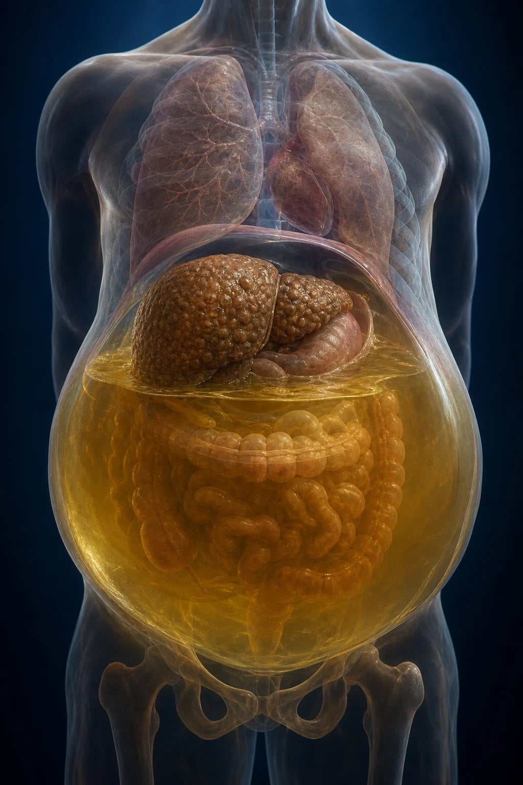
Ascites is the pathological accumulation of excess fluid within the peritoneal cavity. A small volume of peritoneal fluid (up to about 20 mL in a woman of reproductive age, from follicular rupture) is physiological; anything more, or any peritoneal fluid in a man or postmenopausal woman, is abnormal and demands explanation. Clinically detectable ascites on physical examination generally implies at least 1500 mL of fluid; ultrasound can detect as little as 50 mL.[1]
Ascites is the commonest complication of cirrhosis: roughly half of all cirrhotic patients develop ascites within 10 years of diagnosis, and its appearance marks the transition from a compensated to a decompensated phenotype with a steep fall in prognosis — median survival drops from over 10 years (compensated) to about 1.5 to 2 years once ascites appears. For this reason, new ascites in any patient warrants a careful search for liver disease and a diagnostic paracentesis.[1]
Spontaneous bacterial peritonitis (SBP) is the monomicrobial infection of pre-existing ascitic fluid in the absence of an obvious intra-abdominal surgical source (no perforated viscus, no abscess, no contaminated wound). It is defined by an ascitic fluid polymorphonuclear (PMN) neutrophil count of 250 cells/mm3 or more. SBP occurs in 10 to 30 percent of hospitalised cirrhotic patients with ascites, may be entirely asymptomatic at presentation, and carries an in-hospital mortality of 20 to 40 percent despite treatment. After a first episode, recurrence approaches 70 percent at one year without antibiotic prophylaxis, and one-year mortality after the first SBP is around 50 percent — which is why a single episode is an indication to evaluate for liver transplantation.[2][3]
The two concepts — ascites as a syndrome of sodium and water retention, and SBP as its infective complication — are taught together because the paracentesis that diagnoses one is the same tap that excludes or confirms the other, and because they share the long-term consequences of cirrhosis: hepatorenal syndrome, refractory ascites and the need for transplant. [1]
Classification
Ascites is classified along three axes that together direct investigation and management: by serum-ascites albumin gradient (SAAG), by severity grade, and by response to treatment. [1]
By SAAG — the aetiological split
The SAAG is the single most useful ascitic fluid test. It is calculated as serum albumin minus ascitic fluid albumin (both measured on the same day in g/dL) and classifies ascites by mechanism, not by specific disease: [1]
SAAG 1.1 g/dL or more (portal hypertension)
- Cirrhosis (about 80 percent of all ascites)
- Alcoholic hepatitis
- Heart failure (cardiac ascites) and constrictive pericarditis
- Budd-Chiari syndrome (hepatic vein outflow obstruction)
- Massive hepatic metastases or sinusoidal infiltration
- Portal vein thrombosis (late, with cavernous transformation)
- Myxoedema (rare)
SAAG under 1.1 g/dL (non-portal, peritoneal or barrier defect)
- Peritoneal carcinomatosis (ovarian, gastric, colorectal, mesothelioma)
- Peritoneal tuberculosis
- Pancreatic ascites (duct disruption; high amylase)
- Nephrotic syndrome (hypoalbuminaemia and low oncotic pressure)
- Chylous ascites (lymphatic obstruction or trauma; high triglycerides)
- Bile leak or biliary peritonitis (high bilirubin)
- Connective tissue disease serositis; Meigs syndrome
The SAAG works because in portal hypertension the sinusoid is leaky and the hepatic sinusoid freely equilibrates albumin between plasma and ascitic fluid, narrowing the gap to under 1.1 g/dL only when the problem is not portal pressure. Total ascitic protein then refines the picture within the high-SAAG group: a low total protein (under 2.5 g/dL) is typical of cirrhosis (the failing liver plus sinusoidal capillarisation lose opsonins), whereas a high total protein (over 2.5 g/dL) with high SAAG points to cardiac ascites or Budd-Chiari, where the sinusoid is intact and leaks protein-rich fluid.[2]
By severity (the International Ascites Club / EASL grade)
Grade 1 (mild)
- Detected only on ultrasound
- No physical signs; asymptomatic
- Dietary sodium restriction usually sufficient; diuretics if symptomatic
Grade 2 (moderate)
- Moderate symmetrical abdominal distension
- Shifting dullness demonstrable
- Sodium restriction plus spironolactone and furosemide (100:40 ratio)
Grade 3 (large / tense)
- Gross distension with discomfort, dyspnoea, early satiety
- Large-volume paracentesis with albumin for symptom relief
- Then maintenance diuretics and assessment for refractory disease
By response to diuretics — refractory vs uncomplicated
Uncomplicated ascites responds to sodium restriction and diuretics. Refractory ascites is defined by the International Ascites Club as ascites that cannot be mobilised by (diuretic-resistant) or recurs early after (diuretic-intractable) therapeutic paracentesis, despite strict sodium restriction and high-dose diuretics (spironolactone 400 mg per day with furosemide 160 mg per day) for at least one week. Refractory ascites carries a mortality of about 50 percent at 6 months and is an absolute indication to consider TIPS or transplant.[1]
Severity of the underlying cirrhosis — Child-Pugh and MELD-Na
Two scores grade the cirrhosis itself and drive transplant referral: [1]
- Child-Pugh (Child-Turcotte-Pugh): bilirubin, albumin, INR, encephalopathy and ascites, scored 5 to 15 — A (5 to 6) compensated, B (7 to 9) decompensated, C (10 to 15) severe. Ascites contributes directly to the score.
- MELD-Na (Model for End-stage Liver Disease incorporating sodium): MELD-Na = MELD + 1.32 times (137 minus serum sodium) minus (0.033 times MELD times (137 minus serum sodium)), used for transplant allocation; the addition of sodium reflects that dilutional hyponatraemia and ascites both mark advanced disease. [1]
Epidemiology & Risk Factors
Ascites and SBP — the numbers that matter
Who gets ascites
Ascites appears once clinically significant portal hypertension is established — that is, once the hepatic venous pressure gradient exceeds about 10 mmHg, and ascites becomes likely once it exceeds 12 mmHg. The commonest underlying diseases are, in approximate global order, alcohol-related liver disease, hepatitis B, hepatitis C and MASLD (metabolic dysfunction-associated steatotic liver disease, formerly NAFLD); less often autoimmune, cholestatic (primary biliary cholangitis, primary sclerosing cholangitis), metabolic (haemochromatosis, Wilson, alpha-1-antitrypsin) and vascular (Budd-Chiari).[1]
In India, hepatitis B remains the single commonest cause of cirrhosis and therefore of ascites, followed by alcohol and hepatitis C, with MASLD rising rapidly in urban populations. In the West, alcohol and (curable) hepatitis C have historically dominated, and MASLD is now the fastest-growing indication for transplant. [1]
Risk factors for ascites formation
- Advanced liver disease — Child-Pugh B or C, MELD over 15.
- Ongoing liver injury — continued alcohol intake, uncontrolled viraemia, untreated autoimmune inflammation.
- Portal hypertension — large varices, splenomegaly, portal vein collaterals on imaging.
- Renal sodium retention — activated RAAS; dietary non-compliance (high salt intake).
- Drugs — NSAIDs (block prostaglandins, blunt diuretic response, risk AKI), ACE inhibitors and ARBs (hypotension, AKI in decompensation), and over-aggressive diuretic use causing prerenal azotaemia. [1]
Risk factors for SBP
The risk of SBP rises sharply when the ascitic fluid is poor at killing bacteria, which means low total protein and low opsonic activity: [1]
- Ascitic fluid total protein under 1.5 g/dL (15 g/L) — the single strongest predictor; the fluid lacks complement and immunoglobulin to opsonise bacteria.
- Very low protein under 1.0 g/dL (10 g/L) combined with renal impairment (creatinine over 1.2 mg/dL, BUN over 25 mg/dL) or hyponatraemia (under 130 mmol/L) — the combination that triggers primary norfloxacin prophylaxis.[6]
- Prior SBP — recurrence over 70 percent at one year; indicates an inability to clear translocated bacteria.
- Acute variceal bleeding — gut bacterial overgrowth and translocation during the bleed; short-course antibiotic prophylaxis (ceftriaxone) is standard.
- Severe liver disease — Child-Pugh C, high bilirubin.
- Proton pump inhibitor use — alters gut microbiota and increases enteric bacterial overgrowth; an association seen in cohort studies.
Pathophysiology
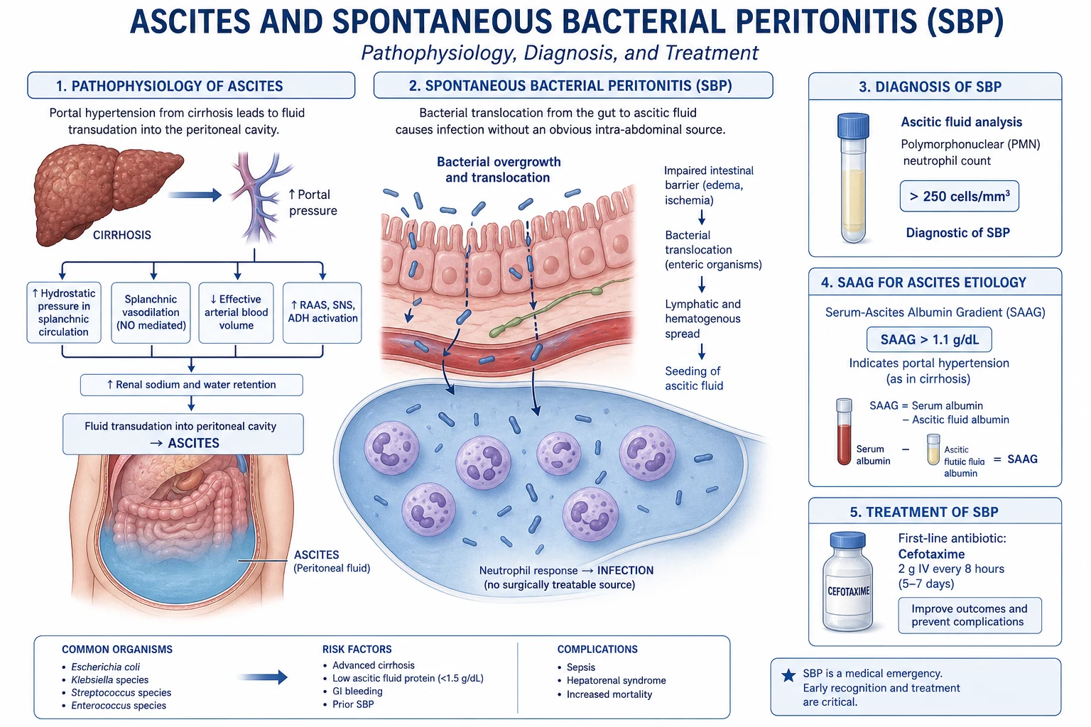
The formation of cirrhotic ascites is best understood through the peripheral arterial vasodilation hypothesis, which integrates four interacting mechanisms. Each is a target of therapy. [1]
The peripheral arterial vasodilation hypothesis
-
Portal hypertension (the initiating event). Cirrhotic architectural distortion plus dynamic stellate-cell contraction raises intrahepatic resistance; the hepatic venous pressure gradient climbs above 10 mmHg. Sinusoidal hydrostatic pressure rises and fluid is driven across the sinusoidal membrane into the space of Disse and the peritoneal cavity. Ascites rarely forms below an HVPG of 12 mmHg. [1]
-
Splanchnic arterial vasodilation (the pivotal step). The high portal pressure triggers a sustained increase in splanchnic production of nitric oxide (NO), carbon monoxide (CO), glucagon, prostacyclin and endocannabinoids. These dilate the splanchnic arteriolar bed, increasing splanchnic blood volume and, paradoxically, reducing the effective arterial blood volume that the heart and great vessels can sense. This is the central insight of the hypothesis: the patient is volume-overloaded but the baroreceptors read underfilling. [1]
-
Neurohormonal activation and renal sodium and water retention (the response). To restore the perceived underfilling, the body activates the renin-angiotensin-aldosterone system (RAAS), the sympathetic nervous system and non-osmotic vasopressin (ADH) release. Aldosterone drives distal sodium reabsorption; the sympathetic tone promotes proximal sodium reabsorption and renin release; ADH inserts aquaporin-2 channels in the collecting duct, retaining free water and producing dilutional hyponatraemia. The result is avid renal sodium and water retention — urine sodium is typically very low (under 10 mmol/day) in untreated cirrhotic ascites. Over time, renal vasoconstriction (from angiotensin II and sympathetic drive, compounded by intrarenal imbalance of prostaglandins and leukotrienes) progresses to hepatorenal syndrome.[4]
-
Hypoalbuminaemia (the permissive factor). The failing liver synthesises less albumin, lowering plasma oncotic pressure and allowing the retained fluid to escape into the peritoneal space rather than staying within the vascular compartment. [1]
The retained fluid distributes between the peritoneal cavity (ascites) and the interstitium (peripheral and sacral oedema). The cycle is self-perpetuating: more vasodilation, more RAAS activation, more sodium retention, more ascites — until either the cause is treated or the kidney fails (HRS). This is why spironolactone (an aldosterone antagonist) is the cornerstone diuretic, and why the only definitive therapy is to relieve portal pressure (TIPS) or replace the liver (transplant).[1]
Pathogenesis of SBP — bacterial translocation
SBP arises by bacterial translocation: enteric bacteria cross the oedematous, hyperpermeable cirrhotic gut wall, reach the mesenteric lymph nodes and then the systemic circulation, and finally seed an ascitic fluid that is too protein-poor to opsonise and kill them. Four defects converge:[2]
- Intestinal bacterial overgrowth — slowed small-bowel transit (dysmotility) and reduced bile acids allow overgrowth, especially of Gram-negative bacilli.
- Increased intestinal permeability — portal hypertension and splanchnic congestion open tight junctions.
- Impaired reticuloendothelial (Kupffer cell) clearance — the cirrhotic liver fails to clear bacteria reaching it via the portal vein.
- Low ascitic fluid opsonic activity — total protein under 1.5 g/dL means low complement (C3) and immunoglobulin, so bacteria are not opsonised or phagocytosed. [1]
The organisms recovered are dominated by Gram-negative enteric bacilli — Escherichia coli and Klebsiella pneumoniae — followed by Gram-positive cocci (Streptococcus pneumoniae, other streptococci, Enterococcus, and Staphylococcus). With widespread quinolone prophylaxis, Gram-positive and resistant Gram-negative organisms have become proportionally more common. By definition, SBP is monomicrobial; a polymicrobial culture with multiple organisms suggests a secondary peritonitis (perforated viscus) and mandates surgical review and anaerobic cover. [1]
Clinical Presentation
Ascites — symptoms
The dominant complaint is progressive abdominal distension and weight gain, often over weeks. As the abdomen enlarges, patients describe early satiety (gastric compression), abdominal discomfort or a dragging sensation, and — when tense — dyspnoea (diaphragmatic splinting) and orthopnoea. Fluid may track into the scrotum causing embarrassing swelling, or through a patent umbilical ring producing an umbilical hernia. With coexistent peripheral oedema, the legs and sacrum swell. Sleep is disturbed by nocturia and inability to find a comfortable position. [1]
Ascites — signs
Examination begins with general inspection and the stigmata of chronic liver disease, then moves to a focused abdominal examination to confirm and quantify the fluid. [1]
Signs of ascites:
- Bulging flanks — fluid pushes the flanks outward in the supine patient.
- Flank dullness to percussion (the earliest sign; needs about 1500 mL).
- Shifting dullness — the most reliable bedside sign; dullness in the flanks in the supine position that shifts when the patient rolls onto one side. Sensitivity about 80 to 90 percent for over 1500 mL.
- Fluid thrill (fluid wave) — elicited by a tap on one flank felt by the examiner's other hand on the opposite flank, with a third hand on the midline to dampen transmission through fat; specific but insensitive (needs over 2000 mL).
- Eversion of the umbilicus and a tense, glistening abdominal wall.
- Scrotal and pedal oedema if fluid has tracked or if hypoalbuminaemia is severe. [1]
Stigmata of chronic liver disease (these identify the cause as cirrhosis): spider naevi, palmar erythema, jaundice, parotid enlargement, gynaecomastia, testicular atrophy, caput medusae (recanalised umbilical vein), muscle wasting (sarcopenia), asterixis (if hepatic encephalopathy), fetor hepaticus, clubbing, Dupuytren contracture, and in alcohol-related disease a haemorrhagic palmar sign and bilateral parotid enlargement. [1]
Look for the underlying cause — raised JVP, pulsatile tender hepatomegaly, a third heart sound and peripheral oedema suggest cardiac ascites; a rigid, palpable mass or supraclavicular node (Virchow) suggests malignancy; fever, night sweats and high-ascitic-ADA suggest tuberculosis; a painful enlarged liver with ascites and a prothrombotic state suggests Budd-Chiari.[1]
Clinical presentation of SBP
SBP is protean. The classical tetrad is fever, abdominal pain, new or worsening hepatic encephalopathy, and abdominal tenderness, but the presentation is often incomplete or atypical: [1]
- Fever (over 37.8 degrees C) — the commonest single feature, present in about half.
- Abdominal pain or tenderness — mild and diffuse in most; localised peritonism is uncommon and suggests secondary peritonitis.
- New or worsening hepatic encephalopathy — confusion, drowsiness, asterixis; may be the only feature in a deteriorating patient.
- Deteriorating renal function — SBP is the commonest precipitant of type-1 hepatorenal syndrome.
- Gastrointestinal bleeding, hypotension, or paralytic ileus.
- Asymptomatic in 10 to 30 percent — picked up only on routine admission paracentesis. This is why a diagnostic tap is done on every cirrhotic with ascites at admission, regardless of symptoms. [1]
The bedside rule the student must hold: a cirrhotic with ascites who deteriorates in any way — fever, confusion, rising creatinine, falling blood pressure, acidosis — has SBP until proven otherwise, and a paracentesis is mandatory within hours.[2]
Differential Diagnosis
Ascites has a wide differential; the SAAG and a handful of confirmatory tests narrow it. The table reproduces the discriminating features for the commoner non-cirrhotic causes. [1]
| Cause | SAAG | Total protein | Discriminating features | Confirmatory test |
|---|---|---|---|---|
| Cirrhosis | 1.1 or more | Low (under 2.5 g/dL) | Stigmata of CLD, nodular liver, splenomegaly; low platelets | Ultrasound + elastography; biopsy if unclear |
| Heart failure / constrictive pericarditis | 1.1 or more | High (over 2.5 g/dL) | Raised JVP, pulsatile liver, S3, peripheral oedema, pericardial knock, Kussmaul sign | ECG, echocardiogram, BNP |
| Budd-Chiari | 1.1 or more | High | Painful hepatomegaly, sudden ascites, prothrombotic state | Doppler or CT of hepatic veins |
| Peritoneal carcinomatosis | Under 1.1 | Variable | Cachexia, weight loss, primary tumour (ovarian, gastric, colorectal, mesothelioma) | Positive cytology; CT; laparoscopy |
| Peritoneal tuberculosis | Under 1.1 | Variable (often high) | Fever, night sweats, lymphadenopathy, high lymphocytes in fluid | High ADA (over 40 U/L); peritoneal biopsy with caseating granulomas |
| Pancreatic ascites | Under 1.1 | Variable | Pancreatitis history, pseudocyst | Markedly high ascitic amylase (over 1000 U/L) |
| Nephrotic syndrome | Under 1.1 | Low | Generalised oedema, frothy urine, heavy proteinuria | 24-hour urine protein; serum albumin |
| Chylous ascites | Under 1.1 | Variable | Milky fluid; lymphoma, trauma, filariasis, surgery | Triglycerides over 200 mg/dL in fluid |
| Meigs syndrome | Under 1.1 (usually) | Variable | Ovarian fibroma, ascites and pleural effusion triad; resolves with tumour removal | Pelvic ultrasound |
Key examinable distinctions: a high SAAG with high total protein is cardiac or Budd-Chiari; a high SAAG with low total protein is cirrhosis. A low SAAG with positive cytology is carcinomatosis; with high ADA and lymphocytes is tuberculosis; with high amylase is pancreatic; with high triglycerides is chylous. Cytology, amylase, triglycerides, ADA and culture are the additional tests that close the differential.[1][2]
Hepatic hydrothorax deserves a separate mention: a pleural effusion (almost always right-sided) in a cirrhotic with ascites, formed when ascitic fluid tracks through small diaphragmatic defects; it is not cardiac or pulmonary and is managed as ascites. Spontaneous bacterial empyema is the pleural analogue of SBP and is treated the same way. [1]
Clinical & Bedside Assessment
History
A focused history identifies the cause and the complications: [1]
- Liver disease duration and aetiology — alcohol (units per day, years), hepatitis B and C risk (transfusion, intravenous drug use, tattoo, sexual, vertical), MASLD (diabetes, obesity, metabolic syndrome), autoimmune, family history (haemochromatosis, Wilson, alpha-1-antitrypsin).
- Course of ascites — onset, rate of increase, prior episodes, response to diuretics, number of paracenteses, episodes of SBP.
- Precipitants of decompensation — infection, gastrointestinal bleed, constipation, alcohol binge, sedatives, over-diuresis (use the HE-FLAG mnemonic from the cirrhosis topic).
- Drugs — NSAIDs, ACE inhibitors, ARBs, beta-blockers, diuretics, nephrotoxins, proton pump inhibitors, lithium.
- Cardiac disease — heart failure, prior cardiac surgery or radiotherapy (constriction).
- Malignancy and TB risk — weight loss, primary tumour, TB exposure.
- Functional impact — dyspnoea, sleep, mobility, ability to work. [1]
Examination
- Vital signs and general inspection — fever (SBP), hypotension and tachycardia (sepsis, bleed), respiratory rate, oxygen saturation, signs of sepsis or shock.
- JVP and cardiac examination — a raised JVP with a third heart sound and peripheral oedema suggests cardiac ascites; a pericardial knock and Kussmaul sign suggest constriction.
- Abdominal examination — confirm and grade ascites (shifting dullness, fluid thrill), assess for tenderness (SBP, secondary peritonitis), organomegaly (huge nodular liver in malignancy or Budd-Chiari; splenomegaly in portal hypertension), masses (carcinomatosis), and umbilical hernia (rupture risk in tense ascites).
- Stigmata of chronic liver disease — listed above.
- Neurology — asterixis, GCS (Glasgow Coma Scale: eye opening E1-E4, verbal V1-V5, motor M1-M6; maximum 15), to detect and grade hepatic encephalopathy.
- Fluid balance — weight, intake and output, urine sodium if available. [1]
Bedside procedures — paracentesis technique
A diagnostic paracentesis is performed in all patients with new ascites and in any cirrhotic with ascites who deteriorates. The standard site is the left lower quadrant, two finger-breadths cephalad and two finger-breadths medial to the anterior superior iliac spine (avoids the inferior epigastric vessels; the left side avoids the caecum and appendix). The midline 2 cm below the umbilicus in the avascular linea alba is an alternative if fluid is confirmed there. Use ultrasound guidance if the fluid is loculated, the patient is obese, or prior taps were dry. Coagulopathy (raised INR, low platelets) is not a contraindication to a diagnostic tap with a fine needle; it is a relative consideration for large-volume paracentesis, but prophylactic plasma or platelets are not routinely needed.[2]
Investigations
The diagnostic paracentesis is the single most important investigation. Send ascitic fluid for the panel below; send bloods and imaging to characterise the underlying disease. [1]
Ascitic fluid panel (the core of the work-up)
Ascitic fluid — the panel and the thresholds
Practical points: [1]
- The cell count and differential must be available within hours; it is the test that triggers or withholds SBP treatment. Hand-carry the specimen.
- Culture yield rises from 40 to 80 percent when ascitic fluid is inoculated directly into blood culture bottles at the bedside (10 mL aerobic, 10 mL anaerobic) rather than sent in a plain tube. Culture-negative neutrocytic ascites (raised PMN, negative culture) is still SBP and is treated the same way.
- Cytology is highly sensitive for peritoneal carcinomatosis (over 95 percent with three separate samples) but insensitive for hepatocellular carcinoma and most primary liver tumours.
- Glucose, LDH and carcinoembryonic antigen help distinguish SBP from secondary peritonitis (polymicrobial, high LDH, low glucose, multiple organisms) when the picture is equivocal. [1]
Bloods
A full screen characterises the underlying disease and the severity: [1]
- Full blood count — anaemia (chronic disease, hypersplenism, bleed), leucocytosis (SBP, infection), thrombocytopenia (hypersplenism from portal hypertension; under 150,000 is a marker of advanced fibrosis).
- Liver function and synthetic markers — bilirubin, AST, ALT, ALP, GGT, albumin (low in cirrhosis and nephrotic syndrome), INR (synthetic function).
- Renal function and electrolytes — urea, creatinine, sodium (dilutional hyponatraemia is prognostic and a MELD-Na input), potassium (diuretic monitoring).
- Aetiology — viral serology (HBsAg, anti-HCV, HIV), autoimmune (ANA, smooth muscle antibody, anti-LKM1, antimitochondrial antibody, immunoglobulins), iron studies (haemochromatosis), caeruloplasmin and 24-hour urinary copper (Wilson), alpha-1-antitrypsin phenotype, alpha-fetoprotein (hepatocellular carcinoma), TSH (myxoedema).
- Inflammatory and culture — blood cultures (before antibiotics), C-reactive protein, urine culture, chest X-ray.
- Coagulation — INR, PT (and a baseline for paracentesis planning). [1]
Imaging
- Abdominal ultrasound is essential in every patient: confirms ascites, estimates volume, assesses liver size and echotexture (small nodular liver in cirrhosis; large infiltrated liver in malignancy), detects splenomegaly (portal hypertension), patency and direction of flow in the portal and hepatic veins (Budd-Chiari, portal vein thrombosis), focal lesions (hepatocellular carcinoma), and guides the paracentesis when fluid is loculated.
- Doppler of the portal and hepatic veins.
- Triple-phase CT or MRI of the liver if hepatocellular carcinoma is suspected (arterial hypervascularity with washout) or to stage malignancy.
- CT of the abdomen and pelvis if peritoneal carcinomatosis or pancreatic source is suspected.
- Echocardiogram and ECG if cardiac ascites or constriction is suspected (raised BNP, pericardial calcification on CT, ventricular interdependence on echo).
- Diagnostic laparoscopy with peritoneal biopsy for tuberculous peritonitis or carcinomatosis when cytology and imaging are inconclusive; biopsy shows caseating granulomas in TB and metastatic deposits in carcinomatosis. [1]
Definitions reproduced accurately
What is the diagnostic threshold for SBP?
Ascitic fluid PMN count of 250 cells/mm3 or more, with no evident intra-abdominal surgical source. The older threshold of 500 WBC with 50 percent or more neutrophils is less sensitive and is now secondary. If the fluid is haemorrhagic, subtract one PMN for every 250 red cells (to correct for neutrophils in the blood contamination).
What is culture-negative neutrocytic ascites (CNNA)?
Raised PMN (250 cells/mm3 or more) with a negative culture in a patient not on antibiotics. It is a variant of SBP — same organisms, same prognosis, same treatment — and reflects the fastidiousness of the organisms or delayed inoculation, not a different disease.
What distinguishes SBP from secondary (surgical) peritonitis?
Secondary peritonitis is polymicrobial (multiple organisms including anaerobes), has a very high LDH, low glucose (under 50 mg/dL), high protein, and may show multiple organisms on Gram stain; it implies a perforated viscus or abscess and needs surgical review and broad anaerobic cover. SBP is monomicrobial with normal glucose and LDH.
Management — Resuscitation
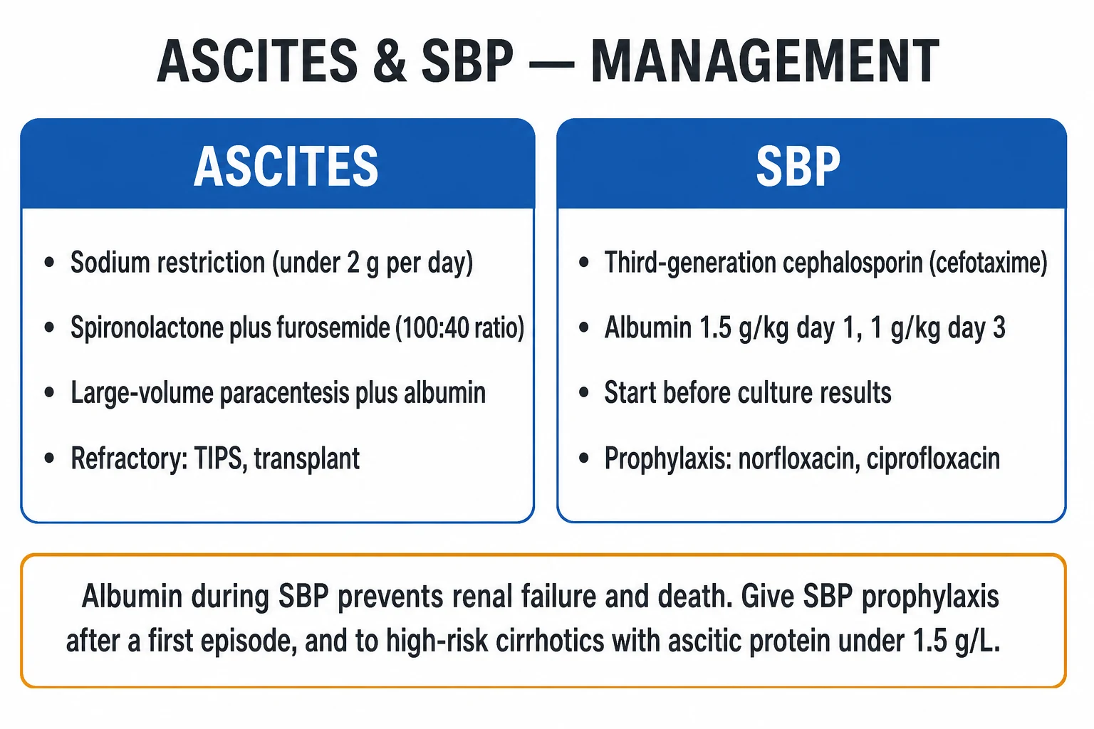
The resuscitation phase applies to three acute scenarios: suspected SBP, tense ascites with respiratory compromise, and acute kidney injury / hepatorenal syndrome. [1]
1. Suspected or confirmed SBP — the time-critical bundle
When the ascitic PMN is 250 cells/mm3 or more, start empirical antibiotics within hours — every hour of delay increases mortality. The bundle is:[2][3]
Diagnostic paracentesis
Cell count + differential stat; culture in blood-culture bottles at bedside before antibiotics
Empirical antibiotic
Cefotaxime 2 g IV every 8 hours, or ceftriaxone 2 g IV once daily — a third-generation cephalosporin; 5 days total; narrow to sensitivities when available
Intravenous albumin
1.5 g/kg on day 1 and 1 g/kg on day 3 — reduces renal impairment (from 33 to 10 percent) and mortality (Sort 1999)
Repeat tap at 48 hours
A fall in PMN of at least 25 percent confirms response; a fall under 25 percent suggests treatment failure or secondary peritonitis — broaden cover, look for a source
Step-down
Switch to an oral agent (ciprofloxacin 500 mg twice daily or amoxicillin-clavulanate) once the patient is afebrile and clinically improved, to complete 5 days
Secondary prophylaxis
Begin norfloxacin 400 mg once daily (or ciprofloxacin 500 mg once daily) after the episode, and refer for transplant assessment
Why albumin in SBP? SBP triggers a sharp fall in effective arterial blood volume (cytokine-mediated vasodilation), which precipitates type-1 hepatorenal syndrome in about a third of untreated patients. The Sort trial established that albumin 1.5 g/kg on day 1 and 1 g/kg on day 3 reduces renal failure and 3-month mortality; the benefit is greatest in patients with creatinine over 1 mg/dL and bilirubin over 4 mg/dL.[3]
2. Tense ascites with respiratory compromise
A single large-volume paracentesis (LVP) relieves symptoms immediately. Removing up to 5 litres is safe without albumin; for volumes over 5 litres, give intravenous albumin at 6 to 8 g per litre removed (for example, 8 litres removed equals 48 to 64 g of 20 percent albumin) to prevent post-paracentesis circulatory dysfunction (PPCD) — a vasodilatory drop in effective arterial volume that triggers RAAS activation, accelerates ascites reaccumulation, and worsens renal function and mortality at 6 months.[1][2]
3. Acute kidney injury and hepatorenal syndrome
If creatinine rises, stop diuretics and all nephrotoxic drugs (NSAIDs, ACE inhibitors, ARBs, aminoglycosides, contrast), give a volume challenge with albumin 1 g/kg per day for two days, and treat any infection. If the creatinine fails to recover, the diagnosis is hepatorenal syndrome-AKI (see Specific Subtypes) — start a vasoconstrictor (terlipressin) plus albumin.[4][9]
Management — Definitive & Stepwise
The definitive ladder moves from diet and drugs to procedures and transplant. [1]
Step 1 — Dietary sodium restriction and fluid management
Restrict dietary sodium to under 88 mmol per day (under 2 g sodium, about 5 g salt per day) — the single most important non-pharmacological step; without it, no diuretic will work. Dietitian-led counselling is essential, with attention to hidden salt in breads, pickles, cheese and processed foods. Fluid restriction is reserved for dilutional hyponatraemia (serum sodium under 125 mmol/L); it is not routinely needed for ascites itself, and restricting fluid without restricting sodium is futile.[1]
Step 2 — Diuretics (the 100:40 ratio)
The pharmacological cornerstone is spironolactone (blocks aldosterone, the dominant sodium-retaining hormone in cirrhosis), started with furosemide to maintain normokalaemia and accelerate response: [1]
The 100:40 diuretic regimen
Practical points on diuretics:
- Monitor weight daily, and sodium, potassium, creatinine and encephalopathy every 2 to 3 days during titration. A 24-hour urine sodium over 78 mmol/day (a "negative" sodium balance) predicts response.
- Spironolactone side effects: hyperkalaemia, gynaecomastia (painful, dose-related; switch to eplerenone or amiloride if intolerable), metabolic acidosis. Onset is slow (2 to 3 days) because of its long half-life.
- Furosemide side effects: hypokalaemia, dehydration, hypotension, prerenal AKI, ototoxicity at high IV doses.
- Stop or hold diuretics if encephalopathy develops, serum sodium falls below 125 to 120 mmol/L despite water restriction, or creatinine rises by over 100 percent to over 2 mg/dL (over-diuresis).[1]
Step 3 — Large-volume paracentesis with albumin
For grade 3 (tense) ascites and for refractory ascites, LVP plus albumin is first-line. A single session can remove 5 to 10 litres; albumin replacement (6 to 8 g per litre removed when over 5 L) prevents PPCD. LVP relieves symptoms immediately but does not treat the underlying sodium retention — so it is combined with maintenance diuretics to slow reaccumulation.[2]
Step 4 — Refractory ascites: TIPS and transplant
When ascites is refractory (fails maximal diuretics or recurs rapidly after LVP), options are serial LVP every 2 to 4 weeks with albumin, transjugular intrahepatic portosystemic shunt (TIPS) in selected patients, and liver transplantation as definitive therapy. [1]
Transjugular intrahepatic portosystemic shunt (TIPS)
Dose
Covered stent placed between hepatic vein and intrahepatic portal vein, dropping portal pressure
A meta-analysis of individual patient data confirmed that TIPS controls ascites better than serial paracentesis (ascites-free at three months in about two-thirds) and may improve transplant-free survival, at the cost of more hepatic encephalopathy; it does not clearly improve overall survival and is not a substitute for transplant evaluation.[5]
Step 5 — Liver transplantation
Ascites — especially refractory ascites, an episode of SBP, or hepatorenal syndrome — is an absolute indication to refer for transplant evaluation, since it marks decompensation with a 50 percent two-year mortality. Allocation is by MELD-Na, which captures both severity and the sodium-retaining phenotype. [1]
Long-term albumin — the ATTIRE evidence
The role of long-term targeted albumin in stable outpatients has been clarified by the ATTIRE trial (China et al, NEJM 2021). Targeted infusions to keep serum albumin over 30 g/L in hospitalised patients with decompensated cirrhosis did not reduce the incidence of new infection, renal dysfunction, or death compared with standard care, tempering enthusiasm for routine long-term high-dose albumin outside its established indications (SBP, LVP, HRS).[7]
Stepwise management ladder — at a glance
1. Diagnostic paracentesis
All new ascites; all cirrhotics with ascites at admission; any deterioration
2. Classify by SAAG and identify cause
SAAG 1.1 or more = portal hypertension; total protein, cytology, ADA, amylase, triglycerides refine
3. Treat the underlying cause
Alcohol abstinence, antivirals for HBV/HCV, immunosuppression for autoimmune, cardiac therapy
4. Sodium restriction and diuretics
Under 88 mmol sodium/day; spironolactone 100 mg with furosemide 40 mg (100:40), titrate to 400/160
5. LVP with albumin for tense ascites
Albumin 6 to 8 g per litre removed over 5 L to prevent PPCD
6. Refractory ascites
Serial LVP; TIPS in selected; transplant referral
7. SBP bundle
Cefotaxime 2 g IV every 8 hours (or ceftriaxone 2 g daily) plus albumin 1.5/1 g/kg
8. SBP prophylaxis
Secondary: norfloxacin 400 mg OD; primary if protein under 1.5 g/dL with renal impairment
9. HRS
Stop nephrotoxins; albumin challenge; terlipressin plus albumin; transplant
10. Monitor
Weight, Na, K, creatinine, encephalopathy; titrate diuretics to maintenance
Specific Subtypes & Scenarios
Spontaneous bacterial peritonitis
Already covered in depth above. Key examinable summary: PMN 250 cells/mm3 or more, monomicrobial, no surgical source; treat with cefotaxime 2 g IV every 8 hours or ceftriaxone 2 g IV daily for 5 days plus albumin 1.5 g/kg day 1 and 1 g/kg day 3; recurrence over 70 percent at one year mandates lifelong norfloxacin 400 mg daily and transplant referral. Up to a third of patients are asymptomatic — the silent deteriorating cirrhotic with ascites is the classic NEET-PG / INICET stem.[2][3]
Refractory ascites
Defined as ascites that cannot be mobilised by, or recurs early after, large-volume paracentesis despite sodium restriction and maximal diuretics (spironolactone 400 mg with furosemide 160 mg) for at least one week. Subdivided into diuretic-resistant (no weight loss on maximal therapy) and diuretic-intractable (complications of diuretics — hepatic encephalopathy, renal impairment, hyponatraemia — preclude effective doses). Management is serial LVP with albumin, TIPS in selected patients, and early transplant referral. Mortality is about 50 percent at 6 months.[1]
Hepatorenal syndrome (HRS-AKI)
Hepatorenal syndrome is a functional, potentially reversible renal failure of advanced cirrhosis (and ascites) with no intrinsic kidney disease — the kidney is structurally normal but vasoconstricted because of intense splanchnic vasodilation and RAAS/sympathetic overactivity. The revised International Club of Ascites criteria (2015) redefine HRS within the broader AKI framework: HRS-AKI is diagnosed when AKI criteria are met (rise in creatinine by at least 0.3 mg/dL within 48 hours, or by at least 50 percent from baseline), no response after 48 hours of diuretic withdrawal and albumin 1 g/kg/day, no shock, no nephrotoxins, no structural kidney disease (normal ultrasound, no proteinuria over 500 mg/day, no microhaematuria).[4][9]
HRS-AKI (formerly type 1 HRS)
- Rapidly progressive AKI — doubling of creatinine to over 2.5 mg/dL in under 2 weeks
- Often precipitated by SBP, large-volume paracentesis without albumin, or over-diuresis
- Median survival 1 to 2 weeks untreated
- Treat with terlipressin plus albumin; norepinephrine in ICU; urgent transplant
HRS-AKD / type 2 HRS
- Slowly progressive, indolent renal dysfunction with refractory ascites
- Driven by chronic splanchnic vasodilation and RAAS activation
- Median survival 4 to 6 months
- Treat ascites (LVP, TIPS), vasoconstrictor plus albumin; transplant referral
Treatment of HRS-AKI:
- Stop diuretics and all nephrotoxic drugs; albumin 1 g/kg per day (maximum 100 g) for two days as a challenge.
- Terlipressin (a vasopressin V1 agonist causing splanchnic vasoconstriction) plus albumin: terlipressin 1 to 2 mg IV every 4 to 6 hours (continuous infusion 2 to 12 mg/day is an alternative) titrated to a creatinine under 1.5 mg/dL, with albumin 20 to 40 g daily. The CONFIRM trial (Wong et al, NEJM 2021) confirmed that terlipressin plus albumin achieves HRS reversal in about 32 percent versus 17 percent with placebo, with the caveat of respiratory failure in about 11 percent; hence terlipressin is reserved for patients without overt fluid overload or respiratory compromise.[8]
- Norepinephrine infusion (0.5 to 3 mg/h, titrated) is an ICU alternative of comparable efficacy.
- Midodrine 7.5 mg three times daily plus octreotide 100 to 200 microgram subcutaneously three times daily plus albumin is a less effective oral-based regimen where terlipressin is unavailable.
- Renal replacement therapy bridges to transplant in candidates; it is not a destination therapy in non-transplant candidates.
- Liver transplantation is the only definitive therapy; combined liver-kidney transplant is considered for those on prolonged dialysis.[4]
Hepatic hydrothorax
A pleural effusion (over 80 percent right-sided) in a cirrhotic with ascites, formed when ascitic fluid tracks through small diaphragmatic defects under the negative intrathoracic pressure. Patients present with dyspnoea; some have minimal or no clinically detectable ascites. Managed as ascites (sodium restriction, diuretics); chest tube insertion must be avoided (high mortality from protein and fluid loss); TIPS is effective for refractory cases. Spontaneous bacterial empyema is the pleural analogue of SBP (pleural fluid PMN 250 or more, positive culture) and is treated with a third-generation cephalosporin.[1]
Cardiac ascites
High SAAG with high total protein (over 2.5 g/dL), raised JVP, pulsatile tender hepatomegaly, and a third heart sound or pericardial knock. Treat the underlying heart failure; avoid TIPS (worsens right heart failure). The distinction from cirrhotic ascites rests on the high ascitic total protein and the cardiac signs. [1]
Malignant and tuberculous ascites
Low SAAG, often with positive cytology (carcinomatosis) or high ADA with caseating granulomas on peritoneal biopsy (tuberculosis). Manage the underlying disease; palliative paracentesis for symptomatic malignant ascites (often rapidly recurring); anti-tubercular therapy (RIPE: rifampicin, isoniazid, pyrazinamide, ethambutol) for six months for peritoneal TB. Both can mimic cirrhotic ascites — the SAAG and the directed fluid tests prevent misdiagnosis. [1]
Umbilical hernia in tense ascites
Common in long-standing tense ascites; rupture (with leak of ascitic fluid and secondary peritonitis) and incarceration are the feared complications. Manage by controlling the ascites first (LVP, diuretics), then elective surgical repair; emergency repair of a ruptured hernia carries high mortality. [1]
Complications & Pitfalls
Disease-related complications
- SBP — 10 to 30 percent of hospitalised cirrhotics; recurrence over 70 percent at one year without prophylaxis.
- Hepatorenal syndrome — type 1 untreated mortality 80 to 90 percent at two weeks; type 2 with refractory ascites 50 percent at six months.
- Hepatic hydrothorax and spontaneous bacterial empyema.
- Umbilical hernia rupture — surgical emergency with high mortality from secondary peritonitis.
- Post-paracentesis circulatory dysfunction (PPCD) — after LVP over 5 litres without albumin; vasodilation, RAAS activation, faster ascites reaccumulation, renal dysfunction; prevented by albumin 6 to 8 g per litre removed.
- Dilutional hyponatraemia — serum sodium under 125 to 130 mmol/L from non-osmotic ADH; prognostic marker; fluid restriction is the mainstay; vaptans are not recommended.
- Abdominal compartment syndrome — rare, from massive tense ascites; reduced renal perfusion and ventilatory compromise; relieved by LVP.
- Progression to ACLF — infection (SBP) is the commonest precipitant of acute-on-chronic liver failure with multi-organ failure. [1]
Procedure-related pitfalls
- Bowel or bladder injury from an inappropriately sited tap — use the left lower quadrant with the landmark technique or ultrasound guidance.
- Bleeding — a theoretical risk with coagulopathy, but prophylactic plasma or platelets are not routinely needed for a diagnostic tap; bleeding is rare (under 1 percent). Avoid the inferior epigastric vessels (lateral to the rectus).
- Leakage of ascitic fluid after LVP — use a Z-track technique; if persistent, a single suture.
- Infection — sterile technique; secondary peritonitis after a contaminated tap is rare.
- PPCD — preventable by albumin replacement for LVP over 5 litres. [1]
Classic errors
- Treating ascites without a diagnostic tap — missing SBP, malignancy or TB.
- Using SAAG without total protein — missing cardiac and Budd-Chiari.
- Over-diuresis — precipitating prerenal AKI, encephalopathy, hyponatraemia.
- Continuing NSAIDs, ACE inhibitors, or beta-blockers in decompensated cirrhosis — worsening renal function.
- LVP over 5 litres without albumin — PPCD.
- Chest tube for hepatic hydrothorax — high mortality; avoid.
- Missing the indication for transplant referral after refractory ascites, first SBP, or HRS. [1]
Prognosis & Disposition
Prognosis at a glance
Determinants of poor outcome are a high Child-Pugh or MELD-Na, refractory ascites, hyponatraemia (sodium under 130 mmol/L), renal impairment (HRS), low ascitic fluid protein, recurrent SBP, poor nutritional status and sarcopenia, and an ongoing precipitant (alcohol, infection, bleed). [1]
Disposition: outpatient management for grade 1 to 2 uncomplicated ascites with close monitoring; admission for tense ascites, any SBP, AKI or HRS, new encephalopathy, or rapid reaccumulation; ICU for septic shock, ACLF, or HRS with multi-organ failure. Every patient with refractory ascites, a first SBP, or HRS should be referred for transplant evaluation at the earliest opportunity.[1]
Special Populations
- Hepatocellular carcinoma with ascites — ascites may arise from portal hypertension, tumour thrombus in the portal or hepatic vein, or peritoneal seeding. Treat the ascites as above, but avoid TIPS if there is advanced HCC or portal vein tumour thrombus; transplant eligibility depends on Milan criteria (single tumour under 5 cm or up to 3 tumours each under 3 cm, no vascular invasion, no extrahepatic spread).
- Pregnancy in cirrhosis — rare (most cirrhotic women are subfertile from hypothalamic-pituitary dysfunction) but possible; managed jointly by hepatology and obstetrics (often maternal-fetal medicine). Avoid teratogenic drugs and NSAIDs; NSAIDs are also best avoided in the third trimester on independent grounds. Decompensation risk rises in the third trimester; plan delivery in a transplant-capable centre.
- Paediatric ascites — biliary atresia (post-Kasai), alpha-1-antitrypsin deficiency, autoimmune hepatitis, and metabolic disease (Wilson, glycogen storage) are the main causes; refer to paediatric hepatology. Doses are weight-based.
- Elderly — limit aggressive diuresis and very large LVP (risk of prerenal AKI, encephalopathy, fluid shifts); weigh the transplant decision against comorbidity and frailty.
- Patients on anticoagulation — diagnostic tap is generally safe; for LVP, weigh bleeding risk and consider holding anticoagulants per their half-life; coagulopathy of cirrhosis is not a routine indication to correct with plasma or platelets before a tap.
- Post-liver-transplant ascites — early postoperative ascites suggests vascular compromise (hepatic artery or portal vein thrombosis), bile leak, or acute rejection; urgent Doppler and transplant-team review. [1]
Evidence, Guidelines & Regional Differences
Landmark trials
Sort et al, NEJM 1999
Population: 126 cirrhotic patients with SBP
Key finding
Renal impairment 10 percent vs 33 percent; in-hospital mortality 10 percent vs 29 percent; 3-month mortality 22 percent vs 41 percent
Fernandez et al, Gastroenterology 2007
Population: Cirrhotic patients with low ascitic protein (under 1.5 g/dL) and advanced liver disease or renal dysfunction
Key finding
SBP reduced (7 percent vs 61 percent at one year); hepatorenal syndrome delayed; survival improved
Salerno et al, Gastroenterology 2007 (meta-analysis)
Population: Individual patient data, TIPS vs LVP for refractory ascites
Key finding
Better ascites control with TIPS; trend to improved transplant-free survival; more encephalopathy
Wong et al, NEJM 2021 (CONFIRM)
Population: 300 patients with HRS-AKI
Key finding
HRS reversal 32 percent vs 17 percent; survival by day 90 not significantly different; respiratory failure 11 percent vs 2 percent
China et al, NEJM 2021 (ATTIRE)
Population: 777 hospitalised patients with decompensated cirrhosis and serum albumin under 30 g/L
Key finding
No significant difference in new infection, renal dysfunction, or death
Regional guideline differences
- SBP diagnosis: ascitic PMN 250 cells/mm3 or more — uniform across EASL, AASLD and BSG.
- Albumin in SBP: 1.5 g/kg day 1 and 1 g/kg day 3 — recommended by EASL and AASLD, with the greatest benefit in those with bilirubin over 4 mg/dL or creatinine over 1 mg/dL.
- LVP albumin replacement: 6 to 8 g per litre removed for volumes over 5 L.
- Same diagnostic thresholds and antibiotic choices; availability and cost drive practice — cefotaxime and ceftriaxone are accessible; TIPS and transplant access are limited.
- Hepatitis B is the commonest underlying cause; antiviral suppression (tenofovir, entecavir) is a critical adjuvant to ascites management.
- Tuberculosis is a more prominent differential than in the West — high ADA in low-SAAG lymphocytic ascites must always trigger a search for peritoneal TB before assuming cirrhosis.
- Quinolone resistance is common; norfloxacin prophylaxis failure may require switching to ceftriaxone during acute episodes.
Controversies and weak evidence
- Long-term outpatient albumin beyond established indications is not supported by ATTIRE; the role of albumin in non-SBP infections and in long-term complication prevention remains under study.
- Terlipressin improves HRS reversal but carries a respiratory-failure risk and no clear survival benefit; selection of patients without fluid overload is critical.
- Primary prophylaxis for SBP — who, when, for how long — remains nuanced: broad norfloxacin use drives quinolone resistance, so current guidance reserves it for the highest-risk patients (low protein with renal impairment, or post-variceal-bleed courses).
- Beta-blockers in refractory ascites — once favoured for variceal prophylaxis, non-selective beta-blockers may worsen survival in refractory ascites (reduced cardiac output in a vasodilated state); many units reduce or stop them when the patient develops refractory ascites or recurrent SBP.[1][2][6]
Exam Pearls
SAAG in one breath
SAAG
both on the same day, in g/dL
portal hypertension — cirrhosis, heart failure, Budd-Chiari, massive metastases
peritoneal or barrier defect — carcinomatosis, TB, pancreatic, nephrotic, chylous
it is a gradient (subtract), not a ratio (divide); superseded the old fluid-to-serum protein ratio
The SBP bundle
PMN-250
ascitic neutrophil count 250 cells/mm3 or more, or 500 WBC with 50 percent or more neutrophils
single organism, no surgical source; polymicrobial means secondary peritonitis
albumin 1.5 g/kg day 1, 1 g/kg day 3 to prevent HRS
cefotaxime 2 g IV every 8 hours or ceftriaxone 2 g daily for 5 days
typical duration; switch to oral when afebrile and improved
norfloxacin 400 mg OD lifelong after a first SBP; refer for transplant
High-yield one-liners: [1]
- Cirrhosis is the commonest cause of ascites (over 80 percent); SAAG 1.1 g/dL or more with low total protein is the signature.
- Diagnostic paracentesis is mandatory in all new ascites and in any cirrhotic with ascites at admission or who deteriorates.
- SAAG 1.1 g/dL or more = portal hypertension; SAAG under 1.1 g/dL = peritoneal or barrier defect.
- High SAAG with high total protein = cardiac or Budd-Chiari; high SAAG with low total protein = cirrhosis.
- SBP = PMN 250 cells/mm3 or more; treat with cefotaxime 2 g IV every 8 hours or ceftriaxone 2 g daily plus albumin 1.5 g/kg day 1 and 1 g/kg day 3.
- Albumin for LVP over 5 L: 6 to 8 g per litre removed to prevent post-paracentesis circulatory dysfunction.
- Diuretic ratio 100:40: spironolactone 100 to 400 mg with furosemide 40 to 160 mg to maintain normokalaemia.
- Refractory ascites = mortality 50 percent at 6 months; serial LVP, TIPS in selected, transplant referral.
- HRS-AKI = no response to 48 hours of diuretic withdrawal and albumin; terlipressin plus albumin (CONFIRM trial), or norepinephrine.
- First SBP episode = one-year mortality about 50 percent — refer for transplant.
- Avoid NSAIDs, ACE inhibitors, ARBs, aminoglycosides, and (often) non-selective beta-blockers in decompensated cirrhosis.
- Never place a chest tube for hepatic hydrothorax — high mortality; manage as ascites.
- Culture-negative neutrocytic ascites is still SBP — treat the same.
- Polymicrobial ascitic culture means secondary peritonitis — look for a perforated viscus and add anaerobic cover. [1]
Exam application bank (NEET-PG / INICET)
One-line answer
Ascites is the pathological accumulation of fluid in the peritoneal cavity. Cirrhosis causes over 80 percent of cases through portal hypertension, hypoalbuminaemia, splanchnic vasodilation (nitric oxide) and renal sodium and water retention (RAAS and sympathetic activation). The serum-ascites albumin gradient (SAAG) of 1.1 g/dL or more separates portal-hypertensive from non-portal causes. Spontaneous bacterial peritonitis (SBP) is a monomicrobial infection of ascitic fluid without an obvious intra-abdominal source, defined by an ascitic polymorphonuclear neutrophil count of 250 cells/mm3 or more, treated with a third-generation cephalosporin (cefotaxime 2 g IV every 8 hours or ceftriaxone 2 g IV daily) plus intravenous albumin (1.5 g/kg on day 1 and 1 g/kg on day 3). Ascites management is built on sodium restriction, spironolactone with furosemide in a 100:40 ratio, large-volume paracent [1]
Worked stems (answer without another resource)
Stem 1 — Classic presentation. Map symptoms to mechanism; name the first investigation and first treatment step with dose/route if drug therapy is standard. [1]
Stem 2 — Unstable / complicated. List red flags that force immediate resuscitation, theatre, ICU, antidote, or reperfusion — and what you do in the first 15 minutes. [1]
Stem 3 — Atypical group. Elderly, pregnancy, child, or immunocompromised: how presentation and thresholds change. [1]
Stem 4 — Differential trap. Name the three closest mimics and one discriminator for each. [1]
Stem 5 — Disposition. Who goes home with safety-netting, who is admitted, who needs HDU/ICU/theatre, and what follow-up is mandatory. [1]
Rapid viva checklist
- Definition + classification
- Pathophysiology chain
- Bedside signs / criteria
- Score with exact components (if any)
- Emergency bundle
- Definitive therapy with doses
- Complications of disease and of treatment
- Special populations
- Guideline/trial name if classic
- Three exam traps
Coverage self-check
If you cannot answer any stem above from this page alone, re-read the matching section — the page is intended to be self-sufficient for final-prof and NEET-PG/INICET questions on Ascites and Spontaneous Bacterial Peritonitis.
References
- [1]European Association for the Study of the Liver EASL Clinical Practice Guidelines for the management of patients with decompensated cirrhosis J Hepatol, 2018.PMID 29653741
- [2]Runyon BA, AASLD Introduction to the revised American Association for the Study of Liver Diseases Practice Guideline management of adult patients with ascites due to cirrhosis 2012 Hepatology, 2013.PMID 23463403
- [3]Sort P, Navasa M, Arroyo V, et al. Effect of intravenous albumin on renal impairment and mortality in patients with cirrhosis and spontaneous bacterial peritonitis N Engl J Med, 1999.PMID 10432325
- [4]Angeli P, Gines P, Wong F, et al. Diagnosis and management of acute kidney injury in patients with cirrhosis: revised consensus recommendations of the International Club of Ascites Gut, 2015.PMID 25631669
- [5]Salerno F, Camma C, Enea M, Rossle M, Wong F Transjugular intrahepatic portosystemic shunt for refractory ascites: a meta-analysis of individual patient data Gastroenterology, 2007.PMID 17678653
- [6]Fernandez J, Navasa M, Planas R, et al. Primary prophylaxis of spontaneous bacterial peritonitis delays hepatorenal syndrome and improves survival in cirrhosis Gastroenterology, 2007.PMID 17854593
- [7]China L, Freemantle N, Forrest E, et al., ATTIRE Trial Investigators A Randomized Trial of Albumin Infusions in Hospitalized Patients with Cirrhosis N Engl J Med, 2021.PMID 33657293
- [8]Wong F, Pappas SC, Curry MP, et al., CONFIRM Study Investigators Terlipressin plus Albumin for the Treatment of Type 1 Hepatorenal Syndrome N Engl J Med, 2021.PMID 33657294
- [9]Nadim MK, Kellum JA, Forni L, et al. Acute kidney injury in patients with cirrhosis: Acute Disease Quality Initiative (ADQI) and International Club of Ascites (ICA) joint multidisciplinary consensus meeting J Hepatol, 2024.PMID 38527522