Gastroenterology · General Medicine
Autoimmune Liver Disease (PBC, PSC & AIH)
Also known as Autoimmune liver disease · Primary biliary cholangitis · PBC · Primary sclerosing cholangitis · PSC · Autoimmune hepatitis · AIH · Overlap syndrome
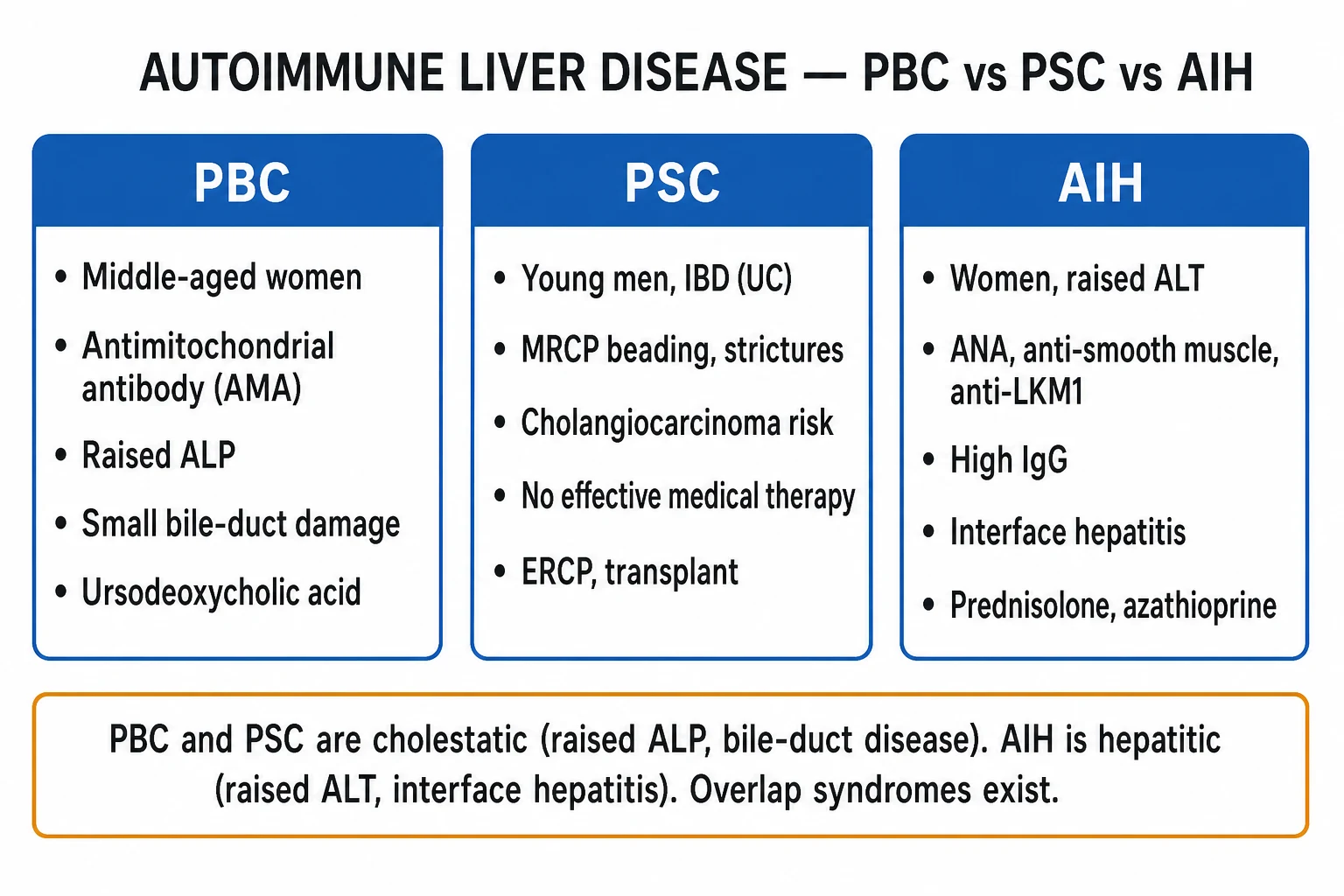
Autoimmune liver disease comprises three distinct immune-mediated conditions. Primary biliary cholangitis (PBC) affects middle-aged women, destroys small intrahepatic bile ducts, is marked by antimitochondrial antibody (AMA) and a raised ALP, and is treated with ursodeoxycholic acid. Primary sclerosing cholangitis (PSC) affects young men (often with IBD, especially ulcerative colitis), causes multifocal biliary strictures (beading on MRCP/ERCP), carries a cholangiocarcinoma risk, has no proven medical therapy and often needs ERCP or transplant. Autoimmune hepatitis (AIH) mainly affects women, causes a hepatitic picture (raised ALT, high IgG, interface hepatitis), with ANA and anti-smooth-muscle (SMA) or anti-LKM1 antibodies, and is steroid-responsive (prednisolone to induce, azathioprine to maintain). Overlap syndromes (e.g. AIH-PBC) are recognised.
On this page & tools
Your progress
Saved locally on this device.
Practise this topic
Exam tags
Red flags
Overview & Definition
Autoimmune liver disease is a group of immune-mediated liver conditions unified by autoantibodies, immune injury and a relapsing or progressive course, but differing in their target and their biochemical pattern. In PBC and PSC the immune attack is aimed at the bile ducts, producing a cholestatic picture dominated by a raised ALP; in autoimmune hepatitis (AIH) the attack is aimed at hepatocytes, producing a hepatitic picture dominated by a raised ALT. Recognising which condition is in front of you is decisive because the treatment differs sharply — ursodeoxycholic acid (UDCA) for PBC, no proven disease-modifying therapy for PSC (endoscopic and transplant management), and immunosuppression for AIH.[1][2]
The three diseases are clinically and immunologically distinct, but they share a final common pathway of chronic inflammation, fibrosis and progression to cirrhosis and its complications (portal hypertension, varices, ascites, encephalopathy, hepatocellular carcinoma). They can also co-exist as overlap syndromes — most often AIH with PBC, less often AIH with PSC — which require combined therapy. A confident diagnosis rests on pattern recognition (cholestatic vs hepatitic), the antibody profile, the immunoglobulin class (IgM in PBC, IgG in AIH), and the histological pattern on liver biopsy. [1]
Autoimmune liver disease — the headline numbers
Classification
The three core autoimmune liver diseases are best understood as a 2 × 2 grid of target (bile ducts vs hepatocytes) and duct size (small vs large). PBC targets the small intrahepatic ducts only, PSC targets both small and large (intra- and extra-hepatic) ducts, and AIH targets hepatocytes directly. Two clinically important variants round out the classification: the overlap syndromes and the IgG4-related sclerosing cholangitis that mimics PSC but is steroid-responsive. [1]
PBC (small-duct)
- Middle-aged woman (40-60), female-to-male 9:1
- Antimitochondrial antibody (AMA) positive; IgM raised
- Cholestatic ALP; normal MRCP (duct loss, not strictures)
- Florid duct lesion / non-suppurative cholangitis on biopsy
- First-line: ursodeoxycholic acid 13-15 mg/kg/day
PSC (large + small duct)
- Young man (30-40), 60-80% have IBD (especially UC)
- AMA negative; p-ANCA positive in up to 80%
- MRCP/ERCP: multifocal strictures + dilatation ('beading')
- Onion-skin periductal fibrosis on biopsy
- No proven medical therapy; ERCP for strictures; cholangiocarcinoma risk
AIH (hepatocyte)
- Woman, any age (bimodal 10-30 and over 50)
- ANA + anti-smooth-muscle (type 1) OR anti-LKM1 (type 2)
- Hepatitic: ALT-dominant; IgG markedly raised
- Interface hepatitis with plasma cells and rosettes
- Steroid-responsive: prednisolone to induce, azathioprine to maintain
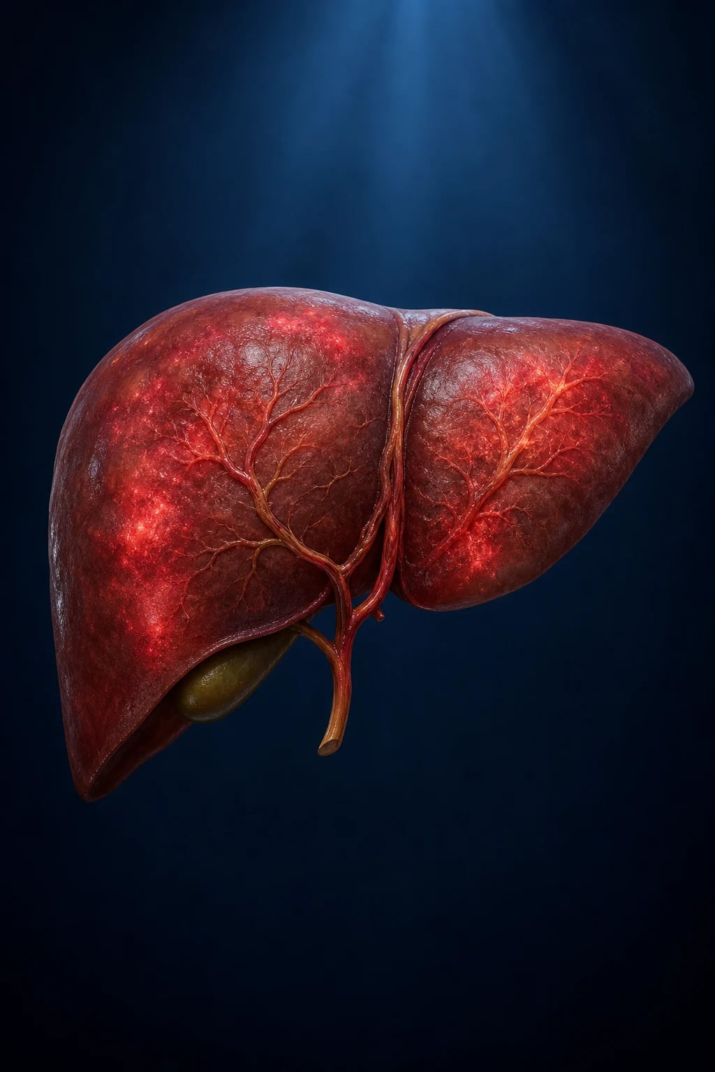
Autoimmune hepatitis is subtyped by its antibody profile. Type 1 (the common adult form) carries ANA and/or anti-smooth muscle antibody (SMA); type 2 (commoner in children and young people, especially in the Mediterranean) carries anti-LKM1 and/or anti-LC1 and tends to present more acutely with a higher relapse rate off therapy. A third specificity marker — anti-SLA (soluble liver antigen) — is highly specific for AIH and predicts relapse. PBC is staged histologically from the florid duct lesion (stage 1) through duct proliferation (stage 2) to fibrosis (stage 3) and cirrhosis (stage 4); PSC is classified as classic (large-duct), small-duct (better prognosis, lower cholangiocarcinoma risk), and the IgG4-related mimic. Overlap syndromes are defined by the Paris criteria, which require features of AIH (raised IgG, autoantibodies, interface hepatitis) coexisting with either PBC (AMA, cholestatic ALP, duct loss) or PSC (cholangiographic beading).[2]
Epidemiology & Risk Factors
PBC is predominantly a disease of middle-aged women — the female-to-male ratio is about 9:1, peak age 40 to 60 years, and prevalence reaches roughly 1 in 1000 women over 40 in some Northern European and North American series. Risk factors include a family history (a first-degree relative confers roughly 10-fold relative risk), a personal history of recurrent urinary tract infection and cigarette smoking, the HLA-DRB1*08 haplotype, and coexisting other autoimmune disease (autoimmune thyroiditis, Sjogren, CREST, rheumatoid arthritis).[1]
PSC shows the opposite demographic: a male predominance of about 2:1, mean age at diagnosis 30 to 40, and a strong association with IBD, present in 60 to 80% of cases — overwhelmingly ulcerative colitis (UC), less often Crohn's colitis. Conversely, only about 5% of UC patients develop PSC. PSC is uncommon in India and in East Asian populations relative to the West, and this epidemiological contrast is a favourite exam point. The HLA associations include HLA-B*08 and HLA-DRB1*03. There is no clearly proven drug, toxin or infective trigger, although a role for the gut microbiome and aberrant lymphocyte trafficking from inflamed colon to liver (the gut-lymphocyte homing hypothesis) is actively investigated. [1]
AIH is again a predominantly female disease (3 to 4:1), presenting at any age but with two peaks — the second and third decades, and a later peak above age 50. Type 2 AIH clusters in children and young adults and is commoner in Mediterranean populations. Genetic susceptibility maps to HLA-DRB1*0301 (DR3) and HLA-DRB1*0401 (DR4) in European populations. Recognised environmental triggers include certain drugs (nitrofurantoin, minocycline, statins, methyldopa, hydralazine) and viral infections (hepatitis A, B, C, Epstein-Barr), which may unmask AIH in a susceptible host. [1]
[1]The natural history is distinct for each: untreated PBC progresses slowly to cirrhosis over 10 to 20 years; PSC has a median transplant-free survival of 10 to 15 years with the ever-present threat of cholangiocarcinoma; and untreated AIH, although uncommonly left untreated today, carries a historical 5-year mortality around 50%, transformed to over 90% 10-year survival by immunosuppression.[1][2]
Pathophysiology
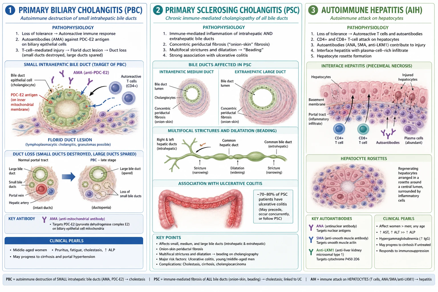
All three diseases arise from a loss of immune self-tolerance, but the antigenic target and the effector mechanism differ. In PBC, the autoantigen is the E2 subunit of the mitochondrial pyruvate dehydrogenase complex (PDC-E2), aberrantly expressed on the apical surface of small intrahepatic bile-duct epithelial cells. Autoreactive CD4+ and CD8+ T cells and natural killer cells, together with the antimitochondrial antibody (AMA), mount a coordinated attack that destroys the small ducts — producing the diagnostic florid duct lesion (non-suppurative destructive cholangitis). Because only small ducts are lost (not strictured), the cholangiogram in PBC is normal — a key contrast with PSC. Duct loss causes cholestasis; retained hydrophobic bile acids injure hepatocytes and drive the clinical syndrome of pruritus (now attributed largely to autotaxin and lysophosphatidic acid, not bile acids alone), fat-soluble vitamin malabsorption, and progressive fibrosis.[1]
In PSC, the trigger is unknown, but the disease reflects chronic inflammation of medium and large intra- and extra-hepatic bile ducts with periductal ('onion-skin') concentric fibrosis, lymphocytic infiltration and progressive duct loss. The fibrosis around ducts narrows the lumen at intervals while segments upstream dilate, giving the radiological multifocal strictures and dilatations ('beading') that are pathognomonic. The gut-lymphocyte homing hypothesis proposes that alpha-4 beta-7-positive mucosal T cells from inflamed colonic mucosa aberrantly traffic to the liver, where they bind MAdCAM-1 aberrantly expressed on bile-duct endothelium — linking PSC mechanistically to its colonic companion. The toxic-bile hypothesis (retained bile salts injuring hepatocytes) further drives fibrosis. The result is cholestasis, recurrent bacterial cholangitis, and a markedly increased risk of cholangiocarcinoma (and also gallbladder and colorectal cancer) driven by decades of chronic inflammation. [1]
In AIH, the target is the hepatocyte itself. There is a loss of self-tolerance with autoreactive CD4+ and CD8+ T cells directed at hepatocyte antigens, accompanied by high-titre non-organ-specific autoantibodies (ANA, SMA, anti-LKM1 directed at CYP2D6, anti-LC1, anti-SLA) and a polyclonal hypergammaglobulinaemia (high IgG). On histology this produces interface hepatitis — lymphoplasmacytic infiltration spilling across the limiting plate between portal tracts and parenchyma — with plasma cells and characteristic hepatocyte rosettes. The shared mechanisms explain the final common pathway of fibrosis and cirrhosis in all three. [1]
Clinical Presentation
The clinical face of autoimmune liver disease is conditioned by which compartment is damaged. Cholestatic disease (PBC, PSC) declares itself through fatigue, pruritus and Sicca symptoms long before jaundice appears, whereas AIH presents more like an acute or subacute hepatitis. [1]
Primary biliary cholangitis most often begins insidiously with fatigue (the commonest and most disabling symptom, often with a postural/autonomic component) and pruritus (characteristically worse at night, often starting on the palms and soles). Sicca symptoms — dry eyes and dry mouth — are extremely common. Skin findings include hyperpigmentation (mostly in sun-exposed areas and from scratching), excoriations (a sign that pruritus is real and severe), xanthelasma and tuberous xanthomata (from hypercholesterolaemia of cholestasis). With progressive disease, jaundice, right-upper-quadrant discomfort, and the stigmata of portal hypertension (ascites, variceal bleed, encephalopathy) emerge. Increasingly PBC is detected asymptomatically on an LFT panel performed for another reason — an isolated raised ALP in a middle-aged woman. [1]
Primary sclerosing cholangitis presents with pruritus, fatigue, and episodic jaundice, and frequently declares itself through episodes of bacterial cholangitis — fever, right-upper-quadrant pain and jaundice (Charcot's triad) — that should prompt ERCP to relieve an obstructing dominant stricture. A very common presentation is the asymptomatic young man with ulcerative colitis found to have a raised ALP on IBD surveillance bloods; this single combination should trigger MRCP. Atypical presentations include progression to cirrhosis and its complications, and the dramatic appearance of cholangiocarcinoma with new weight loss, worsening pruritus, deepening jaundice and a rising CA 19-9. [1]
Autoimmune hepatitis has the widest spectrum. At one end it is an incidental asymptomatic transaminitis; at the other it is acute icteric hepatitis and, in a minority (especially children and young adults), acute liver failure with coagulopathy and encephalopathy — AIH must always be in the differential of the young person with "acute hepatitis of unknown cause". Between these extremes patients describe fatigue, anorexia, malaise, arthralgia and amenorrhoea. Extrahepatic autoimmune disease is frequent and diagnostically helpful: autoimmune thyroiditis, type 1 diabetes, vitiligo, rheumatoid arthritis, Sjogren syndrome, and ulcerative colitis.[2]
[1]Differential Diagnosis
A cholestatic (ALP-dominant) LFT picture can be produced by PBC, PSC, drug-induced cholestasis, choledocholithiasis, extrahepatic malignant biliary obstruction, and IgG4-related sclerosing cholangitis. A hepatitic (ALT-dominant) picture can be produced by AIH, viral hepatitis (HBV, HCV), drug-induced liver injury, Wilson disease, and non-alcoholic steatohepatitis. The discriminator is rarely a single test — it is the convergence of demography, antibodies, immunoglobulins, imaging and histology. [1]
PBC
- Middle-aged woman; fatigue, pruritus, Sicca; AMA positive; IgM raised
- Smooth extrahepatic ducts on MRCP; small-duct florid duct lesion
PSC
- Young man with IBD; p-ANCA; MRCP beading/strictures
- Onion-skin fibrosis; cholangiocarcinoma risk
IgG4-sclerosing cholangitis
- Older man; enlarged pancreas / type 1 autoimmune pancreatitis
- Raised serum IgG4; long irregular stricture; steroid-responsive
Drug-induced cholestasis
- Temporal link to culprit (antibiotics, OCP, anabolic steroids)
- Resolves on withdrawal; eosinophilia may be present
Choledocholithiasis
- Biliary colic, fever, obstructive jaundice; dilated CBD on ultrasound
- MRCP/ERCP shows a filling defect, not multifocal beading
Wilson disease is the single most important hepatitic mimic of AIH in a young patient and must be excluded before committing to lifelong steroids: check caeruloplasmin (low), 24-hour urinary copper (raised), and perform slit-lamp examination for Kayser-Fleischer rings. Drug-induced autoimmune-like hepatitis (nitrofurantoin, minocycline, statins) resolves on withdrawal of the offending drug, although a course of steroids may be needed. The IgG4-related sclerosing cholangitis mimic deserves special emphasis: an older man with an enlarged pancreas, a long, irregular common-bile-duct stricture, and a raised serum IgG4 has a disease that — unlike PSC — responds dramatically to corticosteroids, and misdiagnosing it as PSC denies the patient a curative course of treatment.[1]
Clinical & Bedside Assessment
The bedside examination in suspected autoimmune liver disease has three objectives: to detect the stigmata of chronic cholestasis, to find the extrahepatic autoimmune associations that support the diagnosis, and to recognise decompensation or malignancy that change management. [1]
Look first for the skin and general signs of cholestasis: excoriations and linear scratch marks (proof that pruritus is genuine), hyperpigmentation, xanthelasma and xanthomata, and clubbing (in advanced PBC). Examine the mouth and eyes for Sicca (dry, tacky mucosae, absent salivary pooling), palpate for hepatosplenomegaly, and search for the stigmata of portal hypertension (spider naevi, palmar erythema, ascites with shifting dullness, caput medusae, asterixis). Vitiligo and a thyroid goitre or bruit point to coexisting autoimmune disease. In PSC specifically, look for right-upper-quadrant tenderness and fever (cholangitis) and for weight loss and cachexia (cholangiocarcinoma). [1]
The extrahepatic associations are not incidental — finding them strengthens the diagnosis and guides screening. In PSC, a full colonoscopy with biopsies at diagnosis is mandatory (to find or stage IBD), and annual colorectal cancer surveillance follows if colitis is present. In PBC, screen for and treat autoimmune thyroid disease and Sicca/Sjogren. In AIH, look for type 1 diabetes, thyroiditis and vitiligo and consider screening family members. Bedside and simple investigations that aid assessment include urinalysis (the UTI association in PBC), blood pressure and cardiovascular exam (relevant to portal hypertension and to drug therapy), and a focused assessment for frailty and nutrition in advanced disease. [1]
Investigations
The diagnostic strategy is staged: first define the biochemical pattern (cholestatic vs hepatitic), then order the targeted antibody and immunoglobulin panel, then image the biliary tree, and finally obtain histology where the picture is not diagnostic or an overlap is suspected. [1]
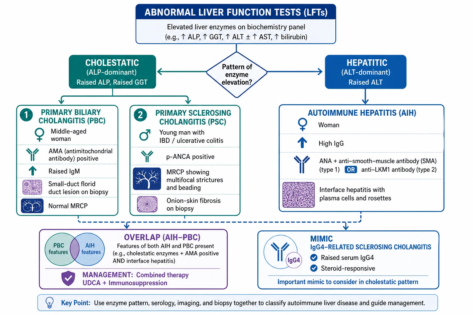
First-line bloods. A full liver screen: LFTs (ALP, ALT, AST, GGT, bilirubin, albumin), full blood count, coagulation (INR), renal function, glucose, and a fasting lipid profile. Then the aetiological panel — viral serology (HBsAg, anti-HCV), ferritin and transferrin saturation (haemochromatosis), caeruloplasmin (Wilson, in patients under 40), alpha-1-antitrypsin, and a careful drug and alcohol history. The autoimmune panel is the centrepiece: [1]
PBC bloods
- Antimitochondrial antibody (AMA) positive — highly specific
- IgM raised; PBC-specific ANA if AMA-negative: anti-gp210, anti-sp100
PSC bloods
- p-ANCA positive in up to 80%; AMA negative
- IgG4 to exclude the IgG4-sclerosing cholangitis mimic
AIH bloods
- Type 1: ANA and/or anti-smooth-muscle (SMA)
- Type 2: anti-LKM1 and/or anti-LC1; anti-SLA highly specific
- IgG markedly raised (over 2 times ULN supports the diagnosis)
Imaging. Ultrasound is first-line to exclude extrahepatic obstruction and to assess the liver texture, spleen, and portal vein. In suspected PSC, MRCP is the diagnostic test of choice (non-invasive), showing the characteristic multifocal strictures and dilatations ('beading') of intra- and extra-hepatic ducts. Importantly, MRCP is normal in PBC (because only small ducts are lost, not strictured) — a decisive point of distinction. ERCP is reserved for therapeutic intervention (brushings, balloon dilatation, stenting of dominant strictures) and for cases where MRCP is non-diagnostic. Cross-sectional CT/MRI and PET-CT are used to evaluate suspected cholangiocarcinoma. Liver biopsy confirms histology when the diagnosis is in doubt, when AMA is negative, when an overlap syndrome is suspected, or to stage fibrosis; it shows the florid duct lesion in PBC, onion-skin fibrosis in PSC, and interface hepatitis with plasma cells and rosettes in AIH. [1]
The simplified diagnostic criteria for AIH (Hennes 2008) turn the work-up into a score: points are awarded for autoantibody titre (ANA/SMA 1:40 = 1 point, over 1:80 = 2; anti-LKM1 over 1:40 = 2; ANA/SMA/anti-LKM1/anti-SLA less than 1:40 = 0), IgG level (over 1.1 times ULN = 1 point, over 1.1 to under 2 times... and over 2 times ULN = 3 points), histology (compatible = 1, typical of AIH = 2), and absence of viral hepatitis (yes = 2). A score of 6 or more = probable AIH and 7 or more = definite AIH.[2]
Surveillance and staging. Liver elastography (FibroScan) stages fibrosis non-invasively; a liver stiffness over about 12 to 15 kPa suggests cirrhosis. Once cirrhotic, begin 6-monthly ultrasound (plus or minus AFP) for hepatocellular carcinoma and an OGD to screen for varices. A DEXA scan screens for the osteoporosis of cholestasis, and fat-soluble vitamin levels (A, D, E, K) are checked in advanced cholestatic disease. In PSC, screen for cholangiocarcinoma with annual MRI/MRCP plus CA 19-9, and perform colonoscopy at diagnosis (to detect IBD) with annual surveillance thereafter if colitis is present. [1]
Management — Resuscitation
Most autoimmune liver disease is chronic, but three scenarios are time-critical and demand an immediate bundle. Acute severe AIH or AIH presenting as acute liver failure is a hepatology emergency: admit, give supportive care, exclude infection (the major contraindication to steroids), and start high-dose corticosteroids — prednisolone 1 mg/kg/day orally (or IV methylprednisolone 60 mg/day if unable to take orally) after bloods and cultures. Recheck INR, bilirubin and encephalopathy daily; if there is no clear improvement within 1 to 2 weeks, urgent transplant referral is mandatory (AASLD).[2]
Acute bacterial cholangitis in PSC is managed with the standard bundle: ABCDE, IV fluid resuscitation, blood cultures before antibiotics, and broad-spectrum antibiotics such as piperacillin-tazobactam 4.5 g IV 8-hourly (or a fluoroquinolone plus cephalosporin per local guidance). Severe or high-risk cholangitis (persistent fever, hypotension, rising inflammatory markers) demands urgent ERCP for biliary decompression — usually balloon dilatation or stenting of a dominant stricture — within 24 to 48 hours. [1]
Severe intractable pruritus can be disabling and even suicidal; treat in a stepwise ladder: cholestyramine 4 g orally before breakfast (and again at lunch, separated from other drugs by 2 to 4 hours because it binds them) is first-line; if ineffective, rifampicin 150 to 300 mg orally twice daily (monitor LFTs); then naltrexone 50 mg orally daily (opioid antagonist), sertraline 50 to 100 mg daily, and finally plasmapheresis or MARS or liver transplant for truly refractory disease. A variceal bleed or decompensation in cirrhotic-stage disease is managed as for any cirrhosis — vasoactive drug (terlipressin 2 mg IV 4-hourly or octreotide infusion) + antibiotic prophylaxis (ceftriaxone 1 g IV daily) + endoscopic band ligation within 12 hours. [1]
[1]Management — Definitive & Stepwise
The definitive management is disease-specific — the single most important principle in autoimmune liver disease, because the wrong therapy (e.g. steroids for PBC, high-dose UDCA for PSC) is at best useless and at worst harmful. [1]
Primary biliary cholangitis[1]
- First-line, all stages: ursodeoxycholic acid (UDCA) 13 to 15 mg/kg/day orally in divided doses. UDCA improves biochemistry, histology and transplant-free survival, and is the only disease-modifying therapy of proven benefit.
- Assess response at 12 months using the Paris II criteria (ALP under 1.5 times ULN and bilirubin under 2 times ULN, with normal AST, at 12 months). If the response is inadequate, add second-line obeticholic acid (OCA) 5 mg orally daily, escalating to 10 mg at 6 months if tolerated. Fibrates (e.g. bezafibrate 400 mg daily) are an off-label alternative or add-on. OCA is contraindicated in decompensated cirrhosis (Child-Pugh B or C) because it can precipitate liver failure and death.
- Symptom control: cholestyramine for pruritus (with dose spacing as above); calcium, vitamin D and a bisphosphonate for osteoporosis; replacement of fat-soluble vitamins (A, D, E, K) when deficient.
- Surveillance: 6-monthly ultrasound for HCC and OGD for varices once cirrhotic; DEXA-guided bone management. [1]
Primary sclerosing cholangitis[1]
- No proven disease-modifying therapy exists. UDCA is NOT routinely recommended (AASLD); high-dose UDCA (28 to 30 mg/kg/day) was associated with increased mortality and adverse events in the Lindor 2009 trial and should be avoided.
- Manage dominant strictures by ERCP with balloon dilatation and/or short-term stenting; brushings of any stricture are mandatory to exclude cholangiocarcinoma.
- Treat episodes of bacterial cholangitis with appropriate antibiotics; consider rotating antibiotic prophylaxis (e.g. ciprofloxacin 500 mg daily or alternating cycles) in recurrent cholangitis.
- Screen for cancer: annual MRI/MRCP plus CA 19-9 for cholangiocarcinoma; colonoscopy at diagnosis and annual colorectal cancer surveillance if IBD is present (the colorectal cancer risk in PSC + UC is markedly elevated); HCC surveillance as for cirrhosis.
- Liver transplant for end-stage disease, recurrent cholangitis, or intractable pruritus; 5-year survival is excellent (around 80 to 85%), although PSC recurs in about 20%. [1]
Autoimmune hepatitis[2]
- Induce remission with prednisolone 60 mg orally once daily (tapered over weeks to a maintenance dose of about 5 to 10 mg daily) plus azathioprine 1 to 2 mg/kg/day orally introduced from day 1 to allow faster steroid tapering. An alternative induction is budesonide 9 mg orally daily as a steroid-sparing option in non-cirrhotic AIH only.
- Maintain remission with azathioprine 1 to 2 mg/kg/day (monotherapy once prednisolone is weaned) for at least 2 to 3 years. Mycophenolate mofetil 1 to 2 g/day orally is the standard alternative in azathioprine-intolerant patients or those with undesirable side effects; tacrolimus and cyclosporin are third-line.
- Define remission as biochemical (transaminases under 2 times ULN and normal IgG) AND histological (inactive interface hepatitis), usually achieved by 18 to 24 months. Attempt treatment withdrawal only after sustained combined remission; relapse is common (around 50% within 1 year, up to 80% by 3 years) and is the rule in type 2 AIH, in whom lifelong maintenance is usual.
- Monitor therapy: FBC and LFTs every 2 to 4 weeks early, then 3-monthly; bone protection on steroids; TPMT activity before azathioprine where available. [1]
Overlap syndromes (AIH with PBC, defined by Paris criteria) are treated with combined UDCA plus immunosuppression (prednisolone then azathioprine). Transplant is reserved for acute liver failure (AIH), refractory pruritus or end-stage disease, with allocation driven by the MELD score (bilirubin + INR + creatinine + sodium). [1]
Worldwide, EASL (Europe) and AASLD (US) guidance drive most practice: UDCA 13-15 mg/kg/day for PBC with OCA second-line (Paris II response); no UDCA for PSC with ERCP for strictures and cholangiocarcinoma surveillance; prednisolone + azathioprine for AIH. The BSG (UK) aligns closely with EASL. Transplant allocation is MELD-based in most systems.
Specific Subtypes & Scenarios
AMA-negative PBC accounts for about 5% of cases and is clinically and histologically identical to AMA-positive disease. The diagnosis rests on PBC-specific ANA — anti-gp210 and anti-sp100 (anti-nuclear rim and dot patterns) — together with a cholestatic ALP and compatible biopsy; management with UDCA is the same. [1]
Small-duct PSC affects only the intrahepatic small ducts, giving a normal cholangiogram and a diagnosis on biopsy (onion-skin fibrosis). It has a better prognosis and a substantially lower cholangiocarcinoma risk than classic large-duct PSC, and may rarely progress to classic PSC. [1]
IgG4-related sclerosing cholangitis is the most important steroid-responsive mimic of PSC. Think of it in an older man with an enlarged pancreas (type 1 autoimmune pancreatitis), weight loss and jaundice, a long, irregular common-bile-duct stricture and a raised serum IgG4. A diagnostic-therapeutic trial of prednisolone 0.6 mg/kg/day for 4 weeks then taper produces dramatic radiological and biochemical improvement; classic PSC, by contrast, does not respond to steroids.[1]
Acute severe AIH presenting as acute liver failure is most often seen in children and young adults. Management is high-dose corticosteroids after exclusion of sepsis, with early transplant referral if there is no biochemical improvement within 1 to 2 weeks; King's College criteria guide transplant timing. [1]
Overlap syndrome (AIH-PBC) presents with mixed features — a cholestatic ALP and AMA (PBC) together with high IgG, ANA/SMA and interface hepatitis (AIH). It is treated with combined UDCA plus immunosuppression. AIH-PSC overlap is rarer and managed with AIH-type immunosuppression plus PSC-type ERCP/transplant as needed. [1]
Pregnancy in AIH is a recurring scenario: aim for remission for at least 6 to 12 months before conception; continue azathioprine (safe in pregnancy); use the lowest effective prednisolone dose; screen for varices in the second trimester; and anticipate a flare 1 to 3 months postpartum, monitoring transaminases and IgG closely. [1]
Complications & Pitfalls
The complications of autoimmune liver disease fall into three groups: those of cirrhosis and portal hypertension (common to all three), those that are disease-specific, and those of treatment. The cirrhotic complications — ascites, spontaneous bacterial peritonitis, variceal bleed, hepatic encephalopathy, hepatocellular carcinoma — are shared and managed as for any cirrhosis. The disease-specific complications distinguish the three: [1]
[1]The treatment complications matter because they drive monitoring. Long-term corticosteroids in AIH cause diabetes, osteoporosis, cataracts, hypertension, weight gain and infection risk — the rationale for early introduction of azathioprine (steroid-sparing) and for bone protection. Azathioprine causes cytopenias, hepatotoxicity, pancreatitis and a small increase in lymphoma risk; check TPMT activity before starting and monitor FBC and LFTs. Obeticholic acid can cause dose-dependent pruritus and, critically, is contraindicated in decompensated cirrhosis, where it precipitates liver failure and death.[1]
The classic pitfalls are the wrong-therapy errors and the missed-mimic errors: [1]
Wrong-therapy pitfalls
- Steroids for PBC — no benefit, harms
- High-dose UDCA for PSC — increased mortality (Lindor 2009)
- Stopping azathioprine abruptly or withdrawing AIH therapy too early — relapse
Missed-diagnosis pitfalls
- Calling a raised ALT in a UC patient 'fatty liver' without MRCP (misses PSC)
- Missing IgG4-sclerosing cholangitis as a steroid-responsive PSC mimic
- Missing Wilson disease in a young patient with 'AIH-like' hepatitis
Missed-surveillance pitfalls
- Not colonoscoping a PSC patient at diagnosis (IBD + colorectal cancer)
- Not screening for cholangiocarcinoma (annual MRCP + CA 19-9)
- Giving OCA to a decompensated cirrhotic
A final pitfall is over-interpreting a low-titre positive ANA in an otherwise healthy person — autoantibodies must always be interpreted in the context of IgG, transaminases and histology, not in isolation. [1]
Prognosis & Disposition
Treated PBC has a substantially improved outlook: early-stage disease treated with UDCA has a near-normal life expectancy, while advanced or untreated disease progresses to cirrhosis and death over 10 to 20 years. Prognosis can be quantified with validated scores — Barcelona, Paris I and II, UK-PBC, and GLOBE — which combine ALP, bilirubin, platelets and other variables to predict long-term outcome and guide the addition of OCA and transplant referral. [1]
PSC carries the worst outlook of the three, with a median transplant-free survival of 10 to 15 years; the development of cholangiocarcinoma (lifetime risk 5 to 15%) transforms the prognosis, with most patients dying within months of diagnosis. Small-duct PSC has near-normal survival and a much lower cholangiocarcinoma risk. After liver transplant, 5-year survival is 80 to 85%, but PSC recurs in about 20% of grafts. [1]
AIH treated with immunosuppression has a 10-year survival over 90%; untreated, the historical 5-year survival was under 50%. Cirrhosis at presentation worsens the outcome, and acute severe AIH has a high transplant-free mortality without transplant. After withdrawal of therapy, relapse occurs in around 50% within 1 year and up to 80% by 3 years, especially in type 2 AIH — so many patients require lifelong maintenance. Transplant referral is triggered by MELD-based criteria, by acute liver failure, by refractory pruritus, or by recurrent cholangitis; disease recurs after transplant in PBC (around 30%), AIH (20 to 30%), and PSC (around 20%).[1][2]
Special Populations
Pregnancy in AIH requires planning: aim for remission for at least 6 to 12 months before conception; continue azathioprine (acceptable and safe in pregnancy); use the lowest effective prednisolone dose; screen for varices in the second trimester; and anticipate a flare 1 to 3 months postpartum, with close transaminase and IgG monitoring. UDCA for PBC is safe in pregnancy. PSC in pregnancy is managed expectantly, with attention to cholangitis and dominant strictures. [1]
Paediatric autoimmune liver disease is more often type 2 AIH and tends to present with acute severe hepatitis or established cirrhosis. Treatment is prednisolone and azathioprine, often lifelong; siblings should be screened. The PSC-AIH overlap is over-represented in children. [1]
The elderly frequently present with advanced PBC or AIH at first contact, with atypical features, higher comorbidity, polypharmacy (raising drug-drug interaction risk), and reduced transplant candidacy on grounds of frailty — bone protection and falls prevention are especially important. [1]
PSC with IBD is a distinct high-risk population: colonoscopy at diagnosis (to detect IBD) and annual colorectal cancer surveillance thereafter if colitis is present, because the colorectal cancer risk in PSC + colitis is markedly elevated and independent of disease duration. Colectomy does not alter the course of PSC. [1]
Post-liver-transplant patients require management of immunosuppression, screening for de novo malignancy and opportunistic infection, and surveillance for recurrence of the native disease (PBC, AIH or PSC). [1]
Evidence, Guidelines & Regional Differences
Modern practice is shaped by the EASL (2017) and AASLD (2018) PBC guidelines — UDCA first-line at 13 to 15 mg/kg/day, response assessed at 12 months (Paris II), and obeticholic acid as second-line — and by the AASLD (2015/2018) PSC guidance, which explicitly does not recommend UDCA and emphasises MRCP for diagnosis, ERCP for strictures, and cholangiocarcinoma surveillance. The AASLD (2019) and EASL (2015) AIH guidance endorses the simplified diagnostic score, prednisolone plus azathioprine as first-line, budesonide as a steroid-sparing alternative in non-cirrhotic disease, and mycophenolate for azathioprine-intolerant patients.[1][2]
Landmark trials underpin these recommendations: Lindor 2009 showed that high-dose UDCA was harmful in PSC (more death and adverse events), establishing the do-not-treat stance; Hirschfield 2015 (POISE) established obeticholic acid as an effective second-line agent in PBC; Manns 2001 and 2010 defined the prednisolone + azathioprine regimen for AIH; and Hennes 2008 derived the simplified diagnostic score for AIH that is used worldwide. The role of fibrates and of peroxisome proliferator-activated receptor agonists (e.g. elafibranor, selonsertib) is being refined in ongoing PBC and PSC trials. [1]
The key regional contrast is epidemiological: PSC is uncommon in India and East Asia, while HBV and HCV dominate the chronic hepatitis landscape, so an AIH diagnosis always requires rigorous exclusion of viral hepatitis before committing to lifelong immunosuppression. INASL guidance addresses viral and alcohol-related liver disease in detail but does not yet issue standalone autoimmune liver disease recommendations. Cost and access limit the use of obeticholic acid, FibroScan and transplant in low-resource settings, while UDCA, prednisolone and azathioprine remain affordable and widely available.
Exam Pearls
Autoimmune liver disease — the FLAIR mnemonic
FLAIR
PBC and AIH are female-predominant; PSC is the male exception with IBD
ALP-dominant = cholestatic (PBC/PSC); ALT-dominant = hepatitic (AIH)
AMA (PBC), p-ANCA (PSC), ANA/SMA/anti-LKM1 (AIH)
IgM raised in PBC; IgG raised in AIH
UDCA (PBC), none proven (PSC), steroids (AIH)
- PBC = middle-aged woman, AMA, raised ALP, small-duct damage, ursodeoxycholic acid; AMA-negative PBC needs anti-gp210 / anti-sp100 or biopsy.
- PSC = young man + IBD (especially UC), MRCP beading, cholangiocarcinoma risk, NO proven medical therapy, ERCP/transplant; high-dose UDCA is harmful.
- AIH = woman, raised ALT, high IgG, ANA/SMA (type 1) or anti-LKM1 (type 2), interface hepatitis with plasma cells and rosettes, prednisolone → azathioprine.
- PBC and PSC are cholestatic (ALP); AIH is hepatitic (ALT). Overlaps (AIH-PBC) are recognised and treated with combined UDCA + immunosuppression.
- Pruritus first-line = cholestyramine; second-line = rifampicin; do NOT use UDCA for pruritus in PSC.
- IgG4-sclerosing cholangitis is the steroid-responsive mimic of PSC (raised IgG4, older man, pancreatitis).
- PSC colonoscopy at diagnosis (IBD) and annual colorectal cancer surveillance; cholangiocarcinoma surveillance with annual MRCP/CA 19-9.
- Obeticholic acid is contraindicated in decompensated cirrhosis (precipitates liver failure).
- Exclude Wilson disease (caeruloplasmin, slit-lamp) before labelling a young hepatitis as AIH.
- Relapse after AIH withdrawal is common (about 50% at 1 year, 80% by 3 years) and almost universal in type 2 — most patients need lifelong maintenance. [1]
Exam application bank (NEET-PG / INICET)
One-line answer
Autoimmune liver disease comprises three distinct immune-mediated conditions. Primary biliary cholangitis (PBC) affects middle-aged women, destroys small intrahepatic bile ducts, is marked by antimitochondrial antibody (AMA) and a raised ALP, and is treated with ursodeoxycholic acid. Primary sclerosing cholangitis (PSC) affects young men (often with IBD, especially ulcerative colitis), causes multifocal biliary strictures (beading on MRCP/ERCP), carries a cholangiocarcinoma risk, has no proven medical therapy and often needs ERCP or transplant. Autoimmune hepatitis (AIH) mainly affects women, causes a hepatitic picture (raised ALT, high IgG, interface hepatitis), with ANA and anti-smooth-muscle (SMA) or anti-LKM1 antibodies, and is steroid-responsive (prednisolone to induce, azathioprine to maintain). Overlap syndromes (e.g. AIH-PBC) are recognised.
Worked stems (answer without another resource)
Stem 1 — Classic presentation. Map symptoms to mechanism; name the first investigation and first treatment step with dose/route if drug therapy is standard. [1]
Stem 2 — Unstable / complicated. List red flags that force immediate resuscitation, theatre, ICU, antidote, or reperfusion — and what you do in the first 15 minutes. [1]
Stem 3 — Atypical group. Elderly, pregnancy, child, or immunocompromised: how presentation and thresholds change. [1]
Stem 4 — Differential trap. Name the three closest mimics and one discriminator for each. [1]
Stem 5 — Disposition. Who goes home with safety-netting, who is admitted, who needs HDU/ICU/theatre, and what follow-up is mandatory. [1]
Rapid viva checklist
- Definition + classification
- Pathophysiology chain
- Bedside signs / criteria
- Score with exact components (if any)
- Emergency bundle
- Definitive therapy with doses
- Complications of disease and of treatment
- Special populations
- Guideline/trial name if classic
- Three exam traps
Coverage self-check
If you cannot answer any stem above from this page alone, re-read the matching section — the page is intended to be self-sufficient for final-prof and NEET-PG/INICET questions on Autoimmune Liver Disease (PBC, PSC & AIH).
References
- [1]European Association for the Study of the Liver. EASL Clinical Practice Guidelines: The diagnosis and management of patients with primary biliary cholangitis J Hepatol, 2017.PMID 28427765
- [2]Manns MP, Czaja AJ, Gorham JD, et al. Diagnosis and management of autoimmune hepatitis Hepatology, 2010.PMID 20513004