Gastroenterology · Gastroenterology
Chronic Pancreatitis
Also known as Chronic pancreatitis · Chronic calcific pancreatitis · Tropical calcific pancreatitis · Fibrocalculous pancreatic diabetes · Autoimmune pancreatitis
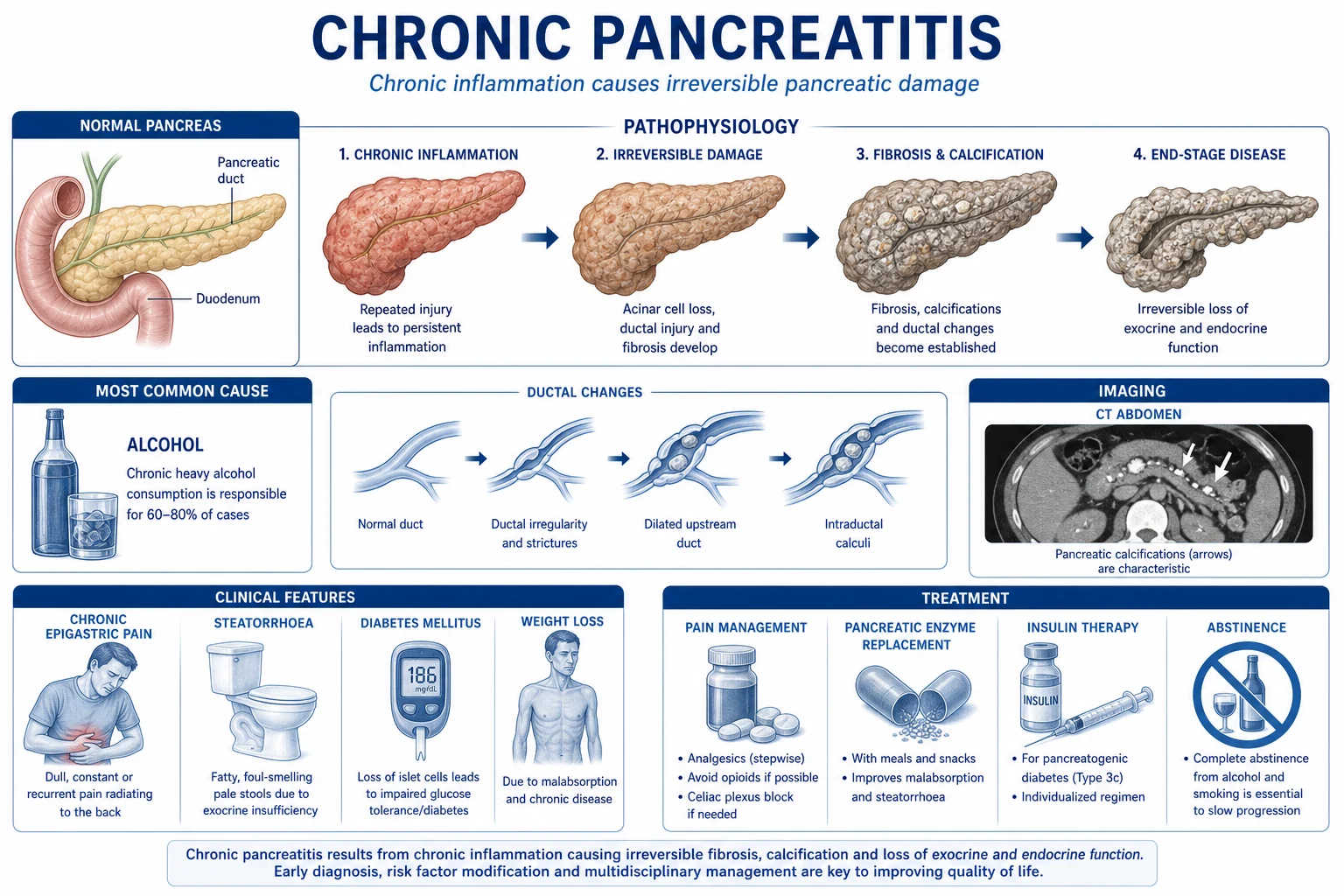
Chronic pancreatitis (CP) is a progressive, inflammatory, fibrotic disease of the pancreas producing irreversible morphological damage with permanent loss of exocrine (acinar) and endocrine (islet) function. Commonest cause worldwide is alcohol (60 to 70 percent); tobacco is an independent co-factor; tropical calcific pancreatitis dominates in southern India; hereditary (PRSS1), autoimmune (IgG4) and obstructive subtypes complete the spectrum (TIGAR-O classification). Presents with the classic triad of recurrent epigastric pain radiating to the back, steatorrhoea, and diabetes with weight loss; atypical painless CP presents with new diabetes and cachexia. Diagnosis rests on a combination of structural imaging (calcification, ductal change, atrophy) and functional testing (faecal elastase under 200); there is no single gold standard. Management is multidisciplinary: alcohol and smoking cessation, stepwise analgesia, pancreatic enzyme replacement (PERT: pancreatin 25,000 to 40,000 units lipase per meal with a PPI), insulin for type 3c diabetes, then endoscopic (ESWL, stenting) and surgical (Puestow drainage for duct over 7 mm, resection for head mass) options for refractory pain; corticosteroids for autoimmune pancreatitis. Complications include pseudocyst, biliary stricture, splenic vein thrombosis with isolated gastric varices (splenectomy curative), pseudoaneurysm with catastrophic bleed, and pancreatic cancer (cumulative risk around 4 percent at 20 years). Prognosis: progressive, life expectancy shortened by 10 to 20 years.
On this page & tools
Your progress
Saved locally on this device.
Practise this topic
Exam tags
Red flags

Overview & Definition
Chronic pancreatitis is a progressive, inflammatory, fibrotic disease of the pancreas characterised by irreversible morphological damage (fibrosis, acinar loss, calcification, ductal distortion and strictures) and permanent loss of exocrine (digestive enzyme) and endocrine (islet hormone) function.[1][2]
The single feature that distinguishes CP from acute pancreatitis (AP) is irreversibility. AP is an acute inflammatory event that, in its mild interstitial form, resolves completely with return of normal gland architecture and function. CP, by contrast, leaves permanent structural and functional damage. The two are, however, related: recurrent AP is now recognised as a driver of CP (the SAPE — Sentinel Acute Pancreatitis Event — model), and an acute painful flare may complicate established CP.[2]
The clinical challenge in CP is not the diagnosis in its full-blown form (a cachectic alcoholic with calcification on AXR and steatorrhoea is unmistakable) but recognising early or atypical disease, excluding pancreatic cancer (the differential that must never be missed), relieving chronic pain without inducing opioid dependence, and managing the endocrine and nutritional consequences of progressive glandular failure.[1]
Classification
CP is classified three ways — by aetiology (TIGAR-O), by combined risk factors (M-ANNHEIM), and by morphology (calcifying vs non-calcifying vs obstructive vs autoimmune). All three appear in exams.[2][4]
TIGAR-O aetiological classification (the high-yield one)
| TIGAR-O class | Examples |
|---|---|
| Toxic-metabolic | Alcohol (commonest, 60 to 70 percent), tobacco (independent co-factor), hypercalcaemia, hypertriglyceridaemia, chronic renal failure, toxins |
| Idiopathic | 10 to 30 percent; early- and late-onset forms; tropical |
| Genetic | PRSS1 (hereditary, autosomal dominant), SPINK1, CFTR, CTRC, CPA1 |
| Autoimmune | Type 1 (IgG4-related, lymphoplasmacytic sclerosing pancreatitis) and Type 2 (idiopathic duct-centric, IBD-associated) |
| Recurrent and severe Acute Pancreatitis | Necrotising AP, recurrent AP (vascular/ischaemic) |
| Obstructive | Pancreatic duct tumour, pancreas divisum with papillary stenosis, post-traumatic stricture, ascariasis (endemic), ampullary stenosis |

M-ANNHEIM classification (combined risk factors)
A unifying system that scores Multiple risk factors together — Alcohol consumption, Nicotine, Nutritional factors, Hereditary, Efferent duct factors, Immunological, and Miscellaneous/metabolic — and combines them with clinical course, imaging and function to grade disease severity.[4]
Morphological classification
- Calcifying CP — the commonest form (alcoholic, tropical, hereditary); intraductal protein plugs calcify into stones, producing ductal dilation, strictures and the 'chain of lakes' ERCP appearance.
- Non-calcifying (obstructive) CP — caused by a discrete ductal obstruction (tumour, stricture, divisum); the duct dilates upstream of the obstruction and calcification is absent or sparse; reverses if the obstruction is relieved.
- Autoimmune pancreatitis (AIP) — diffuse 'sausage-shaped' enlargement with a capsule-like rim, IgG4-positive plasma-cell infiltrate; steroid-responsive (see Specific Subtypes).
- Tropical calcific pancreatitis — early-onset, large ductal calculi, severe diabetes, in southern India (see Specific Subtypes).[1]
Alcoholic CP
- Commonest worldwide (60 to 70 percent)
- Men, 40s to 50s, heavy intake over 5 to 10 years
- Calcifying with diffuse ductal change
- Pain-predominant early; exocrine/endocrine failure late
Tropical calcific CP
- Southern India, Africa, Asia; under 30 years
- Large ductal calculi, early severe diabetes (FCPD)
- Malnutrition, low BMI; cassava hypothesis
- High pancreatic-cancer risk
Autoimmune (type 1, IgG4)
- Older men, painless obstructive jaundice
- 'Sausage' pancreas with capsule-like rim
- Other organ involvement (bile ducts, kidney, retroperitoneum, salivary)
- Dramatic steroid response, high relapse
Hereditary CP
- PRSS1 autosomal dominant; onset under 20 years
- Penetrance about 80 percent; family history
- Lifetime pancreatic-cancer risk about 40 percent by age 70
- Surveillance from age 40 / 20 years after onset
Epidemiology & Risk Factors
CP is uncommon but not rare. Estimated prevalence in the West is 30 to 40 per 100,000 population; the incidence is 4 to 13 per 100,000 per year.[3]
Regional epidemiology is striking and examinable: in southern India (Kerala), the prevalence of tropical calcific pancreatitis reaches over 100 per 100,000 (some series report 114 to 200 per 100,000), and fibrocalculous pancreatic diabetes (FCPD) was once a major cause of diabetes in young Indians. This regional excess is attributed to cassava (tapioca) cyanogenic-glycoside exposure, chronic protein-calorie malnutrition, and a high prevalence of SPINK1 mutations.[1][3]
Risk factors and the aetiology they favour:[1]
| Risk factor | Aetiology favoured |
|---|---|
| Alcohol (over 60 to 80 g/day for over 5 to 10 years) | Toxic-metabolic (commonest overall) |
| Tobacco smoking | Independent co-factor; accelerates calcification and progression; multiplies alcohol risk |
| Young onset / family history | Hereditary (PRSS1, SPINK1), tropical |
| Recurrent AP attacks | Recurrent-and-severe-AP (SAPE) pathway |
| Hypercalcaemia, hypertriglyceridaemia | Toxic-metabolic |
| Pancreatic mass / divisum / trauma | Obstructive |
| Older man + jaundice + raised IgG4 | Autoimmune type 1 |
Sex and age: alcoholic CP shows a male : female ratio of 4 to 5 : 1, presenting in the 40s to 50s. Idiopathic CP is more evenly split and may be early-onset (mean age 20) or late-onset (mean age 60).[3]
Pathophysiology
The healthy pancreas is an exocrine gland producing digestive zymogens (trypsinogen, chymotrypsinogen, amylase, lipase, pro-elastase, pro-phospholipase) and an endocrine gland producing insulin (beta), glucagon (alpha), somatostatin (delta) and pancreatic polypeptide. In CP, both arms are progressively destroyed. Several, non-mutually-exclusive mechanisms have been proposed.[1][2]
The four mechanistic hypotheses
- Toxic-metabolic hypothesis — alcohol and its metabolites (acetaldehyde), and free radicals from CYP2E1 induction, directly injure acinar cells, producing fatty degeneration, vacuolisation and necrosis. Even modest intake in a susceptible (often genetically predisposed) host can trigger disease — explaining why only a minority of heavy drinkers develop CP.[2]
- Oxidative-stress hypothesis — ethanol induces CYP2E1, generating reactive oxygen species that overwhelm glutathione and antioxidant defences, causing lipid peroxidation, membrane damage and periductal fibrosis. This was the rationale for antioxidant therapy (now largely refuted by the ANTICIPATE trial — see Evidence).[6]
- Necrosis-fibrosis (sclerosis) sequence — repeated episodes of focal acinar and periductal necrosis (often clinically silent) heal by fibrosis. With each cycle, more acini are lost and replaced by collagen. This is the substrate that links recurrent AP to CP (the SAPE model).[2]
- Ductal / obstructive / primary-duct hypothesis — proteinaceous plugs, calculi or strictures obstruct ducts, raising intraductal pressure, causing upstream acinar atrophy and ductal dilation. In obstructive CP the change reverses if the obstruction is relieved.
The central effector: the pancreatic stellate cell (PSC)
The final common pathway of all hypotheses is the pancreatic stellate cell. In health, PSCs are quiescent, vitamin-A (retinoid)-storing periacinar cells. In CP, PSCs are activated by alcohol, acetaldehyde, oxidative stress and cytokines (TGF-beta, PDGF, IL-1, TNF-alpha, IL-6) into myofibroblast-like cells that synthesize and deposit collagen types I and III, fibronectin and matrix proteins. Persistent PSC activation produces the dense fibrosis, acinar loss and ductal distortion that are the histological signature of CP.[2]
Mechanisms of pain (four parallel pathways)
Pain in CP is multifactorial and understanding it drives treatment:[1] />
- Ductal hypertension — stones, strictures and proteinaceous plugs obstruct the main pancreatic duct, raising intraductal pressure. Relieved by ESWL, stenting or surgical drainage (Puestow).
- Parenchymal / compartment hypertension — the fibrotic, non-compliant capsule raises interstitial pressure, producing a form of pancreatic compartment syndrome. Relieved by decompression or resection.
- Perineural inflammation — CP nerves are increased in number and diameter, surrounded by immune-cell (eosinophil) infiltrates, and exposed to nerve growth factor (NGF) and inflammatory cytokines. This produces neuropathic, opioid-preferring pain. Partially addressed by neuromodulators.
- Ischaemia — microvascular compromise from fibrosis.[1] />
Functional loss: exocrine and endocrine failure
- Exocrine insufficiency — steatorrhoea appears only when pancreatic lipase secretion falls below 10 percent of normal (i.e. when over 90 percent of acinar reserve is lost). This large functional reserve explains why malabsorption is a late feature.[1]
- Type 3c (pancreatogenic) diabetes — loss of beta-cells (insulin) AND alpha-cells (glucagon) produces a diabetes that is brittle and hypoglycaemia-prone (no glucagon counter-regulation), often with low insulin requirements but hepatic and peripheral insulin resistance. Type 3c accounts for 5 to 10 percent of all diabetes in Western series but is much more common where tropical calcific pancreatitis is prevalent.
TIGAR-O aetiology of chronic pancreatitis
TIGARO
Alcohol (60 to 70 percent), tobacco, hypercalcaemia, hypertriglyceridaemia, chronic renal failure
10 to 30 percent; early-onset and late-onset forms; tropical
PRSS1 (hereditary), SPINK1, CFTR, CTRC, CPA1
Type 1 (IgG4-related lymphoplasmacytic sclerosing); type 2 (duct-centric, IBD)
SAPE pathway — necrotising or recurrent AP driving fibrosis
Tumour, pancreas divisum, stricture, ascariasis, ampullary stenosis
Clinical Presentation
The classic picture is the triad of recurrent epigastric pain radiating to the back, steatorrhoea, and diabetes with weight loss — but the order and completeness of the triad varies with aetiology and stage.[1]
Pain (85 to 90 percent of patients)
- Epigastric, often radiating through to the back; may be relieved by sitting forward or by opioids.
- Post-prandial, lasting hours to days; frequently alcohol-precipitated.
- Relapsing-remitting early, becoming continuous in advanced disease.
- In 10 to 15 percent, CP is painless — the patient presents with diabetes, steatorrhoea or weight loss, or is found incidentally on imaging.[1]
Exocrine insufficiency — steatorrhoea and malabsorption
- Bulky, pale, foul-smelling, oily stools that are difficult to flush and leave an oil ring in the pan (steatorrhoea = faecal fat over 7 g/day).
- Weight loss, anorexia, bloating, flatulence, fat-soluble vitamin deficiency (A, D, E, K) — easy bruising (K), night blindness (A), osteomalacia (D), ataxia/neuropathy (E).
- B12 deficiency (impaired R-protein cleavage) and calcium, magnesium, zinc deficiency.[1]
Endocrine insufficiency — type 3c diabetes
- Polyuria, polydipsia, weight loss, but with a tendency to severe hypoglycaemia because glucagon counter-regulation is lost (alpha-cell destruction).
- Often insulin-requiring but at lower doses than type 1 (residual beta-cell mass plus hepatic/insulin resistance).[1]
Atypical presentations (the exam-tested exceptions)
- Painless CP — new diabetes, steatorrhoea or weight loss; calcification found incidentally.
- Elderly — presents only with cachexia and unexplained weight loss, or as pancreatic cancer (which must always be excluded).
- Tropical calcific CP — young, thin patient from southern India/Africa with early severe diabetes and large ductal stones.
- Autoimmune pancreatitis — painless obstructive jaundice in an older man, mimicking pancreatic cancer.
- Hereditary pancreatitis — recurrent AP from childhood with a positive family history.[7]
General examination
Chronic pancreatitis — key numbers
- Cachexia, temporal wasting, low BMI, muscle wasting (sarcopenia).
- Jaundice (common bile duct stricture in head disease or oedema; cholangitis if infected).
- Epigastric tenderness — often surprisingly mild relative to the reported pain.
- Palpable mass — a pseudocyst or inflammatory head mass.
- Erythema ab igni — reticular brown/purple discolouration of the abdominal skin from chronic heat-pad application (a classic exam-favourite bedside sign of chronic pain).
- Splenomegaly suggests splenic vein thrombosis (left-sided portal hypertension).
- Signs of chronic liver disease (alcohol), injection marks and stigmata of chronic opioid use.[1] />
Differential Diagnosis
Chronic epigastric pain with weight loss, or steatorrhoea with diabetes, is not always CP. The most important differential to exclude is pancreatic adenocarcinoma.[1][7]
| Differential | Distinguishing features |
|---|---|
| Pancreatic adenocarcinoma | Painless progressive obstructive jaundice, short history, rapid weight loss, sudden-onset diabetes, a solid mass on imaging; CA 19-9 raised; biopsy/EUS-FNA. CP and cancer may coexist and CP is itself a cancer risk factor. |
| Recurrent acute pancreatitis | Normal inter-attack pancreatic function and imaging; no irreversible change. Recurrent AP is a TIGAR-O cause of CP and the boundary is blurred. |
| Peptic ulcer disease | Relationship to food, response to PPI, diagnosis by OGD; no steatorrhoea or diabetes. |
| Chronic mesenteric ischaemia | Post-prandial pain, food fear, weight loss, abdominal bruit; no steatorrhoea; CT mesenteric angiography. |
| Functional / narcotic-bowel pain | Long-standing, no objective weight loss or structural abnormality; opioid dependence prominent. |
| Biliary pain | Right upper quadrant, biliary colic pattern, gallstones on US; no exocrine/endocrine failure. |
| Coeliac disease | Steatorrhoea + weight loss + anaemia, but iron/folate deficiency and anti-tTG positive; faecal elastase normal. |
| Small-intestinal bacterial overgrowth | Diarrhoea, B12 deficiency, positive breath test; faecal elastase normal. |
| Inflammatory bowel disease | Diarrhoea ± blood, weight loss, raised faecal calprotectin, colonoscopy. |
| Autoimmune pancreatitis | Painless jaundice, 'sausage' pancreas, IgG4 elevated, steroid-responsive; distinguished by HISORt criteria (see Investigations). |
Always specifically exclude pancreatic cancer and autoimmune pancreatitis when the presentation, epidemiology, or imaging is atypical, because both fundamentally change management.[1][7]
Clinical & Bedside Assessment
The bedside assessment has three aims: (1) confirm malnutrition, (2) detect complications, and (3) identify the treatable aetiology (alcohol, autoimmune, obstruction).[1]
- Vital signs, weight, height, BMI — serial weight is the single most useful clinical measure of progression and nutritional failure. Assess muscle mass (temporal wasting, grip strength) for sarcopenia, and perform a Subjective Global Assessment (SGA).
- Abdominal examination — epigastric tenderness (often mild); palpable mass (pseudocyst); splenomegaly (splenic vein thrombosis); ascites (pancreatic ascites from a disrupted duct).
- Stool inspection — bulky, pale, oily, foul-smelling stool that floats; bedside Sudan III stain confirms excess fat.
- Skin — erythema ab igni (heat-pad sign); easy bruising (vitamin K deficiency); needle-track marks (opioid use); signs of chronic liver disease.
- Neurology — proximal myopathy (osteomalacia, vitamin D); ataxia / peripheral neuropathy (vitamin E, B12, alcohol).
- Eyes — night blindness (vitamin A); jaundice (biliary obstruction).[1]
Investigations
There is no single gold-standard test for CP. Diagnosis rests on a combination of structural imaging (calcification, ductal change, atrophy) and functional testing (exocrine insufficiency). Early disease may have normal imaging and normal function, making diagnosis genuinely difficult.[1][2]
Functional tests (exocrine)
- Faecal elastase-1 — first-line non-invasive test. Enzyme is stable in stool and is NOT affected by pancreatic enzyme replacement therapy (an advantage over faecal chymotrypsin). Cut-offs: under 200 microg/g = pancreatic exocrine insufficiency (moderate); under 100 microg/g = severe insufficiency. Sensitivity depends on disease severity; poor in early disease.
- Secretin stimulation test — the most sensitive direct function test for early CP (insertion of a duodenal tube, IV secretin, measurement of bicarbonate output); invasive and available only in specialist centres.
- Faecal fat (72-hour) — historical; confirms steatorrhoea (over 7 g/day) but does not localise the cause.
- BT-PABA / pancreolauryl / faecal chymotrypsin — older indirect tests, largely superseded by faecal elastase.[1]
Structural imaging
- Abdominal X-ray (AXR) — pancreatic calcification is pathognomonic when present and is found in 30 to 50 percent of cases (higher in alcoholic and tropical CP). Cheap, fast, exam-favourite first test.
- Ultrasound — first-line cross-sectional; shows ductal dilation, calcification, pseudocyst, gallstones; limited by body habitus and bowel gas.
- Contrast CT pancreas — most useful single structural test; shows parenchymal calcification, dilated main pancreatic duct, gland atrophy, pseudocysts, inflammatory masses, vascular complications and excludes pancreatic cancer. Graded by the Cambridge classification.
- MRCP / secretin-MRCP — best non-invasive ductal imaging; secretin enhancement improves sensitivity for early disease and functional reserve.
- Endoscopic ultrasound (EUS) — most sensitive for early or equivocal disease; uses the Rosemont criteria (echogenic strands, lobularity, cysts, ductal irregularity, calcification, dilatation).
- ERCP — once the diagnostic gold standard (the 'chain of lakes' / 'string of pearls' ductal appearance from alternating strictures and dilatation), now used predominantly therapeutically (stone extraction, stenting) because of post-ERCP pancreatitis risk.[7]
Cambridge classification of imaging severity (reproduced)
| Grade | ERCP findings |
|---|---|
| Normal | No abnormal signs |
| Equivocal | Under 3 abnormal branches; main duct normal |
| Mild | 3 or more abnormal side branches; main duct normal |
| Moderate | Abnormal side branches AND main-duct abnormalities |
| Marked/severe | All of the above plus one or more of: large cavity, duct obstruction, severe dilation, strictures, intraductal filling defects (stones) |
Adjunctive tests
- Glucose / HbA1c — type 3c diabetes; often with low C-peptide.
- IgG4 (serum) and other-organ assessment — for autoimmune pancreatitis (salivary glands, bile ducts, kidney, retroperitoneum, lymph nodes).
- Fat-soluble vitamins (A, D, E, K), B12, folate, calcium, magnesium, zinc — deficiency from malabsorption.
- Bone profile, DEXA scan — osteoporosis / osteomalacia.
- Genetic testing — PRSS1 (hereditary), SPINK1, CFTR, CTRC, CPA1 in young-onset, familial, tropical or idiopathic disease.
- Tumour markers (CA 19-9) and EUS-FNA — to exclude pancreatic cancer in any mass.[7]
HISORt criteria for autoimmune pancreatitis (reproduced)
| Mnemonic | Criterion |
|---|---|
| Histology | Lymphoplasmacytic sclerosing pancreatitis, storiform fibrosis, abundant IgG4-positive cells |
| Imaging | Diffuse enlargement ('sausage' gland), capsule-like rim, narrowed main pancreatic duct |
| Serology | Raised serum IgG4 (over 135 mg/dL) |
| Other organ involvement | Intrahepatic / extrahepatic bile-duct strictures, retroperitoneal fibrosis, renal lesions, salivary / lacrimal enlargement, lymphadenopathy |
| Response to therapy | Dramatic regression with corticosteroids |
Diagnosis is made by a combination of these features; a steroid trial is both diagnostic and therapeutic when imaging and serology are suggestive.[1]
Management — Resuscitation

CP is chronic, but patients present with acute painful flares or with complications requiring resuscitation.[1]
- Acute painful flare — manage as for acute pancreatitis: NPO, IV fluid resuscitation (balanced crystalloid), analgesia; look for and treat complications (pseudocyst, cholangitis, pseudoaneurysm, splenic vein thrombosis).
- Stepwise analgesia (WHO ladder) — paracetamol (1 g, oral, 6-hourly, max 4 g/day) first; then an NSAID (e.g. ibuprofen 400 mg 8-hourly or naproxen 500 mg 12-hourly, oral — with caution: NSAIDs risk gastric injury and renal dysfunction in the malnourished/alcoholic patient); then a weak opioid such as tramadol 50 to 100 mg, oral, 4 to 6-hourly (preferred weak opioid — less constipation than codeine); then a strong opioid. Morphine (5 to 10 mg, oral, 4-hourly; or fentanyl transdermal patch 12 to 100 microg/h for chronic stable pain; or methadone in specialist hands for long-term use) is now acceptable — the historical preference for pethidine (meperidine) (avoiding sphincter of Oddi spasm) is no longer tenable. Pethidine is NOT preferred — its metabolite (norpethidine) causes seizures with chronic use.
- Pain-modifying adjuncts — pregabalin 75 to 300 mg 12-hourly or gabapentin 300 to 1200 mg 8-hourly for the neuropathic component of pain; amitriptyline 10 to 25 mg at night.
- Early PERT for pain — pancreatic enzymes reduce cholecystokinin (CCK)-mediated pancreatic stimulation through a negative-feedback loop at the duodenal CCK-releasing peptide, and may reduce pain in some patients (start before escalating opioids).
- Nutritional support — address malnutrition and micronutrient deficiency with oral nutritional supplements, a small, frequent, low-fat (under 20 percent calories from fat), high-protein diet, medium-chain triglycerides (MCTs) (absorbed directly without micelles), and enteral tube feeding (nasogastric or nasojejunal, or PEG/J-tube) when oral intake is inadequate; rarely parenteral nutrition. Give thiamine (100 mg daily) and B-complex to alcoholics; watch for refeeding syndrome (phosphate, potassium, magnesium).[1] />
Management — Definitive & Stepwise
Definitive management is multidisciplinary and targets the four consequences of CP: pain, exocrine insufficiency, endocrine insufficiency, and complications.[1][2]
1. Correctable causes and conservative measures (Tier 1 — start here)
- Complete abstinence from alcohol — slows progression, reduces flares, improves survival. Provide structured withdrawal support, thiamine, and addiction-medicine input.
- Smoking cessation — independent benefit (see Epidemiology).
- Pancreatic enzyme replacement (PERT) — for both exocrine insufficiency AND as a pain modifier. Pancreatin (Creon) 25,000 to 40,000 units of lipase with each main meal, and 10,000 to 25,000 units with snacks, titrated to a maximum of about 75,000 to 80,000 units per meal; give with a PPI (omeprazole 20 to 40 mg daily, or pantoprazole 40 mg daily) or H2-antagonist (ranitidine is withdrawn in many regions — use famotidine 20 mg 12-hourly) to protect the enzymes from gastric-acid degradation. Take WITH food, throughout the meal — never before or after. Capsules may be opened and granules sprinkled for those who cannot swallow.
- Diet — small, frequent, low-fat, high-protein meals; MCT oil; fat-soluble vitamin supplementation (A, D, E, K); calcium and vitamin D for bone health.
- Stepwise analgesia and neuromodulators (as above).[1] />
2. Endoscopic therapy (Tier 2)
- Extracorporeal shock-wave lithotripsy (ESWL) — first-line for large (over 5 mm) calcified pancreatic-duct stones; fragments stones so they can pass or be extracted. Often combined with ERCP and sphincterotomy.
- Endoscopic sphincterotomy and dilation / stenting of dominant strictures (single plastic stent initially, multiple or fully-covered self-expanding metal stents for refractory benign strictures).
- Endoscopic pseudocyst drainage (transgastric or transduodenal via EUS-guided cystgastrostomy).[1]
3. Surgical therapy (Tier 3) — chosen by duct size and head disease
| Procedure | Indication |
|---|---|
| Lateral pancreaticojejunostomy (Puestow / Partington-Rochelle) | Dilated main duct over 7 mm with refractory pain; drains the whole duct into a Roux limb of jejunum |
| Duodenum-preserving pancreatic head resection (Beger, Frey, Berne) | Inflammatory head mass with pain; preserves duodenum and biliary tree; better endocrine outcomes than Whipple |
| Pancreatoduodenectomy (Whipple) | Suspicion of malignancy in a head mass, or where cancer cannot be excluded |
| Distal pancreatectomy | Body/tail disease, pseudoaneurysm, or distal duct disruption |
| Total pancreatectomy with islet autotransplantation (TPIAT) | Intractable painful small-duct CP (especially hereditary, in children/young adults) — removes the source of pain, reinfuses isolated islets into the liver to mitigate brittle diabetes |
Celiac plexus block / neurolysis (EUS- or CT-guided injection of steroid ± bupivacaine/neurolytic) and thoracoscopic splanchnicectomy are third-line options for refractory pain when endoscopic and surgical options are exhausted; benefit is typically short-lived.[1]
4. Type 3c (pancreatogenic) diabetes
- Insulin is usually required, but doses are often lower than in type 1 (residual beta-cell mass plus hepatic insulin resistance). The dominant risk is severe hypoglycaemia (loss of glucagon counter-regulation) — patients must be warned and monitored, and targets individualised.
- Metformin may be added if an insulin-resistance component is present and tolerated.
- Avoid sulfonylureas (hypoglycaemia risk), GLP-1 agonists and DPP-4 inhibitors (pancreatitis concerns), and caution with SGLT2 inhibitors (euglycaemic ketoacidosis).[1]
5. Autoimmune pancreatitis — corticosteroids
- Prednisolone 0.6 to 1 mg/kg/day (typically 40 mg daily) for 2 to 4 weeks, then a gradual taper over weeks to months; the radiological response is typically dramatic (the gland shrinks, the mass regresses, jaundice resolves).
- Type 1 relapses frequently (over 50 percent) and may require maintenance azathioprine, mycophenolate, rituximab or low-dose steroids; type 2 relapses less.
- Always exclude pancreatic cancer (biopsy/EUS-FNA) before committing to a steroid trial where the diagnosis is not secure (HISORt-positive).[1]
6. Complications — management (see Complications for the full list)
- Pseudocyst — observe if asymptomatic and under 6 cm; drain (endoscopic EUS-guided cystgastrostomy first-line, surgical cystgastrostomy, or percutaneous) if symptomatic, infected, over 6 cm, or persisting beyond 6 weeks (when the wall is mature).
- Biliary obstruction — endoscopic biliary stenting for common-bile-duct strictures; surgical bypass if refractory or with resection.
- Splenic vein thrombosis with isolated gastric varices — splenectomy is curative (the definitive exam answer).
- Visceral pseudoaneurysm (splenic, gastroduodenal) — urgent mesenteric angiography and coil embolisation; may need surgery.[1]
Specific Subtypes & Scenarios
- Tropical calcific pancreatitis / fibrocalculous pancreatic diabetes (FCPD) — onset under 30 years in southern India, Africa and Asia; low BMI and malnutrition; large, dense intraductal calculi; early, severe, insulin-requiring diabetes (FCPD, classified under type 3c); cassava (cyanogenic glycoside) hypothesis and a high prevalence of SPINK1 N34S mutation; high pancreatic-cancer risk.[1]
- Hereditary pancreatitis — PRSS1 (cationic trypsinogen) autosomal dominant, penetrance about 80 percent; recurrent AP from childhood (under 20 years); family history of CP or pancreatic cancer; lifetime pancreatic-cancer risk around 40 percent by age 70; surveillance (EUS/MRCP from age 40, or 20 years after onset) in affected families.[5]
- Autoimmune pancreatitis type 1 — older men, painless obstructive jaundice, 'sausage-shaped' pancreas with capsule-like rim, raised IgG4, other-organ involvement (bile ducts, retroperitoneum, kidney, salivary/lacrimal glands, lymph nodes), dramatic steroid response, high relapse; part of IgG4-related disease.[1]
- Autoimmune pancreatitis type 2 — no IgG4 elevation, association with inflammatory bowel disease (especially Crohn's / ulcerative colitis), younger patients, equally steroid-responsive, lower relapse.
- Obstructive CP — discrete ductal obstruction by pancreatic tumour, pancreas divisum with papillary stenosis, post-traumatic stricture, ascariasis (in endemic regions); reverses if the obstruction is relieved.
- Groove (paraduodenal) pancreatitis — inflammation in the pancreaticoduodenal groove with cystic dystrophy of the duodenal wall, typically in young men with heavy alcohol use; mimics pancreatic head cancer.
- Asymptomatic / painless CP — found incidentally; manage exocrine/endocrine failure.
Complications & Pitfalls
- Pain — the commonest and most disabling complication; the most common iatrogenic sequel is opioid dependence / narcotic bowel syndrome.
- Pancreatic pseudocyst — 10 to 30 percent of patients; manage as above; infected pseudocyst = pancreatic abscess.
- Bile-duct stricture — distal common bile duct obstruction from fibrosis or head mass → obstructive jaundice, cholangitis, secondary biliary cirrhosis if chronic.
- Duodenal obstruction — fibrosis/inflammatory mass in the head → gastric-outlet obstruction.
- Pancreatic fistula — disrupted duct → pancreatic ascites or chronic pleural effusion (very high amylase in the fluid); managed by NPO, octreotide, ERCP and stenting, or surgery.
- Splenic vein thrombosis → left-sided (sinistral) portal hypertension → isolated gastric fundal varices (a high-yield exam pearl; splenectomy is curative).
- Visceral pseudoaneurysm (splenic, gastroduodenal, pancreatic) — may erode into a duct or pseudocyst → catastrophic, life-threatening GI bleed; managed by urgent angiography and embolisation.
- Metabolic / nutritional — fat-soluble vitamin deficiency, osteoporosis, osteomalacia, sarcopenia, recurrent hypoglycaemia, metabolic bone disease.
- Pancreatic cancer — cumulative risk about 4 percent at 20 years and roughly 5 percent over a lifetime (higher in hereditary, tropical, and smoking CP); any new pain, weight loss or sudden-onset diabetes must trigger a search for cancer.[7]
Classic pitfalls
- Missing pancreatic cancer in a patient labelled CP — any change in symptoms, mass or new diabetes demands re-evaluation.
- Labelled CP without considering autoimmune pancreatitis — a steroid-responsive, treatable mimic of cancer.
- Forgetting splenic vein thrombosis as the cause of gastric variceal bleeding (and missing the curative splenectomy).
- Opioid over-prescription without a pain-service plan, leading to dependence.
- Under-dosing PERT or giving it without a PPI / not with food.
- Using sulfonylureas in type 3c diabetes (hypoglycaemia).
- Over-relying on faecal elastase in early disease (may be normal).[7]
Prognosis & Disposition
CP is progressive and shortens life expectancy by about 10 to 20 years. The dominant causes of death are diabetes, malnutrition, opioid and alcohol addiction, pancreatic cancer, and cardiovascular disease.[1][3]
Poor prognostic factors: continued alcohol and tobacco use, low BMI and continued weight loss, young age at onset, hereditary or tropical subtype, the presence of calcification, and low social support.[1]
Natural history of pain: paradoxically, pain often diminishes or 'burns out' in very advanced disease as the gland destroys itself — but at the cost of complete exocrine and endocrine failure. This is not a therapeutic goal.[1] />
Follow-up: structured annual review of pain, glucose / HbA1c, nutrition (weight, BMI, micronutrients, faecal elastase), fat-soluble vitamins, bone density (DEXA), PERT adequacy, and adherence. In hereditary and other high-risk subtypes, pancreatic-cancer surveillance with EUS/MRCP is undertaken from an appropriate age.[7]
Special Populations
- Children — CP is rare; consider hereditary (PRSS1), autoimmune, and tropical causes. Emphasise genetic testing, growth and nutrition, PERT and fat-soluble-vitamin replacement, and avoid opioids where possible; TPIAT is increasingly used for hereditary intractable disease.
- Pregnancy — optimise nutrition and glycaemic control before and during pregnancy; prefer non-opioid analgesia (paracetamol, low-dose tramadol) and reserve opioids for severe flares; time any elective drainage or surgery for the postpartum period. PERT and insulin are safe in pregnancy.
- Elderly — atypical, often painless presentation with weight loss and new diabetes; lower threshold to image and to exclude pancreatic cancer; mind renal function and polypharmacy when dosing analgesia and PERT.
- Malnourished and alcoholic patient — thiamine (100 mg daily), B-complex, folate, refeeding-syndrome precautions (monitor phosphate, potassium, magnesium), fat-soluble vitamins, compression-stockings and DVT prophylaxis for the immobile.
- The patient already on long-term opioids — multimodal analgesia, addiction-medicine input, consider TPIAT in carefully selected intractable small-duct disease.[7]
Evidence, Guidelines & Regional Differences
Antioxidants — refuted by ANTICIPATE
The oxidative-stress hypothesis motivated antioxidant supplementation (mixtures of selenium, beta-carotene, vitamin C, vitamin E, methionine). The early, smaller, positive Kirk 2006 trial suggested reduced pain and improved quality of life.[8] However the definitive multicentre randomised ANTICIPATE trial (Siriwardena 2012, Gastroenterology) showed that antioxidants did NOT reduce pain in CP — leading most guidelines to move away from routine antioxidant supplementation.[6]
Endoscopic-first vs surgery — the Dutch trials
The Dutch randomised trials (ESCAPE / Dwyer 2018 and successors) comparing endoscopic to surgical drainage in obstructive painful CP with a dilated duct showed that surgery provided superior, more durable pain relief at medium- and long-term follow-up, though endoscopic therapy remains first-line where expertise exists and in those unfit for surgery. International consensus (haPanIT) now recommends early multidisciplinary evaluation so that surgery is not withheld from patients with dilated-duct disease who fail a single endoscopic attempt.[1]
Total pancreatectomy with islet autotransplantation (TPIAT)
An evolving option for intractable painful small-duct CP, especially hereditary and paediatric disease. It removes the source of pain and re-infuses isolated islets into the liver (via the portal vein) to mitigate the resulting brittle diabetes; outcomes are best in children and in those without pre-existing severe exocrine/endocrine failure.[2]
Regional differences
India (NMC / ICMR)
- Tropical calcific pancreatitis and FCPD are common
- Cassava / malnutrition / SPINK1 hypothesis
- Painful young diabetics must be screened for CP
- High pancreatic-cancer surveillance threshold
UK / Europe (NICE, BSG, ESGE/UEG)
- Alcohol dominates
- Multidisciplinary pancreatitis service standard
- Endoscopic-first; surgery for dilated-duct failure
- Routine DEXA, micronutrient and pain-service input
USA (AGA / APA)
- Alcohol dominates; genetic work-up in young onset
- Earlier surgical consideration (TPIAT more used)
- Cancer surveillance in hereditary from age 40 / 20 y after onset
Exam Pearls
- TIGAR-O is the aetiological classification (Toxic-metabolic, Idiopathic, Genetic, Autoimmune, Recurrent/severe AP, Obstructive). M-ANNHEIM is the combined risk-factor/severity system.[4]
- Splenic vein thrombosis in CP causes left-sided (sinistral) portal hypertension with ISOLATED gastric fundal varices; splenectomy is curative.
- Faecal elastase under 200 microg/g = exocrine insufficiency; under 100 = severe; it is NOT affected by PERT.
- Steatorrhoea appears only when over 90 percent of lipase reserve is lost (large functional reserve).
- Type 3c diabetes is brittle and hypoglycaemia-prone because BOTH insulin (beta-cell) and glucagon (alpha-cell) are lost; avoid sulfonylureas.
- The 'chain of lakes' / 'string of pearls' sign on ERCP/MRCP is the classic CP ductal appearance (alternating strictures and dilatation).
- A 'sausage-shaped' pancreas with a capsule-like rim and painless jaundice in an older man is autoimmune pancreatitis (IgG4) — the steroid-responsive mimic of pancreatic cancer; diagnose by HISORt.
- Pancreatic calcification on AXR in chronic abdominal pain is virtually pathognomonic of CP.
- PERT must be taken WITH food, throughout the meal, with a PPI (protects enzymes from gastric acid).
- Erythema ab igni (heat-pad sign) on the abdomen is a classic bedside clue to chronic pancreatic pain.
- Duct over 7 mm with refractory pain is the indication for a surgical drainage procedure (Puestow).
- Antioxidants do NOT reduce pain (ANTICIPATE, Siriwardena 2012).[6]
- Pancreatic-cancer risk in CP is about 4 percent at 20 years; higher in hereditary (PRSS1, ~40 percent by age 70), tropical and smoking disease.[7][5]
- Pseudoaneurysm of the splenic/gastroduodenal artery presents as catastrophic GI bleed — manage with urgent angiography and embolisation.
Exam application bank (NEET-PG / INICET)
One-line answer
Chronic pancreatitis (CP) is a progressive, inflammatory, fibrotic disease of the pancreas producing irreversible morphological damage with permanent loss of exocrine (acinar) and endocrine (islet) function. Commonest cause worldwide is alcohol (60 to 70 percent); tobacco is an independent co-factor; tropical calcific pancreatitis dominates in southern India; hereditary (PRSS1), autoimmune (IgG4) and obstructive subtypes complete the spectrum (TIGAR-O classification). Presents with the classic triad of recurrent epigastric pain radiating to the back, steatorrhoea, and diabetes with weight loss; atypical painless CP presents with new diabetes and cachexia. Diagnosis rests on a combination of structural imaging (calcification, ductal change, atrophy) and functional testing (faecal elastase under 200); there is no single gold standard. Management is multidisciplinary: alcohol and smoking ce
Worked stems (answer without another resource)
Stem 1 — Classic presentation. Map symptoms to mechanism; name the first investigation and first treatment step with dose/route if drug therapy is standard. [1]
Stem 2 — Unstable / complicated. List red flags that force immediate resuscitation, theatre, ICU, antidote, or reperfusion — and what you do in the first 15 minutes. [1]
Stem 3 — Atypical group. Elderly, pregnancy, child, or immunocompromised: how presentation and thresholds change. [1]
Stem 4 — Differential trap. Name the three closest mimics and one discriminator for each. [1]
Stem 5 — Disposition. Who goes home with safety-netting, who is admitted, who needs HDU/ICU/theatre, and what follow-up is mandatory. [1]
Rapid viva checklist
- Definition + classification
- Pathophysiology chain
- Bedside signs / criteria
- Score with exact components (if any)
- Emergency bundle
- Definitive therapy with doses
- Complications of disease and of treatment
- Special populations
- Guideline/trial name if classic
- Three exam traps
Coverage self-check
If you cannot answer any stem above from this page alone, re-read the matching section — the page is intended to be self-sufficient for final-prof and NEET-PG/INICET questions on Chronic Pancreatitis.
References
- [1]Majumder S, Chari ST. Chronic pancreatitis Lancet, 2016.PMID 26948434
- [2]Kleeff J, Whitcomb DC, Spryer R, Löhr JM, Laque R, Lerch MM, et al. Chronic pancreatitis Nat Rev Dis Primers, 2017.PMID 28880010
- [3]Yadav D, Lowenfels AB. The epidemiology of pancreatitis and pancreatic cancer Gastroenterology, 2013.PMID 23622135
- [4]Schneider A, Lohr JM, Singer MV. The M-ANNHEIM classification of chronic pancreatitis: introduction of a unifying classification system based on a review of previous classifications of the disease J Gastroenterol, 2007.PMID 17351799
- [5]Whitcomb DC. Genetics of alcoholic and nonalcoholic pancreatitis Curr Opin Gastroenterol, 2012.PMID 22885947
- [6]Siriwardena AK, Mason JM, Sheen AJ, Makin AJ, Shah NS. Antioxidant therapy does not reduce pain in patients with chronic pancreatitis: the ANTICIPATE study Gastroenterology, 2012.PMID 22683257
- [7]Kirkegard J, Mortensen FV, Cronin-Fenton D. Chronic Pancreatitis and Pancreatic Cancer Risk: A Systematic Review and Meta-analysis Am J Gastroenterol, 2017.PMID 28762376
- [8]Kirk GR, White JS, McKie L, Stevenson M, Young I, Clements WJ, et al. Combined antioxidant therapy reduces pain and improves quality of life in chronic pancreatitis J Gastrointest Surg, 2006.PMID 16627214