Gastroenterology · Gastroenterology
Cirrhosis
Also known as Chronic liver disease · End-stage liver disease · Hepatic cirrhosis · Laennec cirrhosis · Portal hypertension
Cirrhosis is the diffuse, irreversible distortion of hepatic architecture by fibrosis and regenerative nodules that follows chronic hepatocellular injury. It is the final common pathway of virtually every chronic liver disease (alcohol, viral hepatitis B/C, MASLD, autoimmune, cholestatic, metabolic, vascular). Clinically it is divided into a long compensated phase (asymptomatic with stigmata) and a decompensated phase defined by ascites, variceal haemorrhage, hepatic encephalopathy and jaundice. The pathophysiology centres on portal hypertension (mechanical distortion plus dynamic vasoconstriction) and loss of hepatocellular mass (synthetic and detoxification failure). Diagnosis is clinical, biochemical (low albumin, raised INR, thrombocytopenia), radiological (nodular liver, splenomegaly) and histological; severity is graded by Child-Pugh and MELD-Na. Management treats the cause, prevents and treats the four decompensation events, and escalates to liver transplantation.
On this page & tools
Your progress
Saved locally on this device.
Exam tags
Red flags

Overview & Definition
Cirrhosis (Greek kirrhos, tawny-yellow) is the diffuse, fibrotic, nodular distortion of the entire hepatic parenchyma that follows repeated or chronic hepatocellular injury. Three morphological elements define it histologically: [1]
- Diffuse fibrosis — extracellular collagen (mainly types I and III) laid down in the space of Disse and bridging between portal tracts and central veins.
- Regenerative nodules — proliferating hepatocytes attempting to restore mass but, trapped by fibrous septa, growing as disordered nodules that further distort vascular architecture.
- Distortion of the entire hepatic architecture — not focal scarring; the whole liver is involved. [1]
The two most important practical distinctions the student must hold in mind: [1]
- Cirrhosis is not the same as fibrosis. Simple fibrosis is potentially reversible once the injurious stimulus is removed; advanced cirrhosis has long been considered irreversible, although Baveno VII now recognises that regression to a compensated state ("recompensation") is possible with sustained cause treatment.[2]
- Cirrhosis is not the same as chronic hepatitis. Chronic hepatitis is inflammation lasting over 6 months; cirrhosis is its end-stage, defined by architectural distortion. A liver can be chronically inflamed without being cirrhotic.
Two functional phases dominate clinical thinking and prognosis: [1]
- Compensated cirrhosis — the liver still performs its synthetic, detoxifying and excretory functions despite architectural distortion. The patient is usually asymptomatic, detected by stigmata, abnormal liver tests or incidental imaging. Median survival exceeds 10–12 years.
- Decompensated cirrhosis — defined by the development of one of four decompensation events: ascites, variceal haemorrhage, hepatic encephalopathy, or jaundice. Median survival without transplant falls to about 1.5–2 years. [1]
Why this matters clinically
Cirrhosis is the commonest non-malignant cause of liver-related death worldwide and a leading indication for liver transplantation. Almost every NEET-PG / INICET question on the hepatology paper is, at its core, a question about cirrhosis or one of its complications. The student must be able to: recognise the stigmata, grade the severity (Child-Pugh, MELD), interpret ascitic fluid (SAAG, PMN count), give the acute variceal bleed bundle from memory, and know the four decompensation events and their management. [1]
Classification
Cirrhosis is classified along three axes: morphological, aetiological, and functional. [1]
Morphological classification (gross pathology)
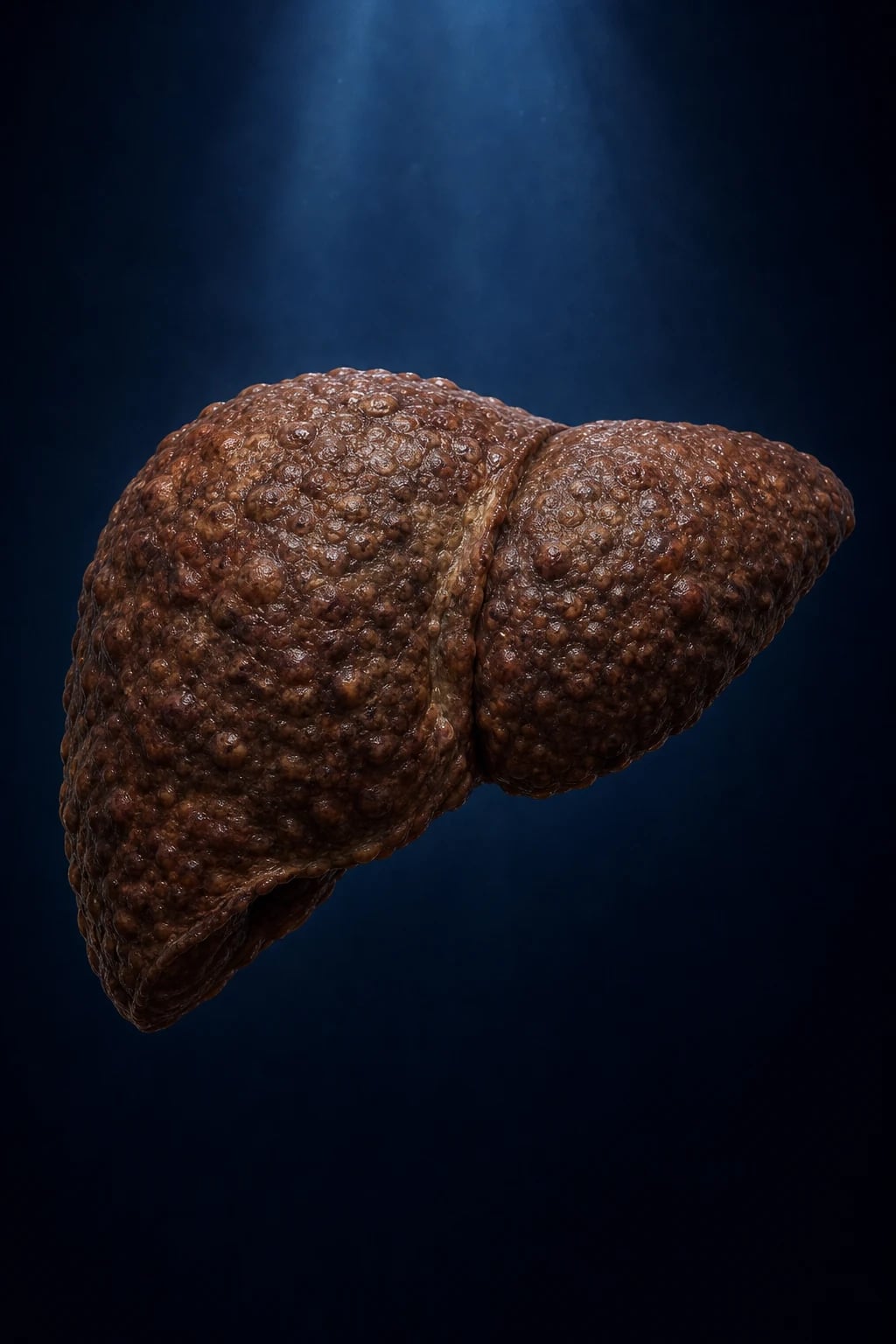
Micronodular
- Nodules under 3 mm, uniform, regular fibrous septa
- Classical of **alcohol-related** cirrhosis and haemochromatosis
- Early stage pattern; with time, nodules enlarge and the picture becomes mixed/macronodular
Macronodular
- Nodules over 3 mm, often varying in size, irregular septa
- Typical of **post-necrotic** cirrhosis — viral hepatitis B/C, autoimmune, Wilson
- Higher risk of **hepatocellular carcinoma** because regeneration is more disordered
Mixed
- Both micro- and macronodules coexist
- The commonest pattern in advanced disease — pure forms become mixed with time
- Morphological form does NOT usually change management; aetiology does
Aetiological classification (the classification that matters)
| Aetiology | Hallmark findings |
|---|---|
| Alcohol-related | AST over ALT (ratio over 2), g-GT markedly raised, macrocytosis, history |
| Hepatitis B (HBV) | HBsAg positive, anti-HBc IgG, high HBV-DNA; commonest single cause in India |
| Hepatitis C (HCV) | Anti-HCV positive, HCV-RNA positive; curable with DAAs |
| MASLD (formerly NAFLD) | Cryptogenic cirrhosis with metabolic syndrome; commonest rising cause in the West |
| Autoimmune hepatitis | High IgG, ANA/SMA/anti-LKM; female predominance |
| Primary biliary cholangitis (PBC) | Female, anti-mitochondrial antibody (AMA), high IgM, raised ALP |
| Primary sclerosing cholangitis (PSC) | Male, inflammatory bowel disease, p-ANCA, multifocal strictures on MRCP |
| Haemochromatosis | High ferritin, transferrin saturation over 45%, HFE gene (C282Y) |
| Wilson disease | Low caeruloplasmin, high urinary copper, Kayser-Fleischer ring |
| Alpha-1-antitrypsin deficiency | Low serum A1AT, PAS-positive diastase-resistant globules on biopsy |
| Vascular — Budd-Chiari, cardiac ("nutmeg") | Hepatic vein outflow obstruction; or chronic right heart failure |
| Cryptogenic | No cause found; many are burnt-out MASLD or undiagnosed autoimmune |
Functional classification — compensated vs decompensated
| Feature | Compensated | Decompensated |
|---|---|---|
| Defining events | None of the four | Ascites, variceal bleed, hepatic encephalopathy, jaundice |
| HVPG | 5–10 mmHg (clinically significant portal hypertension over 10) | Over 10 mmHg; over 12 mmHg for ascites; over 16 for bleed risk |
| Symptoms | Often none; fatigue | Overt — abdominal swelling, confusion, bleeding |
| Median survival | Over 10–12 years | 1.5–2 years without transplant |
| Goals of care | Prevent first decompensation (NSBB, cause treatment, HCC surveillance) | Treat each complication; refer for transplant |
Baveno VII "recompensation" criteria (recognises that effective aetiological treatment can return a decompensated patient to a compensated phenotype):[2]
- Resolution of ascites and hepatic encephalopathy off diuretics / off lactulose (or on stable low doses)
- Sustained cause treatment (e.g. HCV cure, HBV suppression, alcohol abstinence)
- No further decompensation for at least 12 months
- Note: the underlying cirrhosis persists (often compensated cirrhosis with residual portal hypertension); the patient is not cured, but the prognosis improves substantially.
Epidemiology & Risk Factors
Cirrhosis — key numbers
- Global burden. Cirrhosis causes over 1 million deaths per year worldwide and is among the top 15 causes of death globally. The prevalence is rising, driven by MASLD (metabolic dysfunction-associated steatotic liver disease, formerly NAFLD) and hepatitis C, even as alcohol-related cirrhosis and HBV decline in regions with vaccination and antiviral access.
- India. Hepatitis B is the commonest single cause of cirrhosis, followed by alcohol and hepatitis C. HBV is commoner because of vertical and horizontal transmission, lower birth-dose vaccination coverage historically, and high carriage rates. MASLD is rapidly rising with urbanisation, obesity and type-2 diabetes. Cost-limited access to transplantation and TIPS constrains therapy.
- West (US/Europe). Alcohol and HCV historically dominated; with effective DAAs curing HCV, MASLD and alcohol are now the leading indications for transplant. [1]
Risk factors for progression
In a patient with any chronic liver disease, the following accelerate progression to cirrhosis and decompensation: [1]
- Ongoing insult — continued alcohol intake, uncontrolled viraemia (HBV/HCV), untreated autoimmune inflammation
- Metabolic factors — obesity, type-2 diabetes, insulin resistance (the "two-hit" of MASLD)
- Co-infection — HIV/HBV, HIV/HCV, hepatitis D (HDV) superinfection
- Male sex and older age — males progress faster (estrogens may be protective); fibrosis accumulates with age
- Genetics — PNPLA3, TM6SF2, HSD17B13 variants modify MASLD/alcohol progression [1]
Precipitants of acute decompensation / ACLF
In a previously compensated cirrhotic, decompensation is often precipitated by a defined insult. The student must know the list (these drive the search when a cirrhotic deteriorates): [1]
Precipitants of decompensation / HE
HE-FLAG
variceal or any GI bleed — protein load in gut + hypovolaemia
hyponatraemia, hypokalaemia — often from over-diuresis
SBP, pneumonia, UTI, bacteraemia
ammonia absorption from gut
binge, or withdrawal
sedatives, opioids, diuretics; hepatotoxins
When organ failure (liver, kidney, brain, coagulation, circulation, lung) accompanies acute decompensation, the syndrome is acute-on-chronic liver failure (ACLF) — short-term mortality 20–60%. [1]
Pathophysiology
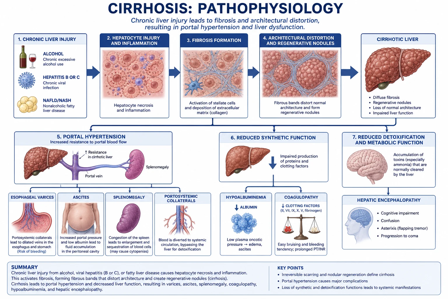
The pathophysiology of cirrhosis has two master processes — fibrogenesis and portal hypertension — that together generate every clinical feature. [1]
1. The fibrogenesis cascade
- Chronic hepatocellular injury (virus, alcohol, fat, cholestasis, autoimmune, metabolic) produces apoptotic hepatocytes and reactive oxygen species.
- Injured hepatocytes, sinusoidal endothelium and Kupffer cells release profibrogenic cytokines — TGF-beta1, PDGF, CTGF, leptin.
- These activate quiescent hepatic stellate cells (the vitamin-A storing Ito cells of Disse space), which transdifferentiate into proliferating, contractile myofibroblasts.
- Activated HSCs:
- Secrete large amounts of type I and type III collagen into the space of Disse (perisinusoidal fibrosis).
- Express tissue inhibitors of metalloproteinases (TIMPs), blocking matrix breakdown.
- Contract around sinusoids, dynamically increasing intrahepatic resistance.
- Sinusoidal endothelium loses its fenestrations (sinusoidal capillarisation), impeding oxygen and metabolite exchange.
- Fibrous septa bridge portal tracts to central veins; regenerative nodules form; the architecture is irreversibly distorted. [1]
Regression. If the injurious stimulus is removed (HCV cure, alcohol abstinence, HBV suppression), activated HSCs undergo apoptosis or senescence, collagenases (MMP-1) break down matrix, and fibrosis can regress — the basis for "recompensation" in Baveno VII.[2]
2. Portal hypertension — the master mechanism
The hepatic venous pressure gradient (HVPG) — the gradient between wedged (sinusoidal) and free hepatic venous pressure — is the gold-standard measure of sinusoidal portal pressure: [1]
- Normal: under 5 mmHg
- Clinically significant portal hypertension (CSPH): HVPG over 10 mmHg — varices form, decompensation risk rises
- Threshold for ascites: over 12 mmHg
- Threshold for variceal bleed: over 16 mmHg (or a varix with red wale signs / large size) [1]
Two mechanisms raise intrahepatic resistance:
- Structural — fibrosis, nodules, distortion (fixed, irreversible component).
- Dynamic / vasoconstrictive — activated HSCs contract (endothelin, thromboxane, angiotensin-II drive contraction; intrahepatic nitric oxide is deficient). This dynamic component is the rationale for NSBB and terlipressin (vasopressin V1 agonist causing splanchnic vasoconstriction). [1]
Splanchnic vasodilation increases portal inflow. Excess nitric oxide and glucagon dilate the splanchnic arteriolar bed, increasing flow into the portal vein and worsening portal pressure despite thecirrhotic obstruction. [1]
3. The hyperdynamic circulation and ascites formation
Splanchnic vasodilation causes peripheral arterial vasodilation → low systemic vascular resistance → low effective arterial blood volume → baroreceptor-mediated activation of RAAS, sympathetic nervous system and non-osmotic vasopressin (ADH). [1]
The consequences:
- Avid sodium retention (aldosterone) — ascites.
- Avid free-water retention (ADH) — dilutional hyponatraemia.
- Vasoconstriction of the renal circulation — hepatorenal syndrome. [1]
Ascites forms when three conditions coexist: (i) sinusoidal portal hypertension (HVPG over 12 mmHg); (ii) splanchnic vasodilation; (iii) hypoalbuminaemia lowering plasma oncotic pressure. Fluid weeps from the sinusoids into the space of Disse and then the peritoneal cavity. The ascites of cirrhosis is a transudate with SAAG over or equal to 1.1 g/dL (a marker of portal hypertension). [1]
4. Hepatic encephalopathy — pathogenesis
Hepatic encephalopathy (HE) is a reversible syndrome of impaired brain function in liver failure and/or portosystemic shunting. The dominant hypothesis is the ammonia hypothesis, modified by systemic inflammation: [1]
- Nitrogenous gut-derived toxins (chiefly ammonia, NH3) bypass hepatic clearance through portosystemic collaterals (or fail to be cleared by the failing liver) and cross the blood-brain barrier.
- Astrocytes metabolise ammonia to glutamine (via glutamine synthetase); accumulated glutamine is osmotically active → astrocyte swelling and cerebral oedema (mainly in acute liver failure; subtle in cirrhosis).
- Increased GABAergic tone and systemic inflammation (cytokines increase blood-brain barrier permeability) synergise with ammonia to depress neuronal function.
- Precipitation: any factor that increases gut ammonia (constipation, GI bleed — protein load), worsens liver failure, or causes systemic inflammation (infection) precipitates HE. [1]
This explains why lactulose (acidifies colon, traps NH3 as NH4+ and acts as an osmotic laxative), rifaximin (gut ammonia-lowering antibiotic), and protein avoidance are NOT recommended (malnutrition worsens outcome). [1]
Clinical Presentation
Compensated cirrhosis
Often asymptomatic; detected incidentally on abnormal liver tests, imaging, or by stigmata found on examination. May report fatigue, anorexia, right upper quadrant discomfort, disturbed sleep, loss of libido. [1]
Stigmata of chronic liver disease (the viva-examiner's shopping list — know each): [1]
- Hands: palmar erythema (thenar/hypothenar, sparing the central palm), Dupuytren contracture, clubbing (especially in PBC), leuconychia (hypoalbuminaemia), asterixis (if encephalopathic).
- Skin: spider naevi (central arteriole with radiating vessels, distributed in the drainage of the superior vena cava — above the nipple line), easy bruising, porphyria cutanea tarda or palmar erythema in alcohol/HCV.
- Face/Head: parotid enlargement, fetor hepaticus (sweet faecal breath from mercaptans), xanthelasma/xanthomata in PBC, sallow complexion.
- Chest/endocrine: gynaecomastia (altered oestrogen-androgen balance — testosterone falls, oestrogen rises due to reduced hepatic clearance), testicular atrophy, loss of secondary sexual hair in men.
- Abdomen: hepatosplenomegaly (early), small shrunken liver (late), caput medusae (recanalised umbilical vein), ascites.
- General: muscle wasting, particularly temporal and proximal (sarcopaenia). [1]
Decompensated cirrhosis — the four decompensation events
- Ascites — the commonest first decompensation. Abdominal distension, weight gain, ankle oedema, later dyspnoea and umbilical herniation.
- Variceal haemorrhage — haematemesis (often massive, painless), melaena, signs of shock; oesophageal varices in 50% of cirrhotics, gastric varices less often.
- Hepatic encephalopathy — see below.
- Jaundice — progressive yellowing of skin and sclera; pruritus in cholestatic cirrhosis. [1]
Hepatic encephalopathy — West Haven grading
| Grade | Mental state | Asterixis | Other |
|---|---|---|---|
| Minimal / covert | Normal on clinical exam; subtle psychometric deficits | Absent | Diagnosed by psychometric tests / PHES score, EEG, or Stroop app |
| I | Mild confusion, euphoria/anxiety, shortened attention, sleep disturbance (reversed sleep-wake) | Slight | Able to do basic tasks |
| II | Lethargy, apathy, disorientation to time, inappropriate behaviour | Present | Constructional apraxia — cannot draw a star / clock / Reitan trail |
| III | Somnolent but rousable, gross disorientation, bizarre behaviour | Usually present | Slurred speech, marked apraxia |
| IV | Coma (unrousable) | Absent (no flap possible) | Response only to pain (IVa) or no response (IVb) |
Overt HE = grades II–IV. Covert/minimal HE = grade I and minimal HE — important because it predicts overt HE and impairs driving. [1]
Spontaneous bacterial peritonitis (SBP)
A cirrhotic with ascites may present atypically — fever, abdominal pain, or simply worsening encephalopathy, worsening renal function, or unexplained ileus. A diagnostic tap is mandatory in any cirrhotic admitted to hospital or with new/worsening ascites. [1]
Atypical presentations
- Elderly: paucity of classical stigmata; decompensation as the first presentation (confusion, falls, infection); high frailty.
- Alcohol-related: symptoms may be dominated by withdrawal, malnutrition, Wernicke, or anaemia; the liver disease may be silent until decompensation.
- Pregnancy with cirrhosis: high risk of variceal bleed during second trimester and delivery (increased blood volume and intra-abdominal pressure); screen and band varices early; prefer regional anaesthesia.
- Diabetic with MASLD: cirrhosis may be entirely silent until HCC or decompensation; cardiovascular disease dominates mortality. [1]
Differential Diagnosis
1. Ascites — the SAAG approach (the single most testable ascites concept)
The serum-ascites albumin gradient (SAAG) = serum albumin minus ascitic albumin (taken on the same day). It classifies ascites by mechanism: [1]
| SAAG | Mechanism | Examples |
|---|---|---|
| Over or equal to 1.1 g/dL (high gradient) | Portal hypertension | Cirrhosis (commonest), heart failure (cardiac ascites), Budd-Chiari syndrome |
| Under 1.1 g/dL (low gradient) | Peritoneal disease / non-portal | Peritoneal carcinomatosis (ovarian, gastric, colon), tuberculous peritonitis, nephrotic syndrome, pancreatic ascites, chylous ascites |
Distinguishing features within high-SAAG ascites:
- Cirrhosis — chronic liver stigmata, low platelets, nodular liver, splenomegaly.
- Cardiac ascites (constrictive pericarditis, right heart failure) — ascitic protein over 2.5 g/dL, jugular venous distension, hepatomegaly, no splenomegaly, normal-sized or large liver.
- Budd-Chiari — painful hepatomegaly, sudden-onset ascites, caudate lobe hypertrophy on imaging. [1]
2. Acute confusion in a cirrhotic — beyond hepatic encephalopathy
A deteriorating cirrhotic is NOT automatically encephalopathic. Distinguish: [1]
| Cause | Distinguishing feature |
|---|---|
| Hepatic encephalopathy | Asterixis, reversed sleep-wake, response to lactulose; ammonia may be raised but correlation is poor |
| Hyponatraemia | Serum sodium under 125; slow correction to avoid osmotic demyelination |
| Sepsis / septic encephalopathy | Source identified (SBP, pneumonia); positive cultures |
| Intracranial lesion | Subdural haematoma (fall, coagulopathy); focal signs; CT head if any focal sign or unexplained deterioration |
| Wernicke encephalopathy | Alcoholism; ataxia, ophthalmoplegia, confusion — give IV thiamine before glucose |
| Drug / sedative effect | Opioids, benzodiazepines accumulate; flumazenil/naloxone trial |
3. Decompensated cirrhosis vs acute liver failure
| Feature | Acute liver failure | Decompensated cirrhosis |
|---|---|---|
| Onset | Under 26 weeks, previously well | Chronic; over months-years |
| Portal hypertension | Absent (no varices, no splenomegaly) | Present |
| INR | High; may reverse with vitamin K | High; does not fully reverse |
| Ascites | Usually absent | Often present |
| Cerebral oedema | Common, life-threatening | Uncommon |
4. Nodular liver — other causes
Budd-Chiari, metastatic disease, macronodular cirrhosis from Wilson/haemochromatosis, regenerative nodular hyperplasia, focal nodular hyperplasia, HCC within a cirrhotic liver. [1]
5. Splenomegaly-dominant presentation
When splenomegaly dominates with modest liver disease, consider extra-hepatic portal vein obstruction (portal vein thrombosis), myeloproliferative neoplasm with portal hypertension, or schistosomiasis (presinusoidal, normal liver). [1]
Clinical & Bedside Assessment
Abdominal examination for ascites
- Bulging flanks — fluid settling laterally.
- Flank dullness to percussion — the most sensitive bedside sign; dullness shifts when the patient rolls because fluid is free-flowing.
- Shifting dullness — percuss from the umbilicus laterally; the air-fluid boundary shifts with repositioning. Requires about 1500 mL.
- Fluid thrill — place one hand flat on one flank, tap the other flank with the opposite hand; a palpable impulse indicates free fluid (only with large-volume ascites, over 2000 mL).
- Ballot the liver and spleen — the spleen is ballotable if splenomegaly from portal hypertension.
- Puddle sign — historical; dullness in the dependent umbilical area when on all fours (low sensitivity, abandoned). [1]
Neurological examination for hepatic encephalopathy
- Asterixis (liver flap) — ask the patient to hold the hands dorsiflexed with fingers spread for 30 seconds; a flap (rapid flexion-extension at the wrist/MCP) appears bilaterally. Also seen in uraemia, hypercapnia (a "metabolic flap").
- Constructional apraxia — Reitan trail-making test, clock-drawing, or star-drawing; failure indicates grade I–II HE.
- Mental state — orientation to time, place, person; grade by West Haven.
- Reflexes — hyperreflexia, extensor plantar in advanced cases. [1]
Stigmata (covered above) — examiner will ask for ten signs
Palmar erythema, spider naevi, gynaecomastia, testicular atrophy, parotid enlargement, Dupuytren contracture, clubbing, leuconychia, caput medusae, jaundice / scratch marks. [1]
Aetiological clues at the bedside
- Tattoos / piercings / IV drug use — HBV, HCV.
- Kayser-Fleischer ring (brown copper deposition at the corneal limbus, best seen by slit-lamp) — Wilson disease.
- Bronze diabetes, arthritis, cardiac failure — haemochromatosis.
- Cushingoid, parotid enlargement, Dupuytren — alcohol.
- Xanthomata, pruritus — PBC.
- IBD, recurrent cholangitis — PSC. [1]
Diagnostic abdominal paracentesis
Indications (mandatory): any cirrhotic with new-onset or worsening ascites, or any cirrhotic admitted to hospital with ascites (the most common pitfall is failing to tap). [1]
Technique: clean skin, sterile gloves; needle (typically 20–22 G) inserted under local anaesthetic in the left lower quadrant, 2 cm medial and 2 cm superior to the anterior superior iliac spine (avoids the inferior epigastric vessels). Send fluid for: cell count (PMN), albumin (for SAAG), culture (in blood-culture bottles — increases yield), total protein, and if indicated, cytology, ADA/TB-PCR, amylase, triglycerides. [1]
Investigations
First-line panel in suspected cirrhosis
| Test | Expected pattern / why |
|---|---|
| FBC | Thrombocytopenia (under 150 x 10^9/L) from hypersplenism ± reduced thrombopoietin; macrocytosis (alcohol); anaemia of chronic disease, folate deficiency, GI bleed |
| LFTs | Low albumin (synthetic failure), raised bilirubin (excretory failure), AST over ALT in alcohol; ALP raised in cholestasis (PBC/PSC) |
| Coagulation | Raised INR / PT (reduced factor V, VII synthesis; vitamin K does not fully correct) |
| U&E, creatinine | Baseline renal function; later hepatorenal syndrome |
| Serum sodium | Dilutional hyponatraemia from non-osmotic ADH |
| g-GT | Markedly raised in alcohol and cholestasis |
| Glucose / HbA1c / lipids | Screen MASLD |
| Serum and ascitic albumin (same day) | To calculate SAAG |
Aetiology screen
- Viral serology: HBsAg, anti-HBc IgG, anti-HBs, HBV-DNA; anti-HCV and HCV-RNA.
- Iron studies: ferritin, transferrin saturation (over 45% suggests haemochromatosis → HFE gene).
- Caeruloplasmin (low) and 24-h urinary copper (high) — Wilson disease (especially under age 40).
- Alpha-1-antitrypsin level and phenotype.
- Immunoglobulins: high IgG (autoimmune), high IgM (PBC).
- Autoantibodies: ANA, SMA, anti-LKM (AIH); anti-mitochondrial antibody (AMA) (PBC, 95%); p-ANCA (PSC).
- Alpha-fetoprotein (AFP) — HCC marker; check at baseline and for surveillance.
- Alcohol markers where history is unreliable: GGT, MCV, carbohydrate-deficient transferrin (CDT). [1]
Severity scores — reproduced verbatim
Child-Pugh (Child-Turcotte-Pugh) score — five components, each scored 1–3: [1]
| Parameter | 1 point | 2 points | 3 points |
|---|---|---|---|
| Total bilirubin (micromol/L) | under 34 | 34–50 | over 50 |
| Serum albumin (g/L) | over 35 | 28–35 | under 28 |
| INR | under 1.7 | 1.7–2.3 | over 2.3 |
| Ascites | None | Mild (controlled medically) | Moderate–severe (refractory) |
| Hepatic encephalopathy | None | Grade I–II | Grade III–IV |
Grades (sum of points): A = 5–6 (1-yr survival ~100%, 2-yr ~85%); B = 7–9 (1-yr ~80%, 2-yr ~60%); C = 10–15 (1-yr ~45%, 2-yr ~35%). [1]
MELD score (Model for End-stage Liver Disease) — predicts 3-month mortality, drives transplant priority: [1]
MELD = 3.78 × ln(bilirubin mg/dL) + 11.2 × ln(INR) + 9.57 × ln(creatinine mg/dL) + 6.43 (laboratory values floored at 1.0; creatinine capped at 4.0 mg/dL; dialysis counted as 4.0). [1]
MELD-Na adds sodium (because hyponatraemia independently predicts mortality in cirrhosis): [1]
MELD-Na = MELD + 1.32 × (137 − Na) − [0.033 × MELD × (137 − Na)] (Na capped between 125 and 137 mmol/L). [1]
Approximate 3-month mortality by MELD: 10 → ~6%; 20 → ~20%; 30 → ~53%; 40 → ~71%. [1]
Serum-ascites albumin gradient (SAAG)
- High gradient (over or equal to 1.1 g/dL) → portal hypertension (cirrhosis, cardiac, Budd-Chiari).
- Low gradient (under 1.1 g/dL) → peritoneal disease (TB, malignancy), nephrotic, pancreatic, chylous.
- Ascitic total protein further refines: in cirrhotic ascites protein is under 2.5 g/dL; in cardiac ascites it is over 2.5 g/dL. [1]
Diagnostic ascitic fluid in suspected SBP
Diagnostic threshold for SBP: polymorphonuclear neutrophil (PMN) count over or equal to 250 cells/mm^3 (equivalently over 250 x 10^6/L) without an obvious intra-abdominal surgical source. [1]
- Culture in blood-culture bottles at the bedside (yield rises to 80%).
- Cytology if peritoneal carcinomatosis suspected.
- ADA and TB-PCR if tuberculous peritonitis suspected.
- Total protein under 1 g/dL raises SBP risk (low opsonic activity) — an indication for primary SBP prophylaxis. [1]
Imaging
- Ultrasound abdomen — first-line. Shows nodular liver surface, splenomegaly, ascites, portal vein patency (rule out thrombosis), and screens for HCC. Doppler shows reversed (hepatofugal) portal flow in advanced disease.
- Transient elastography (FibroScan) — non-invasive measure of liver stiffness. Liver stiffness over 12–15 kPa strongly suggests cirrhosis in most aetiologies; over 20–25 kPa with low platelets defines clinically significant portal hypertension (Baveno VII).
- CT or MRI with multiphase contrast — for HCC diagnosis (arterial enhancement with portal/delayed washout), Budd-Chiari, and pre-transplant assessment.
- Upper GI endoscopy — to screen for oesophago-gastric varices and portal hypertensive gastropathy (every 2–3 years in compensated cirrhosis without varices; sooner if CSPH).
- Liver biopsy — the gold standard for confirming cirrhosis, grading fibrosis, and defining aetiology when non-invasive tests are equivocal (e.g. transjugular route if coagulopathy/ascites). Not always needed when cirrhosis is obvious clinically and radiologically. [1]
HCC surveillance
Every cirrhotic, regardless of cause or viral control, undergoes 6-monthly ultrasound ± serum AFP for life. A nodule with arterial hypervascularity and washout on multiphase CT/MRI is HCC by imaging criteria (LI-RADS) without biopsy. [1]
Management — Resuscitation
Acute variceal haemorrhage — the time-critical bundle
Airway / Breathing
- Protect airway from aspiration (haematemesis + encephalopathy = high aspiration risk)
- Intubate early if encephalopathic or uncontrolled haemorrhage
- Oxygen; two large-bore (14–16 G) cannulae
Circulation
- Restrictive transfusion: target **Hb 7–8 g/dL** — over-transfusion raises portal pressure and worsens bleeding
- Activate massive transfusion protocol; correct coagulopathy and thrombocytopenia judiciously (no routine FFP/platelets unless active bleeding)
- Avoid excess crystalloid
Vasoactive drug (start at first suspicion)
- **Terlipressin 2 mg IV every 4 hours** (vasopressin V1 agonist; splanchnic vasoconstriction) — first-line where available
- OR **octreotide 50 microgram IV bolus then 50 microgram/hour infusion** — alternative
- Continue 2–5 days; reduces transfusion requirement and mortality
Antibiotics
- **Ceftriaxone 1 g IV daily for up to 7 days** (or oral ciprofloxacin/norfloxacin if low risk) — prophylaxis against SBP and bacteraemia
- Antibiotics reduce mortality and rebleeding; given to ALL cirrhotics with GI bleed
Endoscopy
- **Endoscopic band ligation (EBL) within 12 hours** of presentation
- If uncontrolled: **balloon tamponade (Sengstaken-Blakemore / Minnesota tube)** as a bridge to definitive therapy — max 24 h
- **TIPS (transjugular intrahepatic portosystemic shunt)** for failure to control bleeding or early rebleeding
Resuscitation in SBP
- Empiric cefotaxime 2 g IV every 12 hours (or ceftriaxone 1 g IV daily) — start as soon as PMN threshold met, do not wait for culture.
- Intravenous albumin 1.5 g/kg on day 1 and 1 g/kg on day 3 — reduces hepatorenal syndrome and mortality (the Sort 1999 NEJM finding).[1]
- Treat for 5–7 days, repeat tap at 48 h; rising PMN suggests failure — broaden antibiotics.
Resuscitation in hepatic encephalopathy (grades III–IV)
- Secure the airway (intubate if comatose or unable to protect).
- Identify and treat the precipitant — the most important step. Send cultures, check sodium, do a diagnostic tap, rectal exam for melaena, review drug chart.
- Correct electrolytes and dehydration cautiously (avoid rapid sodium correction).
- Avoid sedatives; if benzodiazepine given inadvertently, consider flumazenil. [1]
Resuscitation in hepatorenal syndrome-AKI
- Stop all diuretics and nephrotoxins (NSAIDs, ACE-inhibitors, aminoglycosides, contrast).
- Volume challenge: albumin 1 g/kg/day (max 100 g/day) for 2 consecutive days with withdrawal of diuretics.
- If creatinine fails to improve, the diagnosis is HRS-AKI — escalate to terlipressin + albumin (see below). [1]
Management — Definitive & Stepwise
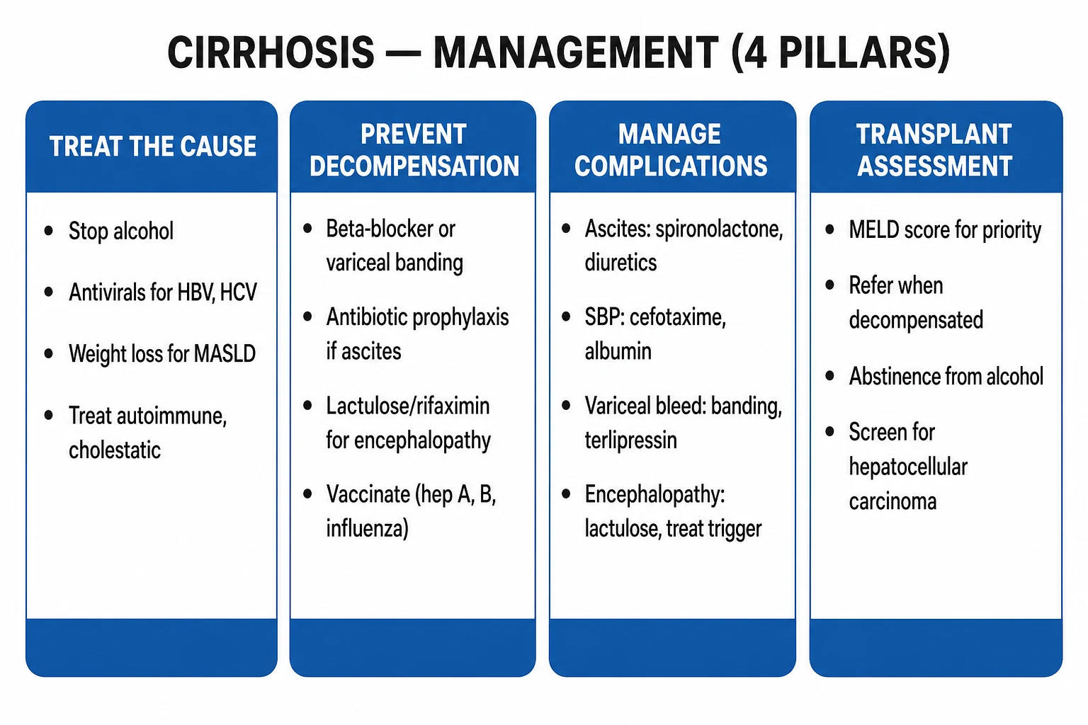
1. Treat the underlying cause (the most important long-term intervention)
| Aetiology | Definitive therapy |
|---|---|
| Alcohol-related | Abstinence transforms prognosis; pharmacotherapy (acamprosate, naltrexone); treat withdrawal with benzodiazepines (CIWA); thiamine before glucose to prevent Wernicke |
| Hepatitis C | Direct-acting antivirals (DAAs) — cure in 8–12 weeks (e.g. sofosbuvir/velpatasvir); HCC surveillance continues despite cure |
| Hepatitis B | Nucleos(t)ide analogues — tenofovir disoproxil fumarate 300 mg daily or entecavir 0.5–1 mg daily; indefinite suppression |
| MASLD | Weight loss 7–10%, treat diabetes, statins as tolerated, bariatric surgery; cardiovascular risk reduction (the leading cause of death) |
| Autoimmune hepatitis | Prednisolone (induction) ± azathioprine (maintenance) |
| PBC | Ursodeoxycholic acid 13–15 mg/kg/day (improves biochemistry and survival) |
| Haemochromatosis | Weekly venesection until ferritin under 50 microgram/L |
| Wilson | Penicillamine or trientine chelation; zinc for maintenance |
| Alpha-1-antitrypsin | No specific therapy; supportive; transplant |
2. Ascites — the stepwise ladder
- Dietary sodium restriction to under 2 g sodium per day (88 mmol/day) — the cornerstone.
- Fluid restriction only if hyponatraemia (sodium under 120–125 mmol/L).
- Diuretics — spironolactone + furosemide in a 100:40 ratio (e.g. spironolactone 100 mg + furosemide 40 mg orally in the morning), titrated up to a maximum of 400 mg + 160 mg daily. The 5:2 ratio maintains normokalaemia; spironolactone (aldosterone antagonist) is more effective than loop alone because of secondary hyperaldosteronism. Monitor weight (target loss 0.5 kg/day, 1 kg/day if peripheral oedema), U&E, and watch for gynaecomastia, hyperkalaemia, dehydration.
- Refractory ascites (failure to mobilise or recurrence despite maximum diuretics):
- Large-volume paracentesis (LVP) — drain over 5 L per session with intravenous albumin 8 g per litre drained (over 5 L) to prevent post-paracentesis circulatory dysfunction.
- TIPS (transjugular intrahepatic portosystemic shunt) — decompresses portal hypertension; best in selected patients without severe cardiopulmonary disease or uncontrolled HE.
- Liver transplantation — definitive. [1]
3. Spontaneous bacterial peritonitis
- Treatment: cefotaxime 2 g IV every 12 hours for 5–7 days + albumin 1.5 g/kg day 1, 1 g/kg day 3.
- Secondary prophylaxis (after one episode of SBP — high recurrence): norfloxacin 400 mg orally daily or ciprofloxacin 500 mg daily indefinitely (or until transplant).
- Primary prophylaxis: in patients with ascitic protein under 1.5 g/dL with impaired renal/hepatic function (Creatinine over 1.2, BUN over 25, Child-Pugh over 9, sodium under 130). [1]
4. Acute variceal bleed — definitive bundle
- Vasoactive (terlipressin 2 mg IV 4-hourly OR octreotide 50 microgram bolus + 50 microgram/h) for 2–5 days.
- Antibiotic prophylaxis — ceftriaxone 1 g IV daily for up to 7 days.
- Endoscopic band ligation within 12 hours (sclerotherapy for gastric varices — cyanoacrylate glue).
- Balloon tamponade (Sengstaken-Blakemore) as a temporary bridge — max 24 h, intubate to protect airway.
- TIPS for failure to control bleeding or early rebleeding in high-risk patients (Child-Pugh C under 14, or Child-Pugh B with active bleeding at endoscopy — early or "pre-emptive" TIPS). [1]
Primary prophylaxis of first variceal bleed (in those with high-risk varices = medium/large varices, or small varices with red wale signs or in Child-Pugh B/C):
- Non-selective beta-blocker (NSBB) — propranolol 20–80 mg BD, nadolol 20–40 mg daily, or carvedilol 6.25–12.5 mg daily titrated to resting HR 55–60 bpm. Carvedilol also reduces intrahepatic resistance via alpha-blockade — preferred by Baveno VII for CSPH without varices.
- OR endoscopic band ligation (EVL) — serial sessions every 2–8 weeks until variceal eradication. [1]
Secondary prophylaxis (after a bleed): NSBB + EVL combined (lowest rebleeding rate); indefinite; consider TIPS if recurrent. [1]
5. Hepatic encephalopathy
- Identify and treat the precipitant — the most important single intervention.
- Lactulose 15–30 mL orally two to four times daily, titrated to 2–3 soft stools per day (lactulose acidifies the colon, converting NH3 to non-absorbable NH4+, and is an osmotic laxative).
- Rifaximin 550 mg orally twice daily — added for secondary prevention of overt HE (reduces recurrence, hospitalisation; Bass 2010 NEJM).
- Nutrition — do NOT restrict protein. Daily protein 1.2–1.5 g/kg; treat sarcopaenia. Vegetable/dairy proteins may be better tolerated.
- TIPS may worsen HE; consider embolisation of large shunts in refractory cases.
- Novel agents under study: L-ornithine L-aspartate (LOLA), branched-chain amino acids, polyethylene-glycol. [1]
6. Hepatorenal syndrome (HRS-AKI)
Diagnostic criteria (ICA): rise in serum creatinine (AKI criteria); no response to albumin challenge (1 g/kg/day x 2); absence of shock, no current nephrotoxins, no structural kidney disease (normal ultrasound, no proteinuria). [1]
- Albumin + terlipressin 1–2 mg IV every 4–6 hours (titrate) for up to 14 days — restores renal function in HRS-AKI (Wong 2021 NEJM confirms terlipressin efficacy; monitor for ischaemia, fluid overload). Where terlipressin is unavailable, noradrenaline infusion in ICU.
- Renal replacement therapy as a bridge.
- Liver transplant (or simultaneous liver-kidney transplant) is definitive. [1]
7. Hepatocellular carcinoma — stage-based (BCLC)
| BCLC stage | Treatment |
|---|---|
| Very early / early (single tumour, preserved function) | Resection, thermal ablation (RFA/MWA), or transplant within Milan criteria (single tumour under 5 cm OR up to 3 nodules each under 3 cm, no vascular invasion, no extrahepatic spread) |
| Intermediate (multinodular, preserved function) | Transarterial chemoembolisation (TACE) |
| Advanced (portal invasion, metastases) | Systemic therapy — atezolizumab + bevacizumab (first-line), durvalumab + tremelimumab, sorafenib, lenvatinib |
| Terminal | Best supportive care |
8. Liver transplantation
Indications: decompensated cirrhosis with MELD over or equal to 15; first episode of SBP; first episode of overt HE; refractory ascites; HCC within Milan criteria (or selected expansions). Absolute contraindications: active alcohol/substance misuse, uncontrolled extrahepatic sepsis, severe cardiopulmonary disease, uncontrolled extrahepatic malignancy. Survival ~85–90% at 1 year, 70–75% at 5 years. [1]
Specific Subtypes & Scenarios
Alcohol-related cirrhosis
The classical micronodular cirrhosis, AST over ALT (ratio over 2), raised g-GT and MCV. Abstinence is the single most powerful intervention — improves survival at all stages. Add thiamine 100 mg IV/IM daily for 3–5 days (then oral) to prevent Wernicke; manage withdrawal with a long-acting benzodiazepine (chlordiazepoxide) by a symptom-triggered (CIWA) regimen. Malnutrition and sarcopaenia are nearly universal — aggressive nutritional support (35–40 kcal/kg/day, 1.2–1.5 g/kg protein) improves survival. [1]
HBV / HCV-related cirrhosis
- Hepatitis C is now curable with direct-acting antivirals (DAAs) in 8–12 weeks (e.g. sofosbuvir/velpatasvir, glecaprevir/pibrentasvir). Achieving sustained virological response (SVR) dramatically reduces decompensation, HCC, and mortality, and may allow recompensation.[2]
- Hepatitis B is suppressed lifelong with high-barrier nucleos(t)ide analogues — tenofovir or entecavir. Stopping risks flare and decompensation.
- HCC surveillance continues regardless of viral control — antiviral cure does not eliminate HCC risk.
MASLD / cryptogenic cirrhosis
The rising tide. Look for metabolic syndrome (central obesity, diabetes, hypertension, dyslipidaemia). Many present as cryptogenic cirrhosis (burnt-out MASLD). Cardiovascular disease is the leading cause of death — manage aggressively with statins (safe and beneficial in cirrhosis), antihypertensives (avoid ACE-inhibitors/NSAIDs), tight glycaemic control, weight loss. Gradual weight loss 7–10% targets steatohepatitis regression. [1]
PBC and PSC cirrhosis
- PBC — middle-aged woman, fatigue, pruritus, anti-mitochondrial antibody (AMA) positive, raised ALP and IgM. Ursodeoxycholic acid 13–15 mg/kg/day improves biochemistry and transplant-free survival. Osteoporosis and fat-soluble-vitamin deficiency need monitoring and replacement.
- PSC — young male with inflammatory bowel disease, recurrent cholangitis, multifocal strictures and dilatation ("beading") on MRCP, p-ANCA positive. High risk of cholangiocarcinoma and colorectal cancer — annual MRCP and colonoscopic surveillance. No proven disease-modifying drug; UDCA for biochemistry; ERCP for dominant strictures; transplant for decompensation. [1]
Acute-on-chronic liver failure (ACLF)
Definition: acute deterioration of pre-existing chronic liver disease/cirrhosis (with or without a precipitant) associated with organ failure and high 28-day mortality. Graded ACL-1/2/3 by the number of organ failures (CLIF-C OF score). Organ failures: liver (bilirubin), kidney (creatinine), brain (West Haven), coagulation (INR), circulation (vasopressors), lung (PaO2/FiO2). Management: treat the precipitant, organ support in ICU, terlipressin/albumin if HRS, early transplant in selected patients. [1]
Refractory ascites and hepatic hydrothorax
- Refractory ascites — serial LVP + albumin, TIPS (especially if rapid re-accumulation), autologous ascites reinfusion, transplant.
- Hepatic hydrothorax — pleural effusion (usually right-sided) through small diaphragmatic defects; treat as ascites (sodium restriction, diuretics); avoid chest drains (high mortality); TIPS or transplant for refractory cases. [1]
Complications & Pitfalls
Portal-hypertensive
- **Variceal haemorrhage** — oesophageal and gastric; massive, life-threatening
- **Portal hypertensive gastropathy / enteropathy** — chronic GI blood loss, anaemia
- **Splenomegaly / hypersplenism** — pancytopenia (mainly thrombocytopenia and leucopenia)
Liver-failure
- **Coagulopathy** — raised INR, factor V/II/VII/IX/X reduction (vitamin-K independent)
- **Jaundice / hyperbilirubinaemia**
- **Hepatic encephalopathy** — overt and covert
- **Hepatorenal syndrome** — HRS-AKI and HRS-AKD
- **Hepatopulmonary syndrome** — triad of liver disease, hypoxaemia, intrapulmonary vasodilation (platypnoea-orthodeoxia)
- **Portopulmonary hypertension** — pulmonary arterial hypertension in portal hypertension
Infection / malignancy
- **Spontaneous bacterial peritonitis** — PMN over 250 cells/mm^3
- **Bacteraemia, pneumonia, UTI** (cirrhotic immunodeficiency)
- **Hepatocellular carcinoma** — surveillance lifelong
Systemic / metabolic
- **Malnutrition / sarcopaenia / frailty**
- **Osteoporosis** (especially cholestatic), **vitamin D and A, D, E, K deficiency**
- **Anaemia** (folate, iron, haemolysis, bleed), **thrombocytopenia**
- **Hyponatraemia**, **hypoglycaemia**, **adrenal insufficiency**
Classic pitfalls (high-yield viva)
- Failing to perform a diagnostic tap in every cirrhotic with new or worsening ascites.
- Over-transfusing a variceal bleed — raises portal pressure and worsens bleeding; target Hb 7–8 g/dL.
- Giving NSAIDs, ACE-inhibitors, aminoglycosides or iodinated contrast — precipitate HRS.
- Protein-restricting an encephalopathic patient — worsens malnutrition and mortality.
- Missing HCC surveillance in a stable compensated cirrhotic.
- Rapidly correcting hyponatraemia — risk of osmotic demyelination.
- Forgetting antibiotics and vasoactive drugs in the acute variceal bleed bundle.
- Forgetting thiamine before glucose in the alcoholic hypoglycaemic patient. [1]
Prognosis & Disposition
Prognosis in cirrhosis is dominated by whether disease is compensated or decompensated (ascites, variceal haemorrhage, hepatic encephalopathy, jaundice). Median survival falls sharply after decompensation. Child-Pugh and MELD(Na) quantify short-term mortality and transplant urgency. [1]
Child-Pugh components (reproduce)
| Points | 1 | 2 | 3 |
|---|---|---|---|
| Bilirubin (mg/dL) | <2 | 2–3 | >3 |
| Albumin (g/dL) | >3.5 | 2.8–3.5 | <2.8 |
| INR | <1.7 | 1.7–2.3 | >2.3 |
| Ascites | None | Mild | Moderate–severe |
| Encephalopathy | None | Grade I–II | Grade III–IV |
Class A 5–6, B 7–9, C 10–15 points — C predicts highest mortality without transplant. [1]
MELD
Uses bilirubin, INR, creatinine (MELD-Na adds sodium). Higher MELD → higher 90-day mortality and higher transplant priority.
Disposition
- Compensated outpatients: aetiology treatment, variceal screening, HCC surveillance (US ± AFP every 6 months)
- Decompensated: admit if tense ascites, SBP suspected, bleed, refractory HE, AKI/HRS, infection
- Transplant evaluation for decompensation, HCC within criteria, or high MELD after aetiology control where possible
Special Populations
Alcohol-related cirrhosis
Abstinence is disease-modifying. Thiamine before glucose if risk of Wernicke. Screen for alcohol withdrawal; nutrition (calories/protein) is treatment, not optional. Consider steroids in severe alcoholic hepatitis (Maddrey DF ≥32) after excluding infection — not for all cirrhosis.
Viral hepatitis B/C
Treat HCV with DAAs when feasible (curative); HBV nucleos(t)ide analogues to suppress replication and reduce decompensation/HCC risk. Vaccinate for HAV/HBV if non-immune.
NAFLD/MASLD cirrhosis
Cardiometabolic risk kills as often as liver failure — manage obesity, diabetes, statins when indicated. Emerging metabolic therapies under specialist use.
Pregnancy
Rare; manage in tertiary centres. Endoscopy for varices before conception if known cirrhosis; beta-blockers individualised; delivery planning for decompensation risk.
Surgery and procedures
Elective surgery risk rises with Child-Pugh B/C. Correct coagulopathy thoughtfully (INR is a poor predictor of bleeding risk in cirrhosis — use platelet/fibrinogen context). Avoid unnecessary invasive procedures.
Acute-on-chronic liver failure (ACLF)
Rapid organ failures (CLIF-C / EASL definitions) after a precipitant (infection, bleed, alcohol binge) — high short-term mortality; early ICU and transplant discussion.
Evidence, Guidelines & Regional Differences
Landmark trials and scores
| Study / consensus | Year | Finding |
|---|---|---|
| Baveno VII (de Franchis, J Hepatol) | 2022 | NSBB / carvedilol first-line for CSPH; "recompensation" criteria; LSM and platelet rule-out for varices (Baveno VI criteria: LSM under 25 kPa AND platelets over 110 → safe to skip endoscopy) [2] |
| AASLD Portal Hypertension Guidance (Kaplan, Hepatology) | 2024 | Risk stratification by LSM + platelets; primary/secondary prophylaxis; acute-bleed bundle (vasoactive + antibiotic + EBL) [1] |
| Sort (NEJM) — albumin in SBP | 1999 | Albumin 1.5 g/kg D1 + 1 g/kg D3 with cefotaxime reduces renal failure and mortality |
| Bass (NEJM) — rifaximin for HE | 2010 | Rifaximin 550 mg BD reduces overt HE recurrence over 6 months |
| Wong (NEJM) — terlipressin in HRS | 2021 | Terlipressin + albumin improves HRS reversal (CONFIRM trial); watch for ischaemia |
| Malinchoc (Hepatology) — MELD | 2000 | Original MELD model from TIPS patients predicts 3-month mortality |
| Kim (NEJM) — MELD-Na | 2008 | Adding serum sodium improves mortality prediction |
| Singal — HCC surveillance | 2022 | 6-monthly ultrasound improves early HCC detection and survival |
Regional differences
- India (INASL / NACO): HBV commonest cause; high HCV burden (often iatrogenic); cost-limited access to transplant and TIPS; generic DAAs have transformed HCV; mass HBV vaccination programmes aim to reduce HBV cirrhosis long-term.
- AASLD (US) / EASL (Europe): drive most international guidelines; emphasise non-invasive fibrosis assessment (elastography), NSBB first-line, recompensation, and Milan-based transplant selection.
- Baveno consensus (Europe-led, international): the central authority on portal hypertension nomenclature, thresholds (HVPG), and management — the spine of every guideline. [1]
Worked NEET-PG Stems — Cirrhosis
- SAAG 1.4, ascitic protein low → portal hypertensive ascites.
- Ascitic PMN 400 → SBP; start third-generation cephalosporin; do not wait for culture.
- Tense ascites → large-volume paracentesis + albumin if large volume removed; then diuretics.
- Haematemesis in Child C → variceal bundle: resus, terlipressin, antibiotic, banding, salvage TIPS.
- Asterixis after constipation + infection → HE; lactulose; fix precipitant; rifaximin if recurrent.
- Rising creatinine in ascites on diuretics → hold diuretics; exclude SBP/hypovolaemia/HRS pathway.
- HCC surveillance → ultrasound ± AFP every 6 months in cirrhosis.
- Child-Pugh variables → bilirubin, albumin, INR, ascites, encephalopathy. [1]
Exam Pearls
- SAAG over or equal to 1.1 g/dL = portal hypertension (cirrhosis / cardiac / Budd-Chiari); under 1.1 = peritoneal (TB / malignancy / nephrotic).
- SBP threshold = PMN over or equal to 250 cells/mm^3. Treat with cefotaxime 2 g IV 12-hourly + albumin (1.5 g/kg D1, 1 g/kg D3).
- Ascites first-line diuretics = spironolactone + furosemide in 100:40 ratio, titrated.
- Variceal bleed bundle = vasoactive (terlipressin 2 mg IV 4-hourly) + antibiotic (ceftriaxone 1 g IV daily) + endoscopic band ligation within 12 hours. Restrictive transfusion to Hb 7–8 g/dL.
- Hepatic encephalopathy = lactulose (titrate to 2–3 soft stools/day) + rifaximin 550 mg BD for prevention. Do NOT restrict protein.
- MELD = bilirubin + INR + creatinine (+ sodium for MELD-Na); predicts 3-month mortality and transplant priority. Child-Pugh adds ascites + encephalopathy to the labs.
- HCC surveillance = 6-monthly ultrasound in ALL cirrhotics; treat early-stage by resection / ablation / transplant (Milan: single tumour under 5 cm, or up to 3 nodules under 3 cm).
- Avoid NSAIDs, ACE-inhibitors, aminoglycosides, and iodinated contrast in cirrhosis — they precipitate HRS and renal failure.
- Hepatorenal syndrome: stop diuretics and nephrotoxins, albumin 1 g/kg/day x 2, then terlipressin; transplant definitive.
- HVPG thresholds: over 5 mmHg portal hypertension; over 10 mmHg clinically significant (varices, decompensation); over 12 mmHg ascites; over 16 mmHg variceal bleed risk.
- Spider naevi are confined to the drainage of the superior vena cava (above the nipple line).
- Palmar erythema spares the central palm.
- AST over ALT (ratio over 2) is classical of alcohol-related liver disease; ALT over AST suggests viral hepatitis.
- Wernicke before glucose: thiamine 100 mg IV before any dextrose in the alcoholic — prevents irreversible ophthalmoplegia/ataxia/confusion.
- Carvedilol is preferred over propranolol for portal hypertension (additional alpha-1 blockade lowers intrahepatic resistance) — but avoid in severe asthma, decompensated heart block, hypotension (SBP under 90).
- Baveno VI rule-out: LSM under 25 kPa AND platelets over 110 x 10^9/L → varices unlikely; can safely skip screening endoscopy. [1]
Exam application bank (NEET-PG / INICET)
One-line answer
Cirrhosis is the diffuse, irreversible distortion of hepatic architecture by fibrosis and regenerative nodules that follows chronic hepatocellular injury. It is the final common pathway of virtually every chronic liver disease (alcohol, viral hepatitis B/C, MASLD, autoimmune, cholestatic, metabolic, vascular). Clinically it is divided into a long compensated phase (asymptomatic with stigmata) and a decompensated phase defined by ascites, variceal haemorrhage, hepatic encephalopathy and jaundice. The pathophysiology centres on portal hypertension (mechanical distortion plus dynamic vasoconstriction) and loss of hepatocellular mass (synthetic and detoxification failure). Diagnosis is clinical, biochemical (low albumin, raised INR, thrombocytopenia), radiological (nodular liver, splenomegaly) and histological; severity is graded by Child-Pugh and MELD-Na. Management treats the cause, preven
Worked stems (answer without another resource)
Stem 1 — Classic presentation. Map symptoms to mechanism; name the first investigation and first treatment step with dose/route if drug therapy is standard. [1]
Stem 2 — Unstable / complicated. List red flags that force immediate resuscitation, theatre, ICU, antidote, or reperfusion — and what you do in the first 15 minutes. [1]
Stem 3 — Atypical group. Elderly, pregnancy, child, or immunocompromised: how presentation and thresholds change. [1]
Stem 4 — Differential trap. Name the three closest mimics and one discriminator for each. [1]
Stem 5 — Disposition. Who goes home with safety-netting, who is admitted, who needs HDU/ICU/theatre, and what follow-up is mandatory. [1]
Rapid viva checklist
- Definition + classification
- Pathophysiology chain
- Bedside signs / criteria
- Score with exact components (if any)
- Emergency bundle
- Definitive therapy with doses
- Complications of disease and of treatment
- Special populations
- Guideline/trial name if classic
- Three exam traps
Coverage self-check
If you cannot answer any stem above from this page alone, re-read the matching section — the page is intended to be self-sufficient for final-prof and NEET-PG/INICET questions on Cirrhosis.
References
- [1]Kaplan DE, Ripoll C, Thiele M, et al. AASLD Practice Guidance on risk stratification and management of portal hypertension and varices in cirrhosis Hepatology, 2024.PMID 37870298
- [2]de Franchis R, Bosch J, Garcia-Tsao G, et al. Baveno VII - Renewing consensus in portal hypertension J Hepatol, 2022.PMID 35120736