Gastroenterology · Gastroenterology
Coeliac Disease
Also known as Coeliac disease · Celiac disease · Gluten-sensitive enteropathy · Non-tropical sprue · Coeliac sprue
Coeliac disease is a chronic, immune-mediated small-intestinal enteropathy triggered by gluten (prolamins — gliadin in wheat, hordein in barley, secalin in rye) in genetically predisposed (HLA-DQ2/DQ8) individuals. Presents across a spectrum from classical malabsorption (diarrhoea, weight loss, bloating, steatorrhoea) to atypical/extraintestinal disease (iron-deficiency anaemia, osteoporosis, aphthous ulcers, short stature, dermatitis herpetiformis, transaminitis) and silent/latent forms found on screening. Diagnosis requires serology while on a gluten-containing diet — anti-tissue transglutaminase IgA (anti-tTG IgA, most sensitive ~90–95%) and anti-endomysial IgA (EMA, most specific ~99%) — PLUS a total IgA to exclude selective IgA deficiency (5–10x more common in coeliac); in IgA deficiency use anti-deamidated gliadin peptide (DGP) IgG. Diagnosis is confirmed by duodenal biopsies (greater than or equal to 6 biopsies from bulb and distal duodenum) showing villous atrophy, crypt hyperplasia and intraepithelial lymphocytosis — graded by the Marsh classification (0 to 3c). Definitive treatment is a strict lifelong gluten-free diet (GFD) (avoid wheat, barley, rye; oats if uncontaminated, Codex threshold under 20 ppm). Correct deficiencies (iron, B12, folate, calcium, vitamin D), vaccinate against encapsulated organisms (functional hyposplenism), and monitor annually. Refractory coeliac disease and enteropathy-associated T-cell lymphoma (EATL) are the feared complications. Never start a gluten-free diet before serology and biopsy — it sero-negatives the work-up.
On this page & tools
Your progress
Saved locally on this device.
Practise this topic
Exam tags
Red flags

Overview & Definition
Coeliac disease is a chronic, systemic, immune-mediated disorder in which ingestion of gluten — the storage protein of wheat (gliadin), barley (hordein) and rye (secalin) — triggers, in genetically susceptible (HLA-DQ2/DQ8) individuals, a T-cell-mediated enteropathy of the proximal small intestine characterised by villous atrophy, crypt hyperplasia, intraepithelial lymphocytosis and a characteristic autoantibody response (anti-tissue transglutaminase IgA and anti-endomysial IgA). The resulting malabsorption produces a wide clinical spectrum, and the only effective treatment is a strict lifelong gluten-free diet.[1][2]
The modern definition has moved decisively beyond the "classical malabsorption" stereotype. Today the majority of adult cases present with atypical or extraintestinal features — iron-deficiency anaemia, osteoporosis, aphthous ulcers, fatigue, transaminitis, dermatitis herpetiformis, infertility, or are detected on screening of high-risk groups (type 1 diabetes, autoimmune thyroid disease, Down syndrome, first-degree relatives). Silent coeliac disease (serology and biopsy positive, no symptoms) and potential coeliac disease (serology positive, normal biopsy) are recognised entities. This shift is the single biggest reason a high index of suspicion and low-threshold serological screening is now standard practice — coeliac is common (about 1 per cent), is lifelong, carries real malignancy risk, and is treatable with diet alone.[2][3]
Classification
Coeliac disease is classified clinically (by the dominant phenotype), serologically/histologically (Marsh-Oberhuber grade), and by response to treatment (responsive vs refractory). A second axis distinguishes IgA-competent from selective IgA-deficient disease, which fundamentally changes the choice of serological test. [1]
Classical coeliac
- Malabsorption-dominant: chronic diarrhoea, steatorrhoea, weight loss, abdominal distension, bloating, failure to thrive
- Marked nutrient deficiency: iron/folate anaemia, hypocalcaemia, vitamin K bruising
- Most common in infants after gluten introduction and in 3rd-5th decade adults
Atypical / extraintestinal coeliac
- Iron-deficiency anaemia (often the only clue), osteoporosis/osteomalacia, aphthous stomatitis, dental enamel defects
- Dermatitis herpetiformis (pathognomonic cutaneous marker), transaminitis, fatigue, short stature, delayed puberty, recurrent miscarriage, unexplained infertility
- Neurological: gluten ataxia, peripheral neuropathy, epilepsy with bilateral occipital calcifications
Silent coeliac
- Serology AND duodenal biopsy both positive but NO symptoms
- Found on screening of high-risk groups (T1DM, Down syndrome, first-degree relatives)
Latent / potential coeliac
- Latent: serology positive on gluten, biopsy normal (may later evolve)
- Potential: serology positive + Marsh 0/1 mucosa; review annually
Refractory coeliac (RCD)
- Persistent/recurrent malabsorptive symptoms AND villous atrophy despite a strict gluten-free diet for over 12 months
- Type 1: polyclonal intraepithelial lymphocytes (IELs), better prognosis
- Type 2: clonal/aberrant IELs (loss of surface CD8, monoclonal TCR) — pre-malignant, high EATL risk
Seronegative coeliac
- Anti-tTG/EMA negative BUT Marsh 3 villous atrophy on biopsy AND responds to gluten-free diet
- Diagnosis of exclusion — must exclude other causes of villous atrophy
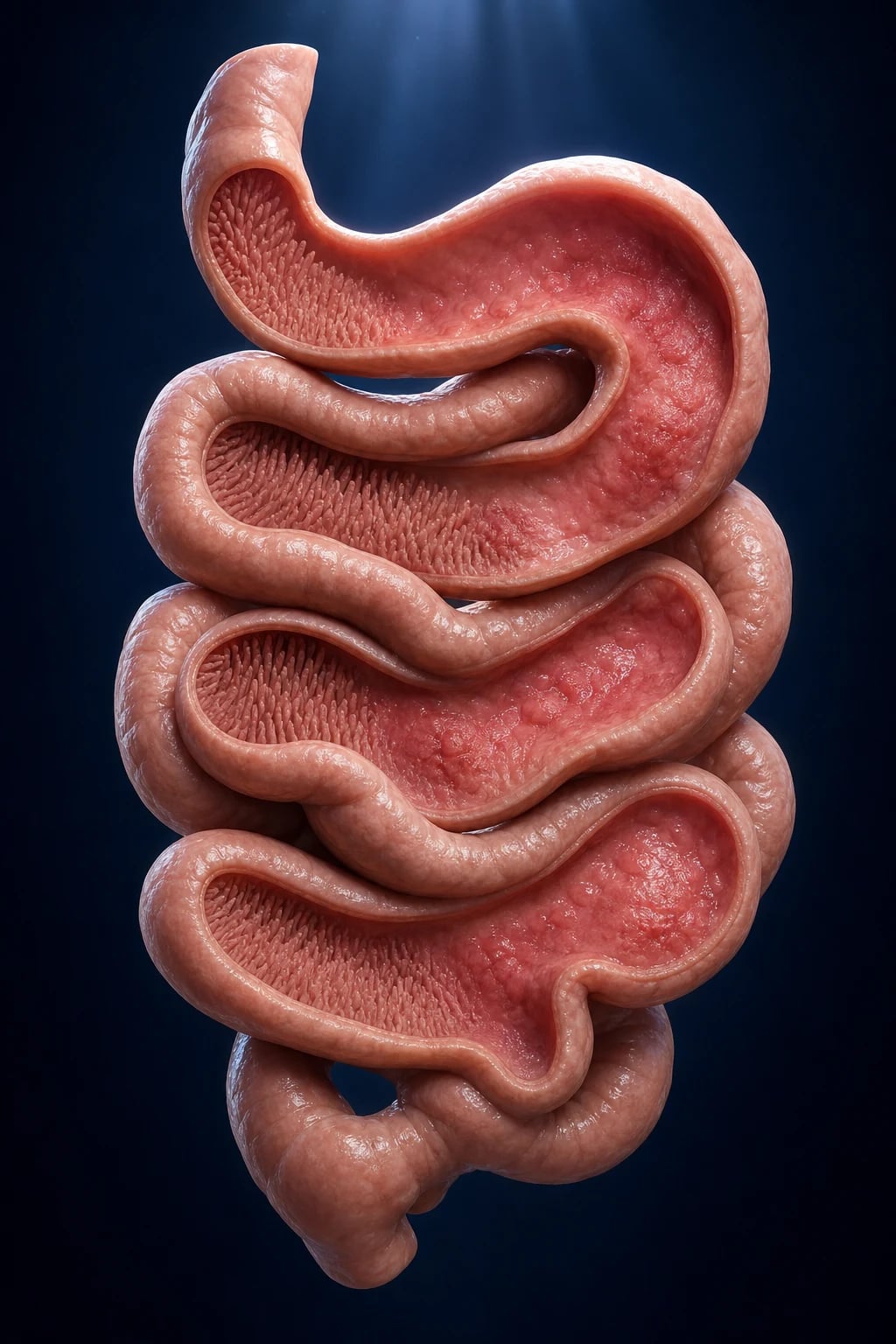
Marsh classification (modified Oberhuber) of small-intestinal histology — the histological grade reported on every duodenal biopsy:[1][2]
| Marsh grade | Intraepithelial lymphocytes (IELs) | Crypts | Villi |
|---|---|---|---|
| 0 (pre-infiltrative) | Normal (under 30 per 100 enterocytes) | Normal | Normal |
| 1 (infiltrative) | Raised (over 30 per 100 enterocytes) | Normal | Normal |
| 2 (infiltrative-hyperplastic) | Raised | Hypertrophic/hyperplastic | Normal |
| 3a (flat-destructive) | Raised | Hyperplastic | Mild atrophy |
| 3b | Raised | Hyperplastic | Marked atrophy |
| 3c | Raised | Hyperplastic | Total/subtotal atrophy (flat mucosa) |
| 4 (atrophic-hypoplastic) | — | Atrophic | Total atrophy — irreversible (rare) |
Epidemiology & Risk Factors
Coeliac disease has a true global prevalence of about 1 per cent (the Singh 2018 meta-analysis of 432 studies: seroprevalence 1.4 per cent, biopsy-confirmed prevalence 0.7 per cent), with substantial regional variation and significant under-diagnosis (the "coeliac iceberg" — for every diagnosed patient, several undiagnosed cases exist).[3]
Coeliac disease — the numbers that examiners ask
Genetic susceptibility is necessary but not sufficient: about 95 per cent of patients carry HLA-DQ2 (DQA105/DQB102) and most of the remainder carry HLA-DQ8 (DQA103/DQB10302). However, up to 30–40 per cent of the general population also carry DQ2/DQ8, so HLA is a rule-OUT test (NPV over 99 per cent), not a rule-in. Concordance is about 75 per cent in monozygotic twins, 30 per cent in HLA-identical siblings, and 10 per cent in first-degree relatives — confirming both genetic and environmental contributions.[1][2]
Risk factors and trigger events: [1]
- Family history — 10 per cent in first-degree relatives; screen even if asymptomatic.
- Female sex — 2-3:1 (often diagnosed during investigation of infertility, anaemia, or osteoporosis).
- Associated autoimmune conditions (screen bidirectionally): type 1 diabetes mellitus (coeliac in 3–8 per cent, screen at diagnosis and periodically), autoimmune thyroid disease (Hashimoto/Graves), autoimmune hepatitis and primary biliary cholangitis, Addison's disease, autoimmune atrophic gastritis, IgA deficiency (5–10x more common in coeliac).
- Chromosomal syndromes: Down syndrome (trisomy 21), Turner syndrome, Williams syndrome — screen routinely.
- Environmental triggers thought to unmask disease in susceptible individuals: gastrointestinal infection (rotavirus in childhood, Campylobacter, reovirus), early infant gluten introduction timing and quantity, microbiome changes, pregnancy, surgery, and stress — none fully deterministic. [1]
Pathophysiology
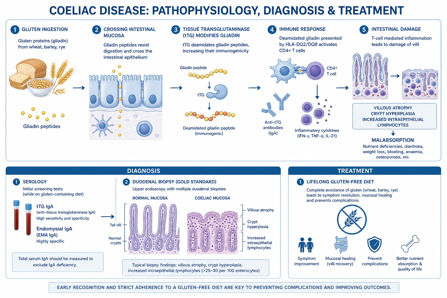
Coeliac disease is the best-characterised immune-mediated mucosal disease in humans — the molecular mechanism is examinable in detail because it explains the serology, the histology, and the targets of therapy.[1][2]
Step 1 — Gluten survives digestion. The prolamin fraction of gluten (gliadin in wheat, hordein in barley, secalin in rye) is rich in proline and glutamine, which makes it resistant to degradation by gastric, pancreatic and brush-border proteases. Large immunoreactive 33-mer peptide (alpha-2 gliadin residues 57–89) and similar fragments reach the small-intestinal lamina propria intact. [1]
Step 2 — tTG deamidation is the pivotal event. In the lamina propria, tissue transglutaminase-2 (tTG2) converts specific glutamine residues in gliadin to glutamate (deamidation). This introduces a negative charge that allows gliadin peptides to bind with high affinity into the antigen-binding groove of HLA-DQ2 or HLA-DQ8 on the surface of antigen-presenting cells. tTG2 also cross-links gliadin and is the autoantigen of the disease — the anti-tTG and anti-endomysial antibodies are diagnostic because they recognise tTG2 itself. [1]
Step 3 — Adaptive immune response (CD4+ Th1). Deamidated gliadin is presented to gliadin-specific CD4+ T-helper cells, which differentiate into a Th1 phenotype secreting interferon-gamma (IFN-gamma). IFN-gamma and other cytokines (IL-21, TNF-alpha) drive epithelial damage, crypt hyperplasia, and lamina propria inflammation. The same interaction activates gliadin-specific B cells that, with T-cell help, produce anti-tTG and anti-endomysial IgA antibodies — these antibodies are pathognomonic diagnostic markers but are NOT the primary mediators of tissue injury (which is T-cell driven). [1]
Step 4 — Innate immune activation (IL-15). Gliadin directly upregulates interleukin-15 in enterocytes and lamina propria macrophages. IL-15 drives the expansion and activation of intraepithelial lymphocytes (IELs), including cytotoxic T cells that express NKG2D and kill stressed enterocytes. Persistent IL-15 signalling is the substrate of refractory coeliac disease type 2 (RCD2), in which IELs acquire clonal, malignant features and progress to enteropathy-associated T-cell lymphoma (EATL). [1]
Step 5 — Histological injury → functional malabsorption. The combined immune attack produces the three histological pillars — villous atrophy, crypt hyperplasia, and intraepithelial lymphocytosis — which reduce the mucosal surface area and disaccharidase content (especially lactase) of the proximal small bowel. This produces malabsorption of iron, folate, calcium, fat-soluble vitamins (A, D, E, K), and, when extensive, of macronutrients → anaemia, osteoporosis/osteomalacia, fat-soluble-vitamin deficiency, diarrhoea, weight loss, and, in children, failure to thrive. [1]
Why gluten-free diet works (and why antibodies fall): removal of the antigen stops deamidation, T-cell activation, and B-cell stimulation; serology falls within months (anti-tTG first, EMA later) and mucosa recovers over 1–2 years (slower in adults, occasionally incomplete). Conversely, gluten re-challenge re-ignites the cascade — which is why diagnosis must be completed on a full-gluten diet.[6]
Clinical Presentation
The classical paediatric "failure to thrive after weaning" and the adult "diarrhoea, weight loss and bloating" are now the minority of presentations — most adult diagnoses today are made through atypical, extraintestinal or screening pathways.[2]
Classical (malabsorptive) presentation: [1]
- Chronic or recurrent diarrhoea, pale, bulky, offensive steatorrhoea
- Abdominal distension/bloating, discomfort, excessive flatus
- Weight loss, muscle wasting, failure to thrive (children), delayed puberty
- Anorexia, nausea, vomiting; rarely constipation (children)
- Lactose intolerance (secondary disaccharidase deficiency — resolves on gluten-free diet)
- Onset after gluten introduction into the infant diet or any time in adult life (3rd–5th decade peak) [1]
Extraintestinal / atypical presentation (often the SOLE presenting feature): [1]
| System | Manifestation | Mechanism |
|---|---|---|
| Haematological | Iron-deficiency anaemia (refractory to oral iron), folate deficiency; B12 less common (ileum spared) | Duodenal iron/folate malabsorption |
| Metabolic/bone | Osteoporosis, osteomalacia, secondary hyperparathyroidism, hypocalcaemia, tetany | Calcium + vitamin D malabsorption |
| Cutaneous | Dermatitis herpetiformis — intensely itchy, symmetric, grouped vesicles on extensor surfaces (elbows, knees, buttocks, scalp); granular IgA on skin biopsy | Epidermal tTG autoimmunity |
| Hepatic | Unexplained transaminitis (raised ALT/AST); autoimmune hepatitis overlap | Immune hepatic involvement |
| Reproductive | Delayed menarche, amenorrhoea, infertility, recurrent miscarriage, low-birth-weight infants | Malnutrition + immune |
| Dental | Dental enamel defects (permanent dentition), recurrent aphthous stomatitis | Malnutrition + immune |
| Neurological | Gluten ataxia (cerebellar, anti-tTG/IgG immune), peripheral neuropathy, epilepsy with bilateral occipital calcifications, headache | Immune/anti-neuronal |
| Musculoskeletal | Arthralgia, short stature (children), myopathy (vitamin D) | — |
| General | Fatigue, irritability, depression, short stature, delayed puberty | Multi-factorial |
Dermatitis herpetiformis (DH) deserves special mention: it is the cutaneous manifestation of coeliac disease, present in about 10–25 per cent of coeliac patients, and virtually all DH patients have coeliac-type enteropathy (often mild/Marsh 1–2). Diagnose with skin biopsy showing granular IgA deposition at the dermal papillae (do NOT biopsy the lesion itself — sample perilesional skin). Treat with gluten-free diet (resolves over months) plus dapsone for symptom control while diet takes effect.[1]
Atypical presentation in specific populations: [1]
- Elderly — may present with osteoporotic fracture, iron-deficiency anaemia, weight loss, or fatigue alone; no GI symptoms; high index of suspicion needed.
- Type 1 diabetes — often asymptomatic, found on screening; unexplained hypoglycaemia or erratic glycaemic control may be the only clue.
- Pregnancy — recurrent miscarriage, intrauterine growth restriction, anaemia; screen women with these plus coeliac risk factors.
- Children — failure to thrive, delayed puberty, short stature, distended abdomen, irritability, dental enamel defects; often missed as "picky eater."
- Selective IgA deficiency — classical coeliac symptoms may be masked; serology with IgA-based tests will be falsely negative (use anti-DGP IgG). [1]
Differential Diagnosis
The finding of chronic diarrhoea with malabsorption or non-specific villous atrophy on duodenal biopsy has a wide differential. The diagnosis of coeliac disease requires the combination of (a) consistent serology, (b) characteristic histology, and (c) response to gluten withdrawal — none alone is sufficient.[2][8]
Irritable bowel syndrome (IBS)
- No weight loss, no anaemia, no steatorrhoea, normal serology, normal biopsy
- Pain dominant, relieved by defecation; coeliac is excluded by tTG + total IgA — never diagnose IBS without screening for coeliac first
Inflammatory bowel disease (esp. Crohn's)
- Terminal ileum/colon involvement, skip lesions, granulomas, strictures, fistulae, perianal disease
- Raised CRP/ferritin, faecal calprotectin raised; capsule/colonoscopy diagnostic
Tropical sprue
- Endemic tropics residents; malabsorption + megaloblastic anaemia (B12 + folate both low); responds to tetracycline + folic acid
- Biopsy: PARTIAL villous atrophy but LESS crypt hyperplasia and LESS IEL — see Sharma 2019 head-to-head comparison
Small intestinal bacterial overgrowth (SIBO)
- Predisposing dysmotility (diabetes, scleroderma), prior surgery, diverticulosis
- Diagnosis: glucose/lactulose hydrogen breath test or duodenal aspirate culture; responds to rifaximin/ciprofloxacin
Olmesartan (and other ARB) sprue-like enteropathy
- Villous atrophy + severe diarrhoea on olmesartan; SEROLOGY NEGATIVE; resolves on drug withdrawal
- Always take a drug history in any seronegative enteropathy
Common variable immunodeficiency (CVID)
- Low IgG/IgA/IgM + recurrent sinopulmonary infections; villous atrophy but NO coeliac serology (panhypogammaglobulinaemia)
- Biopsy may show nodular lymphoid hyperplasia; treat with IV immunoglobulin, not gluten-free diet
Autoimmune enteropathy
- Anti-enterocyte/anti-goblet-cell antibodies; severe secretory diarrhoea; responds to steroids/immunosuppression
- Adults rare, more in infants (IPEX-like)
Other causes of villous atrophy
- Giardiasis, HIV enteropathy, Whipple disease, eosinophilic gastroenteritis, radiation/cytotoxic enteritis, Zollinger-Ellison, Crohn's, amyloidosis, lymphoma (EATL)
- All excluded by serology, special stains (PAS, Ziehl-Neelsen, CD3/CD8 flow), and clinical context
The non-responsive coeliac patient (persistent symptoms or histology despite a strict gluten-free diet for over 12 months) has its own differential: ongoing gluten exposure (most common — dietitian review), refractory coeliac disease (RCD1/RCD2), SIBO, microscopic colitis, IBS overlap, pancreatic insufficiency, and lymphoma (EATL).[6]
Clinical & Bedside Assessment
Bedside assessment in suspected coeliac disease targets nutritional status, evidence of malabsorption, and stigmata of associated autoimmune disease. [1]
General examination: [1]
- BMI and growth centile — wasting, failure to thrive (children), short stature, delayed puberty.
- Hydration and signs of electrolyte disturbance (muscle cramps, tetany from hypocalcaemia).
- Pallor (iron/folate anaemia), koilonychia, angular stomatitis, glossitis (atrophic; beefy red).
- Bruising/petechiae (vitamin K deficiency; thrombocytopenia from hyposplenism).
- Follicular hyperkeratosis, night blindness (vitamin A), proximal myopathy (vitamin D). [1]
Abdominal examination: [1]
- Distension (gas, fluid, dilated bowel loops), mild diffuse tenderness; ascites only if profound hypoproteinaemia.
- Rarely a palpable mass (think lymphoma, complications). [1]
Dermatological: inspect extensor surfaces — elbows, knees, buttocks, scalp, sacrum, face for the intensely itchy, symmetric, grouped vesicles/crusts of dermatitis herpetiformis. Examine the mouth for aphthous ulcers and dental enamel defects. [1]
Systemic and autoimmune screen: thyroid (goitre), skin (vitiligo), joint (arthralgia), stigmata of Down syndrome / Turner syndrome, and signs of type 1 diabetes complications if known diabetic. Look for neurological deficits (ataxia, peripheral neuropathy). [1]
Bedside observations (especially at diagnosis with severe malabsorption): postural blood pressure (volume depletion), heart rate, temperature (intercurrent infection — encapsulated organisms if hyposplenic), and capillary glucose in known diabetics. [1]
Investigations
The diagnosis is serological + histological and must be made while the patient is on a gluten-containing diet (a gluten-free diet sero-negatives the work-up and may normalise the biopsy).[1][6]
Tier 1 — Serology (always with a total IgA): [1]
| Test | Sensitivity | Specificity | Use |
|---|---|---|---|
| Anti-tissue transglutaminase IgA (anti-tTG IgA) | ~90–95% | ~95% | First-line screening — most sensitive |
| Anti-endomysial IgA (EMA) | ~90% | ~97–99% | Most specific — confirmatory; expensive, operator-dependent (monkey oesophagus/Hep-2) |
| Total serum IgA | — | — | Always — exclude selective IgA deficiency (use IgG-based tests if low) |
| Anti-deamidated gliadin peptide IgG (anti-DGP IgG) | ~80–90% | ~95% | Test of choice when IgA deficient (and in children under 2 y) |
| HLA-DQ2/DQ8 typing | — | (NPV over 99%) | Rule-OUT — for discordant serology/biopsy, screening high-risk first-degree relatives, "is this coeliac?" question |
Tier 2 — Confirmatory histology (the gold standard): [1]
- Oesophago-gastro-duodenoscopy (OGD) with multiple duodenal biopsies. Take at least 6 biopsies — 4 from the distal duodenum AND 2 from the duodenal bulb (where disease may be patchy and isolated). Bulb biopsies reduce the false-negative rate substantially.
- Histology (Marsh-Oberhuber): the diagnostic flat lesion is Marsh 3 — villous atrophy with crypt hyperplasia and IEL over 30 per 100 enterocytes. Marsh 1 (IEL alone) is non-specific (H. pylori, NSAIDs, infections, IBD) — never diagnostic alone.
- Orientation on the biopsy is critical (well-oriented villi needed to grade). [1]
Tier 3 — Baseline bloods at diagnosis (deficiency screen): [1]
- FBC + film — iron-deficiency anaemia (microcytic), folate-deficient megaloblastic anaemia, ** Howell-Jolly bodies / target cells / thrombocytopenia / leucopenia (functional hyposplenism)**.
- Ferritin, iron studies, vitamin B12, serum and red-cell folate — all may be low.
- Calcium, phosphate, magnesium, albumin, PTH, 25-hydroxy-vitamin D — hypocalcaemia, osteomalacia, secondary hyperparathyroidism.
- LFTs — unexplained transaminitis (usually resolves on gluten-free diet).
- Coagulation (INR/PT) — vitamin K deficiency.
- TSH and free T4 — screen for associated autoimmune thyroid disease.
- Tissue transglutaminase IgA titre (quantitative baseline for monitoring). [1]
Tier 4 — Complication and comorbidity screen: [1]
- DEXA scan at diagnosis in all adults (osteoporosis/osteopenia risk).
- Capsule endoscopy — for non-responsive/refractory disease, to look for ulcerative jejunoileitis or lymphoma in the distal small bowel beyond reach of OGD.
- CT/MR enterography — for refractory disease, suspected lymphoma, obstruction.
- HLA-DQ2/DQ8 typing — when diagnosis is uncertain (seronegative coeliac, discordant histology, patient already on a gluten-free diet, screening of at-risk relatives).
- Skin biopsy (perilesional) with direct immunofluorescence — for suspected dermatitis herpetiformis: granular IgA at the dermal papillae is pathognomonic. [1]
The no-biopsy paediatric pathway (ESPGHAN 2020 / BSPGHAN): in a symptomatic child with anti-tTG IgA greater than 10x the upper limit of normal PLUS a positive EMA on a separate blood sample PLUS HLA-DQ2/DQ8 positivity, the diagnosis can be made without duodenal biopsy — provided a paediatric gastroenterologist reviews the case. Asymptomatic screened children still need biopsy.[6]
Investigation pitfalls (examiner favourites): [1]
- Falsely negative serology: gluten-free/gluten-low diet (most common), selective IgA deficiency (5–10x over-represented; ~3 per cent of coeliac patients — hence the mandatory total IgA), very young infants (under 2 years — add anti-DGP IgG), immunosuppression, late-stage/refractory disease.
- Falsely positive serology: other autoimmune liver disease, inflammatory bowel disease, heart failure, infections — anti-tTG IgA less specific in these; EMA remains highly specific.
- Villous atrophy not coeliac — always consider olmesartan, tropical sprue, SIBO, CVID, giardiasis, Crohn's, autoimmune enteropathy, lymphoma. A confident diagnosis needs serology + histology + clinical context + response to gluten withdrawal.
- Gluten challenge — if a patient has already started a gluten-free diet, serology and histology may normalise; reinstate gluten (equivalent to at least 3–4 slices of wheat bread per day) for 6–8 weeks before re-testing. In reluctant patients, HLA typing + a single food challenge may help. [1]
Management — Resuscitation

Coeliac disease is chronic — most patients are managed as outpatients. The "resuscitation" framing applies to the severely malabsorbing patient at first presentation: severe dehydration and electrolyte disturbance, profound anaemia (occasionally transfusion-requiring), hypocalcaemia with tetany/seizures, and rarely coeliac crisis (severe acute malabsorption with profuse diarrhoea, dehydration, electrolyte disturbance, metabolic acidosis, weight loss).[1]
Acute supportive measures in severe presentation: [1]
- ABCDE; secure IV access; fluid resuscitation with balanced crystalloid (Hartmann's or Plasma-Lyte) for hypovolaemia; correct hypokalaemia, hypomagnesaemia, hypocalcaemia with IV replacement (monitor and recheck).
- Transfusion of packed red cells for symptomatic severe anaemia (e.g. ferrous sulphate is the definitive therapy, not transfusion, in stable patients).
- Parenteral vitamins for severe deficiency — intravenous or intramuscular vitamin D for osteomalacic tetany, intravenous vitamin K for coagulopathy, intramuscular hydroxocobalamin if B12-deficient.
- Coeliac crisis may require IV corticosteroids (e.g. hydrocortisone 100 mg IV every 6 hours or prednisolone 40–60 mg daily) to dampen the inflammatory response while diet and nutritional support are established — a specialist decision.
- Identify and treat intercurrent infection (especially encapsulated organisms — pneumococcus, Haemophilus influenzae type b, meningococcus — in functional hyposplenism).
- Refer to a gastroenterologist and specialist dietitian for definitive diagnosis and management. [1]
Management — Definitive & Stepwise
The definitive treatment is a strict, lifelong gluten-free diet (GFD), supported by dietitian input, correction of nutritional deficiencies, vaccination, structured follow-up, and patient-society support.[1][2]
1. Lifelong gluten-free diet — the cornerstone
- Exclude completely: wheat (including durum, semolina, spelt, kamut, couscous), barley, rye, and triticale. All sources — bread, pasta, biscuits, cakes, sauces thickened with flour, beer, lager, ale.
- Oats are tolerated by most if uncontaminated by wheat ("pure, uncontaminated oats"); introduce cautiously and monitor serology. Codex Alimentarius and most regulators define "gluten-free" as under 20 parts per million (ppm) of gluten.
- Hidden gluten: processed foods, soups, sausages, soy sauce, stock cubes, communion wafers, lipsticks, some medications — teach label-reading.
- Cross-contamination: separate toasters, utensils, cooking oil; a gluten-free household member needs their own workspace.
- Dietitian referral at diagnosis and at intervals; membership of Coeliac UK / Celiac Disease Foundation / local coeliac society dramatically improves adherence.
- Adherence monitoring: repeat anti-tTG IgA at 3–6 months (should fall) and 12 months (should normalise); persistent elevation suggests ongoing gluten exposure. [1]
2. Correct nutritional deficiencies — agent, dose, route
- Iron deficiency: oral ferrous sulphate 200 mg once to three times daily (or ferrous fumarate 300 mg, ferrous glycine sulphate) until ferritin replete; avoid IV iron unless malabsorption severe or oral not tolerated (now widely used — ferric carboxymaltose, ferric derisomaltose). Recheck ferritin at 3 months.
- Folate deficiency: oral folic acid 5 mg once daily for 4 months (longer if ongoing malabsorption).
- Vitamin B12 deficiency: hydroxocobalamin 1000 micrograms (1 mg) intramuscularly on the standard regimen (alternate days for 2 weeks, then every 3 months if permanent). B12 deficiency is uncommon in coeliac (ileum usually spared) but screen and treat.
- Calcium and vitamin D: oral calcium 1000–1200 mg daily (elemental) plus colecalciferol (vitamin D3) 800–1000 IU daily (or 20 000 IU weekly); alfacalcidol or calcitriol if severe malabsorption/osteomalacia. Target 25-OH-vitamin D greater than 75 nmol/L.
- Multivitamin preparation in newly diagnosed, malnourished patients until mucosal recovery. [1]
3. Preventive care
- Vaccination for functional hyposplenism: pneumococcal (PCV then PPSV23 schedule as per local guidance), annual influenza, Haemophilus influenzae type b, meningococcal (ACWY and B); consider a booster every 5 years for pneumococcal. Counsel the patient to seek urgent care for fever (capsulated-organism sepsis risk).
- DEXA scan at diagnosis; treat osteoporosis with bisphosphonate (e.g. alendronic acid 70 mg once weekly with calcium + vitamin D) per standard osteoporosis pathway.
- Counsel on the hereditary risk (10 per cent in first-degree relatives — they should be screened). [1]
4. Structured annual review
- Annual review with gastroenterologist, GP, or specialist nurse: symptoms, growth (children), BMI, adherence.
- Annual bloods: FBC, ferritin, folate, vitamin B12, calcium, vitamin D, LFTs, TSH, anti-tTG IgA titre (the latter as an adherence marker — should be normal if strict GFD).
- Repeat DEXA at intervals as guided by baseline (typically every 2–5 years).
- Repeat biopsy is NOT routine if responding well; re-biopsy if non-responsive, refractory, or serology/histology discordant. [1]
5. Dermatitis herpetiformis — specific therapy
- Gluten-free diet is the definitive treatment — skin disease resolves over 6–24 months (mucosa heals first).
- Dapsone for symptom control while diet takes effect: start at 50 mg once daily, titrate by 50 mg every 1–2 weeks to itch control (usual 50–150 mg daily). Check G6PD level BEFORE starting (dapsone causes haemolysis/methaemoglobinaemia in deficient patients), monitor FBC and LFTs. Rapid itch relief (within 24–48 hours) is diagnostic.
- Alternatives if dapsone not tolerated: sulfasalazine, colchicine, topical steroids. [1]
6. Refractory coeliac disease — specialist
- Definition: persistent/recurrent malabsorptive symptoms and Marsh 3 villous atrophy despite a strict gluten-free diet for over 12 months, after excluding ongoing gluten exposure (dietitian-led assessment) and other causes of villous atrophy.
- Type 1 RCD (polyclonal IELs): oral budesonide 9 mg once daily (enteric-coated, pH-modified release) or prednisolone 20–40 mg daily tapering; good prognosis.
- Type 2 RCD (clonal/aberrant IELs, surface CD8-negative, monoclonal TCR rearrangement): pre-malignant — high risk of progression to enteropathy-associated T-cell lymphoma (EATL); refer to specialist centre; tioguanine (thioguanine), cladribine, autologous stem-cell transplant considered; poor prognosis (5-year survival 40–60 per cent). [1]
Specific Subtypes & Scenarios
- Dermatitis herpetiformis (DH) — intensely itchy, symmetric, grouped vesicles on extensor surfaces; granular IgA at dermal papillae on perilesional skin biopsy; virtually all DH patients have coeliac enteropathy (often mild). Treat with gluten-free diet plus dapsone 50–100 mg (check G6PD first).[1]
- Seronegative coeliac disease — anti-tTG/EMA negative but Marsh 3 histology, characteristic HLA, and clinical/histological response to gluten-free diet; diagnosis of exclusion requiring rigorous exclusion of other causes of villous atrophy (especially olmesartan and tropical sprue).[6]
- Potential coeliac disease — positive serology with normal (Marsh 0) or only Marsh 1 biopsy; follow-up and re-biopsy; some authorities offer a gluten-free diet if symptomatic or anti-tTG rising.[6]
- Refractory coeliac disease — type 1 vs type 2 (see Management); RCD2 carries a high risk of EATL. Identified by flow cytometry of biopsy IELs (loss of surface CD8) and TCR clonality studies.
- Paediatric coeliac — peak presentation after gluten introduction (4–24 months) or adolescence; failure to thrive, distended abdomen, irritability, delayed puberty, dental enamel defects. ESPGHAN no-biopsy pathway if symptomatic child with anti-tTG IgA greater than 10x ULN + EMA positive on separate sample + HLA-DQ2/DQ8 positive.[6]
- Pregnancy and coeliac — untreated coeliac is associated with infertility, recurrent miscarriage, intrauterine growth restriction, preterm delivery, low-birth-weight infants, anaemia; screen women with recurrent pregnancy loss or unexplained anaemia. Strict gluten-free diet in pregnancy restores fertility and outcomes to normal.
- Coeliac crisis — rare, severe acute malabsorption with profuse diarrhoea, dehydration, electrolyte disturbance, hypoproteinaemia, hypocalcaemia, metabolic acidosis; may require IV fluids, parenteral nutrition, and IV corticosteroids.
- Non-responsive coeliac disease (persistent symptoms despite a gluten-free diet for over 6–12 months) — most commonly ongoing gluten exposure (intentional or inadvertent — re-educate, dietitian review); then SIBO, microscopic colitis, IBS overlap, pancreatic insufficiency, lactose intolerance, refractory disease, lymphoma. Systematic re-evaluation with serology, dietitian review, repeat biopsy, capsule endoscopy.
Complications & Pitfalls
Nutritional and metabolic: [1]
- Iron-deficiency anaemia (the commonest haematological complication), folate-deficient anaemia; rarely B12.
- Metabolic bone disease: osteoporosis and osteomalacia — fracture risk; DEXA at diagnosis.
- Hypocalcaemia, secondary hyperparathyroidism, vitamin K-deficient coagulopathy, fat-soluble-vitamin deficiencies. [1]
Immune and functional: [1]
- Functional hyposplenism — Howell-Jolly bodies, thrombocytopenia, leucopenia; encapsulated-organism sepsis risk (pneumococcus, Hib, meningococcus) — vaccinate.
- Reproductive — delayed puberty, infertility, recurrent miscarriage, low-birth-weight infants. [1]
Malignant (the feared complications): [1]
- Enteropathy-associated T-cell lymphoma (EATL) — the classic coeliac lymphoma; aggressive; arises from RCD2; presents as relapse of malabsorption, abdominal pain, fever, weight loss, perforation, obstruction; diagnosed by capsule/CT enterography and biopsy; poor prognosis.[2]
- Small-bowel adenocarcinoma — risk markedly elevated in coeliac; presents with obstruction, bleeding, weight loss; treat surgically.[5]
- Increased risk of oesophageal and pharyngeal squamous carcinoma, and non-Hodgkin lymphoma of any site.
- A strict gluten-free diet substantially reduces malignancy risk toward baseline (especially after 5 years of adherence).[2]
Other: [1]
- Refractory coeliac disease (RCD1/RCD2) — see above; RCD2 pre-malignant.
- Ulcerative jejunoileitis — benign-appearing ulcers that may be a precursor of EATL; presents with occult bleeding, perforation, obstruction.
- Hyposplenic sepsis — encapsulated organisms; rapid, fulminant. [1]
Classic pitfalls: [1]
- Diagnosing IBS without screening for coeliac — coeliac is present in 3–5 per cent of "IBS" patients; screen first.
- Starting a gluten-free diet before serology + biopsy — the diagnostic work-up is destroyed; gluten challenge is unpleasant and prolonged.
- Forgetting the total IgA — missing selective IgA deficiency gives falsely negative anti-tTG/EMA.
- Treating only the diet, not the patient — deficiencies, hyposplenism vaccination, DEXA, and annual review are mandatory.
- Labelling any villous atrophy as coeliac — seronegative atrophy has a wide differential (olmesartan, tropical sprue, CVID, lymphoma, Crohn's).[7]
- Missing non-responsive/refractory disease or EATL — any relapse of malabsorption on a strict diet needs urgent re-evaluation.
Prognosis & Disposition
With a strict, lifelong gluten-free diet, prognosis is excellent: symptoms improve within weeks to months, anti-tTG IgA titre falls within 3–6 months and normalises by 12–24 months, and the mucosa recovers over 1–2 years (faster in children, slower in adults — sometimes incomplete). Mortality is slightly but significantly increased overall (Lebwohl JAMA 2020: HR 1.21 over a median of 12 years), driven by cardiovascular disease, malignancy (EATL, small-bowel adenocarcinoma), and respiratory disease; strict adherence reduces this risk substantially.[4]
Patients are managed as outpatients under shared care between primary care, gastroenterology, and a dietitian. Discharge to GP-led annual review is reasonable once stable, adherent, and serologically in remission, with gastroenterology re-referral for non-response, refractory disease, or complication suspicion. [1]
Special Populations
- Children — failure to thrive, short stature, delayed puberty, irritability, distended abdomen, dental enamel defects. Use ESPGHAN 2020 no-biopsy pathway in symptomatic children when criteria met. Strict gluten-free diet is essential for normal growth and puberty; families need dietitian and Coeliac Society support.[6]
- Type 1 diabetes mellitus — coeliac in 3–8 per cent; screen at diagnosis and then every 1–2 years (or with symptoms/new glycaemic instability). Untreated coeliac worsens glycaemic control, growth, and bone density.
- Autoimmune thyroid disease / autoimmune hepatitis / Addison's — screen bidirectionally.
- Down syndrome, Turner syndrome, Williams syndrome — screen at diagnosis.
- First-degree relatives — 10 per cent prevalence; screen with anti-tTG IgA + total IgA from age 3 (or with symptoms); HLA-DQ2/DQ8-negative result rules out coeliac for life.
- Selective IgA deficiency — ~3 per cent of coeliac; use anti-DGP IgG; do not rely on IgA-based serology.
- Pregnancy — untreated coeliac causes infertility, miscarriage, IUGR, preterm birth, anaemia; screen at-risk women, enforce strict gluten-free diet pre-conception and through pregnancy; ensure adequate folate, iron, calcium, vitamin D.
- Elderly — may present only with osteoporotic fracture, anaemia, or weight loss; low threshold to screen; dietary change may be harder — emphasise dietitian and Coeliac Society support.
- Thyroid disease overlap — coeliac patients have a higher risk of thyroid carcinoma (Ludvigsson 2013) and other autoimmune thyroid disease; monitor TSH.[9]
Evidence, Guidelines & Regional Differences
Landmark evidence and contemporary guidelines: [1]
- ACG 2013 (Rubio-Tapia et al.) — the foundational US guideline: definition, serology hierarchy (anti-tTG IgA first-line, EMA confirmatory, total IgA always, anti-DGP IgG if IgA-deficient), HLA for discordant cases, greater than or equal to 6 duodenal biopsies including bulb, lifelong gluten-free diet, vaccination for hyposplenism.[1]
- Lebwohl, Sanders, Green, Lancet 2018 — the definitive modern review covering epidemiology, pathophysiology, presentation, and management.[2]
- Singh 2018 meta-analysis — true global prevalence ~1 per cent (seroprevalence 1.4 per cent, biopsy-confirmed 0.7 per cent); under-diagnosis is universal.[3]
- Lebwohl JAMA 2020 — coeliac disease is associated with a small but real increase in all-cause mortality.[4]
- ESPGHAN 2020 (Husby et al.) — the paediatric diagnostic pathway, including the no-biopsy pathway (anti-tTG IgA greater than 10x ULN + EMA positive on separate sample + HLA-DQ2/DQ8 in a symptomatic child) and serology/histology monitoring strategy.[6]
- Sharma 2019 — clinical/endoscopic/histological head-to-head of coeliac vs tropical sprue in endemic regions.[8]
Exam Pearls
COELIAC — the high-yield mnemonic
COELIAC
Histological pillar (with villous atrophy + IELs) = Marsh 3
If pure/uncontaminated — wheat/barley/rye must go
Most SPECIFIC serology (~99%); anti-tTG IgA is most SENSITIVE
Definitive treatment — strict, forever
~3% of coeliac — ALWAYS check total IgA, then use anti-DGP IgG
May be the SOLE presentation — screen any unexplained IDA
EATL (T-cell lymphoma), small-bowel adenocarcinoma — reduced by strict GFD
- Anti-tTG IgA = most sensitive (~90–95%); EMA = most specific (~97–99%). Both IgA-based — always send a total IgA to exclude selective IgA deficiency.[1]
- HLA-DQ2 (95%) / DQ8 (most of the rest) is necessary but not sufficient — rule-OUT test only (NPV over 99%).[1]
- Marsh 3 (villous atrophy + crypt hyperplasia + IEL over 30 per 100 enterocytes) is the diagnostic flat lesion; Marsh 1 (IEL alone) is non-specific and never diagnostic alone.
- Take at least 6 duodenal biopsies including the bulb — patchy disease is missed otherwise.
- Never start a gluten-free diet before serology + biopsy — it destroys the diagnostic work-up.
- Dermatitis herpetiformis: itchy symmetric grouped vesicles on extensors (elbows, knees, buttocks); granular IgA at dermal papillae on perilesional skin biopsy; treat with gluten-free diet + dapsone (check G6PD first).
- Iron-deficiency anaemia may be the ONLY presentation.
- Functional hyposplenism — vaccinate against pneumococcus, Hib, meningococcus, influenza.
- Type 1 diabetes, autoimmune thyroid, Down/Turner syndrome, IgA deficiency, first-degree relatives — screen for coeliac.
- Refractory disease + new weight loss/fever/abdominal pain — think EATL or small-bowel adenocarcinoma — re-biopsy and image.[5]
- Seronegative villous atrophy — always exclude olmesartan (sprue-like enteropathy), tropical sprue, CVID, giardiasis, lymphoma.[7][8]
- Gluten-free threshold: under 20 ppm (Codex Alimentarius, FDA, UK/EU law).
- Codex "gluten-free" = under 20 ppm — "very low gluten" under 100 ppm is a separate claim.
- EATL is a T-cell lymphoma (not B-cell) arising from intraepithelial lymphocytes in RCD2.
- Annual monitoring bloods: FBC, ferritin, folate, B12, calcium, vitamin D, LFTs, TSH, anti-tTG IgA titre (adherence marker).
- Pregnancy + coeliac: untreated = infertility/miscarriage/IUGR; strict GFD restores outcomes to normal.
Exam application bank (NEET-PG / INICET)
One-line answer
Coeliac disease is a chronic, immune-mediated small-intestinal enteropathy triggered by gluten (prolamins — gliadin in wheat, hordein in barley, secalin in rye) in genetically predisposed (HLA-DQ2/DQ8) individuals. Presents across a spectrum from classical malabsorption (diarrhoea, weight loss, bloating, steatorrhoea) to atypical/extraintestinal disease (iron-deficiency anaemia, osteoporosis, aphthous ulcers, short stature, dermatitis herpetiformis, transaminitis) and silent/latent forms found on screening. Diagnosis requires serology while on a gluten-containing diet — anti-tissue transglutaminase IgA (anti-tTG IgA, most sensitive ~90–95%) and anti-endomysial IgA (EMA, most specific ~99%) — PLUS a total IgA to exclude selective IgA deficiency (5–10x more common in coeliac); in IgA deficiency use anti-deamidated gliadin peptide (DGP) IgG. Diagnosis is confirmed by duodenal biopsies (grea
Worked stems (answer without another resource)
Stem 1 — Classic presentation. Map symptoms to mechanism; name the first investigation and first treatment step with dose/route if drug therapy is standard. [1]
Stem 2 — Unstable / complicated. List red flags that force immediate resuscitation, theatre, ICU, antidote, or reperfusion — and what you do in the first 15 minutes. [1]
Stem 3 — Atypical group. Elderly, pregnancy, child, or immunocompromised: how presentation and thresholds change. [1]
Stem 4 — Differential trap. Name the three closest mimics and one discriminator for each. [1]
Stem 5 — Disposition. Who goes home with safety-netting, who is admitted, who needs HDU/ICU/theatre, and what follow-up is mandatory. [1]
Rapid viva checklist
- Definition + classification
- Pathophysiology chain
- Bedside signs / criteria
- Score with exact components (if any)
- Emergency bundle
- Definitive therapy with doses
- Complications of disease and of treatment
- Special populations
- Guideline/trial name if classic
- Three exam traps
Coverage self-check
If you cannot answer any stem above from this page alone, re-read the matching section — the page is intended to be self-sufficient for final-prof and NEET-PG/INICET questions on Coeliac Disease.
References
- [1]Rubio-Tapia A, Hill ID, Kelly CP, Calderwood AH, Murray JA. ACG clinical guidelines: diagnosis and management of celiac disease Am J Gastroenterol, 2013.PMID 23609613
- [2]Lebwohl B, Sanders DS, Green PHR. Coeliac disease Lancet, 2018.PMID 28760445
- [3]Singh P, Arora A, Strand TA, et al. Global Prevalence of Celiac Disease: Systematic Review and Meta-analysis Clin Gastroenterol Hepatol, 2018.PMID 29551598
- [4]Lebwohl B, Green PHR, Söderling J, Roelstraete B, Ludvigsson JF. Association Between Celiac Disease and Mortality Risk in a Swedish Population JAMA, 2020.PMID 32259229
- [5]Emilsson L, Wijesinghe CR, Lee AR, et al. Risk of Small Bowel Adenocarcinoma, Adenomas, and Carcinoids in a Nationwide Cohort of Individuals With Celiac Disease Gastroenterology, 2020.PMID 32679218
- [6]Husby S, Koletzko S, Korponay-Szabó IR, Kurppa K, Mearin ML, Ribes-Koninckx C. European Society Paediatric Gastroenterology, Hepatology and Nutrition Guidelines for Diagnosing Coeliac Disease 2020 J Pediatr Gastroenterol Nutr, 2020.PMID 31568151
- [7]Lebwohl B, Sanders DS, Green PHR. Editorial: sprue-like enteropathy due to olmesartan and other angiotensin receptor blockers--the plot thickens Aliment Pharmacol Ther, 2014.PMID 25303379
- [8]Sharma P, Balgir P, Murari S, et al. Clinical, endoscopic, and histological differentiation between celiac disease and tropical sprue: A systematic review J Gastroenterol Hepatol, 2019.PMID 30069926
- [9]Ludvigsson JF, West J, Ekbom A, et al. Risk of thyroid cancer in a nationwide cohort of patients with biopsy-verified celiac disease Thyroid, 2013.PMID 23281890