Gastroenterology · General Medicine
Colorectal Cancer
Also known as Colorectal cancer · CRC · Bowel cancer · Colon cancer · Rectal cancer
Colorectal cancer (CRC) is an adenocarcinoma of the colonic or rectal mucosa that evolves from a benign adenomatous polyp through the stepwise accumulation of genetic hits — the adenoma-carcinoma sequence (APC, then KRAS, then TP53) over 10 to 15 years. It is the third most commonly diagnosed cancer worldwide and the second leading cause of cancer death. About 85 percent are sporadic; the remainder arise in hereditary syndromes (Lynch syndrome, familial adenomatous polyposis) or long-standing inflammatory bowel disease. Presentation differs by site — left-sided causes obstruction, a change in bowel habit and bright-red bleeding; right-sided presents with iron-deficiency anaemia and occult blood loss. Diagnosis is by colonoscopy and biopsy; staging with CT chest/abdomen/pelvis and MRI rectum; monitoring with CEA. Treatment is surgical (segmental colectomy with lymphadenectomy; total mesorectal excision for rectal cancer) for localised disease, adjuvant FOLFOX or CAPOX for 6 months for stage III, neoadjuvant radiotherapy (short course 25 Gy in 5 fractions, or long course 45 to 50.4 Gy with 5-FU or capecitabine) for T3 or N-plus rectal cancer, and FOLFOX or FOLFIRI plus bevacizumab or anti-EGFR therapy (cetuximab or panitumumab, RAS wild-type only) for metastatic disease. Screening with FIT or colonoscopy from age 45 to 50 exploits the long pre-malignant polyp phase to prevent cancer outright.
On this page & tools
Your progress
Saved locally on this device.
Practise this topic
Exam tags
Red flags
Overview & Definition
Colorectal cancer (CRC) is a malignant adenocarcinoma (over 95 percent of all colorectal malignancies) arising from the glandular epithelium of the colon or rectum. It begins as a benign adenomatous polyp and, through the stepwise accumulation of somatic mutations in key tumour-suppressor genes and oncogenes, progresses to dysplasia and then invasive carcinoma — the adenoma-carcinoma sequence that takes, on average, 10 to 15 years.[1][3] That long, silent window is the single most important biological fact about CRC: it is the reason screening, polypectomy and surveillance can prevent cancer outright, and it is why most early tumours are curable by surgery alone.[1]
CRC is the third most commonly diagnosed cancer in men and the second in women, and the second leading cause of cancer death worldwide. About 85 percent are sporadic (driven by age, diet and lifestyle), 10 percent are hereditary (dominated by Lynch syndrome and familial adenomatous polyposis), and 5 percent complicate long-standing inflammatory bowel disease.[1] The clinical skill in CRC is not the histological diagnosis — that is colonoscopy with biopsy — but recognising the red flags that demand urgent investigation (iron-deficiency anaemia, a change in bowel habit, rectal bleeding in an adult), accurate staging (CT for distant disease, MRI for the rectum), and the multidisciplinary sequencing of surgery, chemotherapy and radiotherapy by stage, site and molecular subtype.[1][2]
Classification
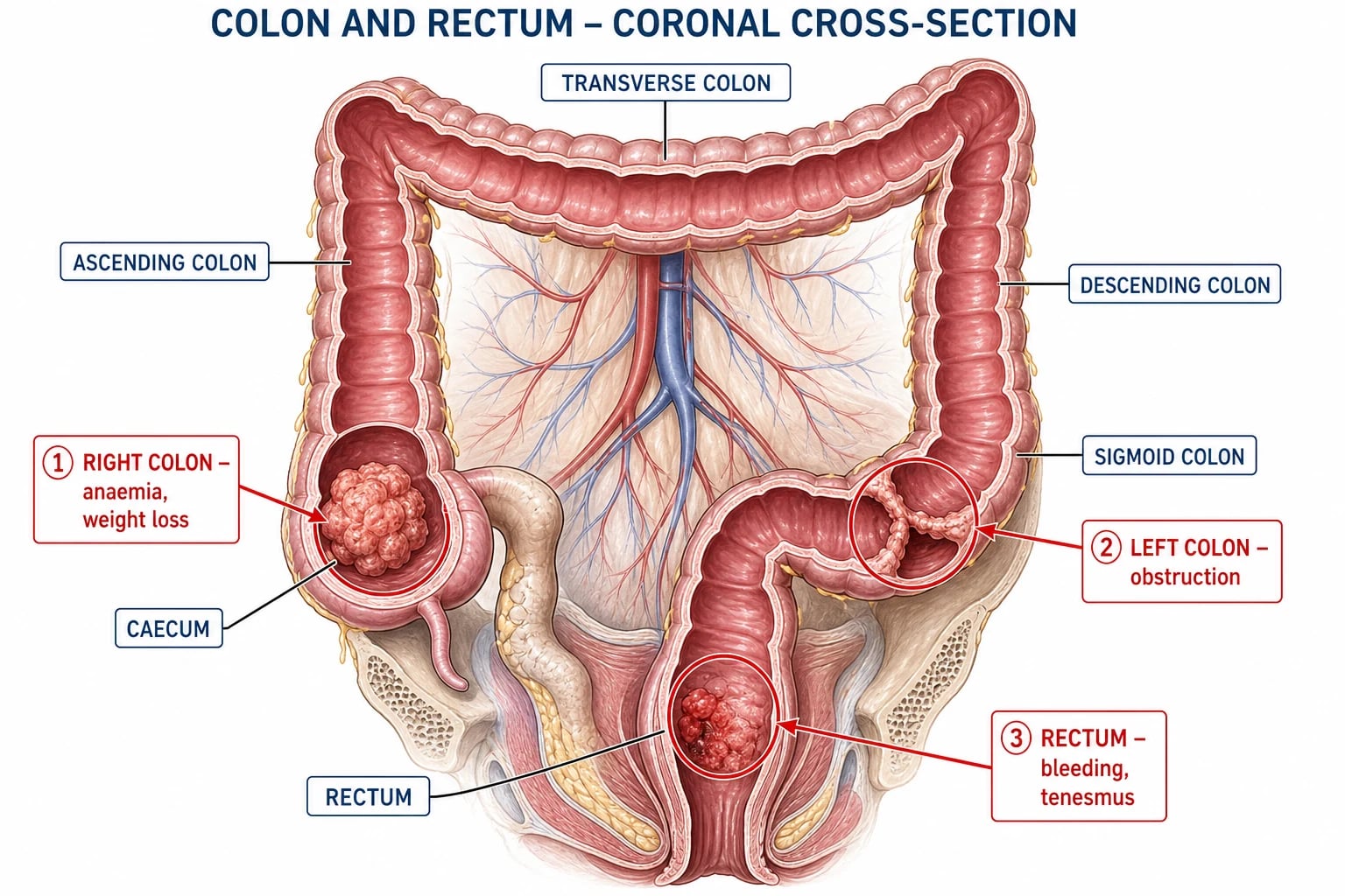
CRC is classified on three interlocking axes: anatomical site (right colon, left colon, rectum — which govern presentation), molecular pathway (chromosomal instability, microsatellite instability, serrated — which govern biology and therapy), and stage (TNM-8 / Dukes — which govern prognosis and treatment). The single most common, examineable classification point is the contrast between right-sided and left-sided tumours, which differ in embryological origin, biology, molecular profile and clinical behaviour. [1]
Right colon
caecum, ascending (midgut)
- **Insidious, late** presentation — anaemia, fatigue, weight loss
- **Iron-deficiency anaemia** and occult bleeding dominate
- **Exophytic, fungating or cauliflower** tumour
- Obstruction **rare** — wide lumen, liquid stool
- Molecular: more often **MSI-H and BRAF mutant**
- **Palpable right iliac fossa mass**
Left colon
descending, sigmoid (hindgut)
- **Altered bowel habit** (constipation/diarrhoea alternating)
- **Bright-red bleeding** mixed in the stool
- **Annular, constricting apple-core or napkin-ring** tumour
- **Large-bowel obstruction** — colicky pain, distension
- **Ribbon-like stools**
- Palpable mass in the left iliac fossa
Rectum
distal 15 cm (hindgut)
- **Fresh rectal bleeding** — often misattributed to haemorrhoids
- **Tenesmus** and a sensation of incomplete evacuation
- **Change in stool calibre** (pencil-thin)
- **Palpable mass on digital rectal examination**
- Relation to the anorectal ring decides APR vs anterior resection
- Highest local recurrence — drives **total mesorectal excision**
Epidemiology & Risk Factors
CRC is the third most commonly diagnosed cancer globally (behind breast and lung) and the second leading cause of cancer death, with nearly 2 million new cases and over 900,000 deaths per year. Incidence is highest in developed nations (Australia/New Zealand, Europe, North America), but is rising rapidly in low- and middle-income countries undergoing the nutrition transition, and — alarmingly — is increasing in adults under 50 in many Western populations, prompting the lowering of the screening start age to 45 in the United States.[1][2] The median age at diagnosis is around 70, and risk rises sharply after age 50; roughly 9 in 10 cases occur in people aged 50 or older.
The proportions by aetiology anchor every CRC answer: about 85 percent are sporadic, 10 percent are hereditary, and 5 percent complicate IBD. The principal modifiable and non-modifiable risk factors are:[1]
- Age — the dominant risk factor; incidence climbs steeply after 50.
- Diet — red and processed meat (haem iron, N-nitroso compounds, heterocyclic amines from high-temperature cooking) increase risk; dietary fibre, fruit, vegetables and dairy are protective (fibre dilutes carcinogens and shortens transit time).
- Lifestyle — smoking, alcohol, obesity (central adiposity and insulin resistance) and physical inactivity all raise risk; their reversal reduces it.
- Family history — one first-degree relative roughly doubles risk; two, or one diagnosed under 50, increases it further.
- Inflammatory bowel disease — long-standing ulcerative colitis (especially pancolitis) and Crohn's colitis raise risk after 8 to 10 years of disease; risk tracks extent, severity and duration of inflammation.
- Hereditary syndromes — Lynch syndrome (3 percent of CRC) and familial adenomatous polyposis (under 1 percent), detailed below.
- Personal history of CRC or adenomatous polyps, acromegaly, ureterosigmoidostomy, pelvic radiotherapy, and type 2 diabetes. [1]
Familial adenomatous polyposis (FAP)
FAP is an autosomal dominant disorder caused by a germline mutation of the APC (adenomatous polyposis coli) gene on chromosome 5q. Affected individuals develop hundreds to thousands of adenomatous polyps (over 100 by adolescence) throughout the colon and rectum from the early teens; without intervention, colorectal cancer is virtually inevitable by age 35 to 40. Attenuated FAP (fewer than 100 polyps, later onset) and MUTYH-associated polyposis (autosomal recessive) are variants. The cornerstone of management is prophylactic colectomy in the late teens to early twenties — proctocolectomy with ileal pouch-anal anastomosis or total colectomy with rectal surveillance — combined with annual sigmoidoscopy of any retained rectum and duodenal and desmoid surveillance.[1]
Lynch syndrome (hereditary nonpolyposis colorectal cancer, HNPCC)
Lynch syndrome is the commonest hereditary CRC syndrome, an autosomal dominant germline defect in a DNA mismatch repair (MMR) gene — MLH1, MSH2, MSH6 or PMS2 (and EPCAM deletions affecting MSH2). Tumours are microsatellite instability-high (MSI-H). Lifetime CRC risk is 40 to 70 percent, with onset averaging the mid-40s (over a decade earlier than sporadic CRC), and a high risk of extracolonic cancers — endometrial (the sentinel cancer in many women), ovarian, gastric, urothelial, small bowel, biliary, brain (Turcot) and sebaceous (Muir-Torre). It is identified clinically by the Amsterdam II criteria and molecularly by the revised Bethesda guidelines, which trigger universal MMR/MSI immunohistochemistry on all newly diagnosed CRC.[10]
Pathophysiology
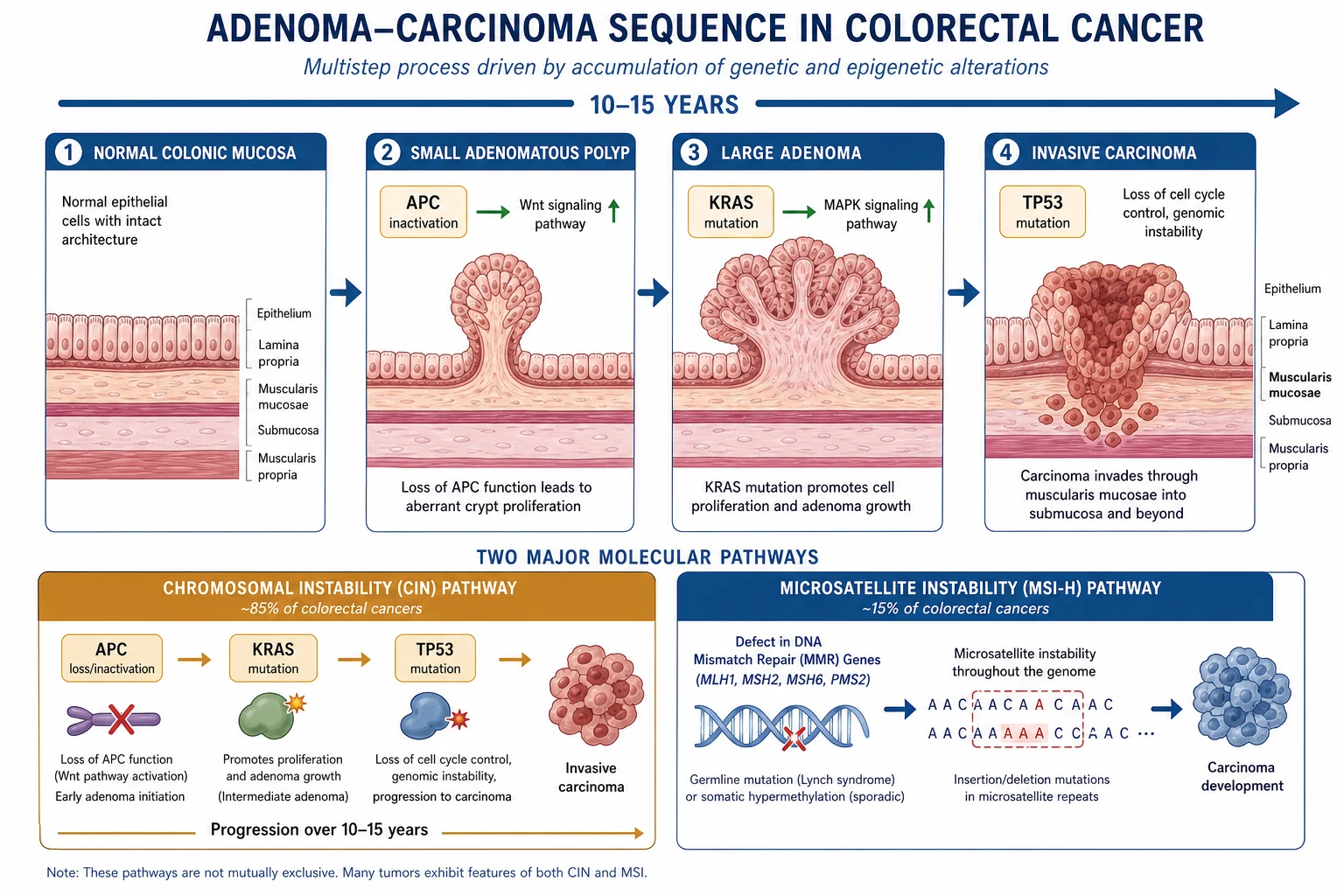
CRC develops through three molecularly distinct pathways that converge on the same endpoint — invasive adenocarcinoma — but differ in precursor lesion, driver mutations, biology and clinical behaviour. Understanding these pathways is essential because they determine prognosis, response to chemotherapy and eligibility for targeted and immunotherapy.[1][3]
1. The chromosomal instability (CIN) / adenoma-carcinoma pathway (about 70 percent)
The classic Vogelstein model of stepwise genetic accumulation:[3]
- APC loss (chromosome 5q) — the initiating event in most CRCs. APC normally degrades beta-catenin in the Wnt signalling pathway; its loss frees beta-catenin to drive adenoma formation (the earliest polyp).
- KRAS activation — a second hit that drives adenoma growth and dysplasia, typically at the intermediate-adenoma stage.
- Loss of heterozygosity at 18q (SMAD2/SMAD4/DCC) — promotes progression to late adenoma.
- TP53 loss (17p) — the final gate that permits the transition from severe dysplasia to frank invasive carcinoma. [1]
The whole sequence takes 10 to 15 years, which is the biological rationale for screening intervals and polypectomy. This pathway produces the conventional tubular and villous adenomas removed at colonoscopy and underlies most left-sided, chromosomally unstable tumours.[1][3]
2. The microsatellite instability (MSI) pathway (about 15 percent)
Caused by defective DNA mismatch repair (MMR) — the enzymes that correct single-nucleotide mismatches and short repeat errors during replication. Their loss produces microsatellite instability-high (MSI-H) tumours with an enormous mutational burden. Two origins:[1][10]
- Sporadic (commoner) — epigenetic silencing (methylation) of the MLH1 promoter, often in association with a BRAF V600E mutation; these are typically right-sided, mucinous, poorly differentiated tumours in older women.
- Lynch syndrome (inherited) — a germline mutation in MLH1, MSH2, MSH6 or PMS2; typically right-sided CRC with a tumour-infiltrating-lymphocyte-rich histology diagnosed in the 40s or 50s. [1]
MSI-H tumours have a better stage-for-stage prognosis (possibly because mutations cripple the tumour cell) and, crucially, respond dramatically to immune checkpoint inhibitors (pembrolizumab, nivolumab) — the modern paradigm of precision oncology. [1]
3. The serrated pathway (about 15 to 30 percent)
The recently recognised third pathway, arising from sessile serrated lesions (SSLs, formerly sessile serrated adenomas) and traditional serrated adenomas in the right colon, rather than from conventional adenomas. It is driven by an initiating BRAF V600E mutation, a CpG island methylator phenotype (CIMP) that silences genes including MLH1 (linking it to the MSI pathway), and Wnt pathway activation. Serrated lesions are flat, right-sided, pale, and easily missed at colonoscopy — a quality target of modern screening. They explain why interval cancers can still arise between scopes.[1]
How CRC spreads
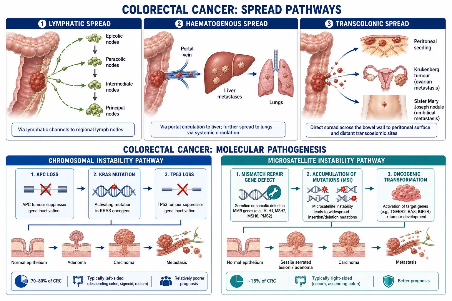
- Direct invasion — through the muscularis propria into pericolic fat and adjacent organs (small bowel, bladder, uterus, stomach).
- Lymphatic — the commonest and earliest route, via epicolic to paracolic to intermediate to principal (named-vessel) lymph nodes, then to the para-aortic chain. Lymph-node involvement is the single most important prognostic factor in non-metastatic disease.
- Haematogenous — tumour cells enter the portal venous system, making the liver the commonest site of distant metastasis, and then reach the lung via the systemic circulation; less commonly bone and brain.
- Transcoelomic — peritoneal seedlings (omental cake, ovarian Krukenberg tumours, ascites), a poor-prognosis pattern.[1]
Clinical Presentation
The presentation of CRC is dominated by tumour site. Because the right and left colon differ in embryology, luminal calibre, stool consistency and molecular biology, a right-sided tumour behaves nothing like a left-sided one. The unifying principle: right-sided bleeds silently and presents late with anaemia; left-sided narrows and presents with obstruction and changed habit; rectal bleeds and tenesmus early.[1]
Right-sided colon cancer (caecum, ascending)
- Iron-deficiency anaemia (the sentinel finding — occult, slow bleeding into a large-lumen viscus), presenting as fatigue, pallor, dyspnoea and angina.
- Vague right iliac fossa discomfort or a palpable mass; weight loss and anorexia.
- Occult blood in the stool — large tumour surface area, liquid stool, so obstruction is rare.
- Often asymptomatic until late, detected via anaemia or screening. [1]
Left-sided colon cancer (descending, sigmoid)
- Change in bowel habit — alternating constipation and diarrhoea, or new constipation in an older adult.
- Bright-red blood mixed into or on the stool (distinguishing it from haemorrhoidal blood on the surface/paper).
- Colicky lower abdominal pain, distension, and ribbon-like stools.
- May present acutely as large-bowel obstruction (colicky pain, distension, absolute constipation, vomiting late) or, rarely, perforation causing peritonitis. [1]
Rectal cancer
- Fresh bright-red bleeding, often wrongly attributed to haemorrhoids — never assume bleeding is haemorrhoidal in an adult without proctoscopy/colonoscopy.
- Tenesmus — a constant urge to defecate with little result.
- Change in stool calibre (pencil-thin stools), mucus discharge, and a sensation of incomplete evacuation.
- A palpable mass on digital rectal examination; the relationship to the anorectal ring (above 5 cm or below) determines whether a sphincter-sparing resection is possible. [1]
General and metastatic features
- Weight loss, anorexia, malaise; less commonly fever (tumour or abscess) or deep venous thrombosis (Trousseau's migratory thrombophlebitis).
- Emergency presentation as large-bowel obstruction (more common on the left) or perforation with faecal peritonitis (high mortality).
- Metastatic disease — hepatomegaly, jaundice, ascites, right upper quadrant pain (liver); cough, dyspnoea, haemoptysis (lung); bone pain or pathological fracture (bone); neurological deficit (brain).[1]
Atypical presentations (examiners test these)
- Iron-deficiency anaemia as the sole finding in an otherwise well older adult — colonoscopy is mandatory.
- Young onset (under 50) — rising incidence; always consider Lynch syndrome and refer for MMR/MSI testing and genetics.
- Painless obstruction or weight loss in the elderly, attributed to "constipation".
- Asymptomatic, screen-detected — increasingly common with FIT and colonoscopy screening. [1]
Differential Diagnosis
A change in bowel habit, rectal bleeding or iron-deficiency anaemia in an adult is colorectal cancer until proven otherwise by colonoscopy. The commoner mimics, and the features that distinguish each from CRC, are:[1]
| Condition | Key distinguishing feature |
|---|---|
| Haemorrhoids | Bright-red blood on the surface of the stool and toilet paper, with a normal colonoscopy; never assume bleeding is haemorrhoidal without full colonic visualisation |
| Diverticular disease | Acute left iliac fossa pain with fever and raised inflammatory markers; CT shows pericolic fat stranding and the diverticula; bleeding is painless, bright-red and brisk and usually stops spontaneously |
| Inflammatory bowel disease (UC/Crohn) | Younger patient, chronic diarrhoea, blood and mucus, weight loss, extra-intestinal features; long-standing colitis raises CRC risk — biopsy distinguishes dysplasia |
| Ischaemic colitis | Acute pain and bloody diarrhoea after a vascular insult; typical watershed distribution on imaging; usually resolves |
| Infective colitis (Campylobacter, Salmonella, Shigella, E. coli, C. difficile, amoebiasis) | Acute onset, fever, recent travel or antibiotic exposure; stool culture and toxin assays |
| Angiodysplasia | Painless, painless bright-red bleeding, typically in the right colon of older patients with CKD/aortic stenosis; colonoscopy or angiography |
| Irritable bowel syndrome | A diagnosis of exclusion in the absence of red flags (no weight loss, anaemia, bleeding, family history); age-appropriate screening must be normal before labelling IBS |
| Colorectal lymphoma, GIST, carcinoid | Rare; diagnosed histologically; carcinoid causes carcinoid syndrome only with metastases |
The two non-negotiable rules: (1) a change in bowel habit or new rectal bleeding in anyone over 40 mandates colonoscopy; and (2) iron-deficiency anaemia in an adult male or postmenopausal woman is colorectal cancer until excluded, and warrants urgent colonoscopy plus upper endoscopy (in a premenopausal woman, investigate if anaemia is significant or persistent). [1]
Clinical & Bedside Assessment
A focused assessment looks for the primary tumour (mass), signs of anaemia (pallor, conjunctival pallor), signs of obstruction or perforation (distension, absent bowel sounds, peritonism), metastatic disease (hepatomegaly, ascites, supraclavicular nodes), and general nutritional state (weight loss, cachexia).[1]
- Digital rectal examination (DRE) — mandatory in every patient with lower-GI symptoms. In rectal cancer assess: a palpable, hard, irregular, ulcerated or annular mass; its mobility (fixed vs mobile); its distance from the anal verge and relation to the anorectal ring (decides operability and sphincter preservation); and whether it involves the sphincter or anterior structures (prostate/vagina). DRE also excludes other causes (palpable masses, faecal impaction, blood).
- Rigid sigmoidoscopy — confirms the level of a rectal tumour from the anal verge and its mobility, complementary to DRE.
- Abdominal examination — palpable mass (right or left iliac fossa), hepatomegaly (metastases), ascites, distension (obstruction), and signs of peritonitis (perforation).
- General examination — pallor (anaemia), cachexia, ** supraclavicular (Virchow) node** (advanced disease), deep vein thrombosis, and digital clubbing (occasionally). [1]
Investigations
The diagnostic and staging pathway has three aims: confirm the diagnosis (colonoscopy with biopsy), stage the extent (CT and MRI), and define the molecular biology (MMR/MSI, RAS/BRAF) that drives therapy. CEA is a monitoring tool, never a screening or sole diagnostic one.[1]
Confirming the diagnosis
- Colonoscopy with biopsy — the gold standard. Directly visualises the tumour, provides tissue for histology, identifies and can remove synchronous polyps or cancers (present in 3 to 5 percent), and allows tattooing of a small lesion to guide later surgical localisation. Complete the examination to the caecum (caecal photographs) and intubate the terminal ileum where possible; this also picks up synchronous tumours.[1]
- CT colonography — an alternative when colonoscopy is incomplete, contraindicated, or refused; sensitive for large polyps and masses but cannot biopsy or tattoo, so any positive finding still requires colonoscopy.
Staging — imaging
- CT chest/abdomen/pelvis — the standard staging modality for all CRC; assesses distant metastases (liver, lung), local invasion, and nodal and peritoneal disease. Performed early in the workup to establish M-stage before deciding on curative vs palliative intent.[1]
- MRI of the rectum — essential and mandatory for rectal cancer, providing high-resolution detail of depth of tumour invasion (T-stage), distance to the mesorectal fascia (the surgical circumferential resection margin — clearance under 1 mm predicts local recurrence), extramural vascular invasion, and suspicious mesorectal nodes. It decides whether neoadjuvant radiotherapy is needed and whether a sphincter-sparing operation is feasible.[1]
- Endorectal ultrasound — an alternative for early rectal tumours (uT1/2) when local excision is contemplated.
- PET-CT — not routine; reserved for selected cases — equivocal lesions on CT, suspected recurrent disease (especially a rising CEA with negative conventional imaging), and to confirm resectability before metastasectomy.[1]
- Staging laparoscopy — used in selected cases of advanced disease to detect occult peritoneal disease not seen on CT before committing to curative surgery.
Tumour biology — molecular testing
- MMR/MSI testing — performed by immunohistochemistry (loss of MLH1, MSH2, MSH6, PMS2) and/or PCR (microsatellite instability). It screens for Lynch syndrome, predicts a better prognosis, and identifies candidates for immune checkpoint inhibitors in advanced disease. Now recommended universally on all newly diagnosed CRC.[10]
- RAS (KRAS and NRAS) and BRAF mutation testing — performed on all metastatic CRC to select patients for anti-EGFR therapy (cetuximab, panitumumab), which benefits only RAS wild-type tumours; BRAF mutation carries a poor prognosis and predicts lack of anti-EGFR benefit.[1]
Tumour marker — CEA
- Carcinoembryonic antigen (CEA) — a glycoprotein useful for baseline measurement before surgery, monitoring treatment response, and detecting recurrence after curative resection. It is NOT a screening test (low sensitivity for early disease) and NOT diagnostic alone (raised in smoking, IBD, pancreatitis, liver disease, other cancers). A rising or persistently elevated CEA after curative resection should trigger imaging to detect recurrence; serial measurement is the cornerstone of surveillance.[13]
Screening — faecal immunochemical test (FIT)
- FIT (faecal immunochemical test) — detects human haemoglobin in stool with high sensitivity for significant colonic neoplasia; it has replaced the older guaiac faecal occult blood test (gFOBT) because it is diet-independent, more sensitive and quantitative. A positive FIT prompts colonoscopy. It is the backbone of population screening programmes.[2]
Colorectal cancer — the numbers that decide the answer
Staging — TNM-8 and Dukes
Staging uses the eighth edition of the TNM (AJCC/UICC) system and the older Dukes classification (still widely used and frequently examined). Reproduced verbatim:[1]
TNM-8 — Tumour (T) [1]
| T | Definition |
|---|---|
| Tis | Carcinoma in situ — intraepithelial or invasion of lamina propria |
| T1 | Invades submucosa |
| T2 | Invades muscularis propria |
| T3 | Invades through muscularis propria into pericolorectal tissue |
| T4a | Penetrates visceral peritoneum |
| T4b | Directly invades or adheres to other organs or structures |
TNM-8 — Nodes (N) and Metastases (M) [1]
| N/M | Definition |
|---|---|
| N0 | No regional lymph-node metastasis |
| N1a | 1 regional node; N1b 2 to 3 nodes; N1c no nodes but tumour deposits in subserosa/mesorectal tissue |
| N2a | 4 to 6 regional nodes; N2b 7 or more nodes |
| M0 | No distant metastasis |
| M1a | Metastasis confined to one organ (e.g. liver, lung) without peritoneal involvement |
| M1b | Metastases in 2 or more organs |
| M1c | Peritoneal surface metastasis with or without other organ involvement |
Stage grouping and the Dukes equivalent (with approximate 5-year survival): [1]
| Stage | TNM-8 | Dukes | 5-year survival |
|---|---|---|---|
| 0 | Tis N0 M0 | — | over 95 percent |
| I | T1 or T2, N0, M0 | A | about 90 percent |
| IIA/IIB/IIC | T3/T4a/T4b, N0, M0 | B | about 70 to 85 percent |
| III A/B/C | any T, N1 or N2, M0 | C | about 40 to 70 percent |
| IV A/B/C | any T, any N, M1 | D | under 15 percent (better with oligometastatic resection) |
Management — Resuscitation (the surgical emergency)
Most CRC is managed electively, but two presentations demand immediate, time-critical care: acute large-bowel obstruction and perforation.[1]
- Acute large-bowel obstruction (classically an obstructing left-sided tumour): ABCs, IV fluid resuscitation, nasogastric decompression, a urinary catheter, DVT prophylaxis, correction of electrolytes and anaemia, broad-spectrum antibiotics, and a water-soluble contrast enema or CT to confirm the level and rule out pseudo-obstruction. Management is surgical — either an emergency Hartmann's procedure (resection with end colostomy and rectal stump, favoured in the unwell or elderly) or a primary anastomosis with or without a defunctioning stoma in a stable, well-prepared patient. In the frail or as a bridge to elective surgery, an endoluminal self-expanding colonic metal stent relieves obstruction and permits staging, bowel preparation and a later single-stage resection.
- Perforation causing faecal peritonitis — aggressive resuscitation, broad-spectrum antibiotics, and emergency laparotomy with resection and stoma; mortality is high.
- Acute lower-GI bleeding (rarer in CRC than diverticular) — resuscitate and transfuse, localise, and control endoscically or surgically. [1]
Management — Definitive & Stepwise
Treatment is determined by stage, tumour site and molecular subtype, decided by a multidisciplinary team (MDT). The pillars are surgery (curative for localised disease), chemotherapy (adjuvant for stage III and high-risk II; first-line for metastatic), radiotherapy (neoadjuvant for locally advanced rectal cancer), and targeted/biological therapy (for metastatic disease).[1]
Surgery — the curative operation
The principle is en-bloc resection of the tumour-bearing segment of bowel with its mesentery and lymphatic drainage, achieving a clear circumferential and distal margin and a minimum of 12 lymph nodes for accurate staging.[1]
- Right hemicolectomy (for caecal, ascending and proximal transverse colon tumours) — resection of the terminal ileum, caecum, ascending colon and right transverse colon, with ligation of the ileocolic, right colic and right branches of the middle colic vessels at their origin, and ileocolic anastomosis.
- Extended right hemicolectomy — for transverse colon and hepatic flexure tumours.
- Left hemicolectomy / sigmoid colectomy (for descending and sigmoid colon tumours) — with ligation of the inferior mesenteric artery (at its origin or below the left colic for a high tie) and inferior mesenteric vein, and a colorectal anastomosis.
- Subtotal or total colectomy — for synchronous tumours, obstructing proximal cancer with unprepared proximal bowel, or hereditary syndromes.
- Rectal cancer resections:
- Low anterior resection (LAR) — for upper and middle rectal tumours above the anorectal ring, preserving the anal sphincter; performed with total mesorectal excision (TME) and a circular-stapled colorectal or coloanal anastomosis, often protected by a temporary defunctioning loop ileostomy.
- Abdominoperineal excision of the rectum (APER / APR) — for low rectal tumours involving or closely related to the sphincter complex, removing the rectum and anus via an abdominal and a perineal approach and leaving a permanent end colostomy.
- Hartmann's procedure — resection (typically sigmoid) with an end colostomy and a closed rectal stump, used for emergency obstruction, perforation or in the frail (may be reversed electively or left permanent). [1]
Adjuvant chemotherapy — colon cancer
- Stage I — surgery alone; no adjuvant chemotherapy.
- Stage II — surgery alone for standard-risk disease. Adjuvant 5-FU plus leucovorin or capecitabine is considered for high-risk stage II tumours, defined as T4 (stage IIC), poorly differentiated, perforation or obstruction, fewer than 12 lymph nodes sampled, lymphovascular or perineural invasion; the decision is individualised (and the MOSAIC data suggest a small benefit with oxaliplatin regimens).[6]
- Stage III (node-positive, M0) — adjuvant FOLFOX or CAPOX for 6 months is the standard of care, building on the landmark MOSAIC trial, which showed that adding oxaliplatin to 5-FU and leucovorin improved disease-free and overall survival in stages II and III colon cancer.[4][5] The IDEA collaboration subsequently showed that a 3-month course is acceptable (non-inferior for disease-free survival) for low-risk stage III (T1 to T3 N1), reducing cumulative oxaliplatin neurotoxicity, while 6 months remains standard for higher-risk disease.[1]
Neoadjuvant therapy — rectal cancer
For locally advanced rectal cancer (defined broadly as T3 or deeper, or node-positive on MRI), preoperative (neoadjuvant) radiotherapy or chemoradiotherapy is given to downsize the tumour, sterilise the pelvic mesorectum and reduce local recurrence. Two regimens, each with landmark evidence:[1][7][8]
- Short-course radiotherapy — 25 Gy in 5 daily fractions of 5 Gy over 1 week, followed by surgery within a week. Established by the Swedish Rectal Cancer Trial (1997), which demonstrated a survival benefit for preoperative radiotherapy in resectable rectal cancer.[8] Favoured for its convenience, lower cost and similar local control when immediate downstaging is not needed.
- Long-course chemoradiotherapy — 45 to 50.4 Gy in 25 to 28 fractions over 5 to 6 weeks with concurrent radiosensitising chemotherapy (infusional 5-FU or oral capecitabine), followed by surgery 6 to 12 weeks later after a "downsizing" interval. The German CAO/ARO/AIO-94 trial (Sauer, 2004) established that preoperative chemoradiotherapy (long course) was superior to postoperative, with better local control, less acute and late toxicity, and more sphincter preservation.[7]
- Total neoadjuvant therapy (TNT) — giving all chemotherapy and chemoradiotherapy before surgery is an emerging standard that improves pathological complete response rates and compliance.
Total mesorectal excision (TME) — the surgical standard for rectal cancer
Introduced by R.J. Heald in 1982, TME is the sharp, anatomical, en-bloc removal of the rectum and its intact mesorectum within the avascular mesorectal fascial plane, ensuring the circumferential resection margin is clear. By removing the tumour and its locoregional lymphatic field intact, TME dramatically reduced local recurrence of rectal cancer from 20 to 30 percent down to under 10 percent when combined with neoadjuvant therapy, and is now the global surgical standard.[9]
Management of metastatic disease (stage IV)
Metastatic CRC is treated with systemic chemotherapy plus biological therapy, with curative-intent resection considered for oligometastatic disease (limited liver or lung metastases).[1][11][12]
- First-line chemotherapy backbones — FOLFOX or FOLFIRI (interchangeable; one is chosen first, the other reserved for second-line), or CAPOX. Adding a biological agent improves response and survival.
- Bevacizumab — a monoclonal antibody against VEGF (vascular endothelial growth factor) that inhibits tumour angiogenesis, added to first- or second-line chemotherapy in metastatic CRC; established by Hurwitz et al. (2004).[11]
- Anti-EGFR monoclonal antibodies — cetuximab and panitumumab — added to chemotherapy (e.g. FOLFIRI or FOLFOX) in the first line, but only in RAS wild-type tumours; the CRYSTAL trial (Van Cutsem, 2009) showed cetuximab plus FOLFIRI improved response and progression-free survival in KRAS wild-type metastatic CRC. RAS mutation testing is mandatory before starting an anti-EGFR agent.[12]
- Metastasectomy — surgical resection of liver and/or lung metastases in selected patients with resectable oligometastatic disease offers the only chance of long-term survival or cure in stage IV, with 5-year survival of 30 to 50 percent in well-selected cases. Strategies include staged or simultaneous resection, portal vein embolisation, and ablation (radiofrequency, microwave).
- Palliative measures — endoscopic colonic stenting, defunctioning stoma, bypass, and best supportive care for obstruction, bleeding or pain.
Surveillance after curative-intent treatment
Surveillance aims to detect resectable recurrence (local, metachronous cancer, or oligometastatic disease) while still curable:[1][13]
- CEA every 3 to 6 months for the first 3 years, then 6-monthly to 5 years; a rising CEA prompts cross-sectional imaging.
- CT chest/abdomen/pelvis annually for up to 3 years (3 to 5 years in higher-risk patients).
- Colonoscopy at 1 year after resection, then at 3 years, then every 5 years if normal (to detect metachronous neoplasia); the perioperative clearing colonoscopy should be complete to the caecum. [1]
Specific Subtypes & Scenarios
Lynch syndrome — surveillance
At-risk relatives undergo colonoscopy from age 20 to 25 (or 2 to 5 years before the earliest affected relative's age), every 1 to 2 years, because Lynch tumours can evolve faster than the classic 10 to 15 year adenoma-carcinoma interval. Women should be counselled about endometrial and ovarian cancer risk with gynaecological surveillance and consideration of prophylactic hysterectomy and bilateral salpingo-oophorectomy after childbearing. Aspirin chemopreduction is supported by long-term data. If CRC develops, subtotal colectomy may be preferred given the high metachronous risk.[10]
FAP — prophylactic surgery
Prophylactic colectomy in the late teens to early twenties — either total proctocolectomy with ileal pouch-anal anastomosis (IPAA) or total colectomy with ileorectal anastomosis (with ongoing rectal surveillance). Annual upper-GI endoscopy for duodenal polyposis and cancer, and vigilance for desmoid tumours (a leading cause of death in FAP).[1]
Obstructing colorectal cancer
Acute left-sided obstruction is managed with resuscitation then emergency surgery (Hartmann's procedure in the unstable; primary anastomosis with defunctioning stoma in the well-prepared), or an endoluminal self-expanding metal stent as a bridge to elective single-stage resection in the frail or where the diagnosis and staging are incomplete.[1]
Inflammatory-bowel-disease-associated CRC
Risk rises after 8 to 10 years of extensive colitis (UC or Crohn's colitis), and is magnified by primary sclerosing cholangitis, severe active inflammation, and a family history of CRC. Surveillance is by colonoscopy with chromoendoscopy and targeted biopsies every 1 to 5 years depending on risk; the goal is to detect and remove dysplasia before it progresses to carcinoma. CRC complicating IBD is more often multifocal and mucinous.[1]
Complications & Pitfalls
- Large-bowel obstruction — the commonest emergency presentation; complete obstruction risks caecal perforation (Laplace's law — the caecum is the widest and most distensible segment).
- Perforation — into the peritoneal cavity (faecal peritonitis, high mortality) or locally (abscess, fistula to bladder or vagina).
- Anastomotic leak — typically presents around day 5 to 7 postoperatively with fever, tachycardia, abdominal pain, ileus, leukocytosis and, later, septic shock; diagnosed by water-soluble contrast enema or CT with rectal contrast; managed with antibiotics, drainage and reoperation (often a defunctioning stoma). Risk is higher after low pelvic anastomosis, in malnutrition, and with neoadjuvant radiotherapy.
- Local recurrence — especially rectal cancer; reduced by TME and neoadjuvant therapy.
- Treatment-related — stoma complications (parastomal hernia, prolapse, retraction, skin excoriation), anterior resection syndrome (frequency, urgency, fragmentation, faecal and urinary incontinence, sexual dysfunction — after low rectal surgery), chemotherapy toxicity (oxaliplatin peripheral neuropathy, capecitabine hand-foot syndrome, neutropenic sepsis), and radiotherapy effects (bowel, bladder and sexual dysfunction).
- Metastatic spread to liver, lung, peritoneum and bone.
- Synchronous and metachronous cancers — the rationale for complete colonoscopy before surgery and surveillance colonoscopy thereafter.[1]
Classic pitfalls: attributing rectal bleeding to haemorrhoids without proctoscopy and colonoscopy; missing iron-deficiency anaemia as a red flag; not testing MMR/MSI on a young-onset tumour; under-staging the rectum by omitting MRI and so omitting neoadjuvant therapy; failing to confirm RAS status before an anti-EGFR agent; assuming a "normal" colonoscopy excludes a small right-sided lesion when the caecum was not reached; and not enrolling patients in surveillance. [1]
Prognosis & Disposition
Prognosis is stage-dependent and, beyond stage, modified by tumour grade, lymphovascular invasion, circumferential resection margin, microsatellite status and sidedness.[1]
Prognosis by stage (5-year survival)
Favourable prognostic factors include early stage, well-differentiated tumour, no lymphovascular invasion, clear margins, MSI-H status (better stage-for-stage prognosis and dramatic response to immunotherapy) and left-sided location. Adverse factors include advanced stage, BRAF mutation, perineural invasion, signet-ring histology, peritoneal metastases and right-sided tumours (which carry a poorer prognosis, partly reflecting higher MSI/BRAF biology).[1]
Disposition after curative-intent treatment is to a structured surveillance pathway (CEA, CT, colonoscopy) and to lifestyle and chemoprevention counselling. Patients with metastatic disease are managed by the MDT with sequential lines of systemic therapy and selective metastasectomy. [1]
Special Populations
- Long-standing IBD — surveillance colonoscopy with chromoendoscopy from 8 to 10 years of symptoms (from diagnosis for primary sclerosing cholangitis), every 1 to 5 years by risk; biopsy for dysplasia.
- Lynch syndrome and FAP — intensive, earlier surveillance from young adulthood, as above.
- Elderly and frail — colonic stenting for obstruction, less aggressive chemotherapy, and individualised decisions on neoadjuvant therapy and surgery; enhanced recovery after surgery (ERAS) pathways improve outcomes.
- Pregnancy — rare; the diagnostic and therapeutic challenge is imaging safely (MRI preferred) and timing surgery (second trimester) and chemotherapy (avoid first trimester).
- Young-onset CRC (under 50) — rising incidence; always suspect a hereditary syndrome, test MMR/MSI, and refer to genetics; present more often with left-sided disease, advanced stage and symptoms.[1]
Evidence, Guidelines & Regional Differences
Landmark trials
- Swedish Rectal Cancer Trial (1997, PMID 9091798) — established that short-course preoperative radiotherapy (25 Gy in 5 fractions) improved survival in resectable rectal cancer.[8]
- CAO/ARO/AIO-94 (Sauer, 2004, PMID 15496622) — preoperative chemoradiotherapy was superior to postoperative for locally advanced rectal cancer, with better local control, less toxicity, and more sphincter preservation.[7]
- MOSAIC (Andre, 2004 and 2009, PMIDs 15175436 and 19451431) — adding oxaliplatin to 5-FU and leucovorin as adjuvant therapy improved disease-free and overall survival in stage III colon cancer — the basis of FOLFOX.[4][5]
- Benson / ASCO (2004, PMID 15199089) — defined high-risk stage II features guiding the adjuvant decision.[6]
- Hurwitz (2004, PMID 15175435) — bevacizumab added to chemotherapy improved survival in metastatic CRC.[11]
- CRYSTAL (Van Cutsem, 2009, PMID 19339720) — cetuximab plus FOLFIRI benefited KRAS wild-type metastatic CRC; underpinned mandatory RAS testing.[12]
Regional screening differences
[1] [1]Across guidelines (USPSTF, NICE, NCCN, ESMO) there is broad convergence on: colonoscopy as the gold-standard diagnostic and screening test; FIT as the population-screening backbone; FOLFOX or CAPOX for 6 months for stage III colon cancer (3 months for low-risk stage III per IDEA); neoadjuvant radiotherapy (short or long course) plus TME for locally advanced rectal cancer; universal MMR/MSI testing; and RAS/BRAF testing before anti-EGFR therapy. Differences are mainly in screening start age (45 US vs 50-60 elsewhere), FIT interval, and the availability of organised programmes.
Prevention and chemoprevention
- Screening and polypectomy — the single most effective intervention; removing adenomas interrupts the adenoma-carcinoma sequence.[1][2]
- Lifestyle — a diet high in fibre, fruit and vegetables, low in red and processed meat, regular exercise, weight control, smoking cessation and moderate alcohol reduce risk.
- Aspirin — observational and randomised data (notably in Lynch syndrome, CAPP2) suggest low-dose aspirin reduces CRC incidence and mortality; it is increasingly recommended for high-risk groups, balancing against bleeding risk.
- Surveillance colonoscopy for post-polypectomy, post-resection, IBD, Lynch and FAP cohorts.[1]
Exam Pearls
- Iron-deficiency anaemia in an adult male or postmenopausal woman = colonoscopy to exclude CRC (examine that spine of every CRC question).
- CEA is for monitoring (recurrence, treatment response), NEVER screening or sole diagnosis.
- Adenoma-carcinoma sequence: APC then KRAS then TP53 over 10 to 15 years (Vogelstein) — the biological basis of screening.[3]
- APC gene on chromosome 5q; FAP = hundreds of polyps; prophylactic colectomy in the late teens to twenties.
- Lynch syndrome = MMR gene defect (MLH1, MSH2, MSH6, PMS2), MSI-H tumours; Amsterdam II criteria; colonoscopy from 20 to 25, every 1 to 2 years.
- Left-sided = obstruction, altered habit, bright-red bleed, apple-core; right-sided = iron-deficiency anaemia, occult loss, mass, exophytic.
- Surgery by site: right hemicolectomy (ileocolic, right colic, middle colic branches); left hemicolectomy (IMA); LAR or APR with TME for rectum; Hartmann's for emergency/frail.
- Adjuvant FOLFOX or CAPOX for 6 months for stage III (MOSAIC); high-risk stage II individualised.
- Neoadjuvant rectal radiotherapy: short course 25 Gy in 5 fractions (Swedish) vs long course 45 to 50.4 Gy with 5-FU or capecitabine (CAO/ARO/AIO-94, Sauer).
- TME reduces rectal cancer local recurrence from 20 to 30 percent down to under 10 percent.
- Anti-EGFR (cetuximab, panitumumab) ONLY if RAS wild-type; bevacizumab (anti-VEGF) for any metastatic CRC.
- Minimum 12 lymph nodes for accurate pathological staging.
- MSI-H predicts better prognosis and response to immunotherapy; BRAF mutant predicts worse outcome.
- Screen from age 45 to 50: FIT yearly or colonoscopy every 10 years; stop around 75 to 85.
- Anastomotic leak presents around day 5 to 7 with fever, pain, ileus and sepsis.
- Surveillance: CEA every 3 to 6 months, CT annually up to 3 years, colonoscopy at 1 then 3 then every 5 years.
Adenoma-carcinoma sequence — AKT
AKT
Loss of APC is the gatekeeper mutation that initiates the polyp
Oncogenic KRAS drives progression from early to intermediate adenoma
Loss of TP53 converts severe dysplasia into invasive adenocarcinoma
Left vs right colon — presentation
ROAR
Right colon: iron-deficiency anaemia, occult blood, palpable right iliac fossa mass, weight loss; obstruction rare
Left colon: annular apple-core tumour, altered bowel habit, bright-red blood, colicky obstruction
Rectum: fresh bleeding, tenesmus, change in stool calibre, palpable mass on DRE
The site of the palpable mass localises the tumour
Exam application bank (NEET-PG / INICET)
One-line answer
Colorectal cancer (CRC) is an adenocarcinoma of the colonic or rectal mucosa that evolves from a benign adenomatous polyp through the stepwise accumulation of genetic hits — the adenoma-carcinoma sequence (APC, then KRAS, then TP53) over 10 to 15 years. It is the third most commonly diagnosed cancer worldwide and the second leading cause of cancer death. About 85 percent are sporadic; the remainder arise in hereditary syndromes (Lynch syndrome, familial adenomatous polyposis) or long-standing inflammatory bowel disease. Presentation differs by site — left-sided causes obstruction, a change in bowel habit and bright-red bleeding; right-sided presents with iron-deficiency anaemia and occult blood loss. Diagnosis is by colonoscopy and biopsy; staging with CT chest/abdomen/pelvis and MRI rectum; monitoring with CEA. Treatment is surgical (segmental colectomy with lymphadenectomy; total mesor
Worked stems (answer without another resource)
Stem 1 — Classic presentation. Map symptoms to mechanism; name the first investigation and first treatment step with dose/route if drug therapy is standard. [1]
Stem 2 — Unstable / complicated. List red flags that force immediate resuscitation, theatre, ICU, antidote, or reperfusion — and what you do in the first 15 minutes. [1]
Stem 3 — Atypical group. Elderly, pregnancy, child, or immunocompromised: how presentation and thresholds change. [1]
Stem 4 — Differential trap. Name the three closest mimics and one discriminator for each. [1]
Stem 5 — Disposition. Who goes home with safety-netting, who is admitted, who needs HDU/ICU/theatre, and what follow-up is mandatory. [1]
Rapid viva checklist
- Definition + classification
- Pathophysiology chain
- Bedside signs / criteria
- Score with exact components (if any)
- Emergency bundle
- Definitive therapy with doses
- Complications of disease and of treatment
- Special populations
- Guideline/trial name if classic
- Three exam traps
Coverage self-check
If you cannot answer any stem above from this page alone, re-read the matching section — the page is intended to be self-sufficient for final-prof and NEET-PG/INICET questions on Colorectal Cancer.
References
- [1]Dekker E, Tanis PJ, Vleugels JLA, et al. Colorectal cancer Lancet, 2019.PMID 31631858
- [2]US Preventive Services Task Force, Davidson KW, Barry MJ, et al. Screening for Colorectal Cancer: US Preventive Services Task Force Recommendation Statement JAMA, 2021.PMID 34003218
- [3]Fearon ER, Vogelstein B. A genetic model for colorectal tumorigenesis Cell, 1990.PMID 2188735
- [4]Andre T, Boni C, Mounedji-Boudiaf L, et al. Oxaliplatin, fluorouracil, and leucovorin as adjuvant treatment for colon cancer N Engl J Med, 2004.PMID 15175436
- [5]Andre T, Quinaux E, Louvet C, et al. Improved overall survival with oxaliplatin, fluorouracil, and leucovorin as adjuvant treatment in stage II or III colon cancer in the MOSAIC trial J Clin Oncol, 2009.PMID 19451431
- [6]Benson AB 3rd, Schrag D, Somerfield MR, et al. American Society of Clinical Oncology recommendations on adjuvant chemotherapy for stage II colon cancer J Clin Oncol, 2004.PMID 15199089
- [7]Sauer R, Becker H, Hohenberger W, et al. Preoperative versus postoperative chemoradiotherapy for rectal cancer N Engl J Med, 2004.PMID 15496622
- [8]Swedish Rectal Cancer Trial. Improved survival with preoperative radiotherapy in resectable rectal cancer N Engl J Med, 1997.PMID 9091798
- [9]Votava Z, Maresova J. Total mesorectal excision - 40 years of standard of rectal cancer surgery Acta Chir Belg, 2020.PMID 32200705
- [10]Umar A, Boland CR, Terdiman JP, et al. Revised Bethesda Guidelines for hereditary nonpolyposis colorectal cancer (Lynch syndrome) and microsatellite instability J Natl Cancer Inst, 2004.PMID 14970275
- [11]Hurwitz H, Fehrenbacher L, Novotny W, et al. Bevacizumab plus irinotecan, fluorouracil, and leucovorin for metastatic colorectal cancer N Engl J Med, 2004.PMID 15175435
- [12]Van Cutsem E, Kohne CH, Hitre E, et al. Cetuximab and chemotherapy as initial treatment for metastatic colorectal cancer N Engl J Med, 2009.PMID 19339720
- [13]Shinkins B, Nicholson BD, Primrose J, et al. What carcinoembryonic antigen level should trigger further investigation during colorectal cancer follow-up? A systematic review and secondary analysis of a randomised controlled trial Health Technol Assess, 2017.PMID 28617240