Gastroenterology · gastroenterology
Functional Dyspepsia
Also known as Functional dyspepsia · Non-ulcer dyspepsia · Idiopathic dyspepsia · Essential dyspepsia
Functional dyspepsia is chronic (at least 3 months) epigastric pain or burning, bothersome postprandial fullness or early satiety, with no structural cause on endoscopy (including normal OGD). It is a disorder of gut-brain interaction driven by gastroduodenal dysmotility, visceral hypersensitivity, duodenal eosinophilic inflammation and low-grade mucosal immune activation, and is divided into two subtypes — postprandial distress syndrome (fullness, early satiety) and epigastric pain syndrome (pain, burning). Diagnosis requires excluding alarm features (age 55 or over, weight loss, bleeding, dysphagia, anaemia, family history) with OGD in selected patients and testing for H. pylori. Management is stepwise — reassurance and lifestyle measures, then test-and-treat H. pylori, a PPI trial, prokinetics, and for refractory disease low-dose tricyclic antidepressants or CBT.
On this page & tools
Your progress
Saved locally on this device.
Exam tags
Red flags

Overview & Definition
Dyspepsia describes any symptom referable to the upper gastrointestinal tract — epigastric pain or burning, postprandial fullness, early satiety, nausea, upper-abdominal bloating or belching. By far the commonest upper-GI complaint in primary care, it is organic (with a structural cause such as peptic ulcer, reflux oesophagitis, or malignancy) in only about 30% of cases; the remaining 70%, after investigation, have no identifiable structural lesion and are labelled functional dyspepsia (FD).[1][2]
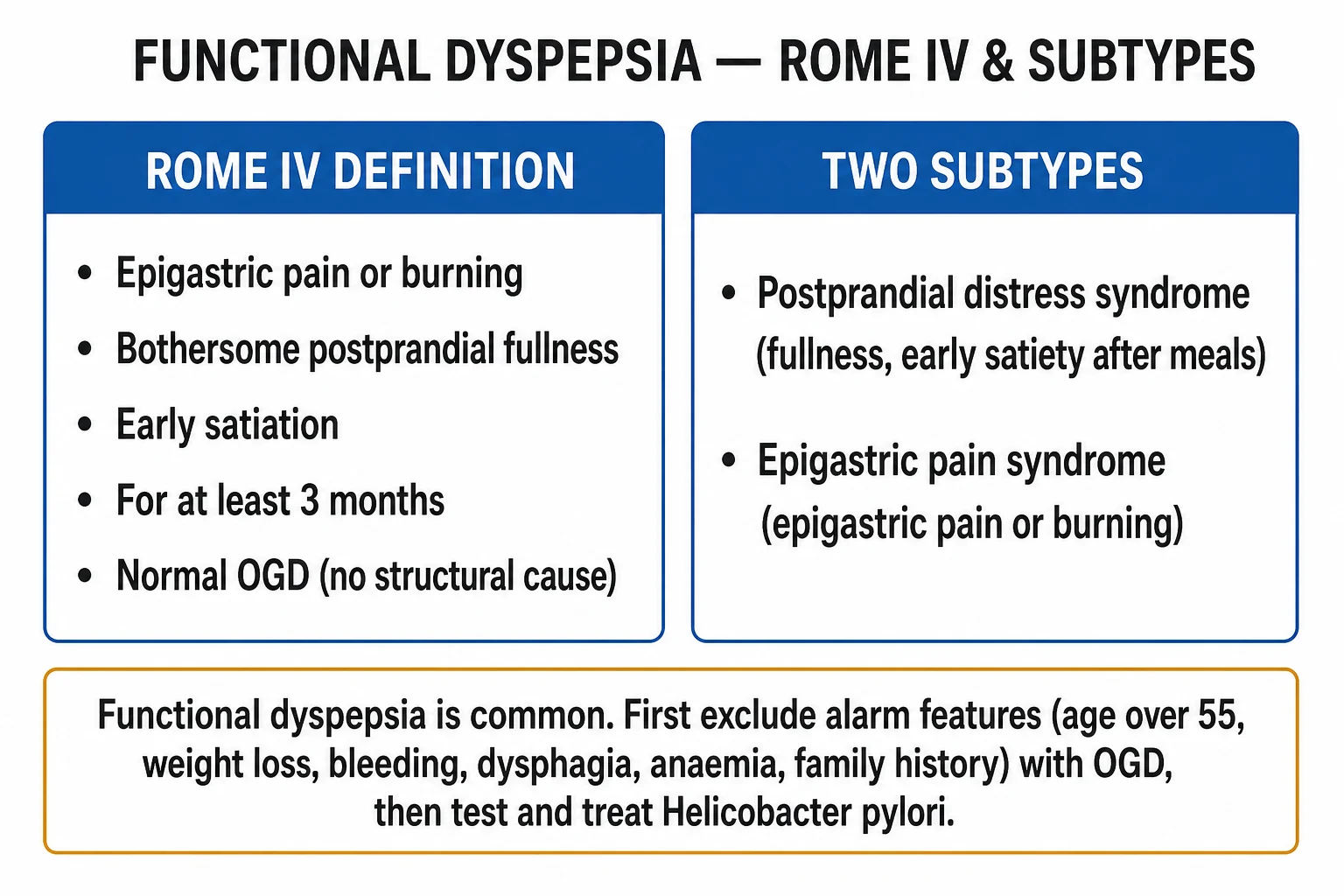
Functional dyspepsia is defined by the Rome IV consensus (Stanghellini et al., 2016) as the presence of one or more of the following bothersome symptoms: [1]
- Epigastric pain
- Epigastric burning
- Bothersome postprandial fullness
- Early satiety [1]
…with the further requirements that symptoms are sufficiently bothersome to interfere with usual activities, that they occur at least 1 day per week, that symptom onset was at least 6 months before diagnosis, that symptoms have been present for at least the last 3 months, and that no structural disease (including at upper endoscopy) explains them.[3]
The historical synonym non-ulcer dyspepsia is obsolete under Rome IV; the preferred term — adopted because it correctly frames the disorder as one of function rather than one of "absence of ulcer" — is functional dyspepsia. FD sits within the broader family of disorders of gut-brain interaction (DGBI), formerly called functional GI disorders, which also encompasses irritable bowel syndrome (IBS) and functional chest pain. Symptoms are genuine: the patient's stomach is structurally normal, but gut-brain signalling, motility, accommodation and mucosal sensitivity are disordered, producing real distress and meaningful impairment of quality of life comparable to peptic ulcer disease or diabetes.[2]
The clinical art of FD is a confident positive diagnosis after a single sensible work-up — explain the diagnosis, exclude structural disease once, address the dominant symptom, and avoid the trap of repeated normal endoscopies that reinforces illness behaviour and iatrogenic harm. The diagnosis is not one of exclusion made after endless negative tests; it is a positive diagnosis defined by the Rome IV criteria and a single normal OGD.[1][6]
Classification — the Rome IV two-subtype model
Rome IV splits functional dyspepsia into two mutually overlapping subtypes, each defined by its dominant symptom cluster and each — critically — predicting the underlying pathophysiology and therefore the first-choice therapy.[3]
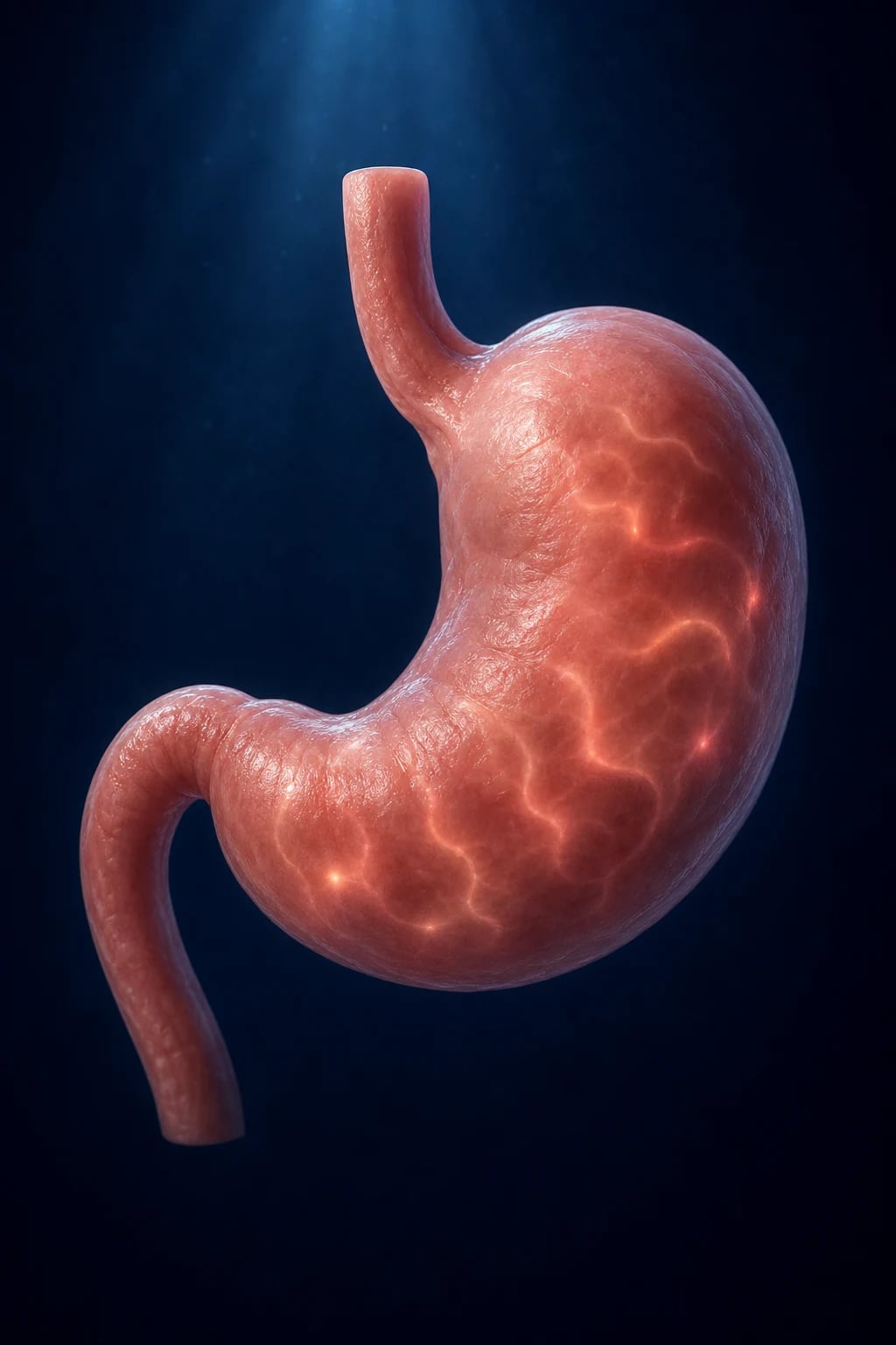
Postprandial distress syndrome (PDS)
- Bothersome postprandial fullness after ordinary-sized meals — at least 3 days per week
- Early satiety preventing finishing a regular meal — at least 3 days per week
- Underlying mechanisms — impaired gastric accommodation (~40%), delayed gastric emptying (~25-35%), duodenal eosinophilia
- First-line therapy — acotiamide 100 mg tds (Japan/India), itopride 50 mg tds, or short-course metoclopramide/domperidone; small low-fat meals; buspirone if anxiety coexists
Epigastric pain syndrome (EPS)
- Bothersome epigastric pain — at least 1 day per week
- Bothersome epigastric burning — at least 1 day per week
- Pain not generalised, not in other abdominal regions, not relieved by defecation
- Underlying mechanisms — visceral hypersensitivity, duodenal acid/eosinophil sensitivity, central sensitisation
- First-line therapy — PPI (omeprazole 20 mg od, 4-8 weeks); low-dose TCA (amitriptyline 10-25 mg nocte) if refractory
Overlap of the two subtypes is common; many patients have features of both PDS and EPS, and many also overlap with IBS (in ~40% of cases) and GORD (~30%). The practical rule is to identify and treat the dominant symptom first, then layer therapy as needed. Rome IV also requires symptoms to be bothersome (interfering with usual activities) — mild occasional symptoms do not qualify. The move from Rome III to Rome IV raised the symptom-frequency threshold (from "at least 2-3 days per week" to "at least 1 day per week" for EPS, but strengthened the bothersome qualifier) and removed heartburn as a dyspepsia symptom, reclassifying it under GORD.[2][3]
Epidemiology & Risk Factors
Functional dyspepsia — the key numbers
Functional dyspepsia is truly common — population-based studies worldwide place prevalence between 10% and 30% of adults, making it one of the single most prevalent chronic medical conditions. It accounts for up to 5% of all primary-care consultations and is the leading GI diagnosis in outpatient practice. There is a female predominance (female-to-male ratio approximately 1.5:1) and a peak onset in the 5th and 6th decades, although onset at any adult age is recognised. FD imposes substantial societal cost through healthcare utilisation, repeated endoscopies, prescription medications, and workplace absenteeism and presenteeism — estimated annual cost in the billions of dollars in developed economies.[2]
Established risk factors include: [1]
- Helicobacter pylori infection — the single most important modifiable organic co-variable; eradication cures a small subset (~7-15% relative benefit over placebo)
- Non-steroidal anti-inflammatory drugs (NSAIDs) and aspirin — irritant + mucosal injury; dose-dependent
- Smoking, obesity, high dietary fat intake — aggravate symptoms, may increase prevalence
- Female sex — consistent female predominance across populations
- Psychological factors — anxiety, depression, somatisation, neuroticism, and childhood adversity/abuse are strongly associated and bidirectionally linked (they are both risk factor and consequence)
- Post-infectious — onset within weeks to months of an acute gastroenteritis (Campylobacter, Salmonella, Giardia, norovirus); accounts for ~5-17% of FD cases
- Genetic predisposition — family aggregation; GNβ3 825C/T polymorphism (encodes G-protein beta-3 subunit, affects signal transduction) association reported but inconsistent across populations
- Dysbiosis and altered gut hormones — disturbed ghrelin (hunger signal, modulates gastric emptying), CCK (cholecystokinin, slows gastric emptying), and GLP-1 (incretin, inhibits accommodation)
- Prior abdominal surgery — may alter gastric motor function [1]
Notably, FD carries quality-of-life impairment comparable to peptic ulcer disease and diabetes mellitus — the patient's suffering is real and measurable, even though no lesion exists. FD patients also have a 2-3-fold increased prevalence of anxiety and depression compared to healthy controls, and the relationship is bidirectional: psychological distress worsens symptoms, and chronic symptoms worsen mood.[2]
Pathophysiology
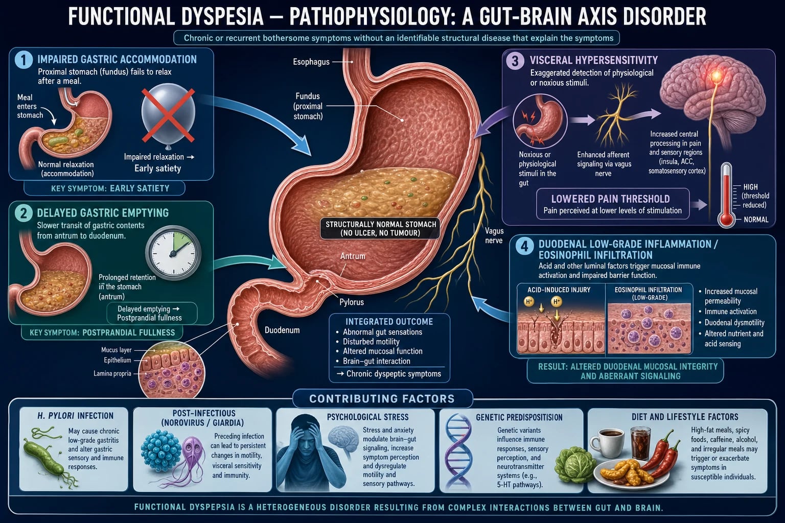
Functional dyspepsia is the prototypical disorder of gut-brain interaction — a bidirectional dysregulation of signalling between the enteric and central nervous systems, operating on a structurally intact gastroduodenal mucosa. No single mechanism explains every patient; instead, several mechanisms coexist in varying proportions, and each tends to drive one of the two Rome IV subtypes. The contemporary view, articulated in the duodenal-gastric model, is that FD may be primarily a duodenal disorder (eosinophilic inflammation, acid sensitivity) that secondarily disrupts gastric sensorimotor function — rather than a purely gastric motility disorder as historically conceived.[2][3][6]
1. Impaired gastric accommodation (a "stiff proximal stomach")
On eating, the proximal stomach (fundus) normally undergoes vagally-mediated receptive relaxation — a reflex that creates a low-pressure reservoir for the meal (gastric accommodation). This reflex is mediated by vagal nitrergic (NO) and cholinergic pathways and involves 5-HT1A receptors on vagal afferents. In about 40% of FD patients this reflex is defective, so the fundus fails to relax. Intragastric pressure rises with each bite, activating stretch-sensitive mechanoreceptors in the gastric wall, producing early satiety and postprandial fullness — the cardinal features of postprandial distress syndrome. The patient therefore typically maintains weight (does not lose it) because they stop eating early, before a significant calorie deficit accrues. Drugs that enhance accommodation (buspirone — 5-HT1A agonist, acotiamide — acetylcholinesterase inhibitor enhancing vagal tone, tandospirone) directly target this defect.[2][6]
2. Delayed gastric emptying
About 25-35% of FD patients — predominantly the postprandial distress subgroup — have objectively delayed solid gastric emptying, measurable on gastric emptying scintigraphy (the gold standard) or 13C-octanoic acid breath test. Retained food in the antrum drives fullness, nausea, bloating and postprandial discomfort. Note the important overlap with diabetic gastroparesis: a diabetic with chronic postprandial fullness may have either FD or gastroparesis — a distinction that requires measurement, not symptom pattern alone. Interestingly, the correlation between delayed emptying and symptom severity is weak, suggesting emptying is one contributor among several. Prokinetics (metoclopramide, domperidone, itopride, acotiamide, mosapride) target this mechanism by enhancing antral contractility and antro-duodenal coordination.[2]
3. Visceral hypersensitivity and central sensitisation
A defining feature of FD (shared with IBS) is a lowered pain threshold to gastric and duodenal distension — patients perceive balloon distension as painful at volumes and pressures that healthy controls barely register. This visceral hypersensitivity arises from peripheral sensitisation of mechanoreceptors (lowered activation threshold due to inflammatory mediators) and abnormal central processing in the insula, anterior cingulate cortex (ACC) and somatosensory cortex, demonstrable on functional MRI — FD patients show amplified brain activation in pain-processing regions and reduced activation in pain-modulating (descending inhibitory) regions. This mechanism — together with duodenal acid hypersensitivity — drives the epigastric pain and burning of epigastric pain syndrome, and explains why neuromodulators (TCAs, mirtazapine) help pain-predominant disease by modulating both peripheral afferent sensitivity and central pain amplification.[2][4]
4. Duodenal low-grade inflammation, eosinophilia and acid sensitivity
A striking and increasingly recognised finding is duodenal eosinophil and mast-cell infiltration in FD — particularly in post-infectious and postprandial-distress subtypes, and more prominent in the duodenal bulb than the distal duodenum. The eosinophilic infiltrate releases mediators (histamine, tryptase, major basic protein, eosinophil-derived neurotoxin) that disrupt the epithelial barrier (increased permeability), sensitise afferent nerves (via protease-activated receptors), and amplify the response to duodenal acid exposure. Duodenal acid hypersensitivity — pain on infusion of physiological acid into the duodenum — overlaps with visceral hypersensitivity and underpins the rationale for PPI therapy in EPS (reducing acid load reaching the sensitised duodenum). This duodenal eosinophilia is now considered by some authorities to be the primary driver of FD, with gastric sensorimotor dysfunction a secondary consequence.[2][6]
Post-infectious FD (after Campylobacter, Salmonella, Giardia or norovirus gastroenteritis) shows similar duodenal eosinophilia and mast-cell infiltrate, indicating an immune-mediated pathway triggered by acute enteric infection. The infiltrate may resolve over months to years, explaining the somewhat better prognosis of post-infectious FD compared to non-post-infectious forms. The risk of developing FD after gastroenteritis is approximately 5-17% in the year following infection — a 2-6-fold increase over background.[2]
5. Helicobacter pylori
H. pylori causes chronic antral gastritis and is strongly associated with peptic ulcer disease and gastric cancer. In FD, H. pylori prevalence is higher than in healthy controls, but the relationship is controversial: eradication cures symptoms permanently in a small subset (relative benefit over placebo approximately 7-15% — the number needed to treat is ~12-15 for one additional symptom cure). The mechanism is presumed to be resolution of antral inflammation and normalisation of acid secretion, but the predictor of who will respond (the "H. pylori-associated dyspepsia" phenotype) is unknown — hence the test-and-treat strategy applied to every FD patient. Crucially, after successful eradication the risk of future peptic ulcer and gastric cancer is also reduced, so eradication in FD is never wasted even when symptoms persist.[1][2]
6. Psychosocial factors, genetics and the gut-brain axis
Psychological stress, anxiety and depression act through the hypothalamic-pituitary-adrenal (HPA) axis and autonomic dysfunction, altering gastric accommodation (stress induces fundic contraction) and amplifying visceral sensitivity via central sensitisation. Early-life adversity and abuse are over-represented in FD cohorts and predict more severe, refractory disease. Genetic variation includes the GNβ3 825C/T polymorphism (affects G-protein signal transduction — the TT genotype confers increased risk in some populations but not others, reflecting gene-environment interaction). Altered ghrelin (reduced in some FD cohorts, correlating with early satiety), CCK hyper-responsiveness (slows emptying, induces fullness), and gut microbiota dysbiosis all modulate susceptibility. These multiple, interacting inputs converge on the gut-brain axis to produce a phenotype in which the stomach looks normal but feels and behaves abnormally.[2]
Clinical Presentation
The core of the diagnosis is symptom-based (Rome IV) plus a normal OGD. The four cardinal symptoms are: [1]
- Epigastric pain — typically dull, aching or gnawing, localised to the epigastrium; not related to defecation and not generalised
- Epigastric burning — often meal-related; may mimic reflux but lacks the retrosternal ascension typical of heartburn
- Bothersome postprandial fullness — an unpleasant sensation of prolonged food retention after ordinary-sized meals (PDS)
- Early satiety — inability to finish a normal-sized meal; the patient feels full shortly after starting (PDS) [1]
Frequently associated features include nausea (common, but vomiting is an alarm feature), belching, upper-abdominal bloating, and a feeling of slow digestion. Mild heartburn-like symptoms are common and create diagnostic overlap with GORD; however, under Rome IV, heartburn is NOT a dyspepsia symptom — if heartburn or acid regurgitation is the dominant complaint, classify and treat as GORD. Many patients have overlap symptoms (dyspepsia + reflux + IBS), which is the rule rather than the exception in functional GI disorders.[1][2]
Crucially, FD does NOT cause: weight loss, dysphagia, recurrent vomiting, haematemesis, melaena, iron-deficiency anaemia, or a palpable mass — the presence of any of these is an alarm feature that mandates OGD to exclude organic disease. A confident FD diagnosis also requires the patient to be systemically well — no fever, no night sweats, no progressive constitutional decline.[1]
The course is chronic and relapsing-remitting, often worsening with stress, anxiety, fatty or spicy meals, NSAIDs and alcohol, and improving with reassurance, holidays, and stable routines. Patients often describe years of intermittent symptoms with multiple normal investigations, and may exhibit health-anxiety, somatisation and depression at presentation. The temporal pattern — episodic flares against a background of chronic low-grade symptoms — is itself characteristic and reassuring.[2]
Atypical presentations
- Elderly — may have organic disease (gastric/oesophageal cancer, peptic ulcer) with minimal or atypical symptoms; always apply the age 55 or over rule and the alarm-feature screen aggressively. A reduced threshold for OGD is appropriate. Polypharmacy (NSAIDs, bisphosphonates, antiplatelets, iron, potassium) is a frequent contributor.
- Diabetic — chronic postprandial fullness may be diabetic gastroparesis rather than FD; consider gastric emptying scintigraphy and review glycaemic control. Autonomic neuropathy and prior vagotomy contribute. Distinguish by measurement, not symptom pattern.
- NSAID/aspirin users — dyspepsia may be drug-induced mucosal injury (chemical gastropathy or peptic ulcer); stop or reduce NSAID, add a PPI, consider gastroprotective alternatives.
- Pregnant — dyspepsia extremely common from reflux and reduced lower-oesophageal-sphincter pressure under progesterone; antacids/alginates first, PPI only if essential and after first trimester.
- Post-gastroenteritis — onset within weeks of an acute enteric infection (Campylobacter, Salmonella, Giardia, norovirus) suggests post-infectious FD, often with somewhat better prognosis. Male sex, predominant bloating and diarrhoea overlap (IBS-D) are typical.
- Young women with weight loss — beware anorexia nervosa or rumination syndrome presenting as "early satiety"; the weight loss is the clue that this is NOT functional dyspepsia. [1]
Differential Diagnosis
A complete differential, with distinguishing features that allow bedside separation:[1][2]
| Differential | Distinguishing features | Key test |
|---|---|---|
| Peptic ulcer disease (DU/GU) | Epigastric pain relieved by food (DU) or worsened by food with weight loss (GU); NSAID/H. pylori history; may bleed | OGD + biopsy GU |
| GORD | Heartburn, acid regurgitation, water brash; worse on lying/bending; PPI response | PPI trial; OGD if alarm features |
| Gastric or oesophageal cancer | Weight loss, dysphagia, anaemia, vomiting, palpable mass, age 55+ | Urgent OGD ± CT staging |
| Gallstone disease / biliary colic | RUQ pain post-fatty food, radiates to right scapula; Murphy sign | Abdominal ultrasound |
| Coeliac disease | Diarrhoea, weight loss, anaemia, dermatitis herpetiformis, FHx | tTG-IgA + total IgA, duodenal biopsy |
| Chronic pancreatitis / pancreatic cancer | Epigastric pain radiating to back, steatorrhoea, weight loss, jaundice | CT, faecal elastase, CA 19-9 |
| Medication-induced dyspepsia | NSAIDs, bisphosphonates, iron, potassium, metformin, antibiotics | Drug history; dechallenge |
| Irritable bowel syndrome | Pain related to defecation, altered bowel habit; overlaps FD ~40% | Rome IV IBS criteria; exclude alarm features |
| Gastroparesis (diabetic, post-vagal) | Nausea, vomiting, early satiety; diabetes, prior gastric surgery | Gastric emptying scintigraphy |
| Atypical angina / ACS | Epigastric discomfort on exertion, diaphoresis; elderly diabetic | ECG, troponin |
| Anxiety / depression / somatisation | Multiple somatic symptoms, health anxiety; no alarm features | Screen PHQ-9, GAD-7 |
| Rumination syndrome | Effortless regurgitation of recently eaten food within minutes of meals; not vomiting | Clinical diagnosis; high-resolution manometry |
| Eosinophilic gastroenteritis | Peripheral eosinophilia, diarrhoea, ascites; rare | OGD + biopsy; blood eosinophil count |
At least three discriminating features should be explicitly sought on every initial assessment: (1) presence of any alarm feature (mandates OGD), (2) relationship of pain to meals and to defecation (separates DU/GU, GORD and IBS), and (3) presence of weight loss (always organic until proven otherwise). The single most dangerous mimic is gastric cancer — missed by ignoring alarm features; the most common mimic is GORD — distinguished by symptom dominance and PPI response.[1]
Clinical & Bedside Assessment
The bedside examination is usually normal — a key diagnostic clue in itself, and one that reinforces the positive Rome IV diagnosis. Epigastric tenderness may be present but is non-specific and non-localising. The aim of examination is not to confirm FD but to seek organic disease: [1]
- General — cachexia, pallor (anaemia), jaundice (biliary/pancreatic), lymphadenopathy (Virchow's node — left supraclavicular — in gastric cancer; Sister Mary Joseph node — periumbilical)
- Abdomen — epigastric tenderness (non-specific), mass (cancer), hepatomegaly (metastases), succussion splash (gastric outlet obstruction or severe gastroparesis — audible splash on shaking the abdomen), guarding/rigidity (peritonism)
- DRE — if bleeding suspected (melaena — black, tarry, foul-smelling stool); mass (rectal cancer)
- Dysphagia assessment — if reported, this is an alarm feature mandating urgent OGD [1]
The decision-defining bedside manoeuvre is the ALARM-FEATURE SCREEN. Memorise it: [1]
WEB-VOMIT
unintentional, significant — always organic until proven otherwise
or other palpable abdominal mass — cancer until excluded
haematemesis, melaena, or unexplained iron-deficiency anaemia
recurrent, persistent, or unexplained — obstruction or cancer
55 years or over at onset — NICE threshold for urgent OGD
iron-deficiency anaemia, unexplained — investigate
dysphagia or odynophagia — oesophageal cancer until excluded
family history of upper-GI cancer — lower OGD threshold
A positive screen mandates urgent OGD (within 2 weeks under the NICE suspected-cancer pathway). A negative screen permits non-invasive management in patients under 55 with new-onset, uncomplicated dyspepsia.[1]
Quantify symptom burden with a validated tool such as the Nepean Dyspepsia Index (NDI) or Leeds Dyspepsia Questionnaire (LDQ) — the NDI is the most widely used and captures both symptom severity and quality-of-life impact. Screen for psychological comorbidity with PHQ-9 (depression — score ≥10 suggests moderate depression) and GAD-7 (anxiety — score ≥10 suggests moderate anxiety) — both strongly predict severity, healthcare use, treatment response and prognosis, and should inform the management plan.[2]
Investigations
The diagnosis of FD is symptom-based (Rome IV) PLUS a normal OGD. The investigation strategy is risk-stratified and minimally invasive wherever possible, with OGD reserved for those at higher risk of organic disease.[1]
Investigation strategy (risk-stratified)
Under 55, NO alarm features
- Non-invasive 'test-and-treat' H. pylori (urea breath test or stool antigen)
- If H. pylori positive — eradicate; confirm eradication 4 weeks later with urea breath test
- Trial PPI 4-8 weeks; lifestyle advice; reassurance
- Avoid routine OGD; avoid repeated endoscopy — a normal OGD does not improve outcome in low-risk patients
Age 55 or over, OR any alarm feature
- Urgent OGD (within 2 weeks — NICE suspected-cancer pathway)
- OGD excludes ulcer, oesophagitis, cancer; biopsy any gastric ulcer (always) to exclude malignancy
- Test H. pylori on biopsy (rapid urease/CLO test) or non-invasively
- Bloods (FBC, coeliac serology, LFTs, U&E, CRP), consider abdominal US/CT if indicated by symptoms
Specific tests
- Upper GI endoscopy (OGD) — the single most useful test in dyspepsia; excludes organic disease and, by being normal, confirms the FD diagnosis. Indications: any alarm feature; age 55 or over at onset (NICE) or 60 or over (ACG); persistent symptoms despite adequate PPI trial; family history of upper-GI cancer. Biopsy any gastric ulcer to exclude malignancy (always — "no gastric ulcer should go unbiopsied"). A normal OGD is itself part of the FD definition. OGD should ideally be performed off PPIs for at least 2 weeks to avoid masking oesophagitis and H. pylori.
- H. pylori testing — choice depends on whether endoscopy is being done:
- Non-invasive (preferred first-line in test-and-treat): urea breath test (13C/14C) or stool antigen (monoclonal, most sensitive); both detect active infection and are suitable for confirmation of eradication
- Serology (IgG) — can NOT distinguish active from past infection; not suitable for confirming eradication; useful only for epidemiological screening
- Biopsy-based (during OGD): rapid urease test (CLO test — yellow-to-red colour change), histology (gold standard for morphology), culture (for antibiotic sensitivity in treatment failure)
- Confirm eradication at least 4 weeks after completing therapy with urea breath test or stool antigen (PPI stopped for at least 2 weeks beforehand to avoid false negatives)
- Bloods — FBC (exclude iron-deficiency anaemia), coeliac serology (tTG-IgA with total IgA — FD can mimic coeliac disease and the distinction matters; coeliac has specific treatment), LFTs, amylase/lipase (pancreatic), glucose/HbA1c (diabetes screen), TSH (thyroid disease can cause GI symptoms), CRP/ESR (inflammation suggests organic disease)
- Abdominal ultrasound — only if biliary symptoms (RUQ pain post-fatty food, radiation to scapula); avoid routine imaging without pointers — low yield in FD
- CT abdomen/pelvis — if weight loss, mass, or suspicion of pancreatic/other malignancy; not routine
- 24-hour pH-impedance study — if reflux symptoms dominate and PPI fails, or before attributing symptoms to FD (to quantify acid exposure objectively)
- Gastric emptying scintigraphy — if gastroparesis is suspected (diabetic, severe nausea/vomiting, succussion splash); measures retention of a radiolabelled solid meal at 2 and 4 hours
- High-resolution oesophageal manometry — rarely; if achalasia is a mimic (dysphagia to liquids and solids, regurgitation) [1]
Avoid repeated endoscopy once OGD is normal and no new alarm features have appeared — repeated normal endoscopies reinforce illness behaviour, cost money, confer no diagnostic yield, and may cause iatrogenic anxiety.[1][2]
Management — Resuscitation
Functional dyspepsia is a chronic non-emergency; there is no time-critical resuscitation bundle in the classical sense. However, several emergent escalations must be recognised — these indicate organic disease has supervened on (or masqueraded as) FD: [1]
- New haematemesis or melaena — manage as acute upper-GI bleeding: two large-bore IV cannulae, fluid resuscitation (crystalloid), bloods including crossmatch and group-and-save, restrict transfusion to Hb < 70 g/L (under 80 g/L if cardiovascular disease); urgent OGD within 24 h. This is a red flag indicating organic disease (ulcer, cancer, varices) has supervened.
- New iron-deficiency anaemia — investigate with OGD ± colonoscopy (the "two-way endoscopy" workup); transfuse per protocol if symptomatic or Hb < 70 g/L; iron replacement (oral ferrous sulphate 200 mg tds, or IV ferric carboxymaltose if intolerant).
- Severe weight loss or new dysphagia — urgent OGD to exclude malignancy; these are never part of FD.
- Suicidal ideation from severe comorbid depression — psychiatric crisis assessment; safety plan; do not assume symptoms are "functional" and dismiss the patient. [1]
In the stable chronic patient, the acute intervention is diagnosis, explanation and reassurance — a powerful therapeutic act in itself that reduces symptom anxiety and healthcare-seeking behaviour.[1]
Management — Definitive & Stepwise
Management is stepwise, symptom-targeted, and explicit. The principle: address the dominant symptom, escalate after an adequate trial (4-8 weeks per step), and reassess for new alarm features at each visit. Each step carries a named drug with dose, route, timing, rationale and monitoring. Both the ACG/CAG guideline and NICE recommend a trial of PPI followed by step-up to prokinetics or neuromodulators, with test-and-treat H. pylori at any stage.[1][2]
Step 1 — Explanation, reassurance and lifestyle
The cornerstone and often the most powerful intervention. Explain in plain language: the diagnosis is real (not "in your head"), the symptoms arise from disordered gut-brain function, the stomach is structurally normal, and the prognosis is benign (no cancer, ulcer, or serious disease). Reinforce that repeated normal endoscopies are not needed and may cause harm. This positive framing — moving from "we found nothing" to "you have a recognised disorder of gut-brain function" — is itself therapeutic and reduces symptom severity and healthcare use. Lifestyle measures:[1][2]
- Small, frequent, low-fat meals — high-fat meals delay gastric emptying and impair fundic accommodation; eat 5-6 small meals rather than 3 large ones
- Avoid triggers: fatty foods, spicy foods, coffee (stimulates acid secretion), citrus, onions, alcohol, NSAIDs and aspirin (mucosal irritants), smoking
- Chew food thoroughly; eat slowly; avoid eating late at night (allow 3 hours before lying down)
- Stress management, regular sleep, moderate exercise — all improve gut-brain function and symptom threshold
- Weight loss if overweight/obese — reduces intra-abdominal pressure and reflux overlap [1]
Step 2 — Test-and-treat Helicobacter pylori
Stool antigen or urea breath test in every FD patient without prior documented infection or recent antibiotic/PPI use (stop PPI 2 weeks before testing). If positive, eradicate:[1]
- First-line (low clarithromycin-resistance areas, penicillin-tolerant): PPI bid + clarithromycin 500 mg bid + amoxicillin 1 g bid — 14 days
- Penicillin-allergic or high clarithromycin resistance: bismuth quadruple therapy (PPI bid + bismuth 525 mg qid + tetracycline 500 mg qid + metronidazole 500 mg qid/tid — 10-14 days), or PPI + clarithromycin + metronidazole
- Confirm eradication at least 4 weeks after completing therapy with urea breath test or stool antigen (PPI stopped 2 weeks beforehand) [1]
Rationale: eradication cures a small but worthwhile subset (relative benefit ~7-15%, NNT ~12-15) and reduces future peptic ulcer and gastric cancer risk — making it cost-effective first-line even when symptom response is modest. Monitoring: drug side-effects (clarithromycin — GI upset, taste disturbance; metronidazole — disulfiram-like alcohol interaction; bismuth — black stools, darkening of tongue).[1][2]
Step 3 — Proton-pump inhibitor (PPI)
For epigastric pain syndrome (EPS) and reflux-like symptoms — the PPI is first-line for EPS:[1]
- Omeprazole 20-40 mg once daily (alternatives: esomeprazole 20-40 mg, pantoprazole 40 mg, rabeprazole 20 mg, lansoprazole 30 mg) for 4-8 weeks
- Response: modest but statistically significant benefit over placebo (relative risk reduction ~14%, NNT ~11); greatest in EPS (pain/burning), least in PDS (fullness/early satiety)
- If responding, taper to lowest effective dose; if asymptomatic, consider intermittent or on-demand therapy rather than continuous long-term PPI
- H2-receptor antagonists (e.g., ranitidine withdrawn in many markets due to NDMA contamination; famotidine 20 mg bid or nizatidine 150 mg bid) are an alternative for PPI-intolerant patients or step-down therapy [1]
Risks of long-term PPI (minimise exposure): Clostridioides difficile infection, community-acquired pneumonia, osteoporotic fracture, hypomagnesaemia (check annually if long-term), vitamin B12 deficiency, rebound acid hypersecretion on cessation. Monitoring: review need annually; deprescribe if symptom-free.[1]
Step 4 — Prokinetics
For postprandial distress syndrome (PDS) — fullness, early satiety, nausea — the prokinetic is first-line for PDS:[1][5]
- Metoclopramide 10 mg three times daily before meals — MHRA: maximum 5 days due to extrapyramidal effects and tardive dyskinesia (black-box warning); avoid in Parkinson's disease, young adults (under 20), pregnancy (first trimester). Stop immediately if extrapyramidal effects occur (acute dystonia, akathisia, parkinsonism).
- Domperidone 10-20 mg three times daily — does not cross the blood-brain barrier (fewer extrapyramidal effects) but QT prolongation and sudden cardiac death risk — ECG before and during, maximum 7 days, avoid with QT-prolonging drugs (macrolides, fluoroquinolones, antipsychotics), hepatic impairment, and structural heart disease.
- Itopride 50 mg three times daily — dopamine D2 antagonist with acetylcholinesterase inhibition; available in India/Asia; favourable cardiac profile (no QT risk); reasonable first-line prokinetic in regions where available.
- Acotiamide 100 mg three times daily before meals — enhances gastric accommodation and accelerates emptying via acetylcholinesterase inhibition (increases vagal ACh release); approved in Japan and India for PDS, not in US, UK, or EU; effective for early satiety and postprandial fullness; well-tolerated (main side effects: nausea, diarrhoea).[5]
- Mosapride 5 mg three times daily — selective 5-HT4 agonist; available in Japan/Asia; prokinetic without QT risk; limited data.
Rationale: enhance gastric emptying and accommodation. Monitoring: extrapyramidal effects (metoclopramide — stop immediately), QT interval (domperidone — ECG), prolactin (galactorrhoea, gynaecomastia — metoclopramide and domperidone cross the pituitary dopamine threshold).[1][5]
Step 5 — Neuromodulators (for refractory pain, nausea, sleep disturbance)
If PPI and/or prokinetic fail after an adequate trial, a neuromodulator is indicated — particularly for pain-predominant (EPS) refractory disease:[4]
- Amitriptyline 10-25 mg at night, titrate to 50-75 mg if tolerated over 4-8 weeks — best evidence among neuromodulators (Ford meta-analysis, Gut 2017); helps pain, sleep, visceral sensitivity; start low and slow to minimise anticholinergic effects (dry mouth, constipation, urinary retention, sedation, blurred vision); avoid in glaucoma, prostate hypertrophy, recent MI; ECG for QT before high doses.[4]
- Mirtazapine 15-30 mg at night — noradrenergic and specific serotonergic antidepressant (NaSSA); particularly useful when weight loss, nausea and insomnia coexist (5-HT3 antagonism + appetite stimulation + sleep enhancement); sedation and weight gain are the main side effects; valuable for the "loss of appetite, poor sleep, nausea" phenotype.
- Buspirone 10-15 mg three times daily — 5-HT1A agonist that enhances gastric accommodation (the key PDS mechanism); useful in PDS with coexisting anxiety; anxiolytic without TCA side effects; takes 2-4 weeks for full effect.
- SSRIs (e.g., sertraline 50 mg od, escitalopram 10 mg od) — preferred when comorbid anxiety/depression dominates (treat the mood disorder); less analgesic than TCAs but better tolerated; useful when pain is secondary to mood rather than visceral hypersensitivity per se.[4]
Rationale: neuromodulation of visceral afferents and central pain processing — these drugs work at doses below those used for major depression, and their benefit is independent of antidepressant effect. Monitoring: anticholinergic effects, QT (TCAs), serotonin syndrome (SSRIs/SNRIs), suicidality (all antidepressants — FDA black box in under-25s).[4]
Step 6 — Psychological therapy
For refractory disease, particularly with psychological comorbidity — growing evidence supports gut-brain therapies:[2][4]
- Cognitive behavioural therapy (CBT) — best trial evidence; addresses symptom catastrophising, hypervigilance, avoidance behaviour, and maladaptive illness beliefs; 8-12 sessions; benefit sustained at 12-month follow-up
- Gut-directed hypnotherapy — growing evidence of benefit; particularly for PDS and for patients with high somatisation scores; teaches visceral relaxation
- Psychodynamic interpersonal therapy — useful for interpersonal/abuse-related triggers and complex psychosomatic presentations [1]
Step 7 — Dietary and complementary therapy
- Low-fat, smaller meals; chew thoroughly; avoid late meals
- Low-FODMAP diet — trial 4-6 weeks then reintroduce; most useful in FD-IBS overlap (reduces fermentable carbohydrate load that drives bloating and gas)
- Peppermint oil — if coexistent spasm (note: may worsen reflux by relaxing the lower oesophageal sphincter)
- Herbal preparations — Iberogast (STW 5), a fixed-combination herbal preparation, shows modest benefit in some trials; carob flour and pink frangula (alder buckthorn) are traditional remedies with limited evidence; artichoke leaf extract has some supportive data
- Fundic-guided gastric accommodation therapy — diaphragmatic breathing exercises to enhance vagal tone and fundic relaxation; emerging non-pharmacological approach for PDS [1]
Escalation triggers: failure of one step after an adequate trial (4-8 weeks), new alarm features (repeat OGD), intolerable drug side-effects, diagnostic uncertainty (refer to gastroenterology). Do not escalate to opioids — they worsen gut motility, cause central sensitisation, and have no role in FD.[1]
[1]Specific Subtypes & Scenarios
Postprandial distress syndrome (PDS)
Dominant: bothersome fullness and early satiety after ordinary-sized meals (at least 3 days/week). Mechanism: impaired gastric accommodation (~40%), delayed gastric emptying (~25-35%), duodenal eosinophilia. First-line: acotiamide 100 mg tds (where available — Japan/India), itopride 50 mg tds, or short-course metoclopramide/domperidone; small low-fat meals; buspirone 10-15 mg tds if anxiety coexists. Second-line: mirtazapine 15 mg nocte (if weight loss/nausea), CBT, gut-directed hypnotherapy. PDS is generally harder to treat than EPS and has weaker evidence for PPI benefit.[2][5]
Epigastric pain syndrome (EPS)
Dominant: bothersome epigastric pain or burning (at least 1 day/week), not related to defecation. Mechanism: visceral hypersensitivity, duodenal acid/eosinophil sensitivity, central sensitisation. First-line: PPI 4-8 weeks (omeprazole 20-40 mg od); if refractory, low-dose amitriptyline 10-25 mg nocte (best-evidenced neuromodulator for EPS). EPS is generally more responsive to therapy than PDS.[1][4]
Overlap (PDS + EPS)
Common — treat the dominant symptom first, then layer a second agent from the other class (e.g., PPI + prokinetic, then add TCA for residual pain). Many patients will need two agents at steady state. [1]
Post-infectious FD
Onset within weeks of an acute gastroenteritis (Campylobacter, Salmonella, Giardia, norovirus). Characterised by duodenal eosinophilia, more common in males, and often overlaps with IBS-D. Better prognosis — symptoms often resolve over months to years. Treat as FD; prokinetics often first-line. The immune-mediated mechanism suggests a role for anti-inflammatory or anti-eosinophil strategies in future, though these are not yet standard.[2]
Refractory FD
Failure of the stepwise ladder after adequate trials of lifestyle, H. pylori eradication, PPI, prokinetic, and neuromodulator. Reassess systematically: (1) repeat OGD if any new alarm feature has appeared; (2) review medication — is the patient still taking NSAIDs or aspirin?; (3) re-confirm the diagnosis — was coeliac serology done? Could this be gastroparesis (gastric emptying study)? Rumination syndrome?; (4) address psychological comorbidity (PHQ-9, GAD-7 → treat anxiety/depression actively); (5) refer to tertiary GI / functional bowel service for CBT, gut-directed hypnotherapy, or trial novel neuromodulation (e.g., gastric percutaneous electrical stimulation — experimental). Do not escalate to opioids.[1][2]
Overlap syndromes (FD-IBS, FD-GORD)
Very common — manage both disorders simultaneously. Low-FODMAP diet for the IBS component; PPI for GORD; prokinetic/TCA for FD; CBT for the shared gut-brain substrate. Patients with triple overlap (FD + IBS + GORD) have the greatest symptom burden and lowest quality of life — they need the most integrated, multidisciplinary care.[2]
Complications & Pitfalls
- No structural complications and no excess mortality — FD is a benign condition in terms of organic harm. However, there are substantial quality-of-life consequences: impaired productivity, absenteeism, anxiety, depression, somatisation, social withdrawal, and medication-related side effects that themselves become problems.
- Misdiagnosis of organic disease — the cardinal and most dangerous pitfall. Missing gastric or oesophageal cancer or peptic ulcer by ignoring alarm features is the error that causes real harm; this is why the alarm-feature screen is non-negotiable on every assessment and at every follow-up visit. Any new alarm feature in a previously diagnosed FD patient mandates repeat OGD.
- Iatrogenic harm from repeated normal endoscopies — reinforces illness behaviour ("there must be something they're missing"), costs money, exposes the patient to procedural risk (perforation, sedation, bleeding), and confers no diagnostic yield once OGD is normal.
- Medication-related harms — a major source of iatrogenic morbidity in FD:
- Long-term PPI — C. difficile colitis, community-acquired pneumonia, osteoporotic fracture, hypomagnesaemia, B12 deficiency, rebound acid hypersecretion, possible dementia association (controversial)
- Metoclopramide — extrapyramidal effects (acute dystonia, akathisia, parkinsonism), tardive dyskinesia (irreversible — MHRA max 5 days)
- Domperidone — QT prolongation, ventricular arrhythmia, sudden cardiac death (max 7 days, ECG monitoring)
- TCAs — anticholinergic effects, sedation, QT prolongation, overdose lethality (cardiotoxicity)
- Hyperprolactinaemia — metoclopramide and domperidone (galactorrhoea, gynaecomastia, amenorrhoea)
- Opioid escalation — avoid; worsens gut motility (narcotic bowel syndrome), causes central sensitisation, and has no role in FD pain management.
- Dismissive labelling — telling a patient "it's just stress" or "it's all in your head" destroys the therapeutic alliance; FD is a real disorder with measurable pathophysiology and requires empathic, validating communication.[1][2]
Prognosis & Disposition
Functional dyspepsia is a chronic relapsing-remitting condition — not dangerous, but often persistent. About half of patients improve or fluctuate over years; a minority (15-20%) have persistent troublesome symptoms requiring long-term symptom-targeted therapy, and a small subset have progressively worsening disease that significantly impairs quality of life. There is no impact on mortality — FD does not shorten life, does not progress to cancer, and does not cause peptic ulcers or bleeding.[2]
Predictors of BETTER outcome
- A confident positive diagnosis with a single sensible work-up (no repeated endoscopy)
- Strong therapeutic alliance and patient understanding of the gut-brain model
- Addressing psychological comorbidity (anxiety, depression) actively
- Limited investigations — avoiding the 'medical shopping' spiral
- Post-infectious FD — symptoms often resolve over 1-5 years
- H. pylori eradication response (in the small subset who respond)
Predictors of WORSE outcome
- Somatisation — multiple unexplained somatic symptoms across systems
- Anxiety, depression, health anxiety — bidirectional with FD severity
- Persistent healthcare-seeking and repeated normal investigations
- Childhood adversity or abuse history
- Severe baseline symptom burden and long duration before diagnosis
H. pylori eradication cures a small but worthwhile subset (~7-15% relative benefit; NNT ~12-15). Reassurance that symptoms will not lead to cancer, ulcer, or serious disease is itself therapeutic and should be delivered explicitly and repeatedly. Most patients can be managed in primary care; secondary care referral is for alarm features, refractory disease, diagnostic uncertainty, or tertiary functional-bowel input. Long-term follow-up is symptom-driven — patients return when symptoms flare, not on a fixed schedule.[1][2]
Prevention
There is no proven primary prevention for FD, but several strategies reduce symptom frequency and severity in established disease and may reduce incidence in high-risk populations:[1][2]
- Eradicate H. pylori where detected — reduces the small subset attributable to H. pylori and reduces future peptic ulcer and gastric cancer risk
- Minimise NSAID and aspirin use — or co-prescribe a PPI when unavoidable; these are major mucosal irritants
- Smoking cessation — smoking is associated with increased FD prevalence and severity
- Moderate alcohol intake — heavy alcohol use aggravates dyspeptic symptoms
- Stress management — mindfulness, CBT skills, regular exercise, adequate sleep; these modulate the gut-brain axis
- Dietary habits — regular small meals, low-fat, avoid excessive coffee; these reduce the mechanical and secretory load on the stomach
- Early treatment of mood disorders — anxiety and depression are bidirectional with FD; treating them early may prevent chronicity [1]
Special Populations
- H. pylori-positive dyspepsia — eradicate using appropriate regimen (see Step 2); confirm eradication with urea breath test 4 weeks after therapy. Eradication also reduces future peptic ulcer and gastric cancer risk, making it a worthwhile intervention even when symptoms persist. In high-prevalence regions (India, ~60-80%), test-and-treat is first-line for all dyspepsia.[1]
- Pregnancy — dyspepsia is extremely common (progesterone relaxes the LES; the gravid uterus displaces the stomach). Antacids/alginates first (Gaviscon, Mucogel — safe); avoid NSAIDs entirely. PPI only if essential and after first trimester (omeprazole — limited human data but generally considered low-risk in second/third trimester; lansoprazole preferred by some). Ranitidine withdrawn in many markets due to NDMA contamination. Avoid domperidone. Metoclopramide (category B) only with caution for severe nausea, short course. Itopride — insufficient safety data; avoid.
- Elderly — lower threshold for OGD; atypical organic presentation is the rule (gastric cancer may present as "weight loss and anorexia" without typical dyspepsia). Review polypharmacy (NSAIDs, bisphosphonates, antiplatelets, iron, potassium — all cause dyspepsia). Check renal function before PPI (hypomagnesaemia risk) and domperidone (accumulation). Review drug-drug interactions (CYP2C19 for omeprazole/esomeprazole). Consider fall risk with TCAs (sedation, orthostatic hypotension).[1]
- NSAID-associated dyspepsia — stop or reduce the NSAID; co-prescribe PPI; consider gastroprotective alternatives (paracetamol, topical NSAID, COX-2 inhibitor with PPI). The dyspepsia may be chemical gastropathy or peptic ulcer — OGD if alarm features.
- Diabetic — distinguish FD from gastroparesis (gastric emptying scintigraphy — retention > 10% at 4 hours is diagnostic); optimise glycaemic control (hyperglycaemia itself delays gastric emptying); avoid metoclopramide beyond 5 days; consider domperidone with ECG monitoring; acotiamide (where available) is a rational prokinetic. Autonomic neuropathy and prior vagotomy contribute.
- Children — FD is uncommon in children; the Rome IV paediatric criteria require symptoms for at least 2 months (vs 3 months in adults). Consider food allergy, H. pylori (especially in endemic regions), constipation, and psychological contributors (school stress, anxiety). Refer to paediatric gastroenterology; avoid off-label adult drugs; weight-based dosing for any medication used.
- Immunocompromised (HIV, transplant, chemotherapy) — consider opportunistic infection (CMV oesophagitis/gastritis, Candida oesophagitis, HSV); lower threshold for OGD; review immunosuppressive therapy (mycophenolate can cause GI ulceration).
Evidence, Guidelines & Regional Differences
- ACG/CAG 2017 Clinical Guideline (Moayyedi et al., PMID 28631728) — the key US guideline. Recommends: test-and-treat H. pylori in all FD; PPI modestly effective (NNT ~11); prokinetics equivocal evidence; TCAs (especially amitriptyline) effective; fundic relaxants/acotiamide limited evidence. Conditional recommendations based on very low quality of evidence for most therapies — reflecting the modest effect sizes in FD.[1]
- Lancet 2020 review (Ford et al., PMID 33049222) — comprehensive contemporary synthesis of pathophysiology (duodenal eosinophilia, gut-brain model) and management. Key message: no single therapy is dramatically effective; stepwise, symptom-targeted therapy is the standard.[2]
- Rome IV consensus (Stanghellini et al., 2016, Gastroenterology, PMID 27147122) — redefined FD and established the two-subtype model (PDS + EPS) that underpins all modern management. Raised symptom thresholds and strengthened the bothersome qualifier; removed heartburn from dyspepsia.[3]
- Ford et al., Gut 2017 (PMID 26567029) — systematic review and meta-analysis of psychotropic drugs in FD; found TCAs effective (NNT ~6) but SSRIs and antipsychotics not superior to placebo. Amitriptyline is the best-evidenced neuromodulator for FD.[4]
- Tack et al., Aliment Pharmacol Ther 2023 (PMID 36859629) — modern review arguing FD may be primarily a duodenal disorder (eosinophilic duodenitis) with secondary gastric sensorimotor dysfunction; reframes the disease model.[6]
- Maastricht VI/Florence 2022 consensus — H. pylori eradication regimens and indications; recommends eradication in FD as a "test-and-treat" strategy, with bismuth quadruple therapy preferred where clarithromycin resistance > 15%.
- NICE CG184 (Dyspepsia and reflux, 2014, updated 2019) — UK guideline; urgent OGD within 2 weeks under the suspected-cancer pathway for age 55 or over with recent-onset dyspepsia, treatment-resistant dyspepsia, or any alarm feature; test-and-treat H. pylori in under-55s without alarm features; step-up therapy (lifestyle → PPI → H2RA → prokinetic → TCA).
- Cochrane systematic reviews (Ford et al.) — PPI, H. pylori eradication, and prokinetics each show a small but statistically significant benefit over placebo (relative risk reduction ~10-15% each); no single agent is dramatically effective — hence the stepwise, combination approach.[2]
Regional deltas:
- Age threshold for OGD — NICE uses 55; ACG uses 60 (with alarm features). Both are pragmatic population-level thresholds balancing cancer yield against endoscopy capacity.
- Acotiamide — approved in Japan and India for PDS; not approved in US, UK, or EU (where itopride, buspirone, and TCAs fill the gap). Acotiamide is the only drug specifically approved for FD (in Japan, since 2013).
- Itopride — widely available in India, China, Southeast Asia; not approved in US/EU. Used as first-line prokinetic in these regions.
- H. pylori eradication regimens — clarithromycin resistance varies by region (> 15% in much of South and East Asia, Southern Europe): use bismuth quadruple therapy as first-line in high-resistance areas. India: high H. pylori prevalence (~60-80%), so test-and-treat is first-line and OGD performed more liberally given higher background gastric cancer risk in older patients.
- Ranitidine — withdrawn globally (2020) due to NDMA contamination; replaced by famotidine and nizatidine as H2-receptor antagonists.
- Metoclopramide — MHRA/EMA/FDA: maximum 5 days (tardive dyskinesia); this is a global restriction.
Controversies
- Role of duodenal eosinophilia — whether anti-eosinophil therapy (montelukast, anti-IL-5) has a role in FD is under investigation; no approved treatment yet targets this mechanism specifically.
- Efficacy of CBT and gut-directed hypnotherapy — growing evidence but limited access and cost; trials show benefit but generalisability to routine practice is uncertain.
- Low-FODMAP diet in FD — evidence strongest for IBS overlap; in pure FD, benefit is uncertain.
- Probiotics — insufficient evidence to recommend routinely; some trials suggest modest benefit.
- Whether FD is "gastric" or "duodenal" — the Tack 2023 review (PMID 36859629) argues for a primary duodenal origin; this debate is ongoing and may reshape treatment targets in the next decade.[6]
Exam Pearls
The six pearls that decide a functional-dyspepsia answer
- Rome IV definition — one or more of epigastric pain, epigastric burning, bothersome postprandial fullness, early satiety; onset 6+ months before diagnosis; present 3+ months; sufficiently bothersome to interfere with usual activities; at least 1 day/week; NORMAL OGD.[3]
- Two subtypes: PDS (fullness/early satiety, 3+ days/week → prokinetic/acotiamide) and EPS (pain/burning, 1+ day/week → PPI → TCA)
- Alarm features — Weight loss, Epigastric mass, Bleeding/anaemia, Vomiting, Over 55, Marrow anaemia, Inability to swallow (dysphagia), Tendency (family history) — mandate urgent OGD
- Strategy — under 55 without alarm features: test-and-treat H. pylori; 55 or over or any alarm feature: urgent OGD within 2 weeks (NICE)[1]
- Management ladder: lifestyle → test-and-treat H. pylori → PPI (EPS) → prokinetic/acotiamide (PDS) → low-dose TCA/amitriptyline → mirtazapine → CBT/gut-directed hypnotherapy
- H. pylori eradication cures a small minority (~7-15% relative benefit; NNT ~12-15) but is cost-effective first-line and reduces future ulcer/cancer risk
- Metoclopramide — black-box tardive dyskinesia, MHRA max 5 days; domperidone — QT prolongation, max 7 days, ECG — a favourite pharmacology-viva trap
- Acotiamide — enhances gastric accommodation (AChE inhibition), approved Japan/India for PDS, not US/UK/EU — high-yield for NEET-PG/INICET[5]
- Amitriptyline 10-25 mg nocte is the best-evidenced neuromodulator for FD (Ford meta-analysis, Gut 2017) — works below antidepressant doses, independent of mood effect[4]
- Gut-brain disorder — impaired Accommodation, Delayed emptying, visceral Pain hypersensitivity, duodenal low-grade inflammation/eosinophilia (mnemonic A-D-P-E)
- Avoid repeated endoscopy once OGD is normal and no new alarm features — re-scoping reinforces illness behaviour and is iatrogenic harm
- Overlap with IBS (~40%) and GORD (~30%) is common — manage both disorders; the patient with triple overlap has the worst quality of life
- FD does NOT cause weight loss, dysphagia, vomiting, bleeding, anaemia, or mass — any of these = alarm feature = OGD
- No opioids for FD pain — they worsen motility and central sensitisation
Exam application bank (NEET-PG / INICET)
One-line answer
Functional dyspepsia is chronic (at least 3 months) epigastric pain or burning, bothersome postprandial fullness or early satiety, with no structural cause on endoscopy (including normal OGD). It is a disorder of gut-brain interaction driven by gastroduodenal dysmotility, visceral hypersensitivity, duodenal eosinophilic inflammation and low-grade mucosal immune activation, and is divided into two subtypes — postprandial distress syndrome (fullness, early satiety) and epigastric pain syndrome (pain, burning). Diagnosis requires excluding alarm features (age 55 or over, weight loss, bleeding, dysphagia, anaemia, family history) with OGD in selected patients and testing for H. pylori. Management is stepwise — reassurance and lifestyle measures, then test-and-treat H. pylori, a PPI trial, prokinetics, and for refractory disease low-dose tricyclic antidepressants or CBT.
Worked stems (answer without another resource)
Stem 1 — Classic presentation. Map symptoms to mechanism; name the first investigation and first treatment step with dose/route if drug therapy is standard. [1]
Stem 2 — Unstable / complicated. List red flags that force immediate resuscitation, theatre, ICU, antidote, or reperfusion — and what you do in the first 15 minutes. [1]
Stem 3 — Atypical group. Elderly, pregnancy, child, or immunocompromised: how presentation and thresholds change. [1]
Stem 4 — Differential trap. Name the three closest mimics and one discriminator for each. [1]
Stem 5 — Disposition. Who goes home with safety-netting, who is admitted, who needs HDU/ICU/theatre, and what follow-up is mandatory. [1]
Rapid viva checklist
- Definition + classification
- Pathophysiology chain
- Bedside signs / criteria
- Score with exact components (if any)
- Emergency bundle
- Definitive therapy with doses
- Complications of disease and of treatment
- Special populations
- Guideline/trial name if classic
- Three exam traps
Coverage self-check
If you cannot answer any stem above from this page alone, re-read the matching section — the page is intended to be self-sufficient for final-prof and NEET-PG/INICET questions on Functional Dyspepsia.
References
- [1]Moayyedi P, Lacy BE, Andrews CN, Enns RA, Howden CW, Vakil N. ACG and CAG Clinical Guideline: Management of Dyspepsia Am J Gastroenterol, 2017.PMID 28631728
- [2]Ford AC, Mahadeva S, Carbone MF, Lacy BE, Talley NJ. Functional dyspepsia Lancet, 2020.PMID 33049222
- [3]Stanghellini V, Chan FK, Hasler WL, Malagelada JR, Suzuki H, Tack J, Talley NJ. Gastroduodenal Disorders Gastroenterology, 2016.PMID 27147122
- [4]Ford AC, Luthra P, Tack J, Schoenfeld PS, Talley NJ, Moayyedi P. Efficacy of psychotropic drugs in functional dyspepsia: systematic review and meta-analysis Gut, 2017.PMID 26567029
- [5]Nakamura K, Kamada M, Watanabe O, et al. Efficacy and Safety of Orally Administered Acotiamide Extended-Release Tablets Among Functional Dyspepsia-Postprandial Distress Syndrome Patients: A Randomized, Double-Blind, Multicenter Study Cureus, 2021.PMID 33968542
- [6]Tack J, Bisschops R, Carbone F. Review article: Functional dyspepsia-a gastric disorder, a duodenal disorder or a combination of both? Aliment Pharmacol Ther, 2023.PMID 36859629