Gastroenterology · General Surgery
Gallstone Disease & Cholecystitis
Also known as Gallstone disease · Cholelithiasis · Cholecystitis · Biliary colic · Choledocholithiasis · Ascending cholangitis
Gallstone disease (cholelithiasis) is the formation of solid crystalline concretions in the gallbladder or biliary tree from precipitated bile components — most commonly cholesterol (the classic risk profile of the 5 Fs — female, forty, fat, fertile, fair, plus rapid weight loss, family history and OCP use) and less often pigment stones from chronic haemolysis (black) or bacterial biliary infection (brown). Sixty to eighty per cent are silent; symptomatic disease spans biliary colic (postprandial RUQ pain, self-limiting), acute cholecystitis (persistent RUQ pain, fever, positive Murphy sign), choledocholithiasis (obstructive jaundice) and the septic emergency of ascending cholangitis (Charcot triad of fever, jaundice and RUQ pain; Reynolds pentad adds hypotension and confusion). Severity is graded by the Tokyo Guidelines 2018 for both cholecystitis and cholangitis. Ultrasound is first-line; MRCP images the ducts non-invasively; ERCP removes common-duct stones and drains an obstructed system. Management is conservative for asymptomatic stones, laparoscopic cholecystectomy (using the critical view of safety) for symptomatic disease, early cholecystectomy within 72 hours for acute cholecystitis, ERCP with sphincterotomy for CBD stones, and antibiotics plus urgent ERCP biliary decompression for cholangitis.
On this page & tools
Your progress
Saved locally on this device.
Exam tags
Red flags
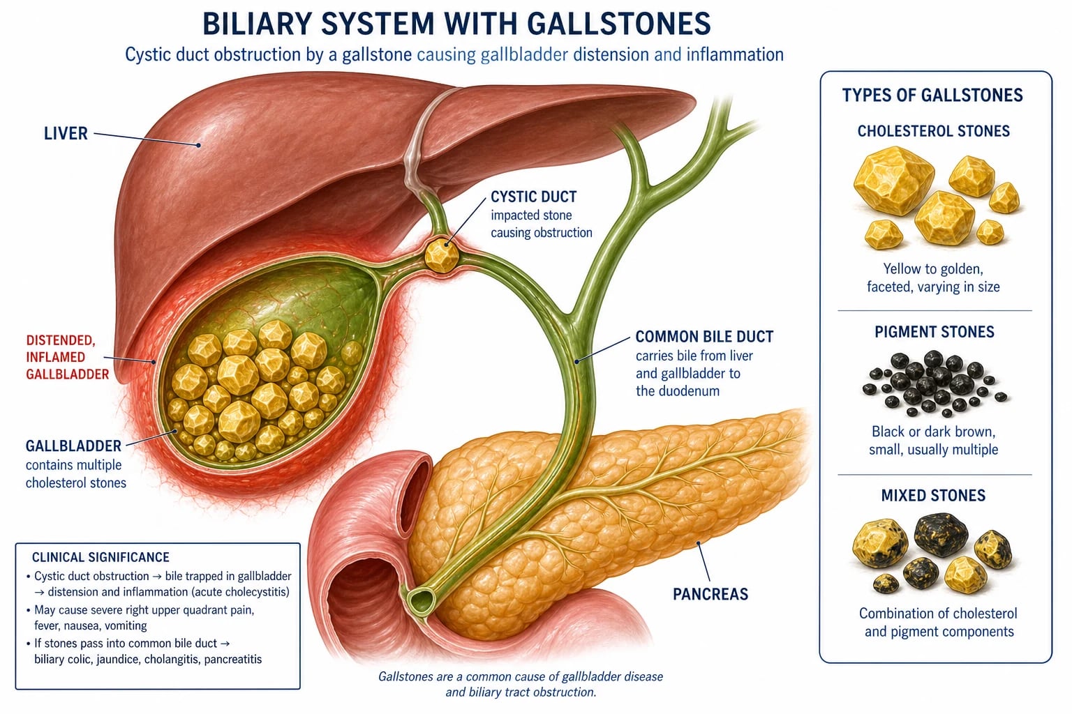
Overview & Definition
Gallstone disease (cholelithiasis) is the presence of solid crystalline concretions — gallstones — within the gallbladder or, when they migrate, the biliary tree. Stones form when one or more of the solutes normally held in solution in bile — cholesterol, unconjugated bilirubin (as calcium bilirubinate) or calcium salts — precipitates and aggregates.[1]
Three anatomical terms define the disease spectrum and are used precisely in exams: [1]
- Cholelithiasis — stones contained within the gallbladder.
- Choledocholithiasis — stones within the common bile duct (CBD), either primary (formed de novo in the duct, classically brown pigment) or secondary (migrated from the gallbladder).
- Cholecystitis — inflammation of the gallbladder wall, almost always triggered by cystic duct obstruction (calculous); rarely acalculous in the critically ill. [1]
Biliary infection layered on obstruction produces ascending (acute) cholangitis — infected, obstructed bile under pressure, a true septic emergency. A stone transiently plugging the ampulla causes gallstone (biliary) pancreatitis. [1]
The clinical task is not making the diagnosis — most cases are obvious on bedside ultrasound — but recognising where the patient sits on the spectrum and acting at the correct level of urgency: [1]
- Reassure the asymptomatic patient (incidental stones need no treatment).
- Offer elective laparoscopic cholecystectomy for biliary colic.
- Treat acute cholecystitis with antibiotics and early (within 72 hours) cholecystectomy.
- Treat ascending cholangitis as a sepsis-six emergency with antibiotics and urgent ERCP biliary decompression.[5][7]
The natural history is favourable for silent stones (1-4% per year become symptomatic),[11] but a long-standing stone burden, especially stones over 3 cm and porcelain (calcified) gallbladder, is associated with gallbladder carcinoma, which is why selected asymptomatic patients are offered prophylactic cholecystectomy.[12]
Classification
Gallstones are classified by their dominant biochemical composition, which in turn reflects their pathogenesis and points to systemic disease in the patient: [1]
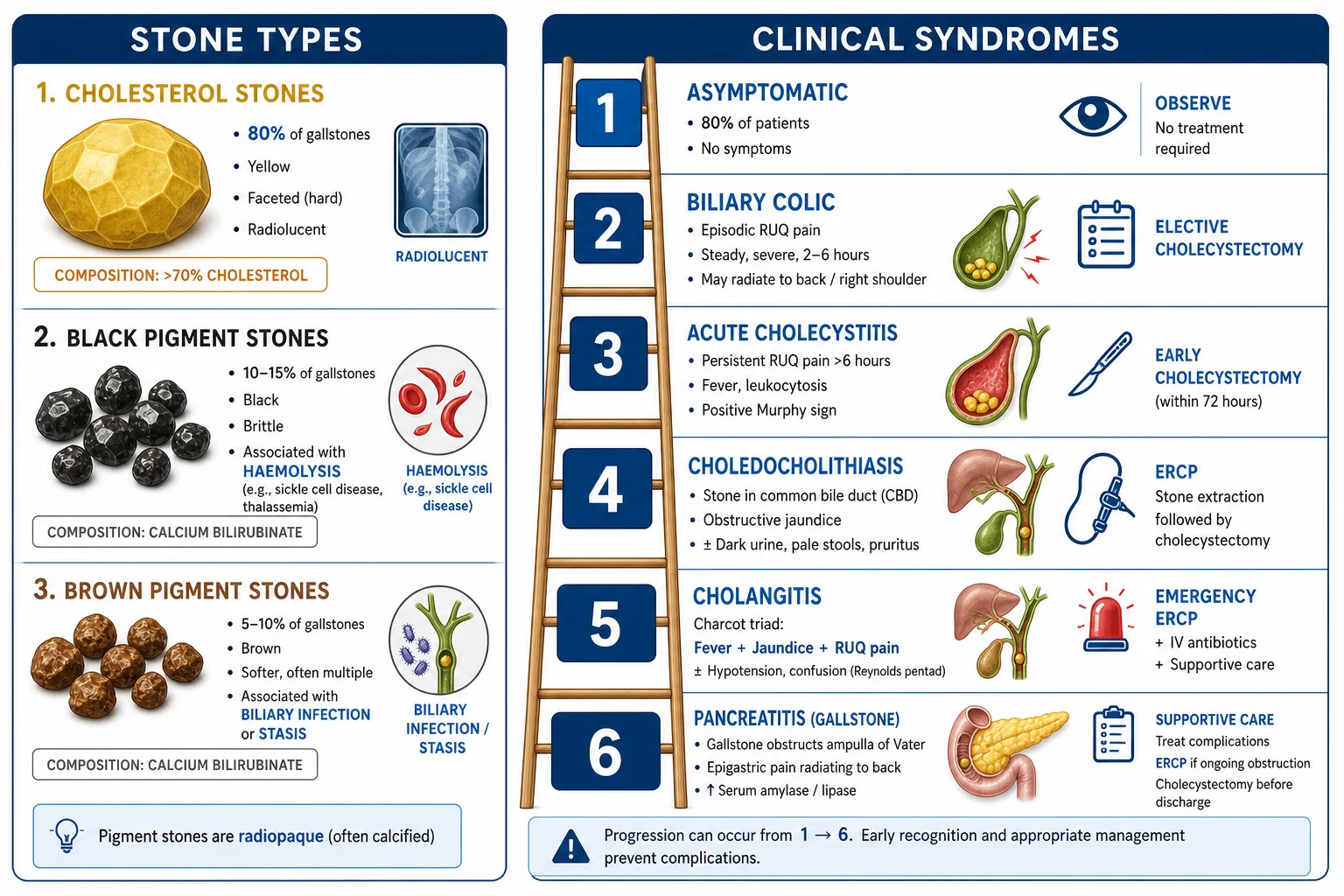
A second axis of classification — by clinical syndrome — is more useful at the bedside, because the same stone causes very different diseases depending on where it lodges: [1]
- Asymptomatic cholelithiasis (60-80%) — incidental stones.
- Biliary colic — symptomatic stones without gallbladder inflammation (transient cystic duct obstruction).
- Acute cholecystitis — sustained cystic duct obstruction with inflammation.
- Choledocholithiasis — CBD stone with obstructive jaundice (or silent).
- Acute cholangitis — infected, obstructed bile.
- Gallstone pancreatitis — ampullary obstruction triggering pancreatic enzyme activation.
- Gallstone ileus / Bouveret syndrome — fistula and mechanical bowel obstruction.
- Mirizzi syndrome — extrinsic compression of the common hepatic duct. [1]
Epidemiology & Risk Factors
Gallstones are among the commonest digestive diseases. Ten to fifteen per cent of adults in developed countries harbour gallstones; the lifetime risk in women approaches 25-30%. Prevalence rises with age (10% at 40 years, 20-30% over 60 years) and the female-to-male ratio is roughly 2:1 in the reproductive years, narrowing after menopause.[1]
The 5 Fs (and a 6th)
Ethnic and geographic variation is striking: the highest prevalence in the world is in Pima Indians of Arizona (up to 70% of adult women), Native Americans, Chileans and Northern Europeans; the lowest is in sub-Saharan Africa. Brown pigment stones predominate in East and South-East Asia (recurrent pyogenic cholangitis, Oriental cholangiohepatitis).[1]
Risk factors beyond the 5 Fs: [1]
- Rapid weight loss (very-low-calorie diets, bariatric surgery — 30-70% form stones within six months).
- Total parenteral nutrition (TPN) — gallbladder stasis from gut starvation.
- Ileal disease or resection / Crohn's disease — loss of the bile-acid enterohepatic pool.
- Cirrhosis — both pigment (haemolysis from hypersplenism) and cholesterol (bile-acid pool changes).
- Diabetes mellitus — hypertriglyceridaemia and autonomic gallbladder dysmotility.
- Drugs — oral contraceptive pill (OCP) and oestrogen therapy (raise biliary cholesterol secretion); clofibrate, ceftriaxone (forms calcium-ceftriaxone sludge in bile, usually reversible), octreotide (Somatostatin analogue — gallbladder hypomotility).
- Spinal cord injury — impaired gallbladder emptying.
- Hypertriglyceridaemia but not hypercholesterolaemia per se.
- Haemolytic anaemias — sickle cell disease, hereditary spherocytosis, beta-thalassaemia, artificial cardiac valves, chronic malaria (black pigment stones).
- Prolonged fasting / critical illness — acalculous cholecystitis. [1]
Pathophysiology
Cholesterol stone formation
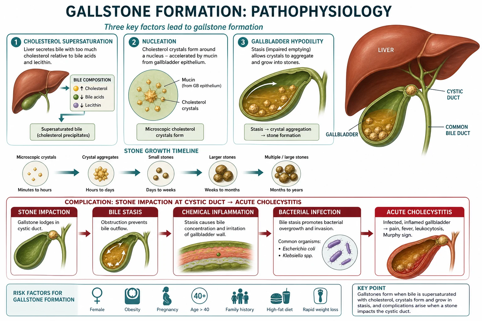
Cholesterol gallstone disease is fundamentally a bile-chemistry and gallbladder-motility disorder. Hepatic bile is normally a micellar solution in which amphipathic bile acids and phospholipids (lecithin) keep hydrophobic cholesterol soluble. When the ratio of cholesterol to bile acids + lecithin rises, bile becomes supersaturated and the cholesterol saturation index (CSI) exceeds 1.[1]
Five defects converge to produce a stone: [1]
- Cholesterol hypersecretion into bile (genetic, OCP, obesity, rapid weight loss).
- Gallbladder hypomotility / stasis — fasting, TPN, pregnancy (progesterone), octreotide, spinal injury.
- Accelerated nucleation — pronucleating proteins (mucin glycoprotein, IgM, aminopeptidase-N, haptoglobin) overwhelm antinucleating factors (apolipoprotein A-I); cholesterol monohydrate crystals form within hours rather than days.
- Mucus hypersecretion by gallbladder goblet cells — provides a gel matrix that traps crystals.
- Altered bile-acid pool (ileal disease/resection) — fewer micelles, more precipitation. [1]
Crystals aggregate on the mucin matrix into macroscopic stones over months to years. Stones are typically few and large (1-3 cm), yellow and faceted.[1]
Pigment stone formation
Black pigment stones arise when there is excess unconjugated bilirubin in bile. Any chronic haemolytic state — sickle cell disease, hereditary spherocytosis, beta-thalassaemia, artificial cardiac valves, chronic malaria — overloads the hepatocyte's glucuronidation capacity; the surplus unconjugated bilirubin is deconjugated in the biliary tree by tissue (non-bacterial) beta-glucuronidase, precipitates as insoluble calcium bilirubinate polymers, and polymerises further with mucin glycoprotein and bicarbonate. Cirrhosis (impaired glucuronidation plus haemolysis from hypersplenism) and TPN are non-haemolytic causes. Black stones are small, dark, fragile, spiculated, and 50% radiopaque.[1]
Brown pigment stones are the product of bacterial infection. Beta-glucuronidase-producing organisms — Escherichia coli, Klebsiella, Enterobacter, Bacteroides, Clostridium perfringens — deconjugate bilirubin within the bile ducts, where they also hydrolyse lecithin to free fatty acids. The result is a soft, brown, laminated stone of calcium bilirubinate mixed with palmitate/stearate soaps and bacterial cytoplasm. Brown stones characteristically form in the ductal system (intra- and extrahepatic) and underlie recurrent pyogenic cholangitis (Oriental cholangiohepatitis) seen across East and South-East Asia.[1]
From stone to inflammation (acute cholecystitis)
When a stone becomes impacted in the cystic duct or Hartmann's pouch, the gallbladder mucosa continues to absorb water and chloride from the trapped bile, concentrating it. Bile salts and the phospholipase A2-generated lysolecithin are direct chemical irritants that inflame the gallbladder wall (chemical cholecystitis). Venous and lymphatic outflow is impeded, producing ischaemia; the stagnant, pressurised bile is then seeded by bacteria ascending from the duodenum — E. coli, Klebsiella, Enterococcus, Enterobacter, and anaerobes (Bacteroides, Clostridium) in roughly descending frequency. The result is a thickened, oedematous gallbladder wall with pericholecystic fluid, a positive sonographic Murphy sign, and the systemic inflammatory response that defines clinical cholecystitis. If untreated, the cycle of ischaemia → secondary infection → wall necrosis progresses to gangrene, perforation and empyema.[3]
From stone to sepsis (ascending cholangitis)
The biliary tree is normally a low-pressure sterile conduit. A stone obstructing the CBD raises intraductal pressure, which disrupts the hepatocyte tight junctions and canalicular membranes, allowing bacteria and endotoxin to reflux into the systemic circulation via the cholangiovenous and cholangiolymphatic pathways. The patient develops bacteraemia (positive blood cultures in 30-60%), fever with rigors, endotoxin-driven hypotension, and the Charcot triad — fever, RUQ pain, jaundice. Once shock and confusion supervene (Reynolds pentad), mortality climbs to 25-50% without prompt decompression.[4]
Clinical Presentation
Asymptomatic (silent) gallstones
Found incidentally on ultrasound performed for another reason (or at laparotomy). The patient has no biliary symptoms. Natural history is benign — 1-4% per year develop symptoms, and the cumulative risk over 20 years is only about 20%. Prophylactic cholecystectomy is not recommended except in selected groups (sickle cell disease, paediatric haemolytic anaemia awaiting transplantation, stones over 3 cm, porcelain gallbladder, polyps over 1 cm, native American high-risk populations).[11]
Biliary colic
The classic presentation of symptomatic uncomplicated stone disease. Despite the name, the pain is constant (not truly colicky), arising 30-60 minutes after a meal — classically fatty food (cholecystokinin releases, gallbladder contracts against an obstructed cystic duct). Key features: [1]
- Location: RUQ or epigastrium, may radiate to the right scapula or right shoulder tip (phrenic nerve C3-C5 irritation) or to the back.
- Quality: deep, gripping, severe, constant.
- Duration: builds over an hour, peaks, and resolves within 2-6 hours as the stone falls back into the gallbladder.
- Associated: nausea, vomiting, diaphoresis; patient is restless and cannot find a comfortable position.
- Crucially: no fever, no Murphy sign, normal WCC. Complete resolution between episodes.[1]
The patient looks well between attacks. Persistent pain beyond six hours, or the appearance of fever or a positive Murphy sign, signals evolution to acute cholecystitis. [1]
Acute cholecystitis
The gallbladder is now inflamed. Symptoms: [1]
- Persistent RUQ pain lasting more than 6 hours (does not settle).
- Fever (typically 38-39 degrees C); rigors suggest cholangitis or empyema.
- Anorexia, nausea, vomiting.
- Positive Murphy sign on examination (see below).
- Leukocytosis, raised CRP, mildly deranged LFTs (the gallbladder wall inflammation can transiently obstruct small intrahepatic ducts).[3]
Choledocholithiasis
A CBD stone produces obstructive (surgical) jaundice — yellowing of sclera and skin, pale stools and dark urine (loss of stercobilin in stool, conjugated bilirubin spilling into urine), and pruritus from bile salt deposition. Pain may be absent (a primary brown stone forming slowly in the duct). LFTs show a predominantly cholestatic picture (raised ALP and GGT, conjugated bilirubin); ALT may transiently exceed 1000 U/L in acute obstruction (the so-called "AST/ALT hepatitis" of acute biliary obstruction, easily mistaken for viral hepatitis). A CBD stone may also precipitate cholangitis or pancreatitis. [1]
Ascending (acute) cholangitis
The septic emergency of biliary disease. Charcot's triad — fever (often with rigors), RUQ pain, and jaundice — is present in only 50-70% of patients (often incomplete in the elderly). Reynolds' pentad adds hypotension/shock and confusion/altered mental state, defining severe (Grade III) cholangitis with mortality of 25-50%. Hypothermia, thrombocytopenia, lactic acidosis and oliguria are ominous. Treatment is resuscitation, broad-spectrum IV antibiotics and urgent biliary decompression (ERCP) — do not delay drainage for conservative trial.[4][7]
Gallstone pancreatitis
A stone transiently obstructs the ampulla of Vater, triggering premature intracellular activation of pancreatic enzymes. The patient presents with sudden severe epigastric pain radiating straight through to the back, nausea, vomiting, and a serum lipase over 3 times the upper limit of normal. Severity and management are those of acute pancreatitis; the biliary-specific decision is whether to perform early ERCP (only if cholangitis or persistent obstruction — see Management). [1]
Gallstone ileus
Mechanical small-bowel obstruction in an elderly woman from a large gallstone (over 2.5 cm) that has eroded through a cholecystoduodenal fistula and lodged at the terminal ileum (the narrowest point). Presentation is insidious, intermittent, with a tender distended abdomen. The classic Rigler triad on imaging — pneumobilia, small-bowel obstruction, and an ectopic gallstone — clinches the diagnosis. Bouveret syndrome is the proximal variant: the stone obstructs the duodenum or proximal jejunum, producing gastric outlet obstruction. [1]
Mirizzi syndrome
A stone impacted in Hartmann's pouch or the cystic duct mechanically compresses (or erodes into) the adjacent common hepatic duct, producing obstructive jaundice that mimics pancreatic head cancer. Pre-operative ERCP is essential to define anatomy and avoid catastrophic bile duct injury during cholecystectomy. [1]
Atypical presentations
- Elderly: vague abdominal pain, confusion, or silent presentation; high index of suspicion for gangrene/empyema in any septic elderly patient.
- Diabetic: autonomic neuropathy blunts pain; higher rates of empyema, gangrene, emphysematous cholecystitis.
- Pregnant: biliary colic is the commonest non-obstetric surgical emergency.
- Immunocompromised / neutropenic: consider acalculous cholecystitis, CMV/Cryptosporidium cholangitis.
- Spinal cord injury: silent stones; autonomic dysreflexia may be the only clue. [1]
Differential Diagnosis
Any cause of RUQ or epigastric pain is in the differential; the skill is identifying the one mimic that, missed, will kill the patient — inferior MI, perforated peptic ulcer, and acute pancreatitis. [1]
[1]RUQ pain differential — the sieve to run through
Clinical & Bedside Assessment
Murphy sign
Examine the RUQ during deep inspiration: place the examining hand under the right costal margin mid-clavicular line and ask the patient to breathe in deeply. The inflamed gallbladder is driven down onto the examining hand, producing sudden pain and an arrest of inspiration. The test is positive only if the same manoeuvre in the left upper quadrant does not reproduce the pain. Sensitivity ~65%, specificity 87-97% for acute cholecystitis; specificity is much lower in the elderly. A sonographic Murphy sign (focal tenderness over the sonographically localised gallbladder) is more specific than the bedside version.[3]
Boas sign
Hyperaesthesia (exaggerated skin sensitivity) in the right subscapular area or referred pain to the right shoulder, due to phrenic nerve (C3-C5) irritation from an inflamed gallbladder in contact with the diaphragm. A non-specific but a viva-favourite sign. [1]
Charcot triad and Reynolds pentad
- Charcot triad = fever + RUQ pain + jaundice → ascending cholangitis (present in 50-70%).
- Reynolds pentad = Charcot triad plus hypotension/shock plus confusion/altered mental state → severe (Grade III) cholangitis. [1]
Courvoisier's law
In a jaundiced patient, a palpable, non-tender, distended gallbladder is unlikely to be caused by gallstones. The reasoning: long-standing stones produce chronic inflammation that scars and contracts the gallbladder, so it cannot distend. A palpably distended gallbladder in jaundice therefore points to malignant obstruction of the distal CBD (pancreatic head cancer, ampullary cancer, cholangiocarcinoma) or, less often, a distal CBD stone that has not yet scarred the gallbladder. The law has exceptions (a Courvoisier gallbladder can occur with a double-impacted duct stone), but it remains an exam cornerstone. [1]
Other bedside findings
- Jaundice — point to CBD obstruction.
- Fever and rigors — cholangitis or empyema.
- Hypotension, tachycardia, confusion — severe sepsis; senior review, ICU.
- Peritonism (guarding, rigidity, rebound) — perforation or peritonitis; surgical emergency.
- Palpable RUQ mass — empyema or mucocele of the gallbladder.
- Scratch marks — chronic cholestatic pruritus.
- Asterixis — hepatic encephalopathy from chronic biliary obstruction (rare in pure stones). [1]
Investigations
The investigation strategy is bedside ultrasound first, then risk-stratify for CBD stones using LFTs and clinical features (ASGE criteria), reserving MRCP for intermediate risk and ERCP for high risk or therapeutic need. [1]
Laboratory
- Full blood count — leukocytosis (over 12-15 × 10⁹/L) in cholecystitis/cholangitis; thrombocytopenia in severe sepsis.
- CRP — raised (over 30 mg/L) in inflammatory complications.
- LFTs — cholestatic pattern (raised ALP and GGT, raised conjugated bilirubin) suggests CBD obstruction; ALT over 150 U/L is highly predictive of choledocholithiasis (ASGE criterion); ALT/AST may transiently exceed 1000 U/L in acute obstruction.
- Serum lipase (preferred) or amylase — over 3 times ULN confirms pancreatitis.
- Coagulation (PT/INR) — deranged in chronic cholestasis or sepsis; needed before ERCP/surgery.
- Lactate — raised over 2 mmol/L in severe sepsis; prognostic.
- Blood cultures (two sets) — before antibiotics; positive in 30-60% of cholangitis.
- U&E — renal impairment in severe sepsis. [1]
Imaging
Diagnostic criteria — Tokyo Guidelines 2018
Acute cholecystitis — TG18 diagnostic criteria:[3]
- Local signs of inflammation: (a) Murphy sign, (b) RUQ mass / pain / tenderness.
- Systemic signs of inflammation: (a) fever over 38 degrees C, (b) leukocytosis, (c) raised CRP.
- Imaging: characteristic findings (gallstones, wall thickening, pericholecystic fluid, distension, positive sonographic Murphy). [1]
Suspected diagnosis = one local + one systemic feature, OR one local + imaging, OR one systemic + imaging. Definite diagnosis = one feature from each of the three groups. [1]
Tokyo Guidelines 2018 severity grading — acute cholecystitis:[3]
- Grade III (severe) — acute cholecystitis with organ dysfunction:
- Cardiovascular — dysfunction requiring dopamine/noradrenaline.
- Neurological — decreased level of consciousness.
- Respiratory — PaO2 under 60 mmHg or SpO2 under 90%.
- Renal — oliguria, creatinine over 2 mg/dL (about 177 micromol/L).
- Hepatic — PT-INR over 1.5.
- Haematological — platelets under 100 × 10⁹/L.
- Grade II (moderate) — any one of: WCC over 18 × 10⁹/L, palpable tender RUQ mass, fever over 39 degrees C, duration of symptoms over 72 hours, or marked local inflammation (gangrenous cholecystitis, pericholecystic abscess, hepatic abscess, biliary peritonitis).
- Grade I (mild) — does not meet Grade II or III criteria; otherwise healthy patient with no organ dysfunction. [1]
Acute cholangitis — TG18 diagnostic criteria:[4]
- Systemic inflammation: (a) fever/rigors, (b) abnormal WCC, (c) abnormal CRP, (d) abnormal procalcitonin.
- Cholestasis: (a) jaundice, (b) abnormal liver function tests (ALP, GGT, AST, ALT, bilirubin over 2 mg/dL).
- Imaging: biliary dilation, evidence of cause (stricture, stone, stent). [1]
Suspected = one item from each of the three. Definite = cholestasis plus biliary dilation plus a cause identified. [1]
Tokyo Guidelines 2018 severity grading — acute cholangitis:[4]
- Grade III (severe) — organ dysfunction requiring ICU/urgent drainage:
- Cardiovascular — hypotension requiring noradrenaline/dopamine.
- Neurological — disturbance of consciousness.
- Respiratory — PaO2/SpO2 under 90%.
- Renal — oliguria under 0.5 mL/kg/h, creatinine over 2 mg/dL.
- Hepatic — PT-INR over 1.5.
- Haematological — platelets under 100 × 10⁹/L.
- Grade II (moderate) — at least two of: abnormal WCC, fever over 39 degrees C, age 75 years or older, total bilirubin 5 mg/dL or more (about 85 micromol/L), hypoalbuminaemia (below 0.7 × lower limit of normal).
- Grade I (mild) — not Grade II or III; initially responsive to medical therapy. [1]
ASGE 2019 risk stratification for choledocholithiasis
A critical framework for deciding who needs ERCP before cholecystectomy: [1]
- High risk (over 50%, proceed to ERCP): (1) CBD stone seen on transabdominal ultrasound; OR (2) CBD dilation over 6 mm with gallbladder in situ (combined with); OR (3) total bilirubin over 4 mg/dL (about 70 micromol/L) at presentation plus a dilated duct or other strong predictor.
- Intermediate risk (10-50%, do EUS or MRCP): abnormal liver biochemical tests, age over 55 years, or CBD dilation.
- Low risk (under 10%, proceed directly to cholecystectomy): none of the above. [1]
This algorithm avoids unnecessary ERCP (with its pancreatitis and bleeding risks) in low-risk patients. [1]
Management — Resuscitation
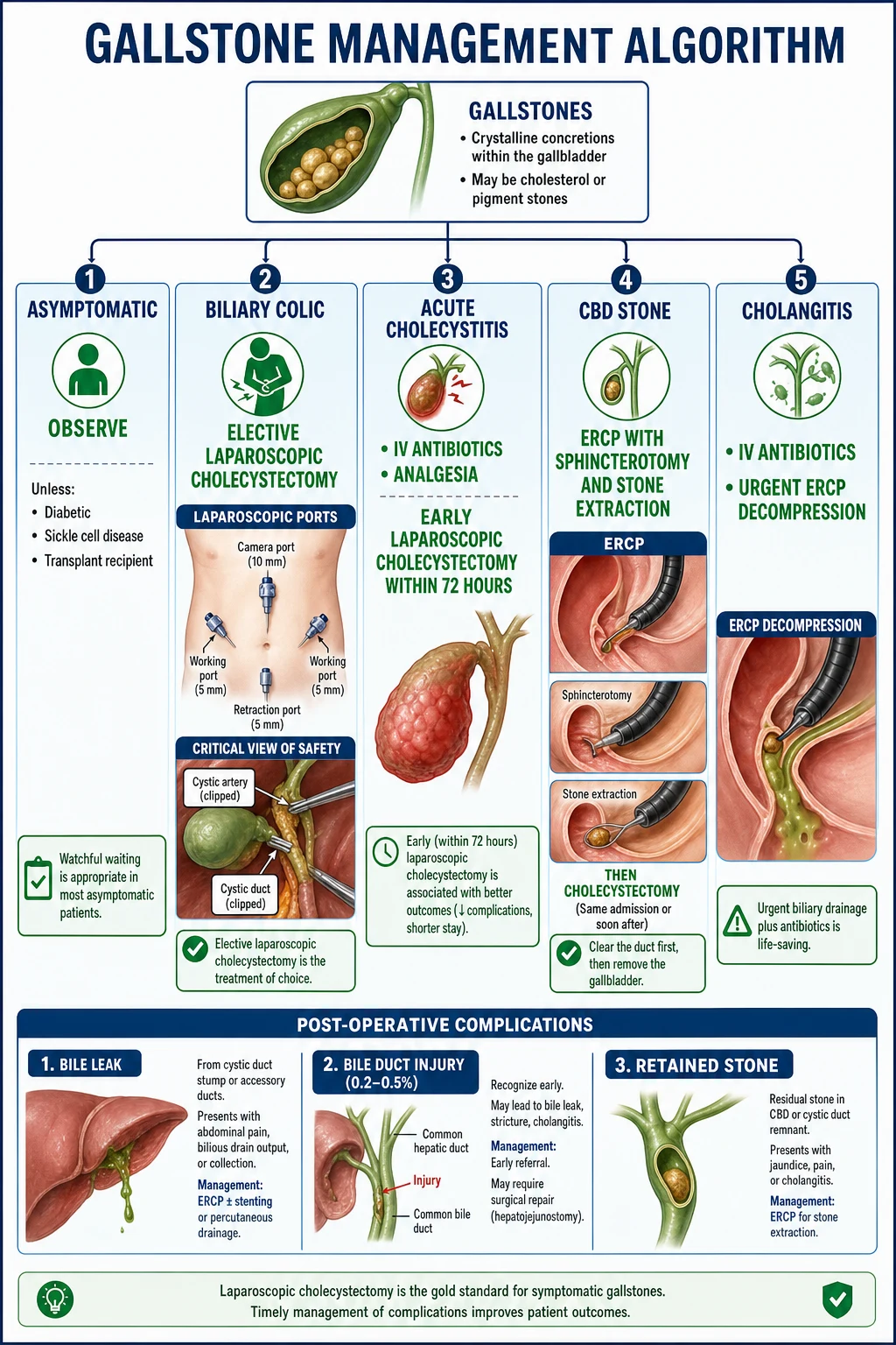
The resuscitation bundle depends on the syndrome — but the principle is uniform: analgesia, fluid resuscitation, antibiotics if inflammation/sepsis, and a clear plan for definitive care.[5][6]
Biliary colic (emergency department)
- Analgesia: an NSAID first-line — diclofenac 75 mg IM (orally/PR if mild), which both relieves pain and reduces sphincter of Oddi tone. Add an opioid if severe: morphine 5-10 mg IV titrated (or fentanyl 50-100 micrograms IV); pethidine is traditional but offers no proven advantage over morphine.
- Antiemetic: ondansetron 4 mg IV or metoclopramide 10 mg IV.
- Investigations: FBC, CRP, LFTs, lipase, USS.
- Disposition: discharge once pain settles with elective surgical referral for laparoscopic cholecystectomy. Educate about red flags (fever, persistent pain, jaundice). [1]
Acute cholecystitis
- Admit, NPO, IV fluid resuscitation (balanced crystalloid, e.g., lactated Ringer's or Hartmann's; 1.5-3 L over the first 24 hours titrated to urine output and vital signs).
- IV opioid analgesia (morphine 5-10 mg every 2-4 hours or fentanyl PCA).
- IV antibiotics per TG18, covering the typical Gram-negative bacilli and anaerobes:[6]
- Community-acquired, mild-to-moderate: co-amoxiclav 1.2 g IV every 8 hours, OR cefuroxime 1.5 g IV every 8 hours + metronidazole 500 mg IV every 8 hours.
- Severe or healthcare-associated: piperacillin-tazobactam 4.5 g IV every 8 hours, OR a third-generation cephalosporin (ceftriaxone 2 g IV daily) + metronidazole.
- Penicillin allergy: ciprofloxacin 400 mg IV every 12 hours + metronidazole.
- Duration: 4-7 days for uncomplicated cholecystitis (TG18).
- Surgical referral for early laparoscopic cholecystectomy within 72 hours (see Definitive Management).
Ascending cholangitis (the septic emergency)
- Apply the Sepsis Six within one hour: (1) high-flow oxygen, (2) IV fluids 30 mL/kg crystalloid bolus, (3) blood cultures + serum lactate, (4) IV broad-spectrum antibiotics within one hour, (5) catheterise for urine output, (6) watch the lactate trend.
- Antibiotics (broader than for uncomplicated cholecystitis): piperacillin-tazobactam 4.5 g IV every 8 hours ± gentamicin (5-7 mg/kg once daily, monitor levels), OR ceftriaxone 2 g IV daily + metronidazole 500 mg IV every 8 hours. Add an antipseudomonal agent in healthcare-associated infection.
- Vasopressors (noradrenaline) for persistent hypotension; intubate if respiratory failure; ICU for Grade III.
- Urgent ERCP within 24 hours for Grade III cholangitis and within 24-48 hours for Grade II; biliary decompression (sphincterotomy + stone extraction + stent or endoscopic nasobiliary drain) is the only intervention that lowers mortality.[4][7]
Gallstone pancreatitis
- Aggressive goal-directed IV fluids (lactated Ringer's preferred; 1.5 mL/kg/h for the first 12-24 hours, titrated to haematocrit and urine output) — see acute-pancreatitis topic.
- Analgesia (opioid), NPO only if severe; otherwise early enteral feeding.
- ERCP within 24 hours only if cholangitis or persistent biliary obstruction (Cochrane Tse & Yuan 2012 — no benefit of routine ERCP otherwise).[9]
Empyema / gangrene / perforation
- IV antibiotics, fluid resuscitation, urgent imaging (CT if perforation suspected), and urgent percutaneous cholecystostomy (unfit) or surgical exploration. [1]
Management — Definitive & Stepwise
Asymptomatic gallstones
Observe. No cholecystectomy, no ursodeoxycholic acid, no further imaging beyond reassurance. Exceptions (prophylactic cholecystectomy):[11]
- Sickle cell disease and other major haemolytic anaemias (children).
- Transplant candidates (heart, kidney) before immunosuppression.
- Stones over 3 cm — gallbladder carcinoma risk.[12]
- Porcelain gallbladder — up to 25% carcinoma risk.
- Gallbladder polyps over 1 cm or growing polyps.
- Selected native American / high-risk populations.
- Anticipated prolonged TPN or bariatric surgery (selected).
Biliary colic
Elective laparoscopic cholecystectomy. Once symptomatic, 30% recur within a year and 50% within five years; surgery prevents progression to cholecystitis, pancreatitis and cholangitis. [1]
Acute cholecystitis
Early laparoscopic cholecystectomy within 72 hours of symptom onset. The Lyu 2018 updated meta-analysis of RCTs confirms earlier surgery gives shorter total hospital stay, fewer complications, lower readmission rate, and equivalent conversion and bile duct injury rates compared with delayed (interval) cholecystectomy.[10]
Grade III (severe) cholecystitis with organ dysfunction is managed initially with urgent percutaneous cholecystostomy + IV antibiotics, with delayed interval cholecystectomy once the patient stabilises (TG18).[3]
The laparoscopic cholecystectomy — critical view of safety
The critical view of safety (CVS) is the modern standard for safe dissection and the single most effective measure to prevent bile duct injury (whose incidence is 0.2-0.5%).[2] Three requirements must be met before any structure is clipped or divided:
- Clear Calot's triangle — the triangle bounded by the cystic duct inferiorly, common hepatic duct medially, and the inferior edge of the liver superiorly.
- Identify the cystic duct and the cystic artery as the only two tubular structures crossing the triangle.
- Clear the hepatocystic triangle of all fat and fibrous tissue so that the gallbladder infundibulum (Hartmann's pouch) is dissected off the liver bed — confirming that what is being clipped is only the cystic duct, not the common hepatic or common bile duct. [1]
Only then are the cystic duct and artery clipped and divided. Routine intra-operative cholangiography is advocated by some units but not universally adopted; indocyanine green (ICG) fluorescence cholangiography is gaining ground as an aid. Conversion to open cholecystectomy (overall 2-10%, higher in acute/severe disease) is mandatory if the anatomy cannot be safely defined — persisting in a hostile field is the principal cause of bile duct injury.[2]
Choledocholithiasis
The standard sequence is ERCP with sphincterotomy + balloon/basket trawl (± mechanical lithotripsy for large stones), followed by laparoscopic cholecystectomy during the same admission to prevent recurrence. Alternative in expert centres: laparoscopic common bile duct exploration (LCBDE) at cholecystectomy (transcystic or choledochotomy), which clears the duct and removes the gallbladder in one operation. If a stone cannot be cleared, a plastic or self-expanding metal biliary stent maintains drainage (bridge to definitive therapy). [1]
Acute cholangitis
Antibiotics + urgent ERCP biliary decompression within 24 hours (Grade III) or 24-48 hours (Grade II). ERCP achieves sphincterotomy, stone removal, and insertion of a biliary stent or endoscopic nasobiliary drain (ENBD). If ERCP fails or is unavailable, percutaneous transhepatic biliary drainage (PTBD) or, rarely, surgical drainage. Cholecystectomy is performed later, once sepsis has resolved.[4][5]
Gallstone pancreatitis
- Supportive care as for acute pancreatitis (goal-directed fluids, analgesia, monitoring for organ failure).
- ERCP within 24 hours only if cholangitis or persistent biliary obstruction (Cochrane Tse & Yuan 2012 — routine ERCP does not reduce complications or mortality).[9]
- Cholecystectomy during the index admission (after recovery from severe pancreatitis, or before discharge if mild) to prevent recurrence.
- Definitive management of any necrosis follows the step-up approach (see acute-pancreatitis topic).
Mirizzi syndrome
Pre-operative MRCP/ERCP to define anatomy and place a stent if the common hepatic duct is involved. Open cholecystectomy (or subtotal cholecystectomy leaving the Hartmann pouch behind) with intra-operative cholangiogram; a cholecystocholedochal fistula (Csendes type II-V) may require bile-duct repair or hepaticojejunostomy. [1]
Gallstone ileus
Enterolithotomy — a small longitudinal enterotomy proximal to the obstruction to milk out the stone; the enterotomy is closed transversely. Run the entire small bowel to exclude additional stones (in 5-10% of cases there is more than one). Cholecystectomy and fistula repair are usually deferred to a second stage, as the inflamed tissue is hostile and most fistulae close spontaneously. Emergency surgery has high mortality (10-20%). [1]
Ursodeoxycholic acid dissolution
Ursodeoxycholic acid (UDCA) 10-15 mg/kg/day orally for 6-24 months dissolves only small (under 1 cm), radiolucent, cholesterol stones in a functioning gallbladder (HIDA filling + normal CCK ejection). Recurrence is 50% within 5 years. Rarely used as first-line; reserved for patients unfit for surgery. [1]
Contact dissolution and lithotripsy
Methyl tert-butyl ether (MTBE) contact dissolution and extracorporeal shock wave lithotripsy (ESWL) are historical niche therapies; rarely offered today. [1]
Escalation triggers
- Organ dysfunction in cholecystitis or cholangitis → ICU; urgent drainage.
- Persistent biliary obstruction → ERCP.
- Failure of conservative management in cholecystitis → surgery or percutaneous drain.
- Suspicion of gangrene, perforation, empyema, emphysematous cholecystitis → urgent imaging (CT) and surgery.
- Anatomical uncertainty at laparoscopy → convert to open rather than risk bile duct injury. [1]
Specific Subtypes & Scenarios
Acalculous cholecystitis
Acute gallbladder inflammation without stones, accounting for 5-10% of acute cholecystitis. Occurs in the critically ill (ICU, severe sepsis, multi-organ failure), after major trauma or burns, post-major surgery, on prolonged TPN, in AIDS, and in severe vasculitis. Pathology is gallbladder ischaemia rather than obstruction. Presentation is often masked by the underlying critical illness; leukocytosis, fever, deranged LFTs, and an unexplained septic picture in an ICU patient warrant urgent USS. Mortality is 30-50%. Treatment: IV antibiotics + percutaneous cholecystostomy (definitive in many; cholecystectomy only after recovery). [1]
Mirizzi syndrome (Csendes classification)
A stone in Hartmann's pouch or the cystic duct compresses (type I) or erodes into (types II-V) the common hepatic duct. Csendes types: [1]
- Type I — external compression only.
- Type II-IV — cholecystobiliary fistula involving less than one-third (II), up to two-thirds (III), or complete destruction (IV) of the CBD.
- Type V — any type plus a cholecystoenteric fistula. [1]
Pre-operative ERCP defines the anatomy and avoids intra-operative catastrophe. Subtotal cholecystectomy or bile-duct repair/hepaticojejunostomy depending on type. [1]
Gallstone ileus and Bouveret syndrome
See above. The Rigler triad (pneumobilia + SBO + ectopic gallstone) on plain AXR or CT makes the diagnosis. Treatment is enterolithotomy (with or without cholecystectomy at the same sitting — most surgeons defer). [1]
Empyema
Pus in the gallbladder from suppurative cholecystitis. The patient is septic, toxic, and often elderly or diabetic. A palpable RUQ mass is common. Treatment: IV antibiotics + percutaneous cholecystostomy (drainage first; cholecystectomy once settled). [1]
Gangrenous cholecystitis
Ischaemic necrosis of the gallbladder wall, heralded by sepsis and a deteriorating clinical picture. Diabetic and elderly patients are at highest risk. Emergency cholecystectomy (often open); subtotal cholecystectomy if dissection is hazardous. [1]
Perforated cholecystitis
Free perforation → biliary peritonitis (surgical emergency). Localised perforation → pericholecystic abscess (drainage + cholecystectomy). Subacute perforation may produce a cholecystoenteric fistula (the precursor of gallstone ileus). [1]
Emphysematous cholecystitis
Gas-forming organisms (Clostridium perfringens, E. coli, Klebsiella) in the gallbladder wall, visible on plain X-ray or CT. Diabetic and elderly male patients over-represented; 50% acalculous. Emergency cholecystectomy + broad-spectrum antibiotics (cover anaerobes). [1]
Mucocele of the gallbladder
Chronic cystic duct obstruction with continued mucin secretion produces a distended, non-tender, palpable gallbladder containing clear mucus. Elective cholecystectomy. [1]
Porcelain gallbladder
Calcification of the gallbladder wall on plain X-ray or CT. Up to 25% association with adenocarcinoma (especially the selective mucosal calcification pattern). Prophylactic cholecystectomy. [1]
Gallbladder polyps
Most are cholesterol polyps (benign). Adenomatous polyps over 1 cm carry malignant potential — cholecystectomy. Polyps under 1 cm in a stone-free asymptomatic gallbladder: surveil with annual USS. [1]
Recurrent pyogenic cholangitis (Oriental cholangiohepatitis)
Brown pigment stones throughout the intra- and extrahepatic ducts with ductal dilation and lobar atrophy (classically left lateral segment). Endemic in East and South-East Asia; presents with recurrent cholangitis. Management: ERCP clearance, antibiotics, hepatectomy for atrophic lobes. [1]
Post-cholecystectomy syndrome
Persistent biliary-type symptoms in 5-40% after cholecystectomy. Causes: sphincter of Oddi dysfunction (type I benefits from sphincterotomy; type III does not — EPISOD trial), retained/recurrent CBD stone, biliary dyskinesia, gastritis or peptic ulcer, irritable bowel syndrome. Investigate with LFTs, MRCP, EUS, and consider sphincter of Oddi manometry. [1]
Complications & Pitfalls
[1]Pitfalls
- Misdiagnosing inferior MI as biliary colic — always ECG the older patient with epigastric pain before labelling biliary.
- Missing ascending cholangitis — a "mildly unwell" elderly patient with low-grade fever and a slightly raised bilirubin may have Grade III cholangitis; have a low threshold for urgent ERCP.
- Performing laparoscopic cholecystectomy in a hostile field — convert to open or perform subtotal (fundus-first) cholecystectomy to avoid bile duct injury.
- Inappropriate ERCP in gallstone pancreatitis — routine ERCP does not help (Cochrane).[9]
- Citing "Murphy sign" in biliary colic — by definition biliary colic has no fever, no Murphy sign, normal WCC. The presence of these is cholecystitis.
- Treating asymptomatic stones with surgery — observe unless special indication.
- Failing to risk-stratify for CBD stones — apply ASGE 2019 criteria to avoid unnecessary ERCP.
Prognosis & Disposition
- Asymptomatic gallstones: benign — 1-4% per year become symptomatic; 20% cumulative risk over 20 years. Most patients are never operated.[11]
- Biliary colic: recurrence 30% at one year, 50% at five years if untreated; excellent outcome after laparoscopic cholecystectomy.
- Acute cholecystitis (early cholecystectomy): mortality under 1% in fit adults; Grade III mortality 10-30%.[3]
- Ascending cholangitis: mortality 2-7% in mild-moderate, 25-50% in Grade III; early ERCP halves mortality.[4]
- Gallstone pancreatitis: mild over 95% survival; severe necrotising up to 30% mortality.
- Laparoscopic cholecystectomy: recovery 1-2 weeks; return to work 1-2 weeks; bile duct injury 0.2-0.5%; conversion 2-10%.
- Bile duct injury: prompt specialist HPB repair gives 80-90% long-term good outcome; delayed recognition risks secondary biliary cirrhosis.[2]
Disposition
- Biliary colic → discharge from ED with elective surgical referral.
- Acute cholecystitis (Grade I-II) → admit surgical ward, antibiotics, early lap chole.
- Acute cholecystitis (Grade III) → ICU/percutaneous drain first.
- Ascending cholangitis → admit (HDU/ICU if severe), antibiotics, urgent ERCP.
- Gallstone pancreatitis (mild) → surgical/medical ward, cholecystectomy before discharge.
- Gallstone pancreatitis (severe) → ICU, step-up approach for necrosis.
- Gallstone ileus / perforation / empyema / emphysematous → emergency surgery. [1]
Special Populations
Pregnancy
Biliary colic is the commonest non-obstetric surgical emergency in pregnancy (progesterone slows gallbladder emptying). Management: [1]
- First-line: conservative — NPO, IV fluids, analgesia (NSAIDs avoided in 3rd trimester; use paracetamol and opioid).
- Laparoscopic cholecystectomy is safe in the 2nd trimester (weeks 14-26) if recurrent colic or acute cholecystitis; minimally raises preterm labour and fetal loss.
- Defer to postpartum if a single mild episode and the patient is stable.
- ERCP if choledocholithiasis or cholangitis — use lead shielding for fetus, deferral of fluoroscopy where possible; EUS-guided ERCP techniques are emerging. [1]
Diabetes mellitus
- Higher rates of empyema, gangrene, emphysematous cholecystitis.
- Atypical presentation (autonomic neuropathy blunts pain); a septic, mildly symptomatic diabetic with deranged LFTs warrants aggressive imaging.
- Lower threshold for surgery; perioperative glucose optimisation (IV insulin sliding scale).
- TG18 considers diabetes a comorbidity warranting admission and IV antibiotics for any acute cholecystitis. [1]
Cirrhosis
- Increased bleeding risk from portal hypertension, coagulopathy, and portocaval shunting at the gallbladder fossa.
- Child-Pugh A tolerates laparoscopic cholecystectomy.
- Child-Pugh C is a relative contraindication (mortality up to 25%) — prefer percutaneous cholecystostomy.
- Subtotal cholecystectomy often preferred to avoid dissection in the gallbladder hilum. [1]
Children and adolescents
- Pigment stones predominate, from haemolytic disease (sickle cell, hereditary spherocytosis, beta-thalassaemia).
- Prophylactic cholecystectomy recommended in sickle cell disease once stones detected.
- Laparoscopic cholecystectomy is well tolerated. [1]
Elderly
- Atypical and late presentation, more comorbidity.
- Higher incidence of gangrene, empyema, perforation, emphysematous cholecystitis.
- Aggressive evaluation of any septic elderly patient with deranged LFTs. [1]
Post-bariatric / rapid weight loss
- 30-70% form gallstones within six months.
- Ursodeoxycholic acid 600 mg/day orally for six months reduces risk.
- Cholecystectomy if symptomatic. [1]
Immunocompromised (AIDS, post-transplant, neutropenia)
- Acalculous cholecystitis, opportunistic biliary infection (CMV, Cryptosporidium, Microsporidium, MAC).
- Broader work-up; lower threshold for imaging. [1]
Anticoagulated patient
- Warfarin — hold pre-operatively and bridge with LMWH if high thrombotic risk; INR under 1.5 before cholecystectomy.
- DOACs — omit morning of surgery, resume once haemostasis secure.
- ERCP with sphincterotomy carries bleeding risk — consider balloon dilation of the sphincter instead, or correct anticoagulation first. [1]
Evidence, Guidelines & Regional Differences
Tokyo Guidelines 2018 (TG18)
The international consensus standard for diagnosis, severity grading, antimicrobial therapy, and management of acute cholecystitis and cholangitis. Key papers:[3][4][5][6][7][8]
- Yokoe 2018 (PMID 29032636) — diagnostic criteria and severity grading of acute cholecystitis.[3]
- Kiriyama 2018 (PMID 29032610) — diagnostic criteria and severity grading of acute cholangitis.[4]
- Miura 2018 (PMID 28941329) — initial management and flowchart.[5]
- Wakabayashi 2018 (PMID 29095575) — surgical management and the critical view of safety.[2]
- Okamoto 2018 (PMID 29045062) — cholecystitis management flowchart.[8]
- Gomi 2018 (PMID 29090866) — antimicrobial therapy.[6]
- Mayumi 2018 (PMID 29090868) — management bundles (early source control, antibiotics, organ support).[7]
Other landmark evidence
- Lyu 2018 meta-analysis (PMID 30167953) — early (within 72 h) laparoscopic cholecystectomy for acute cholecystitis is superior to delayed.[10]
- Cochrane review — Tse & Yuan 2012 (PMID 22592743) — early routine ERCP does not improve outcomes in acute gallstone pancreatitis unless cholangitis or persistent obstruction.[9]
- ASGE 2019 risk-stratified approach — high/intermediate/low-risk classification for choledocholithiasis directs ERCP vs MRCP vs cholecystectomy.
- NICE CG188 (UK, 2014) — laparoscopic cholecystectomy for symptomatic disease; pre-op MRCP only if intermediate/high risk; ursodeoxycholic acid not recommended routinely.
- EPISOD trial (Cotton 2014) — endoscopic sphincterotomy does not benefit type III sphincter of Oddi dysfunction (curbed ERCP over-use).
Exam Pearls
The high-yield gallstone pearls for NEET-PG / INICET
Exam application bank (NEET-PG / INICET)
One-line answer
Gallstone disease (cholelithiasis) is the formation of solid crystalline concretions in the gallbladder or biliary tree from precipitated bile components — most commonly cholesterol (the classic risk profile of the 5 Fs — female, forty, fat, fertile, fair, plus rapid weight loss, family history and OCP use) and less often pigment stones from chronic haemolysis (black) or bacterial biliary infection (brown). Sixty to eighty per cent are silent; symptomatic disease spans biliary colic (postprandial RUQ pain, self-limiting), acute cholecystitis (persistent RUQ pain, fever, positive Murphy sign), choledocholithiasis (obstructive jaundice) and the septic emergency of ascending cholangitis (Charcot triad of fever, jaundice and RUQ pain; Reynolds pentad adds hypotension and confusion). Severity is graded by the Tokyo Guidelines 2018 for both cholecystitis and cholangitis. Ultrasound is first-li
Worked stems (answer without another resource)
Stem 1 — Classic presentation. Map symptoms to mechanism; name the first investigation and first treatment step with dose/route if drug therapy is standard. [1]
Stem 2 — Unstable / complicated. List red flags that force immediate resuscitation, theatre, ICU, antidote, or reperfusion — and what you do in the first 15 minutes. [1]
Stem 3 — Atypical group. Elderly, pregnancy, child, or immunocompromised: how presentation and thresholds change. [1]
Stem 4 — Differential trap. Name the three closest mimics and one discriminator for each. [1]
Stem 5 — Disposition. Who goes home with safety-netting, who is admitted, who needs HDU/ICU/theatre, and what follow-up is mandatory. [1]
Rapid viva checklist
- Definition + classification
- Pathophysiology chain
- Bedside signs / criteria
- Score with exact components (if any)
- Emergency bundle
- Definitive therapy with doses
- Complications of disease and of treatment
- Special populations
- Guideline/trial name if classic
- Three exam traps
Coverage self-check
If you cannot answer any stem above from this page alone, re-read the matching section — the page is intended to be self-sufficient for final-prof and NEET-PG/INICET questions on Gallstone Disease & Cholecystitis.
References
- [1]Portincasa P, Moschetta A, Palasciano G. Cholesterol gallstone disease Lancet, 2006.PMID 16844493
- [2]Wakabayashi G, Iwashita Y, Hibi T, et al. Tokyo Guidelines 2018: surgical management of acute cholecystitis: safe steps in laparoscopic cholecystectomy for acute cholecystitis (with videos) J Hepatobiliary Pancreat Sci, 2018.PMID 29095575
- [3]Yokoe M, Hata J, Takada T, et al. Tokyo Guidelines 2018: diagnostic criteria and severity grading of acute cholecystitis (with videos) J Hepatobiliary Pancreat Sci, 2018.PMID 29032636
- [4]Kiriyama S, Kozaka K, Takada T, et al. Tokyo Guidelines 2018: diagnostic criteria and severity grading of acute cholangitis (with videos) J Hepatobiliary Pancreat Sci, 2018.PMID 29032610
- [5]Miura F, Okamoto K, Takada T, et al. Tokyo Guidelines 2018: initial management of acute biliary infection and flowchart for acute cholangitis J Hepatobiliary Pancreat Sci, 2018.PMID 28941329
- [6]Gomi H, Solomkin JS, Takada T, et al. Tokyo Guidelines 2018: antimicrobial therapy for acute cholangitis and cholecystitis J Hepatobiliary Pancreat Sci, 2018.PMID 29090866
- [7]Mayumi T, Okamoto K, Takada T, et al. Tokyo Guidelines 2018: management bundles for acute cholangitis and cholecystitis J Hepatobiliary Pancreat Sci, 2018.PMID 29090868
- [8]Okamoto K, Suzuki K, Takada T, et al. Tokyo Guidelines 2018: flowchart for the management of acute cholecystitis J Hepatobiliary Pancreat Sci, 2018.PMID 29045062
- [9]Tse F, Yuan Y. Early routine endoscopic retrograde cholangiopancreatography strategy versus early conservative management strategy in acute gallstone pancreatitis Cochrane Database Syst Rev, 2012.PMID 22592743
- [10]Lyu Y, Cheng Y, Wang B, et al. Early versus delayed laparoscopic cholecystectomy for acute cholecystitis: an up-to-date meta-analysis of randomized controlled trials Surg Endosc, 2018.PMID 30167953
- [11]Behari A, Kapoor VK. Asymptomatic Gallstones (AsGS) - To Treat or Not to? Indian J Surg, 2012.PMID 23372301
- [12]Tewari M. Contribution of silent gallstones in gallbladder cancer J Surg Oncol, 2006.PMID 16724346